2. Results and Discussion
Isoquinolinequinone
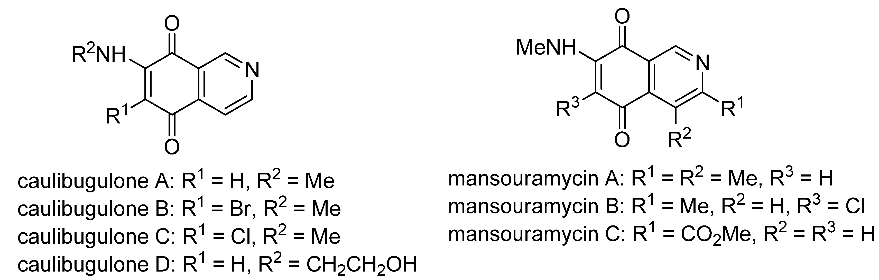
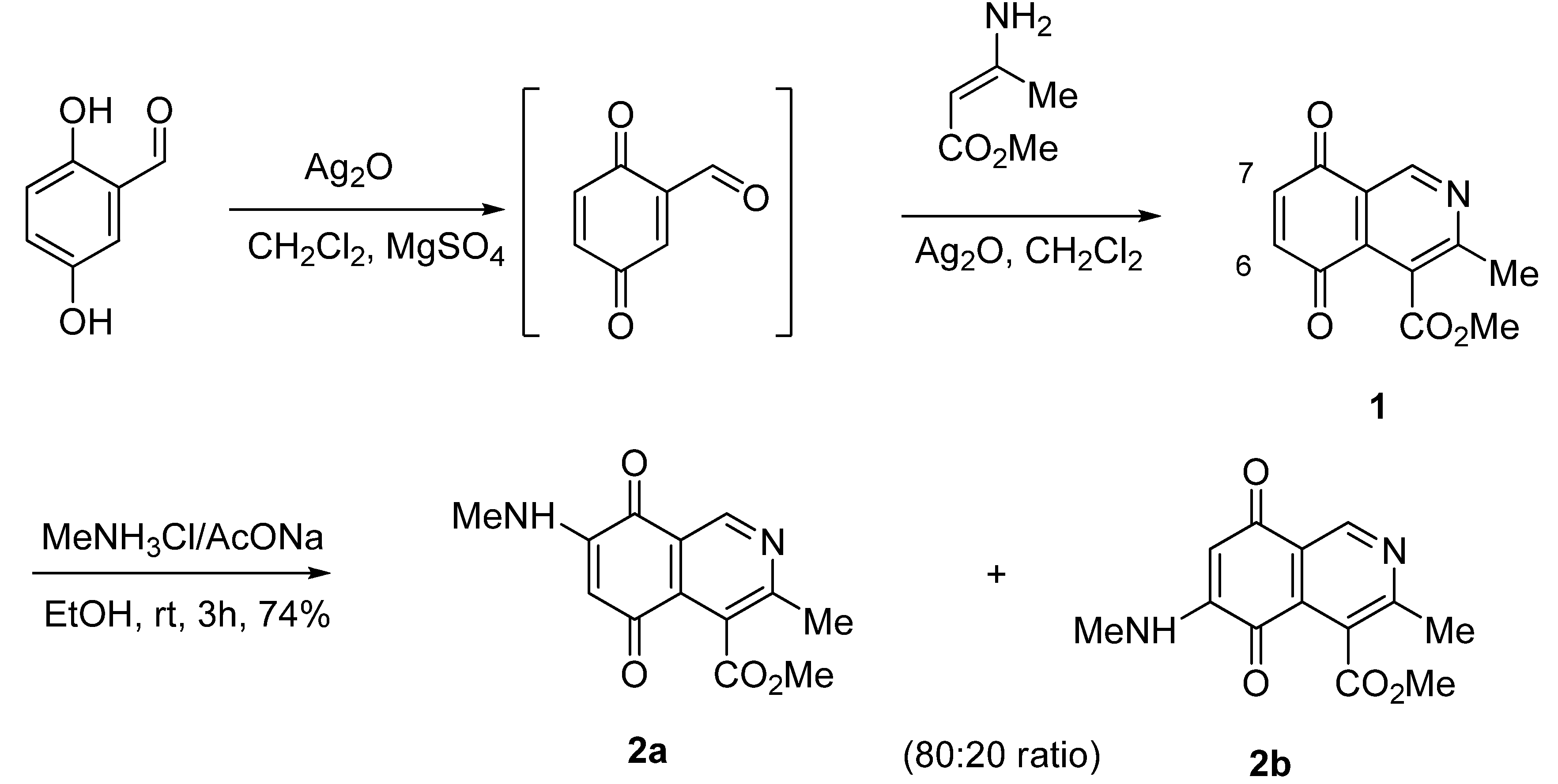
1, unsubstituted at the 1-position and containing a methoxycarbonyl group on the heterocyclic ring, as in mansouramycin C, was selected as a suitable precursor of the designed aminoisoquinolinequinones. The synthesis of the target compound
1 was accomplished in 86% yield from commercially available 2,5-dihydroxybenzaldehyde and methyl aminocrotonate, through a one-pot procedure previously developed in our laboratory [
19] (
Scheme 1).
Scheme 1.
Synthesis and reaction of isoquinolinequinone 1 with methylamine.
Scheme 1.
Synthesis and reaction of isoquinolinequinone 1 with methylamine.
The amination reaction of quinone
1 was firstly examined with methylamine hydrochloride-sodium acetate in ethanol at room temperature. The reaction went to completion in 3 h to give an 80:20 mixture of regioisomers
2a and
2b (as evaluated by
1H-NMR) in 74% yield. Further separation by column chromatography on silica gel afforded pure compound
2a in 52% yield (
Scheme 1). Our efforts to isolate a pure sample of regioisomer
2b by chromatography were unsuccessful. Then we examined the amination reaction of quinone
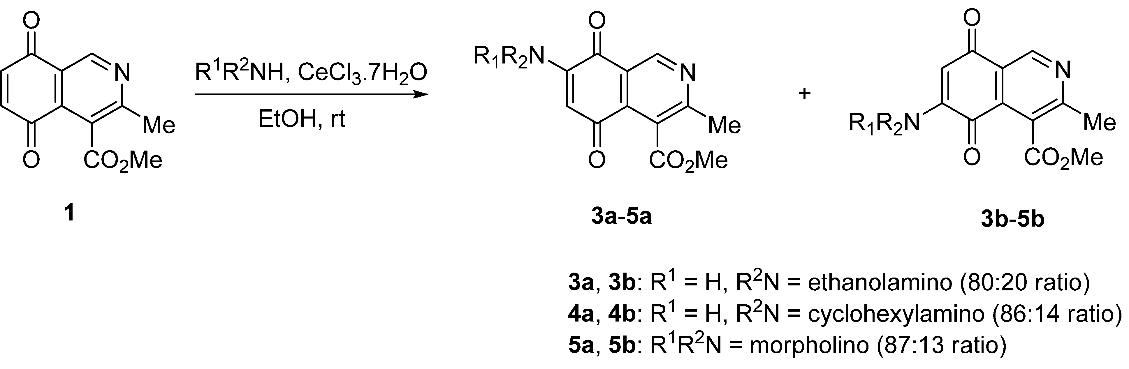
1 with 2-aminoethanol under the same conditions used with methylamine. Analysis of the progress of the reaction by TLC reveals that the amination reaction of
1 proceeds slowly (>7 h) and extensive decomposition of quinone
1 was detected. According to precedents on the use of a Lewis acid to facilitate the reaction of quinones with amines [
20,
21,
22,
23,
24], compound
1 was allowed to react with 2-aminoethanol in ethanol in the presence of 5 mmol% of CeCl
3·7H
2O. Under these conditions, the reaction went to completion in 3 h to give an 80:20 mixture of regioisomers
3a and
3b (as evaluated by
1H-NMR) in 65% yield. Further separation by column chromatography on silica gel afforded the major regioisomer
3a pure in 47% yield (
Scheme 2). Attempts to isolate a pure sample of the minor regioisomer
3b by chromatography were unsuccessful.
Scheme 2.
Reaction of isoquinolinequinone 1 with amines induced by acid-catalysis.
Scheme 2.
Reaction of isoquinolinequinone 1 with amines induced by acid-catalysis.
The amination reactions of quinone
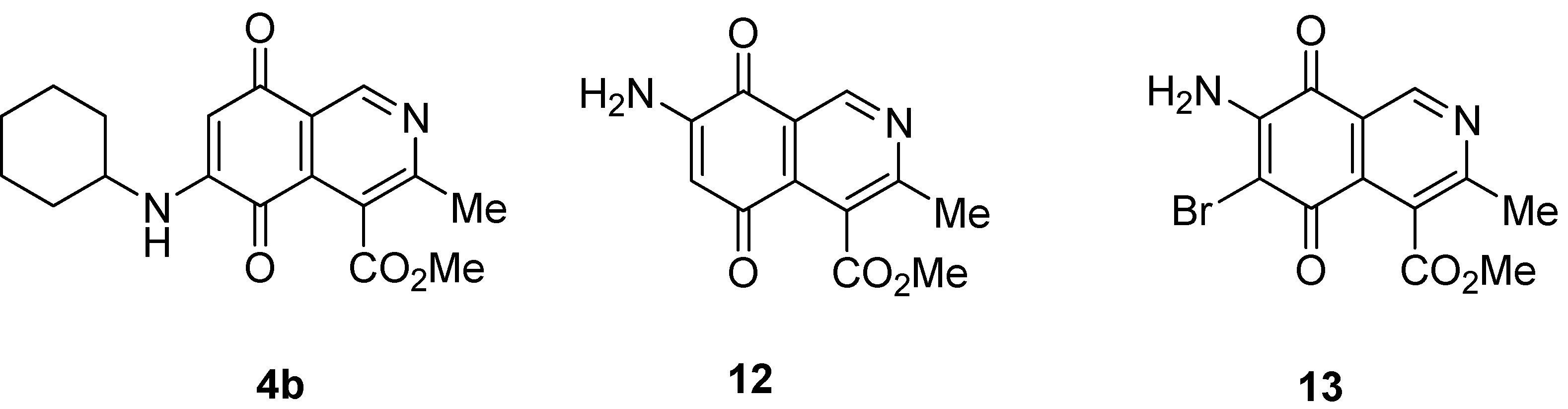
1 with cyclohexylamine and morpholine under the above acid-induced conditions were also explored. The experiments provided access to a mixture of the corresponding alkylaminoisoquinolinequinones
4a,
b and
5a,
b (
Scheme 2). In these cases, pure samples of the regioisomers of each pair were isolated by column chromatography.
According to the amination reaction of quinone
1 with the studied alkyamines we can conclude that the addition of nitrogen nucleophiles across the quinone double bond occurs with high regioselective preference to give the 7-substituted regioisomer as the main product (
Scheme 2).
The structure of the new compounds was established on the basis of their nuclear magnetic resonance (
1H-NMR,
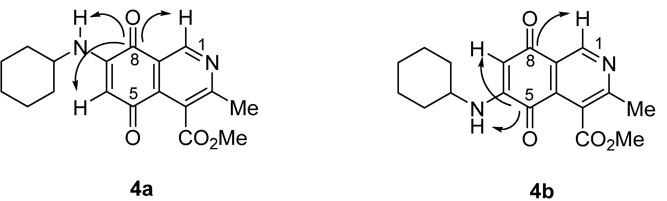
13C-NMR, 2D-NMR) and high resolution mass spectra (HRMS). The position of the nitrogen substituent in these aminoquinones was determined by means of HMBC experiments. For example, the location of the nitrogen group at C-7 in compound
4a was deduced by the
3JC,H couplings between the carbon at C-8 (δ 180.83) with the protons at C-1 (δ 9.17), at C-6 (δ 5.77) and that of the NH group (δ 5.96). In the case of aminoquinone
4b, the location of the nitrogen substituent at C-6 was established by
3JC,H coupling between the carbon at C-5 (δ 181.71) with the proton at C-7 (δ 5.79) and the proton of the NH group (δ 5.75). Also, the
3JC,H coupling between the carbon at C-8 (δ 181.49) with the proton at C-1 (δ 9.29) can be observed (
Figure 2).
Figure 2.
HMBC correlations of regioisomers 4a and 4b.
Figure 2.
HMBC correlations of regioisomers 4a and 4b.
Aminoisoquinolinequinones
2a,
3a,
4a and
5a were submitted to halogenations with
N-halosuccinimide to yield the corresponding alkylamino-haloisoquinolinequinones (
Table 1).
Table 1.
Preparation of aminohaloisoquinolinequinones 6–11.
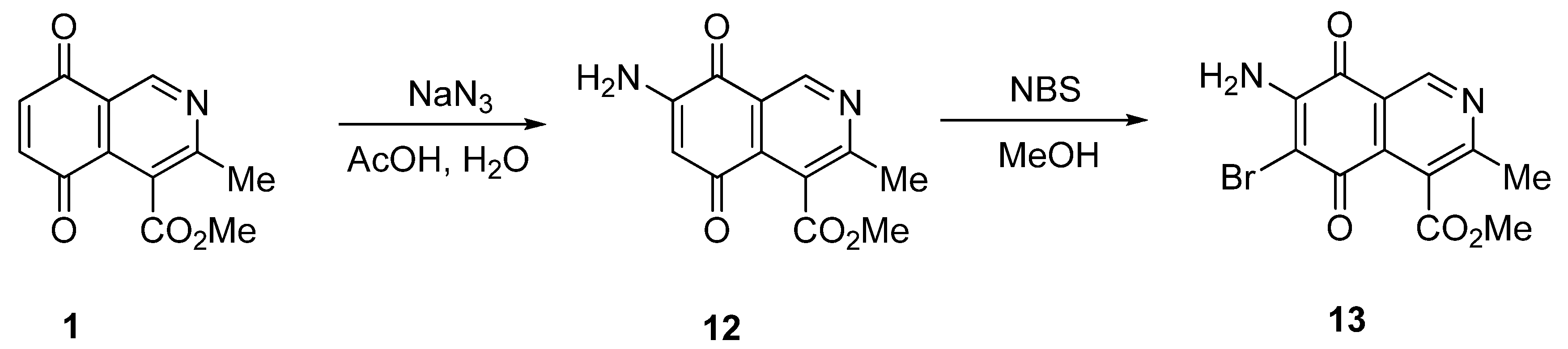
We also explored the preparation of amino- and aminobromoisoquinolinequinones in order to include them in the biological assays. Treatment of quinone
1 with sodium azide in acetic acid, according to a previously reported procedure [
15], afforded aminoisoquinolinequinone
12 as the sole regioisomer in 51% yield (
Scheme 3). The structure of compound
12 was established by means of HMBC experiments. Further bromination of
12 with
N-bromosuccinimide, provides aminobromoisoquinolinequinone
13 in 86% yield.
Scheme 3.
Preparation of amino- and aminobromoisoquinolinequinones 12 and 13.
Scheme 3.
Preparation of amino- and aminobromoisoquinolinequinones 12 and 13.
Isoquinolinequinones
1,
2a,
3a,
4a,
4b,
5a,
6–13 were evaluated for their
in vitro cytotoxic activity against MRC-5 (healthy lung fibroblasts) and four human cancer cell lines: AGS (gastric), SK-MES-1 (lung), J82 (bladder), and HL-60 (leukemia), using the conventional microculture tetrazolium reduction assay [
25,
26,
27]. The broad variety of the synthesized compounds was designed in order to gain insight into the influence of nitrogen and halogen groups on the quinone nucleus of the isoquinolinequinone pharmacophore on the biological activity.
Table 2 summarizes the data from these evaluations. According to the IC
50 values collected in
Table 2, it is evident that insertion of the nitrogen substituents in quinone
1, as in
2a,
3a,
4a,
4b,
5a and
5b, increases the cytotoxic activity in all the evaluated cell lines, compared to those of precursor
1. The initial structure-activity relationship (SAR) was focused on the nature and location of alkylamino at the quinone nucleus of the isoquinolinequinone pharmacophore. The data of
Table 2 for compounds
2a,
3a,
4a and
5a reveal that the shape and polarity of the alkylamino group at the 7-position in the isoquinolinequinone moiety does not significantly influence the biological activity. Concerning the effect of the position of the alkylamino groups, the IC
50 values for the regioisomers
4a/4b and
5a/5b indicate that substitution of the nitrogen group at the 6-position of the quinone pharmacophore induces a greater effect on the antitumor activity than at the 7-position. It can be seen that the effect of such substitution on the antitumor potency on gastric adenocarcinoma and lung cancer cells reaches values nearly seven times higher compared to regioisomer
4a.
Table 2.
Cytotoxic activity of 1 and their aminoisoquinoline-5,8-quinone derivatives.
Next, the SAR analysis was focused on the effects of insertion of chlorine and bromine atoms at the 6-position of the 7-substituted alkylaminoisoquinolinequinones 2a, 3a, 4a and 5a. Comparison of the IC50 values of halo derivatives 6–11 with those of their corresponding precursors indicates that the substitution of halogen atom at the 6-position enhances the antitumor potency in all the evaluated cell lines, except for compound 11 on lung fibroblasts and gastric cancer cells.
According to the data of
Table 2, aminoisoquinolinequinone
12 showed more cytotoxic activity (0.93–2.16 µM) on the cancer cell lines than the
N-alkyl 7-substituted analogs
2a,
3a,
4a and
5a. It is noteworthy that compound
12 exhibits higher antitumor potency on lung and bladder cell lines than the anticancer drug etoposide.
Comparison of the cytotoxic potency of compound 12 (0.93−2.16 µM) with that of its bromoderivative 13 (IC50: 0.21−0.49 µM) reveals that insertion of the bromine atom at the 6-position of 12 induces a high increase of the cytotoxic activity in all the evaluated cell lines. It is noteworthy that compounds 12 and 13 exhibit higher antitumor potency and selectivity index (SI = IC50 fibroblasts/IC50 cancer cells. 12: 0.8–1.9; 13: 0.5–1.3) in almost all cell lines than the anticancer drug etoposide (SI: 0.1–0.6). Among the tested analogs of the series, compounds 4b, 12 and 13 are the most significant antitumor members. Compound 13 was selected as a promising lead compound due to its high potency on the tested tumor cell lines and selectivity index.
3. Experimental
3.1. General
All reagents were commercially available reagent grade and were used without further purification. Melting points were determined on a Stuart Scientific SMP3 apparatus and are uncorrected. 1H-NMR spectra were recorded on Bruker AM-200 and Avance-400 instruments in deuterochloroform (CDCl3). 13C-NMR spectra were obtained in CDCl3 at 50 and 100 MHz. Bidimensional NMR techniques and DEPT were used for signal assignment. Chemical shifts are expressed in ppm downfield relative to tetramethylsilane and the coupling constants (J) are reported in Hertz. The HRMS spectra were obtained on a Thermo Finnigan spectrometer, model MAT 95XP. Silica gel Merck 60 (70–230 mesh) was used for preparative column chromatography and TLC aluminum foil 60F254 for analytical TLC.
3.2. Chemistry
4-Methoxycarbonyl-3-methylisoquinoline-5,8-quinone (1). A suspension of 2,5-dihydroxybenzaldehyde (1 mmol), methyl 3-aminocrotonate (1 mmol), Ag2O (2 mmol) and MgSO4 (0.5 g) in CH2Cl2 (25 mL) was stirred at rt for 3 h. The mixture was filtered, the solids were washed with CH2Cl2 and the solvent removed under reduced pressure. The residue was column cromatographed over silica gel (90:10 CH2Cl2/AcOEt) to yield pure quinone 1 (86%) as yellow solid, mp 109–111.5 °C; IR νmax 1731 (C=O ester), 1668 (C=O quinone); 1H-NMR (400 MHz, CDCl3): δ 2.65 (s, 3H, Me), 4.03 (s, 3H, CO2Me), 7.04 (s, 2H, 6- and 7-H), 9.22 (s, 1H, 1-H); 13C-NMR (100 MHz): δ 22.69, 53.11, 122.18, 125.15, 133.39, 138.38, 138.58, 148.57, 161.83, 167.68, 183.38, 183.60; HRMS (M+): m/z calcd for C12H9NO4: 231.05315; found: 231.05229.
7-Methylamino-4-methoxycarbonyl-3-methylisoquinoline-5,8-quinone (2a). A suspension of quinone 1 (1 mmol), methylamine hydrochloride (2 mmol) and AcONa (2 mmol) in ethanol (20 mL) was stirred at rt for 1.5 h. The solvent was removed under reduced pressure to give an orange solid containing regioisomers 2a and 2b (74%) in 80:20 proportion. The major regioisomer 2a was isolated by column chromatography over silica gel (90:10 CH2Cl2/AcOEt): red solid (52 %), mp 209.5–211 °C; IR νmax 3279 (N-H), 1740 (C=O ester), 1681 (C=O quinone); 1H-NMR: (400 MHz, DMSO): δ 2.66 (s, 3H, Me), 2.95 (d , 3H, J = 5.4 Hz, NHMe), 4.04 (s, 3H, CO2Me), 5.75 (s, 1H, 6-H), 6.09 (br s, 1H, NH), 9.18 (s, 1H, 1-H); 13C-NMR (100 MHz) δ: 23.02, 29.36, 53.21, 101.16, 122.08, 126.38, 136.38, 148.10, 148.82, 163.02, 168.94, 180.24, 180.55; HRMS (M+): m/z calcd for C13H12N2O4: 260.0797; found: 260.0731.
7-(2-Hydroxyethylamino)-4-methoxycarbonyl-3-methylisoquinoline-5,8-quinone (3a). A suspension of quinone 1 (1 mmol), 2-aminoethanol (2 mmol), CeCl3.7H2O (0.05 mmol) and ethanol (25 mL) was left with stirring at rt for 3 h. The solvent was removed under reduced pressure to give an orange solid containing regioisomers 3a and 3b (65%) in 80:20 proportion. The major regioisomer 3a was isolated by column chromatography over silica gel (90:10 CH2Cl2/AcOEt); red solid (47%), mp 195.5–197 °C; IR νmax 3399 (O-H), 3186(N-H), 1700 (C=O ester), 1600 (C=O quinone); 1H-NMR (400 MHz, DMSO): δ 2.52 (s, 3H, Me), 3.27 (dt, 2H, J = 5.8, 11.7 Hz, NHCH2), 3.59 (dt, 2H, J = 5.6, 11.2 Hz, CH2OH), 3.88 (s, 3H, CO2Me), 4.89 (t, 1H, J = 5.7 Hz, OH), 5.79 (s, 1H, 6-H), 7.75 (t, 1H, J = 5.9 Hz, NH), 9.06 (s, 1H, 1-H); 13C-NMR (100 MHz): δ 22.35, 44.77, 52.63, 58.47, 99.87, 122.43, 125.18, 135.92, 147.36, 149.04, 161.13, 168.25, 178.91, 180.24; HRMS (M+): m/z calcd for C14H14N2O5: 290.0903; found: 290.0891.
6- and 7-(Cyclohexylamino)-4-methoxycarbonyl-3-methylisoquinoline-5,8-quinone (4a,4b). A suspension of quinone 1 (1 mmol), cyclohexylamine (2 mmol), CeCl3·7H2O (0.05 mmol) and ethanol (25 mL) was left with stirring at rt for 2 h. The solvent was removed under reduced pressure to give an orange solid containing regioisomers 4a and 4b in 86:14 ratio. These compounds were isolated by column chromatography over silica gel (90:10 CH2Cl2/AcOEt).
Compound 4a (less polar, 80%): orange solid, mp 161.5–163.5 °C; IR νmax 3,246 (N-H), 2,927 and 2,856 (CH), 1741 (C=O ester), 1685 (C=O quinone); 1H-NMR (400 MHz, CDCl3): δ 1.35 (m, 5H, CH2), 1.68 (m, 1H, CH2), 1.81 (m, 2H, CH2), 2.03 (m, 2H, CH2), 2.65 (s, 3H, Me), 3.30 (m,1H, CH), 4.03 (s, 3H, CO2Me), 5.77 (s, 1H, 6-H), 5.96 (d, 1H, J = 7.5 Hz, NH), 9.17 (s, 1H, 1-H); 13C-NMR (100 MHz): δ 22.99, 24.54 (2C), 25.42, 31.83 (2C), 51.44, 53.16, 101.25, 122.11, 126.23, 136.34, 146.52, 148.03, 162.89, 168.93, 180.13, 180.83; HRMS (M+): m/z calcd for C18H20N2O4: 328.14227; found: 328.14083.
Compound 4b (9.5%): red solid, mp 158.5–160 °C; IR νmax 3246 (N-H); 2927 and 2856 (CH); 1741 (C=O ester); 1685 (C=O quinone); 1H-NMR (400 MHz, CDCl3): δ 1.35 (m, 5H, CH2), 1.68 (m, 1H, CH2), 1.81 (m, 2H, CH2), 2.03 (m, 2H, CH2), 2.65 (s, 3H, Me), 3.30 (m, 1H, CH), 4.05 (s, 3H, CO2Me), 5.75 (d, 1H, J = 7.8 Hz, NH), 5.79 (s, 1H, 6-H), 9.28 (s, 1H, 1-H), 13C-NMR (100 MHz): δ 22.64, 24.54 (2C), 25.47, 31.86 (2C), 51.39, 53.26, 101.23, 123.05, 124.92, 132.44, 146.31, 148.73, 159.91, 168.51, 181.49, 181.71;HRMS (M+): m/z calcd for C18H20N2O4: 328.14227; found: 328.14070.
6- and 7-Morpholino-4-methoxycarbonyl-3-methylisoquinoline-5,8-quinone (5a,5b). A suspension of quinone 1 (1 mmol), morpholine (2 mmol), CeCl3·7H2O (0.05 mmol) and ethanol (25 mL) was left with stirring at rt for 2 h. The solvent was removed under reduced pressure to give an orange solid containing regioisomers 5a and 5b in 87:13 proportion. These compounds were isolated by column chromatography over silica gel (90:10 CH2Cl2/AcOEt).
Compound 5a (less polar, 72%): dark orange solid, mp 169.5–171.5 °C; IR νmax 1722 (C=O ester), 1,696 and 1,638 (C=O quinone); 1H-NMR (400 MHz, CDCl3): δ 2.65 (s, 3H, Me), 3.59 (t, 4H, J = 4.7 Hz, N-CH2), 3.87 (t, 4H, J = 4.7 Hz, O-CH2), 4.03 (s, 3H, CO2Me), 6.01 (s, 1H, 6-H), 9.14 (s, 1H, 1-H); 13C-NMR (100 MHz): δ 22.90, 49.21 (2C), 53.28, 66.49 (2C), 110.92, 123.98, 125.36, 134.86, 148.72, 152.78, 161.98, 168.72, 181.16, 181.75; HRMS (M+): m/z calcd for C16H16N2O5: 316.10593; found: 316.10521.
Compound 5b (17%): dark red solid, mp 159.5–162 °C; IR νmax 1724 (C=O ester), 1,678 and 1,634 (C=O quinone); 1H-NMR (400 MHz, CDCl3): δ 2.66 (s, 3H, Me), 3.47 (t, 4H, J = 4.8, N-CH2), 3.85 (t, 4H, J = 4.8 Hz, O-CH2), 4.03 (s, 3H, CO2Me), 6.01 (s, 1H, 6-H), 9.23 (s, 1H, 1-H); 13C-NMR (100 MHz): δ 22.89, 49.10 (2C), 53.25, 66.44 (2C), 111.30, 122.43, 125.41, 135.43, 148.35, 153.21, 160.76, 168.54, 182.10, 182.30; HRMS (M+): m/z calcd for C16H16N2O5: 316.10593; found: 316.10445.
3.3. General Procedure for the Synthesis of 6-Bromo (or 6-chloro)-7-aminoisoquinolinequinone Derivatives
A solution of the 7-aminoisoquinolinequinone derivative (1 mmol), the corresponding N-bromosuccinimide (NBS) or N-chlorosuccinimide (NCS) (1 mmol) and methanol (15 mL) was left with stirring at rt after completion of the reaction as indicated by TLC. The solvent was removed under reduced pressure and the residue was column cromatographed over silica gel (90:10 CH2Cl2/AcOEt) to yield the corresponding 7-amino-6-haloisoquinolinequinone derivative.
6-Bromo-4-methoxycarbonyl-7-methylamino-3-methylisoquinoline-5,8-quinone (6). Prepared from 2a and NBS (30 min, 80%): dark red solid, mp 170–172 °C; IR νmax 3331 (N-H); 1730 (C=O ester), 1687 (C=O quinone); 1H-NMR (400 MHz, CDCl3): δ 2.65 (s, 3H, Me), 3.48 (d , 3H, J = 5.8 Hz, NHMe), 4.05 (s, 3H, CO2Me), 6.39 (br s, 1H, NH), 9.16 (s, 1H, 1-H); 13C-NMR (100 MHz): δ 23.09, 33.34, 53.39, 121.29, 126.56, 134.72, 146.86, 148.62, 163.23, 168.42, 174.40, 178.84 (the signal of the C-6 was not observed in this experiment); HRMS (M+): m/z calcd for C13H11N2O4Br: 337.99023; found: 337.98951.
6-Chloro-4-methoxycarbonyl-7-methylamino-3-methylisoquinoline-5,8-quinone (7). Prepared from 2a and NCS (3 h, 85%): dark red solid, mp 158–160 °C; IR νmax 3331 (N-H); 1773 (C=O ester), 1693 (C=O quinone); 1H-NMR (400 MHz, CDCl3): δ 2.65 (s, 3H, Me), 3.48 (d , 3H, J = 6.0 Hz, NHMe), 4.05 (s, 3H, CO2Me), 6.30 (br s, 1H, NH), 9.15 (s, 1H, 1-H); 13C-NMR (100 MHz): δ 23.09, 32.82, 53.37, 121.25, 126.52, 135.11, 144.83, 148.50, 163.41, 168.36, 174.83, 179.27 (the signal of the C-6 was not observed in this experiment); HRMS (M+): m/z calcd for C13H11N2O4Cl: 294.0407; found: 294.04002
6-Bromo-2-(hydroxyethylamino)-4-methoxycarbonyl-3-methylisoquinoline-5,8-quinone (8). Prepared from 3a and NBS (30 min, 75%): dark red solid, mp 134–136 °C; IR νmax 3463 (O-H), 3320 (N-H), 1720 (C=O ester), 1685 (C=O quinone); 1H-NMR (400 MHz, DMSO): δ 2.63 (s, 3H, Me), 3.92 (dt, 2H, J = 5.1, 11.2 Hz, CH2OH), 4.06 (s, 3H, CO2Me), 4.11 (dt, 2H, J = 5.3, 10.8 Hz, NHCH2), 6.65 (br s, 1H, NH), 9.13 (s, 1H, 1-H); 13C-NMR (100 MHz): δ 22.98, 47.25, 53.40, 61.36, 121.49, 126.41, 134.60, 146.84, 148.61, 163.02, 168.52, 174.19, 178.66, (the signal of the C-6 was not observed in this experiment); HRMS (M+): m/z calcd for C14H13N2O5Br: 368.00079; found: 368.00005.
6-Chloro-2-(hydroxyethylamino)-4-methoxycarbonyl-3-methylisoquinoline-5,8-quinone (9). Prepared from 3a and NCS (4 h, 78%): dark red solid, mp 123–125 °C; IR νmax 3461 (O-H), 3325 (N-H), 1716 (C=O ester), 1687 (C=O quinone); 1H-NMR (400 MHz, CDCl3): 1H-NMR (400 MHz, CDCl3): δ 2.65 (s, 3H, Me), 3.92 (m, 2H, NHCH2), 4.05 (s, 3H, CO2Me), 4.07 (m, 2H, CH2OH), 6.58 (br s, 1H, NH), 9.16 (s, 1H, 1-H); 13C-NMR (100 MHz): δ 23.09, 4.82, 53.42, 61.71, 121.38, 126.48, 134.99, 144.53, 148.59, 163.35, 168.41, 174.67, 179.22, (the signal of the C-6 was not observed in this experiment); HRMS (M+): m/z calcd for C14H13N2O5Cl: 324.0513; found: 324.05073.
6-Bromo-7-(cyclohexylamino)-4-methoxycarbonyl-3-methylisoquinoline-5,8-quinone (10). Prepared from 4a and NBS (15 min, 84% yield): dark red solid, mp 132.5–134.5 °C; IR νmax 3312 (N-H), 3008 and 2858 (C-H), 1735 (C=O ester), 1687 (C=O quinone); 1H-NMR (400 MHz, CDCl3): δ 1.34 (m, 5H, CH2), 1.68 (m, 1H, CH2), 1.81 (m, 2H, CH2), 2.03 (m, 2H, CH2), 2.64 (s, 3H, Me), 4.05 (s, 3H, CO2Me), 4.58 (m,1H, CH), 6.24 (br s, 1H, NH), 9.15 (s, 1H, 1-H); 13C-NMR (100 MHz): δ 23.05, 24.52 (2C), 25.29, 34.50 (2C), 53.32, 61.58, 121.36, 126.59, 134.80, 148.65, 149.99, 163.16, 168.38, 174.32, 178.90, (the signal of the C-6 was not observed in this experiment); HRMS (M+): m/z calcd for C18H19N2O4Br: 406.05280; found: 406.05196.
6-Bromo-7-morpholino-4-methoxycarbonyl-3-methylisoquinoline-5,8-quinone (11). Prepared from 5a and NBS (30 min, 70%): violet solid, mp 127.5–129 °C; IR νmax 2955 and 2856 (C-H), 1722 (C=O ester), 1696 and 1638 (C=O quinone); 1H-NMR (400 MHz, CDCl3): δ 2.66 (s, 3H, Me), 3.70 (t, 4H, J = 4.5 Hz, N-CH2), 3.90 (t, 4H, J = 4.5 Hz, O-CH2), 4.05 (s, 3H, CO2Me), 9.19 (s, 1H, 1-H); 13C-NMR (100 MHz): δ 22.87, 52.59 (2C), 53.42, 67.51 (2C), 115.51, 122.43, 126.05, 133.51, 149.00, 152.08, 162.25, 168.21, 176.25, 180.24; HRMS (M+): m/z calcd for C16H15N2O5Br: 394.01643; found: 394.01457.
7-Amino-4-methoxycarbonyl-3-methylisoquinoline-5,8-quinone (12). A solution of sodium azide (1.2 mmol) in water (5 mL) was added, with stirring, to a previously heated solution (~40 °C) of quinone 1 (1 mmol) in glacial acetic acid (10 mL). The mixture was stirred at rt for 3.5 h, diluted with water and then extracted with AcOEt (3 × 15 mL). The organic phase was washed with saturated aqueous solution of NaHCO3, dried (MgSO4) and the solvent was removed under reduced pressure. The residue was column cromatographed over silica gel (80:20 CH2Cl2/AcOEt) to yield aminoquinone 12 as an orange solid (51%), mp 189–191 °C; IR νmax 3376 and 3140 (N-H), 1732 (C=O ester), 1691 (C=O quinone); 1H-NMR (400 MHz, DMSO): δ 2.66 (s, 3H, Me), 4.05 (s, 3H, CO2Me), 5.38 (br s, 2H, NH2), 6.03 (s, 1H, 6-H), 9.20 (s, 1H, 1-H); 13C-NMR (100 MHz): δ 23.37, 52.68, 102.44, 122.47, 125.17, 135.92, 147.19, 150.88, 161.00, 168.34, 179.30, 180.65; HRMS (M+): m/z calcd for C12H10N2O4: 246.06406; found: 246.06207.
7-Amino-6-bromo-4-methoxycarbonyl-3-methylisoquinoline-5,8-quinone (13). Prepared from 12 and NBS (20 min, 86%): mp 146–148 °C; IR νmax 3423 and 3326 (N-H), 1726 (C=O ester), 1683 (C=O quinone); 1H-NMR (400 MHz, CDCl3): δ 2.67 (s, 3H, Me), 4.06 (s, 3H, CO2Me), 5.45 (br s, 1H, NH), 6.26 (br s, 1H, NH), 9.22 (s, 1H, 1-H); 13C-NMR (100 MHz): δ 23.18, 53.48, 105.07, 121.08, 126.91, 134.81, 147.20, 148.50, 163.58, 168.26, 174.53, 177.24; HRMS (M+): m/z calcd for C12H9N2O4Br: 323.97455; found: 323.97419.
3.4. Anticancer Assay
The cell lines used in this work were obtained from the American Type Culture Collection (ATCC. Manasas, VA, USA). They included MRC-5 normal human lung fibroblasts (CCL-171), AGS human gastric adenocarcinoma cells (CRL-1739), SK-MES-1 human lung cancer cells (HTB-58) and J82 human bladder carcinoma cells (HTB-1). After the arrival of the cells, they were proliferated in the corresponding culture medium as suggested by the ATCC. The cells were stored in medium containing 10% glycerol in liquid nitrogen. The viability of the cells after thawing was higher than 90%, as assessed by trypan blue exclusion test. Cells were sub-cultured once a week and the medium was changed every two days. Cells were grown in the following media: MRC-5, SKMES-1, and J82 in Eagle's minimal essential medium (EMEM) and AGS cells in Ham F-12. The EMEM medium contained 2 mM L-glutamine, 1 mM sodium pyruvate and 1.5 g/L sodium hydrogen carbonate. Ham F-12 was supplemented with 2 mM L-glutamine and 1.5 g/L sodium hydrogen carbonate. All media were supplemented with 10% heat-inactivated FBS, 100 IU/mL penicillin and 100 μg/mL streptomycin in a humidified incubator with 5% CO2 in air at 37 °C. For the experiments, cells were plated at a density of 50,000 cells/mL in 96-well plates. One day after seeding, the cells were treated with the medium containing the compounds at concentrations ranging from 0 up to 100 μM during 3 days and finally the 3-(4,5-dimethylthiazol-2-yl)-2,5-diphenyltetrazolium bromide (MTT) reduction assay was carried out. The final concentration of MTT was 1 mg/mL. The compounds were dissolved in DMSO (1% final concentration) and complete medium. Untreated cells (medium containing 1% DMSO) were used as controls. Each experiment was carried out in sextuplicate.


