Article

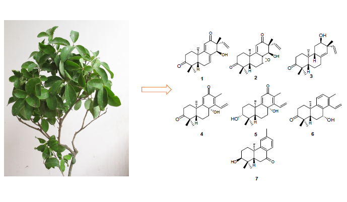
The Diterpenes Ovoideal A–G from Tirpitzia ovoidea

Department of Natural Medicines, School of Pharmaceutical Sciences, Peking University Health Science Center, Xueyuan Road, Beijing 100191, China
*
Author to whom correspondence should be addressed.
Molecules 2014, 19(11), 18966-18979; https://doi.org/10.3390/molecules191118966
Submission received: 23 September 2014 / Revised: 4 November 2014 / Accepted: 5 November 2014 / Published: 18 November 2014
(This article belongs to the Section Medicinal Chemistry)

Abstract

:
Seven new diterpenes, named ovoideal A (1), B (2), C (3), D (4), E (5), F (6) and G (7), have been isolated along with eleven known diterpenes 818 from the petroleum ether soluble fraction of an ethanol extract of the aerial parts of Tirpitzia ovoidea. The structures of the new compounds were elucidated primarily by 1D and 2D NMR spectroscopy, as well as by the HR-ESI-MS spectrometry. All compounds were isolated from the Linaceae family for the first time. The in vitro cytotoxic activity of compounds 1, 35, 818 was evaluated against the Hela, HepG2 and K562 cell lines. Among them, compounds 3, 9, 11, 12, 13, 14, 15, 17, 18 showed moderate inhibitory activities.

Graphical Abstract

1. Introduction

Tirpitzia ovoidea Chun et How ex Sha (Linaceae family) is distributed in Chongzuo, Jingxi, Longzhou, Mashan in Guangxi of China, while Vietnam has some distribution too [1]. The dried aerial parts, containing stems, branches and leaves of this species, is known in China as Baihuachai and has been used as a folk medicine in Guangxi. Baihuachai was used to promoting blood circulation for removing blood stasis, relieving muscle rigidity and activating collaterals and in the treatment of rheumatism, traumatic hemorrhage, injuries from falls, fractures, contusions and strains [1]. The Mulao people use it to treat chronic hepatitis, traumatic infections, fractures, hepatalgia and anepithymia caused by liver cancer [2]. As a folk medicine it has precise therapeutic effects, while there has been no research on its chemical constituents and pharmacological activities. In particular, diterpenoids are thought by many to be the largest pool of chemically diverse and physiologically interesting metabolites [3,4], displaying properties such as anti-leishmania activity [5], cytotocity [6], anti-microbial activity [7], anti-HIV activity [8] and neuro-protection activity [9]. In our research eighteen diterpenes, including seven new compounds (ovoideal A, B, C, D, E, F and G, Figure 1 and Figure 2) were isolated from the petroleum ether soluble fraction of an ethanol extract of the stems, branches and leaves of Tirpitzia ovoidea and their structures were elucidated. All of them were isolated from Linaceae family for the first time. Fourteen of the isolated compounds were evaluated for their cytotoxic activity.
Figure 1. Chemical structures of compounds 118.
Molecules 19 18966 g001
Figure 2. Main 1H-1H COSY and HMBC correlations of compounds 17.
Molecules 19 18966 g002

2. Results and Discussion
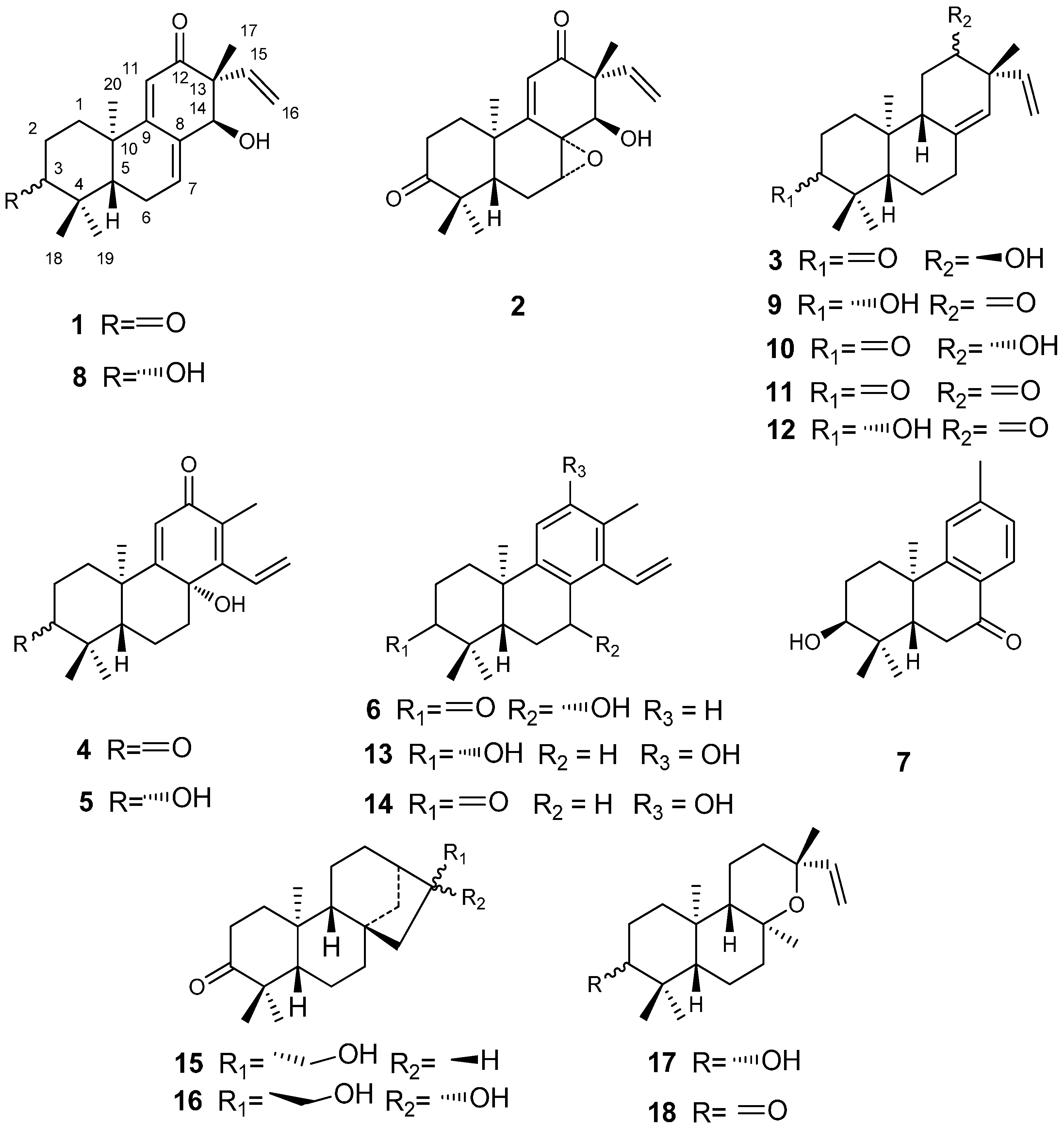
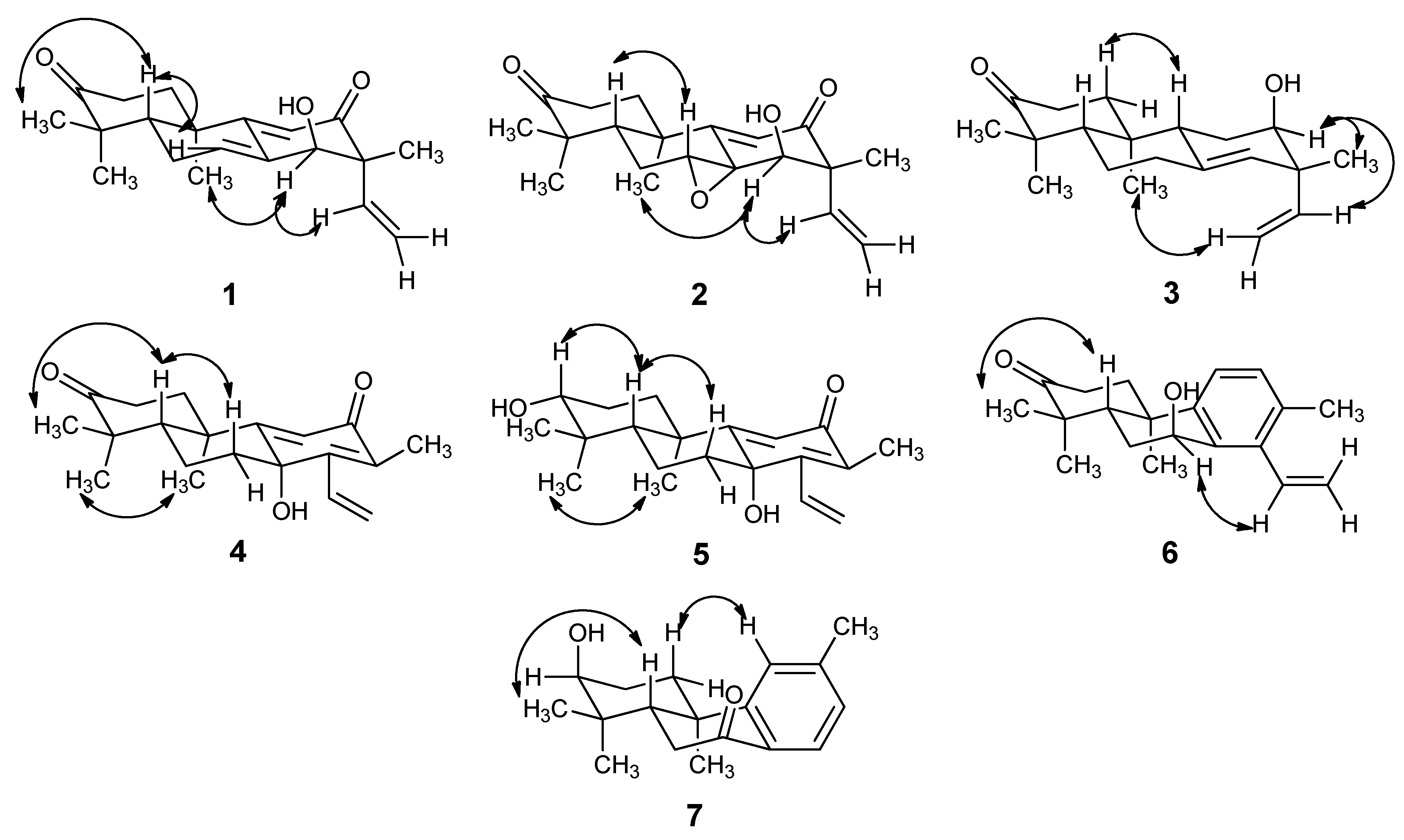
2.1. Chemistry

The petroleum ether soluble fraction of the ethanol extract of the aerial parts of Tirpitzia ovoidea was subjected to repeated column chromatography to afford seven new diterpenes, ovoideals A–G (compounds 17), and 11 known compounds. The structures of the known compounds were determined by comparing their spectroscopic data with literature values [10,11,12,13,14,15,16,17], and they were thus identified as ent-3β,14α-hydroxypimara-7,9(11),15-triene-12-one (8), ent-3β-hydroxypimara-8(14),15-diene-12-one (9), ent-12β-hydroxypimara-8(14),15-diene-3-one (10), ent-pimara-8(14),15-diene-3,12-dione (11), ent-3β,12α-dihydroxypimara-8(14),15-diene (12), spruceanol (13), sonderianol (14), ent-17-hydroxy-16α-kaur-3-one (15), abbeokutone (16), ent-3β-hydroxy-13-epi-manoyl oxide (17), and ent-13-epi-manoyl oxide-3-one (18). Compounds 13 and 14 were evaluated for their cytotoxic activities before [6], and they showed strong inhibition against the Hela, HT29, MCF-7, MM96L and K562 cell lines (their ED50 values were between 2.8 to 18 μg/mL). Compound 17 showed strong inhibition against the Raji, MOLT3 and H9 cell lines [18].
Compound 1 (Figure 1) was obtained as colorless crystals and its molecular formula was determined to be C20H26O3 by HRESIMS, which displayed a quasi-molecular ion peak at m/z 315.1985 ([M+H]+).The 1H-NMR spectrum (Table 1), and HSQC spectroscopic data analyses of 1 indicated the presence of an ABX coupling system at δ 5.81 (1H, dd, J = 17.5, 10.9 Hz, H-15), 5.20 (1H, dd, J = 17.5, 0.8 Hz, H-16a) and 5.14 (1H, dd, J = 10.9, 0.8 Hz, H-16b). It displayed resonances for the C-15, C-16 vinyl moiety [19,20,21], which is characteristic of pimarane diterpenoids [11], indicating that compound 1 was likely to be of this skeletal class. The 1H-NMR spectrum also indicated the presence of oxygenated methine protons at δ 4.29 (s, H-14) and four tertiary methyl protons at δ 1.12 (3H, s, CH3-17), δ 1.20 (3H, s, CH3-18), δ 1.09 (3H, s, CH3-19) and δ 1.32 (3H, s, CH3-20). The 13C-NMR (Table 2) and HSQC spectrum showed 20 carbons, including four methyl, four methylene (one olefinic), five methine (three olefinic, one oxygenated), and seven quaternary carbons (two olefinic, two carbonyl).
The 1H-1H COSY spectrum (Figure 2), in combination with HSQC data, showed cross-peaks at δ 1.84/2.95 (H-1a/H-2b), 2.44/5.75 (H-6/H-7), 5.14/5.81, 5.20/5.81 (H-15/H-16), thus confirming the following structural fragments: -CH2CH2- (C1-C2), -CH2CH- (C6-C7), and -CH=CH2- (C15-C16). When the spectroscopic data of 1 were compared with ent-3β,14α-hydroxypimara-7,9(11),15-triene-12-one [10], the resonances of a hydroxyl group were replaced with a carbonyl group. The most deshielded signal at δC 216.9 belonged to a carbonyl group had the long-range HMBC (Figure 2) couplings of H-2 (216.9/2.95, 216.9/2.32), H-1 (216.9/2.27, 216.9/1.84), H-18 (216.9/1.09) and H-19 (216.9/1.20), indicating that this carbonyl position was assigned at C-3. Cross-peaks in the NOESY (Figure 3) spectrum between H-14 and H-20, H-14 and H-15 at δ 4.29/1.32, 4.29/5.81, revealed that H-14, C20-CH3, and the vinyl group are on the same face of the molecule. This allowed assignment of relative stereochemistry of compound 1. Thus the structure of compound 1 was identified as ent-14α-hydroxypimara-7,9(11),15-triene-3,12-dione and was named ovoideal A.
Figure 3. Main NOESY correlations of compounds 17.
Molecules 19 18966 g003
Compound 2 (Figure 1) was obtained as colorless crystals and its molecular formula was determined to be C20H26O4 by HRESIMS, which displayed a quasi-molecular ion peak at m/z 329.1755 ([M−H]). Compared the 1H-NMR and 13C-NMR (Table 1 and Table 2) spectrum of 2 with 1, we concluded that 2 was shown to be 7,8-epoxide of ent-14-hydroxy-pimara-9(11), 15-diene-3,12-dione. The configuration of the epoxy ring was assigned as α in the 2. The assignment due to H-7 in 2 had the 2D NOESY (Figure 3) correlation with H-5. Furthermore, the skeleton of ent-pimarane had decided the stereochemistry of C-8. Meanwhile, the configuration of the hydroxyl group of C-14 was determined as α-oriented by virtue of the correlation of H-14 and H-15 in NOESY spectrum. We suggest the structure of compound 2 is ent-14α-hydroxy-7α,8α-epoxypimara-9(11),15-diene-3,12-dione, named ovoideal B.
Compound 3 (Figure 1) was obtained as a light yellow gum and its molecular formula was determined to be C20H30O2 by HRESIMS, which displayed a quasi-molecular ion peak at m/z 301.2190 ([M−H]). Comparing the 1H-NMR and the 13C-NMR spectra (Table 1 and Table 2) of 3 with those of the known compound 11, we could note that their A and B rings were the same and they just differed in C ring. It was clear that the carbonyl group at C ring in 3 was reduced into a hydroxyl group. The signal at δC 72.4 belonged to the carbon connected with the hydroxyl group which had long-range HMBC couplings (Figure 2) of H-11 (72.4/1.65), H-14 (72.4/5.09), H-15 (72.4/5.71), H2-16 (72.4/4.98, 4.99) and H-17 (72.4/1.04), indicating that this hydroxyl group was located at C-12. The stereochemistry was determined by the analysis of 2D NOESY (Figure 3) spectrum, which showed correlations between H-12 and H-17, H-12 and H-15, H-16 and H-20, indicating that the hydroxyl group was α-oriented. Consequently we suggest the structure of compound 3 is ent-12α-hydroxypimara-8(14),15-diene-3-one, named ovoideal C.
Compound 4 (Figure 1) was obtained as colorless crystals and its molecular formula was determined to be C20H26O3 by HRESIMS, which displayed a quasi-molecular ion peak at m/z 315.1959 ([M+H]+). 1H-NMR spectrum (Table 1) showed signals at δ 1.94 (3H, s) for an olefinic methyl group, δ 1.09 (3H, s), 1.16 (3H, s), 1.57 (3H, s) for three tertiary methyl protons, δ 6.06 (1H, s) for an olefinic proton. It also gave signals for an ABX system at δ 6.64 (1H, dd, J = 17.9, 11.8 Hz, H-15), 5.57 (1H, dd, J = 17.9, 1.76 Hz, H-16a) and 5.67 (1H, dd, J = 11.9, 1.8 Hz, H-16b), corresponding to a monosubstituted vinyl group attached to an olefinic carbon nucleus. The combination of these groups in a single nucleus suggested this compound was a member of the cleistanthane series [22]. The 13C-NMR spectrum (Table 2) revealed all 20 carbons, which in combination with HSQC measurements were found to be four methyl, five methylene (one olefinic), three methine (two olefinic), and eight quaternary carbons (three olefinic, three oxygenated). 1H-1H COSY (Figure 2) spectrum, in combination with HSQC data, showed cross-peaks at δ 1.97/2.47, 1.97/2.87 (H-1/H-2), 1.52/1.62 (H-5/ H-6), 1.27/1.62, 1.27/2.13 (H-6/H-7), 6.64/5.56, 6.64/5.67 (H-15/H-16), confirming the following structural fragments: -CH2CH2- (C1-C2), -CHCH2CH- (C5-C6-C7), and -CH=CH2- (C15-C16). The most deshielded carbonyl carbon at δC 218.0, has the long-range HMBC correlations with H-1 (2.07, 1.97), H-2 (2.87, 2.47), H-18 (1.09) and H-19 (1.16), thus confirmed the location of this carbonyl group was at C-3. Also, another carbonyl group was located at C-12 from the cross-peak at 189.0/1.94 with H-17. The carbon at δC 71.5, which was connected with a hydroxyl group, was assigned to C-8 because of the cross-peaks with H-6, H-7, H-11, H-15 and H-17. The coupling constant values of H-5 was 2.6 and 12.6, demonstrating that H-5 was axial-oriented [23]. Also cross-peaks in the NOESY (Figure 3) spectrum at 1.16/1.57 (H-19/H-20) revealed that C-20 methyl group was axial-oriented. Therefore we propose that the 8β-hydroxy-cleistanth-9(11),13,15-triene-3,12-dione structure for compound 4, which was named ovoideal D.
Table 1. 1H-NMR Data of Compounds 17.
POSITIONδH Mult (J)
1 a2 b3 b4 a5 b6 b7 b
1a1.84 m1.83 td1.43 m1.97 m1.58 m2.02 m2.09 m
1b2.27 td2.25 m1.89 m2.07 td1.73 m2.47 m2.09 m
2a2.32 m2.42 ddd2.25 dt2.47 ddd1.78 m2.66 dd (6.4, 8.8)1.84 m
2b2.95 m2.74 td2.58 td2.87 ddd1.78 m2.66 dd (6.4, 8.8)2.16 m
3 3.21 dd (5.1, 10.9) 3.57 brs
4
51.88 t (5.4)2. 02 m1.49 m1.52 dd (2.6, 12.6)0.94 dd (2.2, 12.6)2.43 dd (4.8, 10.8)2.34 dd (5.0, 13.1)
6a2.44 m2.02 m1.56 m1.62 m1.66 m1.92 m2.63 m
6b2.44 m2.25 m1.56 m2.13 m1.96 ddd1.94 m2.63 m
7a5.75 s3.80 d (1.6)2.09 m1.27 m1.25 td5.01 t
7b 2.36 dd (14.4, 2.7)2.39 m2.37 dt
8
9 1.89 m
10
116.51 brs6.20 s1.65 m6.06 s6.03 s7.14 d (8.0)7.18 s
12 3.63 brs 7.17 d (8.0)
13 7.10 d (7.9)
144.29 s4.13 brs5.09 s 7.90 d (7.9)
155.81 dd (10.9 17.5)5.95 dd (10.8, 17.2)5.71 dd (10.1, 17.7)6.64 dd (17.9, 11.8)6.56 dd (11.8, 17.9)6.96 dd (11.2, 17.6)
16a5.20 dd (17.5, 0.8)5.31 brd (17.2)4.98 d (10.1)5.56 dd (17.9, 1.76)5.52 dd (1.8, 17.9)5.38 dd (2.0, 17.6)
16b5.14 dd (10.9, 0.8)5.41 brd (10.8)4.99 d (17.7)5.67 dd (11.9, 1.8)5.64 dd (1.8, 11.8)5.67 dd (2.0, 11.6)
171.12 s1.29 s 1.04 s1.94 s1.91 s2.29 s2.39 s
181.09 s1.12 s1.05 s1.09 s0.99 s1.21 s1.03 s
191.20 s1.15 s1.02 s1.16 s0.91 s1.13 s1.02 s
201.32 s1.31 s0.91 s1.57 s1.39 s1.22 s1.25 s
a In MeOD: δ (ppm), J (Hz) 400 MHz for 1H-NMR; b In CDCl3: δ (ppm), J (Hz) 400 MHz for 1H-NMR.
Table 2. 13C-NMR Data of Compounds 17.
POSITIONδC
1 a2 b3 b4 a5 b6 b7 b
136.535.137.137.536.137.530.5
235.334.034.535.327.334.625.5
3216.9213.8216.5218.078.3217.375.2
448.846.837.949.139.737.637.7
550.740.355.255.954.243.242.7
625.522.423.119.717.528.635.6
7118.153.135.040.539.764.9199.1
8134.555.513771.570.9138.5144.8
9164.7162.545.6171.1169.0144.9155.9
1038.637.547.742.041.346.737.6
11134.4127.826.0122.2121.3124.7128.6
12204.5201.172.4189.3187.5130.6124.1
1356.355.243.6130.1129.6133.8127.1
1475.869.5125.4159.1155.8133.9127.5
15140.6138.6146.0134.0132.3135.1
16116.7117.3114.0123.6123.2121.2
1715.916.323.312.512.220.622.1
1825.124.925.626.428.327.027.5
1922.722.822.122.315.521.021.7
2020.121.814.320.220.724.023.3
a In MeOD: δ (ppm) 100 MHz for 13C-NMR; b In CDCl3: δ (ppm) 100 MHz for 13C-NMR.
Compound 5 (Figure 1) was obtained as a white amorphous powder and its molecular formula was determined to be C20H28O3 by HRESIMS, which displayed a quasi-molecular ion peak at m/z 317.2115 ([M+H]+). Comparing the 1H-NMR (Table 1) and 13C-NMR (Table 2) spectra of 5 with those of compound 4, we found that 5 just differed from 4 in the presence of a hydroxyl group instead of a carbonyl group. The carbon connected with the hydroxyl group at δC 78.3, has the long-range HMBC (Figure 2) correlations with H-1, H-2, H-18 and H-19, indicated the location of this hydroxyl group at C-3. In NOESY spectrum there were correlations between H-3 and H-5, so we suggest the structure of compound 5 to be 3β,8β-dihydroxycleistanth-9(11),13,15-triene-12-one, named ovoideal E.
Compound 6 (Figure 1) was obtained as colorless crystals and its molecular formula was determined to be C20H26O2 by HRESIMS, which displayed a quasi-molecular ion peak at m/z 297.1862 ([M−H]). Comparing the 1H-NMR (Table 1) and 13C-NMR (Table 2) spectra of 6 with those of the known compound 14, we noted that 6 differed from 14 only in the location of the hydroxyl group, as the carbon connected with the hydroxyl group at δC 64.9, has long-range HMBC (Figure 2) correlations with H-5 and H-6; also the proton of this carbon correlated with C-8 and C-9, so we propose the peri location of this hydroxyl group at C-7. In the NOESY spectrum, there were correlations between H-7 and H-15, suggesting that the hydroxyl group was β-oriented. Considering the stereochemistry of compound 14, we propose that the structure of compound 6 is 7β-hydroxycleistanth-8,11,13,15-tetraene-3-one, named ovoideal F.
Compound 7 (Figure 1) was obtained as colorless crystals and its molecular formula was determined to be C18H24O2 by HRESIMS, which displayed a quasi-molecular ion peak at m/z 273.1855 ([M+H]+). The molecular formula indicated the presence of seven double-bond equivalents in the molecule. The 13C-NMR spectrum (Table 2) further showed that 7 had one ketonic carbonyl carbon conjugated with the aromatic ring (δ 199.1), three tertiary and three quaternary olefinic (of the aromatic ring), four methyl, three methylene, two methane, and two saturated quaternary carbons. The 1H-NMR spectrum (Table 1) showed four singlets of three protons each at δ 1.02 (H-17), 1.03 (H-16), 1.25 (H-18) and 2.39 (H-15). One one-proton singlet, two one-proton doublets were observed at 7.18 (s), 7.10 (dd, J = 7.9) and 7.90 (dd, J = 7.9), attributed to protons at C-11, C-13 and C-14, respectively. These downfield splitting signals for H-11, H-13 and H-14 and their coupling constant values showed that there existed a 1,2,4-trisubstituted benzene ring in the molecule. These data, along with the seven double-bond equivalents, indicated that 7 was a diterpenoid with a tricyclicpodocarpane type of skeleton [24]. The 1H-1H COSY (Figure 2) spectrum, in combination with HSQC data, showed cross-peaks at δ 1.84/2.09 (H-1/H-2), 1.84/3.57 (H-2/H-3), 2.34/2.63 (H-5/H-6), 7.10/7.90 (H-13/H-14), thus confirming the following structural fragments: -CH2CH2 CH- (C1-C2-C3), -CHCH2- (C5-C6), and ‑CH=CH- (C13-C14), so the aromatic methyl substituent was at C-12. The assignment of the location of the carbonyl group was at C-7 by virtue of the HMBC correlations with C-7 and H-14. We also observed cross-peaks at 75.2/1.84, 75.2/1.02, confirming that the hydroxyl group was located at C-3. The H-3 peak was a broad singlet, revealing that the hydroxyl group was axial. On the basis of the general stereochemistry of the compounds isolated from this plant, we propose that the absolute configuration of 7 is 3S,5S,10R, so we suggest the structure of compound 7 to be 3α-hydroxy-12-methyl-podocarpa-8,11,13-triene-7-one, named ovoideal G.

2.2. Cytotoxity Activity

Many diterpenes have strong anti-tumor activities on human cancer cell lines [25,26], so the in vitro cytotoxity activities for all these compounds, except for 2, 6 and 7, against Hela, HepG2 and K562 were evaluated in our study.
Table 3. IC50 values (μM) of compound 3, 9, 1115, 1718 for human cell lines.
Compound aIC50 (μM)
HelaHepG2K562
392.380.2>100
985.0>100>100
1184.2>10086.4
1283.054.766.3
1387.459.091.0
1489.5>10045.1
1592.166.258.6
1791.4>100>100
1895.0>100>100
Taxol b<0.1<0.18.18
5-Fu b64.1233.6982.0
a Compounds not listed in the table were inactive (IC50 > 100 μM) against all cell lines; b Taxol and 5-Fu were used as positive controls.
Clonogenic-type assays for comparing the growth of treated cells showed some inhibitory activity for compounds 3, 9, 1115, 17, 18 in a range of tumor cell lines (Table 3). The anti-cancer activity of compounds 3, 11, 12, 15 and 18 are reported for the first time in this study.

3. Experimental Section

3.1. General

Optical rotations were recorded on a JASCO DIP-140 digital polarimeter (Tokyo, Japan). IR spectra (KBr) were obtained on a Nicolet Nexus 470 FTIR Spectrometer (Madison, WI, USA). NMR data were acquired using Bruker AVANCE DRX 400 spectrometer (Fällanden, Switzerland) at 400 MHz (1H) and 100 MHz (13C), with tetramethylsilane (TMS) as an internal standard, and chemical shifts were indicated in δ values (ppm). HR-ESI-MS spectra were measured on a Waters Xevo G2 Q-TOF mass analyzer (Milford, MA, USA). Column chromatography (CC) was performed with silica gel (100–200 and 200–300 mesh, Qingdao Marine Chemical Factory, Qingdao, China) and Sephadex LH-20 (GE Healthcare Bio-Science AB, Uppsala, Sweden). TLC was conducted on silica gel GF254 plates (10–40 μm; Qingdao Marine Chemical, Inc., Qingdao, China). Spots were observed by UV light as well as by spraying with 5% anisaldehyde-H2SO4-EtOH followed by heating. Semipreparative HPLC was performed using a Thermo Scientific ODS Hypersil column (250 mm × 21.2 mm i.d.) on a Shimadzu HPLC system (Tokyo, Japan) consisting of a LC-20AD pump and a SPD-M20A UV/vis detector. Tumor cells (Hela: CCL-2TM, HepG2: CCL-185TM and K562: HB-8065TM) were purchased from American Type Culture Collection (ATCC, Rockville, MD, USA), were incubated in a NuAir CO2 incubator (Nuair, Tampa, FL, USA) and observed in a CKX41 inverted microscope (Motic, Xiamen, China). Optical density (OD) values were read under a Model 680 microplate reader (BIO-RAD, Hercules, CA, USA). 5-Fluorouracil (5-FU) (128K1409) and taxol (BCBB4101) were purchased from Sigma Inc. (Saint Louis, MO, USA).

3.2. Plant Material

The aerial parts of Tirpitzia ovoidea were collected from Wuming, Guangxi, China in April 2012 and identified by Associate Professor Ming-Ying Shang. A voucher specimen (No. 7349) was deposited in the Herbarium of Pharmacognosy, School of Pharmaceutical Sciences, Peking University Health Science Center.

3.3. Extraction and Isolation

The air-dried and powdered aerial parts of Tirpitzia ovoidea (29 kg) were sequentially extracted with refluxing 95% ethanol (290 L) and 50% ethanol (232 L) for 2 h each. The extract was combined and concentrated under reduced pressure to obtain a crude extract (1,695 g). After suspension in water, the crude extract was extracted with petroleum ether, EtOAc and n-BuOH, successively. The extracts were concentrated under reduced pressure at 40 °C yielding petroleum ether (200 g), ethyl acetate (100 g) and n-butanol (563 g) fractions, respectively. The petroleum ether soluble fraction (190 g) was subjected to silica gel (200–300 mesh) column chromatography eluting with PE-EtOAc (17:8), to generate fractions V1-V6. Fraction V2 (88.1 g) was subjected to column chromatography over polyamide, eluted with H2O-MeOH (9:1→0:1), to give sub-fractions V2-1 to V2-5. Sub-fraction V-2-2 was subjected to silica gel column chromatography and semipreparative HPLC to afford compound 18 (16.42 mg) and compound 11 (43.22 mg).
V3 (7.6 g) was chromatographed on Sephadex LH-20 eluting with n-hexane-CH2Cl2-MeOH (4:5:1), to yield V3-1 to V3-9. Sub-fraction V3-6 was subjected to repeated silica gel column chromatography and then semipreparative HPLC to yield compound 17 (55.50 mg). Sub-fraction V3-7 was subjected to vacuum silica gel column chromatography, eluted with n-hexane-EtOAc (19:1→0:1) to generate sub-fractions V3-7-1 to V3-7-8. Sub-fractions V3-7-5 and V3-7-7 were subjected to preparative thin layer plate chromatography with n-hexane-EtOAc (7:3) as the developing solvent, and then purified by semipreparative HPLC to afford compounds 7 (3.55 mg), 6 (1.02 mg), 3 (61.98 mg) and 10 (3.96 mg). Sub-fraction V3-7-6 was purified over SephadexLH-20, eluting with MeOH to yield compound 9 (68.41 mg). Sub-fraction V3-9 was subjected to vacuum silica gel column chromatography eluting with n-hexane-EtOAc (17:3) to give V3-9-2 and V3-9-5, which were purified by semipreparative HPLC to get compounds 14 (61.29 mg) and 4 (7.08 mg).
V5 was subjected to medium-pressure silica gel column chromatography, eluting with PE-EtOAc (78:22→55:45) to afford V5-1 to V5-9. Sub-fraction V5-5 was subjected to Sephadex LH-20 chromatography eluting with n-hexane-CH2Cl2-MeOH (4:5:1) to generate sub-fractions V5-5-1 to V5-5-9. Sub-fraction V5-5-8 was subjected to silica gel column chromatography eluting with n-hexane-EtOAc (gradient from 80%), then purified over Sephadex LH-20, eluting with MeOH to yield compound 13 (25.47 mg). Sub-fraction V5-7 was subjected to silica gel column chromatography eluting with n-hexane-EtOAc (gradient 3:1→1:1) to afford V5-7-1 to V5-7-5. Sub-fraction V5-7-3 was applied to a vacuum silica gel column, and purified by semipreparative HPLC to afford compound 1 (45.70 mg).
Fraction V4 was subjected to column chromatography over polyamide, eluted with H2O-MeOH (9:1→0:1), to give sub-fractions V4-1 to V4-7. Sub-fractions V4-2, V4-3, V4-4 and V4-5 were chromatographed on a vacuum silica gel column with n-hexane/EtOAc (1:0→95:5), then purified by semipreparative HPLC to afford compounds 2 (2.74 mg), 5 (7.10 mg), 15 (29.59 mg) and 8 (54.30 mg).
V6 was subjected to polyamide column chromatography, eluted with H2O/MeOH (9:1→0:1), to give sub-fractions V6-1 to V6-6. Sub-fraction V6-2 was subjected to medium-pressure silica gel column chromatography, eluting with n-hexane-EtOAc (9:1→1:1), and then purified by semipreparative HPLC to yield compound 16 (14.16 mg). V6-3 was chromatographed on a vacuum silica gel column with n-hexane/EtOAc (4:1→3:2), then purified by semipreparative HPLC to afford compound 12 (17.12 mg).

3.4. Compound Characterization

ent-14α-Hydroxypimara-7,9(11),15-triene-3,12-dione (ovoideal A, 1): Colorless crystals (MeOH); [ α ] D 23 +8.6 (c 0.007, MeOH); UV, λmax(MeOH) 290 nm; IR, νmax 3462, 2973, 2935, 2870, 1709, 1664, 1643, 1583, 1453, 1431, 1386, 1365, 1269, 1221, 1111, 1082, 1011, 988, 918 cm−1; 1H-NMR and 13C-NMR data, see Table 1; HRESIMS positive mode m/z [M+H]+ 315.1985 (calcd 315.1960).
ent-14α-Hydroxy-7α,8α-epoxypimara-9(11),15-diene-3,12-dione (ovoideal B, 2): Colorless crystals (MeOH); UV, λmax (MeOH) 238 nm; 1H-NMR and 13C-NMR data, see Table 1; IR, νmax 3367, 2971, 2936, 1705, 1667, 1605, 1558, 1455, 1387, 1268, 1079, 1048, 921, 880, 827 cm−1; HRESIMS negative mode m/z [M−H] 329.1755 (calcd 329.1753).
ent-12α-Hydroxypimara-8(14),15-diene-3-one (ovoideal C, 3): Light yellow gum (MeOH); UV, λmax (MeOH) 190 nm; IR, νmax 3461, 3080, 2941, 2869, 2107, 1704, 1633, 1535, 1456, 1432, 1385, 1367, 1313, 1196, 1143, 1106, 1054, 997, 973, 917, 891, 846, 731 cm−1; 1H-NMR and 13C-NMR data, see Table 1; HRESIMS negative mode m/z [M−H] 301.2190 (calcd 301.2168).
8β-Hydroxycleistanth-9(11),13,15-triene-3,12-dione (ovoideal D, 4): Colorless crystals (MeOH); [α]23D −10.0 (c 0.001, MeOH); UV, λmax (MeOH) 200 nm; IR, νmax 3441, 2961, 2922, 2866, 1706, 1649, 1619, 1459, 1432, 1376, 1190, 1040, 1012, 957, 934, 888 cm−1; 1H-NMR and 13C-NMR data, see Table 1; HRESIMS positive mode m/z [M+H]+ 315.1959 (calcd 315.1960).
3β,8β-Dihydroxycleistanth-9(11),13,15-triene-12-one (ovoideal E, 5): Colorless gel (MeOH); UV, λmax (MeOH) 207 nm; IR, νmax 3429, 2943, 2868, 1650, 1616, 1456, 1362, 1191, 1095, 1043, 1003, 929, 888 cm−1; 1H-NMR and 13C-NMR data, see Table 1; HRESIMS positive mode m/z [M+H]+ 317.2115 (calcd 317.2117).
7β-Hydroxycleistanth-8,11,13,15-tetraene-3-one (ovoideal F, 6): Colorless crystals (MeOH); UV, λmax (MeOH) 220 nm; IR, νmax 3369, 2969, 2929, 1703, 1671, 1605, 1553, 1455, 1383, 1263, 1076, 1047, 920, 880, 825 cm−1; 1H-NMR and 13C-NMR data, see Table 1; HRESIMS negative mode m/z [M−H] 297.1862 (calcd 297.1855).
3α-Hydroxy-12-methyl-podocarpa-8,11,13-triene-7-one (ovoideal G, 7): Light yellow crystals (MeOH); UV, λmax (MeOH) 260 nm; IR, νmax 3423, 2963, 2874, 1672, 1604, 1559, 1456, 1411, 1388, 1295, 1267, 1210, 1186, 1137, 1051, 992, 943, 881, 825, 690, 642, 590, 545, 507 cm−1; 1H-NMR and 13C-NMR data, see Table 1; HRESIMS positive m/z [M+H]+ 273.1855 (calcd 273.1855).

3.5. Cytotoxicity Activity

The cytotoxic effects of compounds were estimated against the Hela, HepG2 and K562 cancer cell lines by using the MTT assay method. The cell suspensions were distributed into 96-well cell culture plates and cultured at 37 °C under a 5% CO2 atmosphere in an incubator for 24 h. The compounds were dissolved with limited DMSO and diluted to five different concentrations (0.1, 1, 10, 100, 200 µM) with RPMI 1640 before adding to the corresponding well. After 48 h cultivation, MTT was added to each well for 4 h cultivation. Finally, the supernatant was discarded and limited DMSO was added to the well to dissolve the blue-violet crystal, then the optical density (OD) values were read on the microplate reader at 492 nm. All tests and analyses were carried out in triplicate. DMSO, 5-FU and taxol were applied as the blank control and positive control, respectively.

4. Conclusions

Seven new diterpenes: ent-14α-hydroxypimara-7,9(11),15-triene-3,12-dione, named ovoideal A (1), ent-14α-hydroxy-7α,8α-epoxypimara-9(11),15-diene-3,12-dione, named ovoideal B (2), ent-12α-hydroxypimara-8(14),15-diene-3-one, named ovoideal C (3), 8β-hydroxycleistanth-9(11),13,15-triene-3,12-dione, named ovoideal D (4), 3β,8β-dihydroxycleistanth-9(11),13,15-triene-12-one, named ovoideal E (5), 7β-hydroxycleistanth-8,11,13,15-tetraene-3-one, named ovoideal F (6) and 3α-hydroxy-12-methylpodocarpa-8,11,13-triene-7-one, named ovoideal G (7), were isolated from the petroleum ether soluble fraction of ethanol extract of the aerial parts of Tirpitzia ovoidea along with the eleven known diterpenes 818. The cytotoxity of these compounds was tested against the Hela, HepG2 and K562 cancer cell lines using the MTT assay method, which indicated some compounds showed moderate inhibitory activities.

Supplementary Materials

Supplementary materials can be accessed at: https://www.mdpi.com/1420-3049/19/11/18966/s1.

Supplementary Files

Supplementary File 1

Acknowledgments

This work was financially supported by National Key Technology R&D Program “New Drug Innovation” of China (No. 2013ZX09103002-006, 2013ZX09508104). The authors thank Xu Feng for helpful support during the material collection.

Author Contributions

Ming-Ying Shang, Shao-Qing Cai and Dan Su designed research; Dan Su performed research, analyzed the data and wrote the paper; Feng Xu and Xue-Yan Yang also participated in the research. All authors read and approved the final manuscript.

Conflicts of Interest

The authors declare no conflict of interest.

References

  1. Editorial Committee of Chinese Flora. Flora of China; Science Press: Beijing, China, 1998; Volume 43, p. 96. [Google Scholar]
  2. Editorial Committee of the National assembly of Chinese Herbal Medicine. The Compilation of Chinese Herbal Medicine; People’s Medical Publishing House: Beijing, China, 1975; p. 746. [Google Scholar]
  3. Fujita, E.; Node, M.; Herz, W.; Grisebach, H.; Kirby, G.W.; Tamm, C.E.; Springer-Verlag, V. Diterpenoids of Rabdosia species. Prog. Chem. Org. Nat. Prod. 1984, 46, 77–157. [Google Scholar]
  4. Takeda, Y.; Otsuka, H. Studies in Natural Products Chemistry. Stud. Nat. Prod. Chem. 1995, 15, 111–185. [Google Scholar]
  5. Garcia-Granados, A.; Linan, E.; Martinez, A.; Rivas, F.; Mesa-Valle, C.M.; Castilla-Calvente, J.J.; Osuna, A. In Vitro Action of ent-Manoyl Oxides against Leishmania donovani. J. Nat. Prod. 1997, 60, 13–16. [Google Scholar] [CrossRef]
  6. Maslovskaya, L.A.; Savchenko, A.I.; Gordon, V.A.; Reddell, P.W.; Pierce, C.J.; Parsons, P.G.; Williams, C.M. Isolation and Confirmation of the Proposed Cleistanthol Biogenetic Link from Croton insularis. Org. Lett. 2011, 13, 1032–1035. [Google Scholar] [CrossRef] [PubMed]
  7. Kalpoutzakis, E.; Chinou, I.; Mitaku, S.; Skaltsounis, A.L.; Harvala, C. Antibacterial Labdane-type Diterpenes from the Resin “Ladano” of Cistus creticus subsp. Creticus. Nat. Prod. Lett. 1998, 11, 173–179. [Google Scholar] [CrossRef]
  8. Sutthivaiyakit, S.; Nakorn, N.N.; Kraus, W.; Sutthivaiyakit, P. A novel 29-nor-3,4-seco-friedelane triterpene and a new guaiane sesquiterpene from the roots of Phyllanthus oxyphyllus. Tetrahedron 2003, 59, 9991–9995. [Google Scholar] [CrossRef]
  9. Wang, X.J.; Wang, L.Y.; Fu, Y.; Wu, J.; Tang, X.C.; Zhao, W.M.; Zhang, H.Y. Promising effects on ameliorating mitochondrial function and enhancing Akt signaling in SH-SY5Y cells by (M)-bicelaphanol A, a novel dimeric podocarpane type trinorditerpene isolated from Celastrus orbiculatus. Phytomedicine 2013, 20, 1064–1070. [Google Scholar] [CrossRef] [PubMed]
  10. Denton, R.W.; Harding, W.W.; Anderson, C.I.; Jacobs, H.; McLean, S.; Reynolds, W.F. New Diterpenes from Jatropha Divaricata. J. Nat. Prod. 2001, 64, 829–831. [Google Scholar] [CrossRef] [PubMed]
  11. Sakai, T.; Nakagawa, Y. Diterpenic Stress Metabolites from Cassava Roots. Phytochemistry 1988, 27, 3769–3779. [Google Scholar] [CrossRef]
  12. Heluani, C.S.; Catalan, C.A.N.; Hernandez, L.R.; Burgueno-Tapia, E.; Joseph-Nathan, P. 13C-NMR Assignments and Conformational Evaluation of Diterpenes from Croton sarcopetalus Muell. Magn. Reson. Chem. 1998, 36, 947–950. [Google Scholar] [CrossRef]
  13. Krebs, H.C.; Duddeck, H.; Malik, S.; Beil, W.; Rasoanaivo, P.; Andrianarijaona, M. Chemical Composition and Antitumor Activities from Givotia Madagascariensis. Z. Naturforsch. 2004, 59b, 58–62. [Google Scholar]
  14. Afranio, A.C.; Edilberto, R.S. Two Cleistanthane Type Diterpenes from Croton sonderianus. Phytochemistry 1982, 21, 2571–2574. [Google Scholar] [CrossRef]
  15. Borges-Argaez, R.; Medina-Baizabal, L.; May-Pat, F.; Pena-Rodriguez, L.M. A New ent-kaurane from the Root Extract of Chiococca alba. Can. J. Chem. 1997, 75, 801–804. [Google Scholar] [CrossRef]
  16. Agrawal, P.K.; Bishnoi, V.; Singh, A.K. NMR Chemical Shift Correlations in 16,17-dihydroxy-kauranoids: Implication for Stereochemical Assignments. Phytochemistry 1995, 39, 929–930. [Google Scholar] [CrossRef]
  17. Garcia-Granados, A.; Martinez, A.; Molina, A.; Onorato, M.E.; Rico, M.; Buruaga, A.S.; Buruaga, J.M.S. Diterpenoids from Sideritis varoi Subspecies Cuatrecasasii: 13C-NMR of ent-13-epi-manoyl oxides Functionalized at C-12. Phytochemistry 1985, 24, 1789–l792. [Google Scholar] [CrossRef]
  18. Demetzos, C.; Mitaku, S.; Couladis, M.; Harvala, C.; Kokkonopoulos, D. Natural Metabolites of ent-13-epi-Manoyl Oxide and Other Cytotoxic Diterpenes from the Resin “LADANO” of Cistus creticus. Planta Med. 1994, 60, 590–591. [Google Scholar] [CrossRef] [PubMed]
  19. Wenkert, E.; Beak, P. The Stereochemistry of Rimuene. J. Am. Chem. Soc. 1961, 83, 998–1000. [Google Scholar] [CrossRef]
  20. Borkosky, S.; Bardon, A.; Catalan, C.A.N.; Diaz, J.G.; Herz, W. Direrpenes from Verninanthura amplexicaulis. Phytochemistry 1995, 40, 1477–1479. [Google Scholar] [CrossRef]
  21. Teresa, J.D.P.; Barrero, A.F.; Muriel, L.; Feliciano, A.S.; Grande, M. New Natural Diterpene Acids from Juniperus communis. Phytochemistry 1980, 19, 1153–1156. [Google Scholar] [CrossRef]
  22. Gunasekera, S.P.; Cordell, G.A.; Farnsworth, N.R. Potential Anticancer Agents. XIV. Isolation of Spruceanol and Montanin from Cunuria sprucuana (Euphorbiaceae). J. Nat. Prod. 1979, 42, 658–662. [Google Scholar] [CrossRef] [PubMed]
  23. Spencer, T.A.; Smith, R.A.J.; Storm, D.L.; Villarica, R.M. Total Syntheses of (±)-Methyl Vinhaticoate and (+)-Methyl Vouacapenate. J. Am. Chem. Soc. 1971, 93, 4856–4864. [Google Scholar] [CrossRef]
  24. Ara, I.; Siddiqui, B.S.; Faizi, S.; Siddiqui, S. Three New Diterpenoids from the Stem Bark of Azadirachta indica. J. Nat. Prod. 1990, 53, 816–820. [Google Scholar] [CrossRef]
  25. Shi, Y.P.; Jia, Z.J. New progress about chemistry and biological activity of diterpenoids from genus Euphorbia in China. Chem. J. Chin. Univ. 1997, 7, 1107–1112. [Google Scholar]
  26. Shi, H.M.; Min, Z.D.; Tu, P.F.; Li, X.B. Chemistry and biological activity of diterpenoids from genus Euphorbia in China. Prog. Chem. 2008, 20, 375–387. [Google Scholar]
  • Sample Availability: Samples of the compounds 118 are available from the authors.

