Article

Identification of Catechol as a New Marker for Detecting Propolis Adulteration

1
College of Animal Sciences, Zhejiang University, Hangzhou 310058, China
2
Faculty of Pharmacy, University of Sydney, Sydney, NSW 2006, Australia
*
Author to whom correspondence should be addressed.
Molecules 2014, 19(7), 10208-10217; https://doi.org/10.3390/molecules190710208
Submission received: 18 June 2014 / Revised: 2 July 2014 / Accepted: 10 July 2014 / Published: 14 July 2014
(This article belongs to the Special Issue Natural Antioxidants and Ageing)

Abstract

:
Adulteration of propolis with poplar extract is a serious issue in the bee products market. The aim of this study was to identify marker compounds in adulterated propolis, and examine the transformation of chemical components from poplar buds to propolis. The chemical profiles of poplar extracts and propolis were compared, and a new marker compound, catechol, was isolated and identified from the extracts of poplar buds. The polyphenol oxidase, catechol oxidase, responsible for catalyzing oxidation of catechol was detected in poplar buds and propolis. The results indicate catechol can be used as a marker to detect propolis adulterated with poplar extract.

Graphical Abstract

1. Introduction

The honeybee is a perennial species that exploits virtually all habitats on Earth, and their evolutionary and existent success are not only because of collecting nectar and pollen for fulfilling their nutritional needs, but also the ability to produce bee products: beeswax, venom, propolis and royal jelly [1,2]. As a case of ‘self-medication’ by the bee colonies, propolis has a role in the social immunity of honeybee, reducing the risk of disease and parasite transmission through the colony [3]. Since the ancient time, propolis has been used as a folk medicine for human health and preventing diseases, and it is gaining wider acceptance in popular medicine all over the world. A great number of studies have focused on the pharmacological and biological properties of propolis, including anti-inflammation [4], anticancer [5], antioxidative [6], immunomodulatory [7], antimicrobial, antibacterial, antiviral, and antifungal effects [7,8].
The materials available to bees for manufacturing propolis are plant buds, and substances actively secreted by plants as well as substances exuded from wounds in plants, but bees have a marked preference for one or a few sticky sources [3,9]. According to different geographical locations, poplar, conifer, birch, pine, alder, willow, palm, Baccharis dracunculifolia and Dalbergia ecastaphyllum are identified as the plant sources of propolis [10,11]. Among a multitude of botanical sources, Populus species are considered as the main plant origin of propolis all over the world, especially in the temperate zone [3]. Most propolis collected from Europe, North America, template Asia, has a similar chemical composition, color and smell as the extract of Populus bud. The yield of propolis is relatively low, and does not meet the demand of the growing market, leading to the adulteration of propolis with the extract of the poplar buds. This counterfeiting behavior has seriously disrupted the propolis industry, raising concerns on the quality, efficacy and safety of fake propolis.
A number of studies have been carried out to compare the chemical compositions of propolis and poplar extract. It has been shown that the flavonoids in these two natural products are very similar. Wu et al. found the differences between propolis and poplar extract are caused by the amounts of long-chain alkyl compounds [12]. Zhang et al. developed a HPLC method to use salicin to screen counterfeit propolis [13]. However, salicin is susceptible to acid and can be hydrolyzed to glucose and saligenin which are not detectable by the method. Therefore a new marker compound for distinguishing propolis from poplar extract is urgently needed.
The original plant resin can be modified by enzymes from honeybee [14], which leads to the differences of the chemical constitutes between the propolis and its botanical sources. For example, β-glucosidase has been purified from ventriculus, honey sac, and hypopharyngeal glands of Apis mellifera [15], and propolis. It hydrolyzes flavonoid mono-glucosides in plant resins during propolis collection and processing [16,17].
Catechol (o-diphenol, Figure 1) occurs naturally in fruits, vegetables and plants, along with polyphenol oxidase (PPO), an enzyme localized on the thylakoids of chloroplasts, in vesicles or other bodies of non-green plastid types [18]. PPO catalyzes two different reactions: hydroxylation of monophenols to o-diphenols and oxidation of p- and o-diphenols to p- and o-quinones [19]. The specific isozyme which works on o-diphenol substrates such as catechol is catechol oxidase (EC 1.10.3.1). Upon mixing polyphenol oxidase with the substrate in exposure to oxygen, the colorless catechol is oxidized to reddish-brown melanoid pigments o-diquinones, derivatives of benzoquinone [20].
In our previous studies, we reported that in addition to salicin, there was an unknown chemical component (shown as peak A in Figure 2) in poplar exact (gum) but not in propolis samples [13]. The aim of this study was to identify this marker compound to detect the adulteration of propolis. In addition, we also studied the presence of polyphenol oxidase, which may be a possible reason catechol is not present in propolis.
Figure 1. The chemical structure of catechol.
Molecules 19 10208 g001
Figure 2. HPLC chromatogrsams of (1) catechol standard and (2–8) different poplar extracts.
Molecules 19 10208 g002

2. Results and Discussion

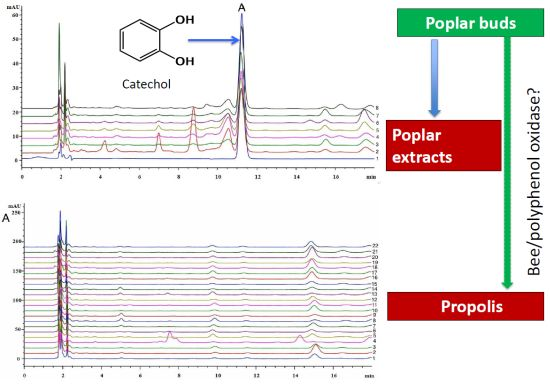
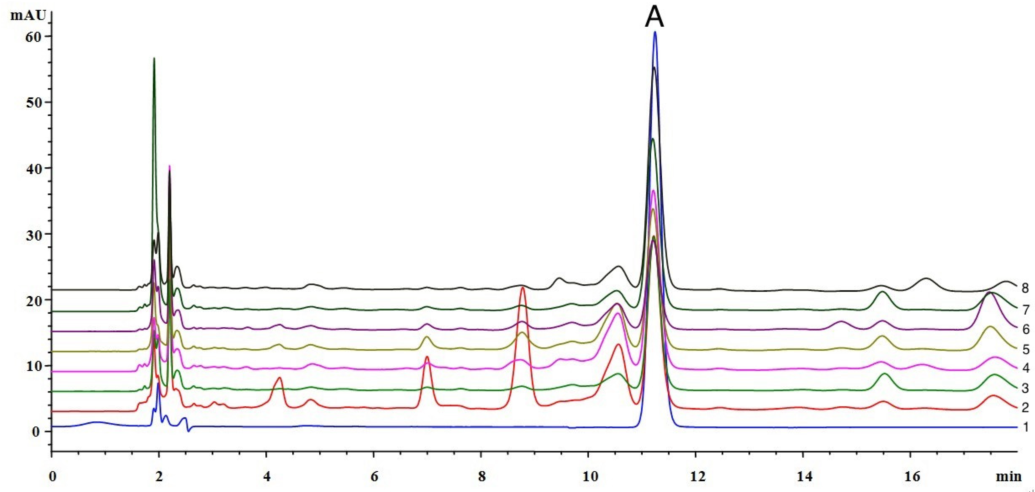
2.1. The Identification of Catechol in Poplar Extract

The unknown compound existing in the poplar extract was isolated as a colorless crystalline solid and had a molecular formula C6H6O2, as determined by the negative mode ESI-MS [m/z 109.1 (M−H)]. According to the 1H-NMR, a typical AA'BB' coupling system signals at δH 6.75 (2H, m) and 6.65 (2H, m) were assigned to an ortho-disubstituded benzene ring. Correspondingly, the 13C-NMR showed three carbon signals at δC 146.3, 120.9, and 116.4, which indicated symmetry in the structure (Table 1). The negative mode ESI-MS, 1H-NMR, 13C-NMR profiles are listed in the Supplementary Information. The chemical data was consistent with an ortho-diphenol structure, catechol (Figure 1). Subsequently, the structure was confirmed by comparing with a catechol standard by HPLC (Figure 2).
Table 1. 1H-NMR and 13C-NMR data of catechol in CD3OD.
Atoms1H-NMR13C-NMR
1 and 2-146.3 (q)
3 and 66.65 (2H, dd, J = 3.8, 7.3 Hz)120.9 (d)
4 and 56.75 (2H, dd, J = 3.8, 7.3 Hz)116.4 (d)
Values are in (δ) ppm. Figures in parenthese are coupling constants (J) in Hz.

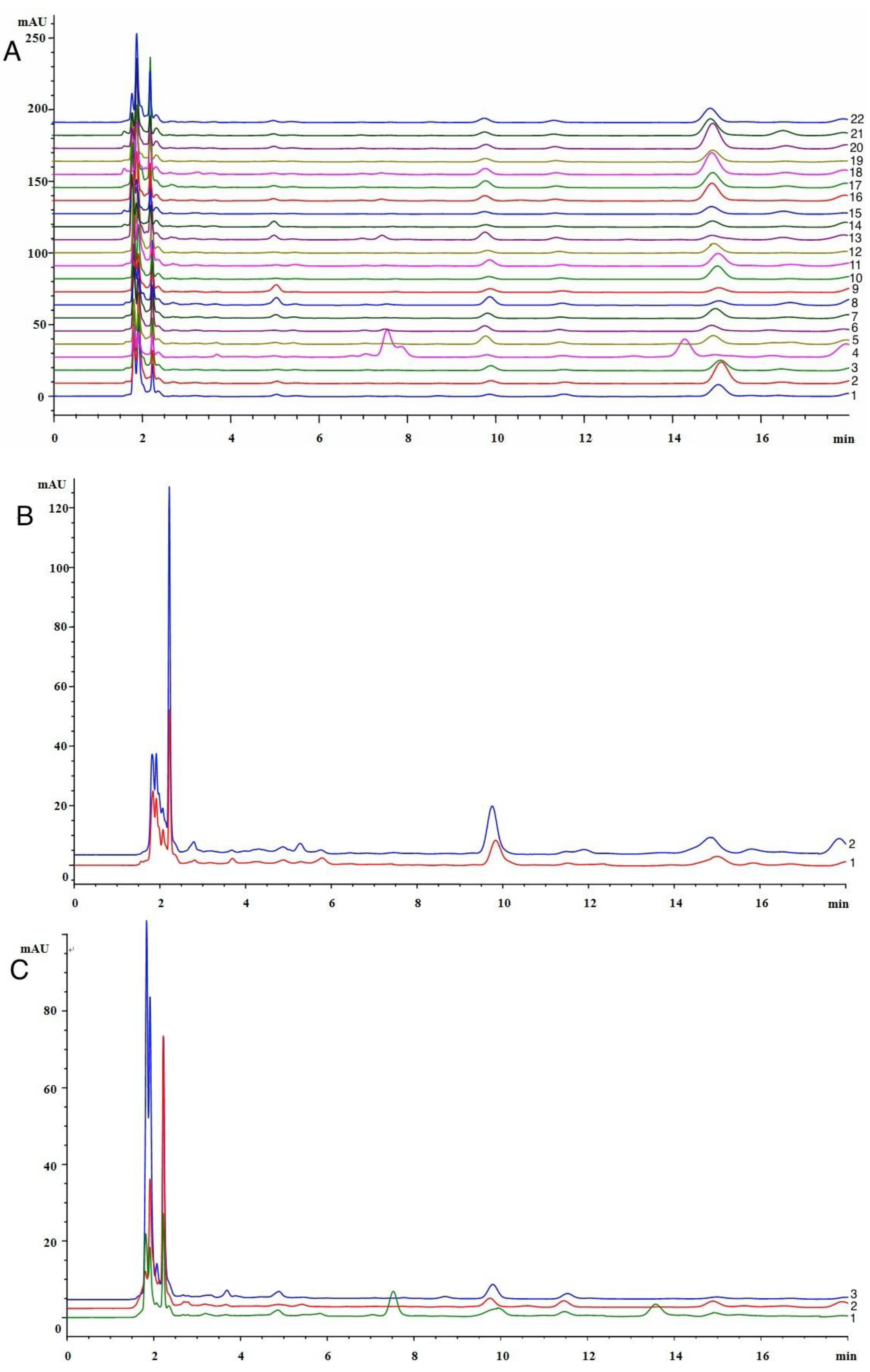
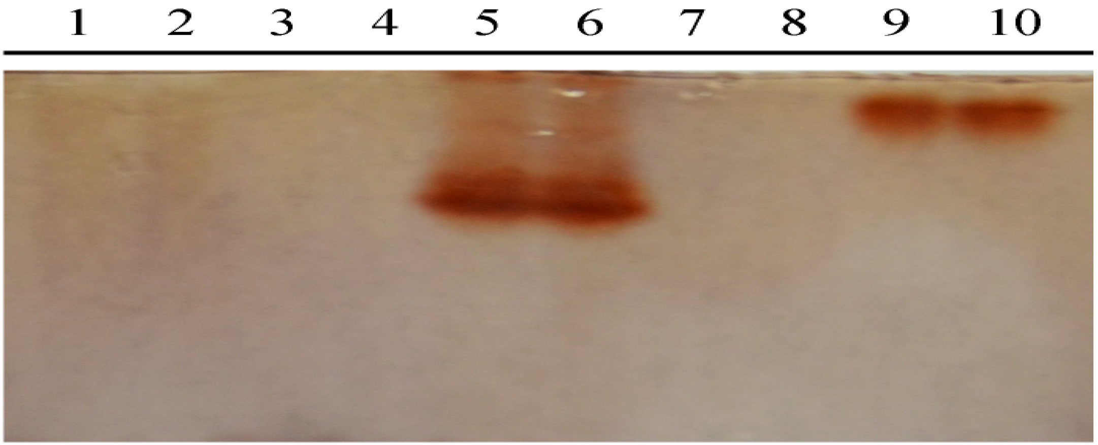
2.2. Determination of Catechol in Poplar Tree Extract and Propolis Samples

Catechol was detected in all poplar extract samples (Figure 2), and its content was relatively high, between 0.052 and 0.132 mg/g. However, catechol was undetectable in 22 Chinese propolis samples collected in different geographical locations and seasons, which were detected by a HPLC method and classed into poplar type propolis in our previous study [21] (Figure 3A). It was not detected in Baccharis dracunculifolia type (Figure 3B) and Eucalyptus type propolis either (Figure 3C). These results indicate that catechol is stable in the process of producing poplar extracts, and chemical analysis. These properties make catechol an ideal marker compound to detect propolis adulterated with poplar extract.
Figure 3. HPLC chromatograms of (A) 22 poplar type propolis; (B) two Baccharis dracunculifolia type proplis; (C) three Eucalyptus type propolis.
Molecules 19 10208 g003

2.3. Detection the Polyphenol Oxidase (PPO) in Propolis and Poplar Buds

We hypothesized catechol from poplar buds is metabolized by enzymes in propolis, we therefore tested the existence of PPO in propolis, together with poplar buds, poplar extract, bee heads and bee bodies. Using catechol as a substrate, the SDS-PAGE gel with polyphenol oxidases turned reddish-brown after staining with the PPO chromogenic reagent. The SDS-PAGE gel (Figure 4), showed brown bands in the lanes of propolis and poplar buds, but not in poplar extracts, bee heads and bee bodies, indicating that PPO existed in the propolis and poplar buds, but not in the poplar extracts, bee heads and bee bodies. However, based on the different band position, the two PPOs in propolis and poplar buds were clearly recognized as isozymes with different molecular mass: the molecular mass of PPO in poplar buds was higher than that in propolis.
Figure 4. SDS-PAGE results of detecting polyphenol oxidase from protein extracts of poplar buds, poplar extracts, propolis, bee heads and bee bodies.
Molecules 19 10208 g004
Notes: Lanes represent: 1–2, bees’ heads; 3–4, bees’ bodies; 5–6, propolis; 7–8, poplar tree gum; 9–10, poplar tree buds.
Polyphenol oxidase was identified from propolis for the first time when it has been purified from black poplar leaves [22] and its activity has been determined in poplar seedlings [23]. The enzyme might have been made inactive when alcohols was added [20], or by heating during the decoction process. Furthermore, the enzyme was not detected in either bee heads or bee bodies, indicating the origin of polyphenol oxidase in propolis is likely its raw material poplar buds, or other plants. However, the PPOs that exist in propolis and poplar buds are isozymes with different molecular masses, as the PPOs protein maybe hydrolyzed by some proteases existing in honeybee bodies [24] and digestive enzymes secreted from honeybee glands such as the salivary [25], or hypopharyngeal gland [26], without changing its activity. There is considerable evidence that PPO exists in a latent form in rudimentary thylakoids of leucoplasts, proplastids, or amyloplasts in healthy green tissues [20]. The vast majority of phenolic compounds in higher plant cells are located in the vacuole—A cellular location isolated from PPO. PPO is apparently not involved in biosynthesis of phenolics, but is involved with the production of o-quinones during pathogen invasion because it normally functions as a phenol oxidase playing a key role in plant defense only in vivo in senescent or damaged cells [18,27]. Honeybees collect plant tissues by beaks and chew them to produce propolis, during the process the cells are destroyed and PPO is activated, so that catechol could be oxidized into quinone in the propolis. In addition, potential natural o-diphenol substrates for PPO enzymes include caffeic acid derivatives and flavonoids, which can be transformed during the propolis collecting and producing process [19]. These results indicate that honeybees play an indispensable role in the process of propolis production, by adding their enzymes to modify the chemical compositions in plants, and also activating the enzymes in plant issues. However, literature about how honeybees transform chemical components of plant to produce propolis is still very limited, and further studies on the involvement of enzymes from plants and honeybees are warrant.

3. Experimental Section

3.1. Chemicals and Reagents

HPLC-grade methanol and acetonitrile were purchased from Merck (Darmstadt, Germany). Ethanol, petroleum ether, chloroform, ethyl acetate, phosphoric acid, acetic acid were analytical grade and purchased from Chemical Reagent Factory of Zhejiang University (Hangzhou, China). Ultra-pure water was purified by a Yjd-upws Ultra-Pure water system (Shanghai, China). Catechol was from Wiley Subscription Services, Inc. (San Francisco, CA, USA). 2% H2O2 and para-phenylenediamine were purchased from Yichen Shiye Corporation (Shanghai, China). Sephadex LH-20 was purchased from the Chemical Faculty of Zhejiang University (Hangzhou, China). All reagents used for HPLC analysis were filtered and degassed prior to use.

3.2. Materials

The buds of Populus × canadensis were collected from Zhejiang, China, in April 2013. Twenty-two poplar type propolis samples were scraped from the frame of the different beehives from different geographical locations of China from May to November 2013. Detailed sample information is presented in Table 2. Three Eucalyptus type propolis samples were obtained from Jims’ Bee Products (Young, Australia). Two Baccharis dracunculifolia type propolis samples were from Fengnaibao Company (Nanjing, China). The honeybees for detecting polyphenol oxidase were sampled from the beekeeping factory of Zhejiang University in September 2013. All samples were kept at −80 °C until analyzed.
Table 2. The geographical origins and collection time of poplar type propolis.
No.Geographical OriginDate of Collection
1Shangshui, HenanAugust 2013
2Qiuxian, HebeiMay 2013
3Shijiazhuang, HebeiJune 2013
4Yicheng, HubeiJune 2013
5Laodongkou, HubeiJune 2013
6Meishan, SichuanJuly 2013
7Wusong, JilinAugust 2013
8Baishan, JilinAugust 2013
9Ji’an, JilinJuly 2013
10Dashiqiao, LiaoningJuly 2013
11Zhuanghe, LiaoningJuly 2013
12Faku, LiaoningJuly 2013
13Kongliu, XinjiangAugust 2013
14Yilan, HeilongjiangMay 2013
15Shuangyashan, HeilongjiangAugust 2013
16Fuyang, AnhuiJune 2013
17Huaibei, AnhuiJune 2013
18Tongcheng, AnhuiAugust 2013
19BeijingJuly 2013
20Penglai, ShandongMay 2013
21Longkou, ShandongJuly 2013
22Dong’e, ShandongAugust 2013

3.3. Extraction and Isolation

Fresh buds of Populus canadensis were lyophilized at freeze dryer (Freeze dryer system 7960032, Labconco, Kansas, KA, USA) for three days before being ground. Around 300 g of the powder was extracted with 500 mL of pure ethanol by an ultrasonic cleaner for 1 h (SK20GT, Shanghai Kedao Ultrasonic Instrument Corporation, Shanghai, China). Then the extracting solution was filtered and concentrated to remove the ethanol in a rotary evaporator (RE-2000A, Shanghai Yarong Biochemistry Instrument Factory, Shanghai, China) at 35 °C until the volume of the extract solution reached less than 100 mL. Ultra-pure water (300 mL) was added to the ethanol extract solution and filtered. The water extract solution was condensed in a rotary evaporator at 35 °C, and then partitioned with petroleum ether, chloroform, and ethyl acetate, successively. The ethyl acetate fraction was subjected to Sephadex LH-20 gel column chromatography with 3,000 mL of ethanol to yield 20 fractions. The 10th fraction was separated by HPLC twice to yield catechol. The HPLC was an Agilent 1200 series (Agilent Technologies, Santa Clara, CA, USA), equipped with a G 1322 A vacuum degasser, a G1311A quaternary pump, a G1314B autosampler, a G1316A thermostatted column compartment, and a Sepax HP-C18 column (150 × 4.6 mm, 5 μm). The solvent was acetonitrile and 0.5% aqueous phosphoric acid (V/V) = 5:95; and the detector wavelength was 213 nm. The eluted fraction at tR = 11.5 min was further purified on HPLC under the same conditions.

3.4. MS and NMR

The identification of the purified catechol was carried out by MS, 1H- and 13C-NMR spectroscopy. HPLC-ESIMS analysis was carried out on an Agilent 6460 Triple Quad LC/MS fitted with an ESI source. The NMR spectra were performed with tetramethylsilane (TMS) as internal standard and CD3OD as solvent using a Bruker AVANCE III 600 MHz NMR spectrometer (Bruker, Stuttgart, Germany) with a standard broadband 5 mm BBFO probe.

3.5. Application of Catechol to Distinguish Poplar Extract and Propolis

Twenty-two poplar type propolis samples, three Eucalyptus type propolis samples, two Baccharis dracunculifolia type propolis, and seven poplar extract samples and the catechol standard were analyzed by the HPLC method described previously [13].

3.6. Polyphenol Oxidase (PPO) Preparation and 1-D SDS-PAGE

Propolis (1 g), poplar buds (1 g), bee heads (20), bee bodies (20) were extracted respectively with Tris buffer (3 mL, 0.1 M, pH 8.3), and ground in a mortar. The crude extract solutions were centrifuged at 12,000 ×g for 20 min at 4 °C, the supernatant were then assayed for PPO. Five μL of crude enzyme extract solution was used per lane for the SDS-PAGE (a 5% stacking gel overlying a 12% resolving gel) with two replicates for each sample. The SDS-PAGE gel was stained in the PPO chromogenic reagent A (2 g para-phenylenediamine in 18 mL acetic acid) and the PPO chromogenic reagent B (60 mL ultra-pure water, 1.5 mL 1% catechol and 0.3 mL 2% H2O2) for 3 min.

4. Conclusions

Catechol is a major marker compound in poplar extract that is used in the adulteration of propolis, and it can be used to detect propolis adulterated with poplar extract. The HPLC analytical process is simple and efficient for application by researchers and industry, providing a new method for authentication of propolis. Our results also suggest that catechol is likely oxidized by polyphenol oxidase, therefore absent in propolis.

Supplementary Materials

Supplementary File 1

Acknowledgments

This work was supported by the grant from the National Natural Science Foundation of China (No. 31272512) and the earmarked fund for Modern Agro-industry Technology Research System from the Ministry of Agriculture of China (CARS-45).

Author Contributions

S.H.: conception, design, data collection, data analysis, and manuscript preparation; C.P.Z: review of the manuscript; G.Q.L.: essential resources and equipment and review of the manuscript; Y.Y.S.: data collection; K.W.: data collection; F.L.H: conception and review of the manuscript.

Conflicts of Interest

The authors declare no conflict of interest.

References

  1. Bankova, V.S. Recent trends and important developments in propolis research. Evid. Based. Complement. Alternat. Med. 2005, 2, 29–32. [Google Scholar] [CrossRef]
  2. Falcão, S.I.; Tomás, A.; Vale, N.; Gomes, P.; Freire, C.; Vilas-Boas, M. Phenolic quantification and botanical origin of Portuguese propolis. Ind. Crop. Prod. 2013, 49, 805–812. [Google Scholar] [CrossRef]
  3. Salatino, A.; Fernandes-Silva, C.C.; Righi, A.A.; Salatino, M.L.F. Propolis research and the chemistry of plant products. Nat. Prod. Rep. 2011, 28, 925–936. [Google Scholar] [CrossRef]
  4. Wang, K.; Ping, S.; Huang, S.; Hu, L.; Xuan, H.Z.; Zhang, C.P.; Hu, F.L. Molecular mechanisms underlying the in vitro anti-inflammatory effects of a Ffavonoid-rich ethanol extract from Chinese propolis (poplar type). Evid. Based. Complement. Alternat. Med. 2013, 2013, 127672:1–127672:11. [Google Scholar]
  5. Banskota, A.H.; Tezuka, Y.; Kadota, S. Recent progress in pharmacological research of propolis. Phytother. Res. 2001, 15, 561–571. [Google Scholar] [CrossRef]
  6. Ahn, M.R.; Kumazawa, S.; Usui, Y.; Nakamura, J.; Matsuka, M.; Zhu, F.; Nakayama, T. Antioxidant activity and constituents of propolis collected in various areas of China. Food Chem. 2007, 101, 1383–1392. [Google Scholar] [CrossRef]
  7. Sforcin, J.M.; Bankova, V. Propolis: Is there a potential for the development of new drugs? J. Ethnopharmacol. 2011, 133, 253–260. [Google Scholar] [CrossRef]
  8. Petelinc, T.; Polak, T.; Demšar, L.; Jamnik, P. Fractionation of phenolic compounds extracted from propolis and their activity in the yeast saccharomyces cerevisiae. PLoS One 2013, 8, e56104. [Google Scholar]
  9. Crane, E. The honeybees’ plant resources. In Beekeeping: Science, Practice and World Recourses; Heinemann: London, UK, 1988. [Google Scholar]
  10. Bankova, V.S.; de Castro, S.L.; Marcucci, M.C. Propolis: Recent advances in chemistry and plant origin. Apidologie 2000, 31, 3–15. [Google Scholar] [CrossRef]
  11. Toreti, V.C.; Sato, H.H.; Pastore, G.M.; Park, Y.K. Recent progress of propolis for its biological and chemical compositions and its botanical origin. Evid. Based. Complement. Alternat. Med. 2013. [Google Scholar] [CrossRef]
  12. Wu, Y.W.; Sun, S.Q.; Zhao, J.; Li, Y.; Zhou, Q. Rapid discrimination of extracts of Chinese propolis and poplar buds by FT-IR and 2D IR correlation spectroscopy. J. Mol. Struct. 2008, 883, 48–54. [Google Scholar]
  13. Zhang, C.P.; Zheng, H.Q.; Liu, G.; Hu, F.L. Development and validation of HPLC method for determination of salicin in poplar buds: Application for screening of counterfeit propolis. Food Chem. 2011, 127, 345–350. [Google Scholar] [CrossRef]
  14. Burdock, G. Review of the biological properties and toxicity of bee propolis (propolis). Food Chem. Toxicol. 1998, 36, 347–363. [Google Scholar] [CrossRef]
  15. Pontoh, J.; Low, N. Purification and characterization of β-glucosidase from honey bees (Apis mellifera). Insect Biochem. Molec. 2002, 32, 679–690. [Google Scholar] [CrossRef]
  16. Zhang, C.P.; Zheng, H.Q.; Hu, F.L. Extraction, partial characterization, and storage stability of β-Glucosidase from Propolis. J. Food Sci. 2011, 76, 75–79. [Google Scholar] [CrossRef]
  17. Zhang, C.P.; Liu, G.; Hu, F.L. Hydrolysis of flavonoid glycosides by propolis beta-glycosidase. Nat. Prod. Res. 2012, 26, 270–273. [Google Scholar] [CrossRef]
  18. Vaughn, K.C.; Lax, A.R.; Duke, S.O. Polyphenol oxidase: The chloroplast oxidase with no established function. Physiol. Plant. 1988, 72, 659–665. [Google Scholar] [CrossRef]
  19. Webb, K.J.; Cookson, A.; Allison, G.; Sullivan, M.L.; Winters, A.L. Gene expression patterns, localization, and substrates of polyphenol oxidase in red clover (Trifolium pratense L.). J. Agric. Food Chem. 2013, 61, 7421–7430. [Google Scholar] [CrossRef]
  20. Vaughn, K.C.; Duke, S.O. Function of polyphenol oxidase in higher plants. Physiol. Plant. 1984, 60, 106–112. [Google Scholar] [CrossRef]
  21. Zhang, C.-P.; Huang, S.; Wei, W.-T.; Ping, S.; Shen, X.-G.; Li, Y.-J.; Hu, F.-L. Development of high-performance liquid chromatographic for quality and authenticity control of chinese propolis. J. Food Sci. 2014, in press. [Google Scholar]
  22. Trémolières, M.; Bieth, J.G. Isolation and characterization of the polyphenoloxidase from senescent leaves of black poplar. Phytochemistry 1984, 23, 501–505. [Google Scholar] [CrossRef]
  23. Tang, F.; Zhao, W.L.; Gao, X.W. Communication between plants: Induced resistance in poplar seedlings following herbivore infestation, mechanical wounding, and volatile treatment of the neighbors. Entomol. Exp. Appl. 2013, 149, 110–117. [Google Scholar]
  24. Matsuoka, T.; Kawashima, T.; Nakamura, T.; Kanamaru, Y.; Yabe, T. Erratum to: Isolation and characterization of proteases that hydrolyze royal jelly proteins from queen bee larvae of the honeybee, Apis mellifera. Apidologie 2014, 45. [Google Scholar] [CrossRef]
  25. Feng, M.; Fang, Y.; Han, B.; Zhang, L.; Lu, X.; Li, J. Novel aspects of understanding molecular working mechanisms of salivary glands of worker honeybees (Apis mellifera) investigated by proteomics and phosphoproteomics. J. Proteomics 2013, 87, 1–15. [Google Scholar] [CrossRef]
  26. Feng, M.; Fang, Y.; Li, J. Proteomic analysis of honeybee worker (Apis mellifera) hypopharyngeal gland development. BMC Genomics 2009, 10. [Google Scholar] [CrossRef]
  27. Cao, C.; Wang, Z.; Yan, S.; Ma, L.; Yang, C. Effects of Lymantria dispar feeding and mechanical wounding on defense-related enzymes in Populus simonii × Populus nigra. Afr. J. Biotechnol. 2011, 10, 7034–7039. [Google Scholar]
  • Sample Availability: Not available.

