Application of Iron Magnetic Nanoparticles in Protein Immobilization

Abstract
:1. Introduction
2. Methods for Preparation of Magnetic Nanoparticles

| Methods | Advantages | Disadvantages | |
|---|---|---|---|
| physical methods | gas-phase deposition | easy to perform | difficult to control the particle size |
| electron beam lithography | well controlled inter-particle spacing | expensive and highly complex machines requiring | |
| wet chemical preparation methods | sol−gel synthesis | precisely controlled in size, aspect ratio, and internal structure | weak bonding, low wear-resistance, high permeability |
| oxidation method | uniform size and narrow size distribution | small-sized ferrite colloids | |
| chemical coprecipitation | simple and efficient | not suitable for the preparation of high pure, accurate stoichiometric phase | |
| hydrothermal reactions | easy to control particle size and shapes | high reaction temperature, high pressure | |
| flow injection synthesis | good reproducibility and high mixing homogeneity together with a precise control of the process | need continuous or segmented mixing of reagents under a laminar flow regime in a capillary reactor | |
| electrochemical method | easy to control particle size | reproducibility | |
| aerosol/vapor phase method | high yields | extremely high temperatures | |
| sonochemical decomposition reactions | narrow particle size distribution | mechanism not still understood | |
| supercritical fluid method | efficient control of the particle size, no organic solvents involved | critical pressure and temperature | |
| synthesis using nanoreactors | the possibility to precisely control the NP size | complex condition | |
| microbial methods | microbial incubation | high yield, good reproducibility, and good scalability, low cost | time-consuming |
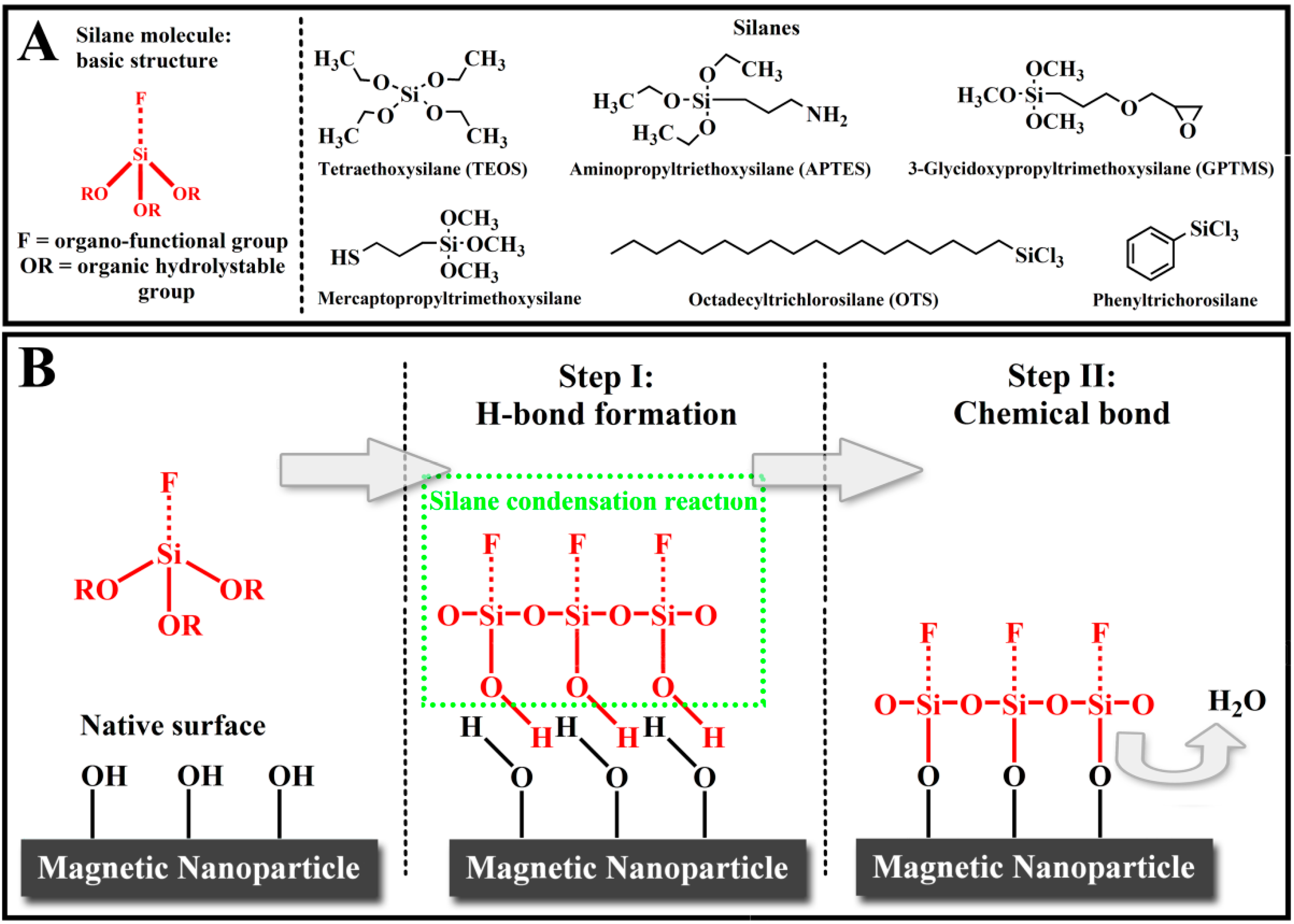
3. Modification of Iron Magnetic Nanoparticles

4. Immobilization Methods
4.1. Physical Immobilization
4.2. Covalent Conjugation
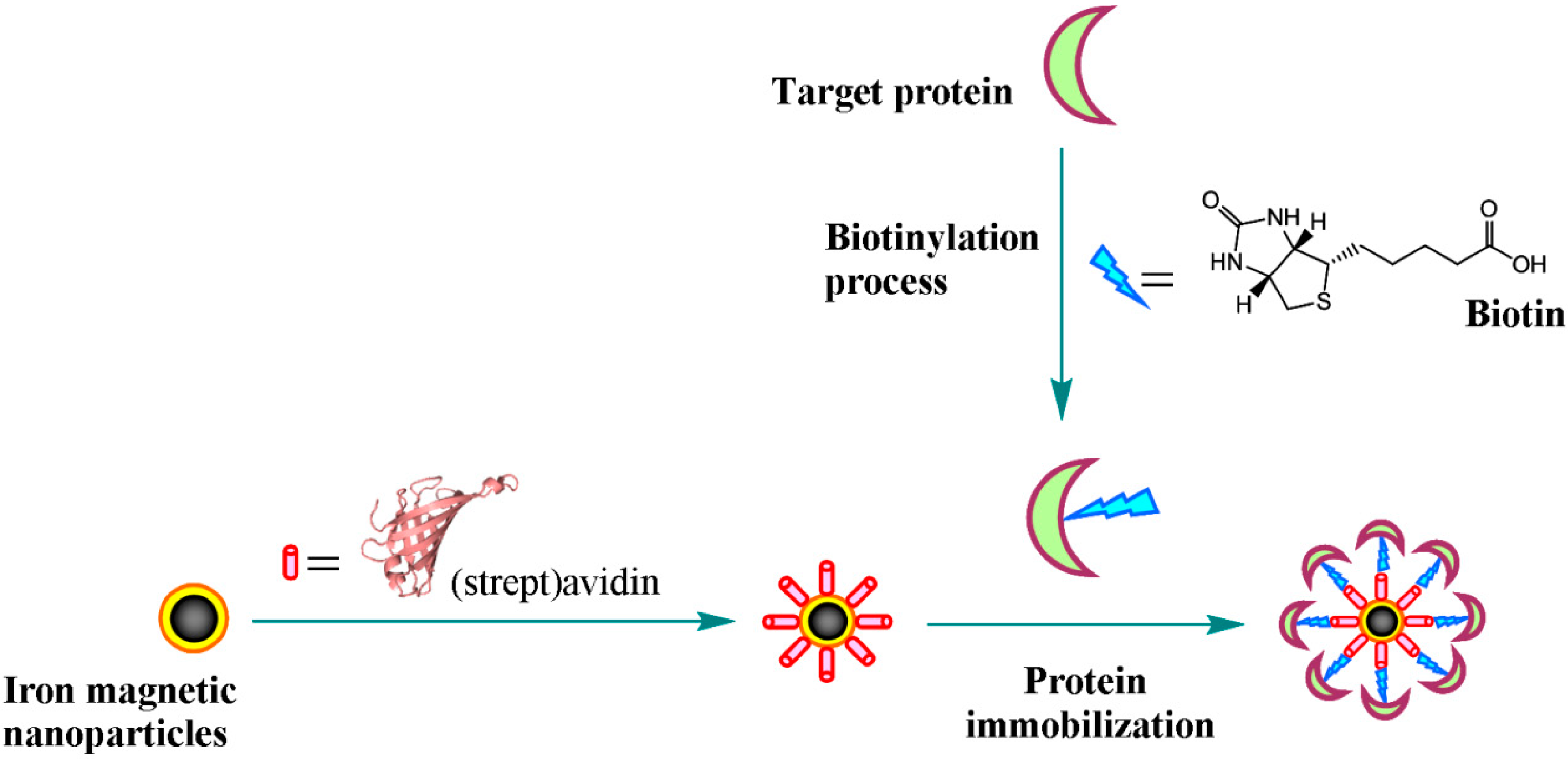
4.3. Biologically Mediated Specific Interaction

5. Aplications of Magnetic Nanoparticles Immobilized Proteins
5.1. Bioseparation
| Protein | Magnetic carrier | Ligand | Elution method | Reference number |
|---|---|---|---|---|
| Lysozyme | Fe3O4 @ PEG @ CM-CTS | -COOH | PBS containing NaCl | [150] |
| Fe3O4 @ SiO2 @ GPS @ Tris | Tris | n/a | [151] | |
| Fe3O4 @ PAA | -COOH | Phosphate buffer containing NaSCN | [152] | |
| Magnetic PHEMA beads @ | Cibacron Blue F3GA | Tris/HCl buffer containing NaCl | [153] | |
| SOD | Fe3O4 @ IDA@Cu2+ | IDA@Cu2+ | Potassium phosphate in the presence of NH4Cl | [154] |
| Lipase | Fe3O4 @ PAA | -COOH | Phosphate buffer (pH 9) | [155] |
| His-tag proteins | Fe3O4 @ PMIDA-Ni2+ | PMIDA-Ni2+ | Sodium phosphate, NaCl and imidazole | [156] |
| Lactoferrin | Fe3O4 @ PGMA-EA @ heparin | Heparin | NaCl | [157] |
| BHb | Fe3O4 @ SiO2 @ GPS @ IDA-Zn2+ | DA-Zn2+ | n/a | [158] |
| Antibody | Fe3O4 @ cellulose @ protein A | Protein A | n/a | [159] |
5.2. Medical Science
5.2.1. Targeted Drug Delivery
5.2.2. Bisensor
5.2.3. Bioimaging
5.3. Food Analysis
6. Concluding Remarks and Prospects
Acknowledgments
Author Contributions
Conflicts of Interest
References
- Kim, J.; Gratea, J.W.; Wang, P. Nanostructures for enzyme stabilization. Chem. Eng. Sci. 2006, 61, 1017–1026. [Google Scholar] [CrossRef]
- Tischer, W.; Wedekind, F. Immobilized Enzymes: Methods and Applications. Top. Curr. Chem. 1999, 200, 95–126. [Google Scholar] [CrossRef]
- Zhang, S.; Gao, S.; Gao, G. Immobilization of β-galactosidase onto magnetic beads. Appl. Biochem. Biotech. 2010, 160, 1386–1393. [Google Scholar] [CrossRef]
- Martin, C.R.; Kohli, P. The emerging field of nanotube biotechnology. Nat. Rev. Drug. Discov. 2003, 2, 29–37. [Google Scholar] [CrossRef]
- Yim, T.J.; Kim, D.Y.; Karajanagi, S.S.; Lu, T.M.; Kane, R.; Dordick, J.S. Silicon nanocolumns as novel nanostructured supports for enzyme immobilization. J. Nanosci. Nanotechnol. 2003, 3, 479–482. [Google Scholar] [CrossRef]
- Romdhane, I.B.B.; Romdhane, Z.B.; Gargouri, A.; Belghith, H. Esterification activity and stability of Talaromyces thermophilus lipase immobilized onto chitosan. J. Mol. Catal. B-Enzym. 2011, 68, 230–239. [Google Scholar] [CrossRef]
- Stepankova, V.; Bidmanova, S.; Koudelakova, T.; Prokop, Z.; Chaloupkova, R.; Damborsky, J. Strategies for stabilization of enzymes in organic solvents. ACS Catal. 2013, 3, 2823–2836. [Google Scholar] [CrossRef]
- Garcia-Galan, C.; Berenguer-Murcia, A.; Fernandez-Lafuente, R.; Rodrigues, R.C. Potential of different enzyme immobilization strategies to improve enzyme performance. Adv. Synth. Catal. 2011, 353, 2885–2904. [Google Scholar] [CrossRef]
- Brady, D.; Jordaan, J. Advances in enzyme immobilization. Biotechnol. Lett. 2009, 31, 1639–1650. [Google Scholar] [CrossRef]
- Iyer, P.V.; Ananthanarayan, L. Enzyme stability and stabilization-Aqueous and non-aqueous environment. Process Biochem. 2008, 43, 1019–1032. [Google Scholar] [CrossRef]
- Sheldon, R.A.; Schoevaart, R.; van Langen, L.M. Cross-Linked Enzyme Aggregates. Methods Biotechnol. 2006, 22, 31–45. [Google Scholar] [CrossRef]
- Mateo, C.; Palomo, J.M.; van Langen, L.M.; van Rantwijk, F.; Sheldon, R.A. A new, mild cross-linking methodology to prepare cross-linked enzyme aggregates. Biotechnol. Bioeng. 2004, 86, 273–276. [Google Scholar] [CrossRef]
- Fernandez-Lafuente, R. Stabilization of multimeric enzymes: Strategies to prevent subunit dissociation. Enzyme Microb. Technol. 2009, 45, 405–418. [Google Scholar] [CrossRef]
- Bezbradica, D.; Mijin, D.; Mihailović, M.; Knežević-Jugović, Z. Microwave-assisted immobilization of lipase from Candida rugosa on Eupergit® supports. J. Chem. Technol. Biot. 2009, 84, 1642–1648. [Google Scholar] [CrossRef]
- Schlossbauer, A.; Schaffert, D.; Kecht, J.; Wagner, E.; Bein, T. Click Chemistry for High-Density Biofunctionalization of Mesoporous Silica. J. Am. Chem. Soc. 2008, 130, 12558–12559. [Google Scholar] [CrossRef]
- Wang, Y.; Caruso, F. Mesoporous Silica Spheres as Supports for Enzyme Immobilization and Encapsulation. Chem. Mater. 2005, 17, 953–961. [Google Scholar] [CrossRef]
- Yáñez-Sedeño, P.; Pingarrón, J.M. Gold nanoparticle-based electrochemical biosensors. Electrochim. Acta 2005, 382. 4, 884–886. [Google Scholar]
- Tang, D.; Su, B.; Tang, J.; Ren, J.; Chen, G. Nanoparticle-Based Sandwich Electrochemical Immunoassay for Carbohydrate Antigen 125 with Signal Enhancement Using Enzyme-Coated Nanometer-Sized Enzyme-Doped Silica Beads. Anal. Chem. 2010, 82, 1527–1534. [Google Scholar] [CrossRef]
- Kim, J.; Kim, B.C.; Lopez-Ferrer, D.; Petritis, K.; Smith, R.D. Nanobiocatalysis for protein digestion in proteomic analysis. Proteomics 2010, 10, 687–699. [Google Scholar] [CrossRef]
- Laurent, S.; Forge, D.; Port, M.; Roch, A.; Robic, C.; Elst, L.V.; Muller, R.N. Magnetic iron oxide nanoparticles: Synthesis, stabilization, vectorization, physicochemical characterizations, and biological applications. Chem. Rev. 2008, 108, 2064–2110. [Google Scholar] [CrossRef]
- Sun, C.; Lee, J.S.H.; Zhang, M. Magnetic nanoparticles in MR imaging and drug delivery. Adv. Drug Delivery Rev. 2008, 60, 1252–1265. [Google Scholar] [CrossRef]
- Xu, P.; Zeng, G.M.; Huang, D.L.; Feng, C.L.; Hu, S.; Zhao, M.H.; Lai, C.; Wei, Z.; Huang, C.; Xie, G.X.; et al. Use of iron oxide nanomaterials in wastewater treatment: A review. Sci. Total Environ. 2012, 424, 1–10. [Google Scholar] [CrossRef]
- Xu, P.; Zeng, G.M.; Huang, D.L.; Lai, C.; Zhao, M.H.; Wei, Z.; Li, N.J.; Huang, C.; Xie, G.X. Adsorption of Pb(II) by iron oxide nanoparticles immobilized Phanerochaete chrysosporium: Equilibrium, kinetic, thermodynamic and mechanisms analysis. Chem. Eng. J. 2012, 203, 423–431. [Google Scholar] [CrossRef]
- Tang, W.W.; Zeng, G.M.; Gong, J.L.; Liang, J.; Xu, P.; Zhang, C.; Huang, B.B. Removal of heavy metals from aqueous solutions using nanomaterials affected by humic/fulvic acid: A review. Sci. Total Environ. 2014, 468–469, 1014–1027. [Google Scholar] [CrossRef]
- Colombié, S.; Gaunand, A.; Lindet, B. Lysozyme inactivation under mechanical stirring: Effect of physical and molecular interfaces. Enzyme Microb. Technol. 2001, 28, 820–826. [Google Scholar] [CrossRef]
- Wu, H.; Fan, Y.; Sheng, J.; Sui, S.F. Induction of changes in the secondary structure of globular proteins by a hydrophobic surface. Eur. Biophys. J. Biophys. 1993, 22, 201–205. [Google Scholar]
- Durán, J.D.G.; Arias, J.L.; Gallardo, V.; Delgado, A.V. Magnetic colloids as drug vehicles. J. Pharm. Sci. 2008, 97, 2948–2983. [Google Scholar] [CrossRef]
- Jain, T.K.; Reddy, M.K.; Morales, M.A.; Leslie-Pelecky, D.L.; Labhasetwar, V. Biodistribution, Clearance, and Biocompatibility of Iron Oxide Magnetic Nanoparticles in Rats. Mol. Pharmaceutics 2008, 5, 316–327. [Google Scholar] [CrossRef]
- Liu, Y.; Jia, S.; Wu, Q.; Ran, J.; Zhang, W.; Wu, S. Studies of Fe3O4-chitosan nanoparticles prepared by co-precipitation under the magnetic field for lipase immobilization. Catal. Comm. 2011, 12, 717–720. [Google Scholar] [CrossRef]
- Gao, J.; Gu, H.; Xu, B. Multifunctional Magnetic Nanoparticles: Design, Synthesis, and Biomedical Applications. Acc. Chem. Res. 2009, 42, 1097–1107. [Google Scholar] [CrossRef]
- Shubayev, V.I.; Pisanic, T.R., 2nd; Jin, S. Magnetic nanoparticles for theragnostics. Adv. Drug Deliv. Rev. 2009, 61, 467–477. [Google Scholar] [CrossRef]
- Zhao, G.X.; Wen, T.; Yang, X.; Yang, S.B.; Liao, J.L.; Hu, J.; Shao, D.D.; Wang, X.K. Preconcentration of U(VI) ions on few-layered graphene oxide nanosheets from aqueous solutions. Dalton Trans. 2012, 41, 6182–6188. [Google Scholar] [CrossRef]
- Zong, P.F.; Wang, S.F.; Zhao, Y.L.; Wang, H.; Pan, H.; He, C.H. Synthesis and application of magnetic graphene/iron oxides composite for the removal of U(VI) from aqueous solutions. Chem. Eng. J. 2013, 220, 45–52. [Google Scholar] [CrossRef]
- Reddy, L.H.; Arias, J.L.; Nicolas, J.; Couvreur, P. Magnetic Nanoparticles: Design and Characterization, Toxicity and Biocompatibility, Pharmaceutical and Biomedical Applications. Chem. Rev. 2012, 112, 5818–5878. [Google Scholar] [CrossRef]
- King, J.G.; Williams, W.; Wilkinson, C.D.W.; McVite, S.; Chapman, J.N. Magnetic properties of magnetite arrays produced by the method of electron beam lithography. Geophys. Res. Lett. 1996, 23, 2847–2850. [Google Scholar] [CrossRef]
- Rishton, A.; Lu, Y.; Altman, R.A.; Marley, A.C.; Bian Hahnes, C.; Viswanathan, R.; Xiao, G.; Gallagher, W.J.; Parkin, S.S.P. Magnetic tunnel junctions fabricated at tenth-micron dimensions by electron beam lithography. Microelectron. Eng. 1997, 35, 249–252. [Google Scholar] [CrossRef]
- Lee, C.S.; Lee, H.; Westervelt, R.M. Microelectromagnets for the Control of Magnetic Nanoparticles. Appl. Phys. Lett. 2001, 79, 3308–3310. [Google Scholar] [CrossRef]
- Mathur, S.; Barth, S.; Werner, U.; Hernandez-Ramirez, F.; Romano-Rodriguez, A. Chemical Vapor Growth of One-dimensional Magnetite Nanostructures. Adv. Mater. 2008, 20, 1550–1554. [Google Scholar] [CrossRef]
- Da Costa, G.M.; de Grave, E.; de Bakker, P.M.A.; Vandenberghe, R.E.J. Synthesis and Characterization of Some Iron Oxides by Sol-Gel Method. Solid State Chem. 1994, 113, 405–412. [Google Scholar] [CrossRef]
- Itoh, H.; Sugimoto, T.J. Systematic control of size, shape, structure, and magnetic properties of uniform magnetite and maghemite particles. J. Colloid Interface Sci. 2003, 265, 283–295. [Google Scholar] [CrossRef]
- Amemiya, Y.; Arakaki, A.; Staniland, S.S.; Tanaka, T.; Matsunaga, T. Controlled formation of magnetite crystal by partial oxidation of ferrous hydroxide in the presence of recombinant magnetotactic bacterial protein Mms6. Biomaterials 2007, 28, 5381–5389. [Google Scholar] [CrossRef]
- Vereda, F.; Rodríguez-González, B.; de Vicente, J.; Hidalgo-Álvarez, R.J. Evidence of direct crystal growth and presence of hollow microspheres in magnetite particles prepared by oxidation of Fe(OH)2. J. Colloid Interface Sci. 2008, 318, 520–524. [Google Scholar] [CrossRef]
- Massart, R. Preparation of aqueous magnetic liquids in alkaline and acidic media. IEEE Trans. Magn. 1981, 17, 1247–1248. [Google Scholar] [CrossRef]
- Estévez, M.; Vargas, S.; Castaño, V.M.; Rodríguez, J.R.; Lobland, H.E.H.; Brostow, W. Novel wear resistant and low toxicity dental obturation materials. Mater. Lett. 2007, 61, 3025–3029. [Google Scholar] [CrossRef]
- Khollam, Y.B.; Dhage, S.R.; Potdar, H.S.; Deshpande, S.B.; Bakare, P.P.; Kulkarni, S.D.; Date, S.K. Microwave hydrothermal preparation of submicron-sized spherical magnetite (Fe3O4) powders. Mater. Lett. 2002, 56, 571–577. [Google Scholar] [CrossRef]
- Chen, F.; Gao, Q.; Hong, G.; Ni, J. Synthesis and characterization of magnetite dodecahedron nanostructure by hydrothermal method. J. Magn. Magn. Mater. 2008, 320, 1775–1780. [Google Scholar] [CrossRef]
- Salazar-Alvarez, G.; Muhammed, M.; Zagorodni, A.A. Novel flow injection synthesis of iron oxide nanoparticles with narrow size distribution. Chem. Eng. Sci. 2006, 61, 4625–4633. [Google Scholar] [CrossRef]
- Cabrera, L.; Gutierrez, S.; Menendes, N.; Morales, M.P.; Herrasti, P. Magnetite nanoparticles: Electrochemical synthesis and characterization. Electrochim. Acta 2008, 53, 3436–3441. [Google Scholar] [CrossRef]
- Marques, R.F.C.; Garcia, C.; Lecante, P.; Ribeiro, J.L.; Noé, L.; Silva, N.J.O.; Amaral, V.S.; Millan, A.; Verelst, M. Electro-precipitation of Fe3O4 nanoparticles in ethanol. J. Magn. Magn. Mater. 2008, 320, 2311–2315. [Google Scholar] [CrossRef]
- González-Carreño, T.; Morales, M.P.; Gracia, M.; Serna, C.J. Preparation of uniform γ-Fe2O3 particles with nanometer size by spray pyrolysis. Mater. Lett. 1993, 18, 151–155. [Google Scholar] [CrossRef]
- Strobel, R.; Pratsinis, S.E. Direct synthesis of maghemite, magnetite and wustite nanoparticles by flame spray pyrolysis. Adv. Powder Technol. 2009, 20, 190–194. [Google Scholar] [CrossRef]
- Enomoto, N.; Akagi, J.; Nakagawa, Z. Sonochemical powder processing of iron hydroxides. Ultrason. Sonochem. 1996, 3, 97–103. [Google Scholar] [CrossRef]
- Dang, F.; Enomoto, N.; Hojo, J.; Enpuku, K. Sonochemical synthesis of monodispersed magnetite nanoparticles by using an ethanol-water mixed solvent. Ultrason. Sonochem. 2009, 16, 649–654. [Google Scholar] [CrossRef]
- Eckert, C.A.; Knutson, B.L.; Debenedetti, P.G. Supercritical fluids as solvents for chemical and materials processing. Nature 1996, 383, 313–318. [Google Scholar] [CrossRef]
- Teng Lam, U.; Mammucari, R.; Suzuki, K.; Foster, N.R. Processing of Iron Oxide Nanoparticles by Supercritical Fluids. Ind. Eng. Chem. Res. 2008, 47, 599–614. [Google Scholar] [CrossRef]
- Breulmann, M.; Colfen, H.; Hentze, H.P.; Antonietti, M.; Walsh, D.; Mann, S. Elastic magnets: Template-controlled mineralization of iron oxide colloids in a sponge-like gel matrix. Adv. Mater. 1998, 10, 237–240. [Google Scholar] [CrossRef]
- Liu, J.F.; Lua, M.F.; Chaia, P.; Fua, L.; Wang, Z.L.; Cao, X.Q.; Meng, J. The magnetic and structural properties of hydrothermal-synthesized single-crystal Sn1−xFexO2 nanograins. J. Magn. Magn. Mater. 2007, 317, 1–7. [Google Scholar] [CrossRef]
- Narayanan, K.B.; Sakthivel, N. Biological synthesis of metal nanoparticles by microbes. Adv. Colloid Interface Sci. 2010, 156, 1–13. [Google Scholar] [CrossRef]
- Moon, J.W.; Roh, Y.; Lauf, R.J.; Vali, H.; Yeary, L.W.; Phelps, T.J. Microbial preparation of metal-substituted magnetite nanoparticles. J. Microbiol. Methods 2007, 70, 150–158. [Google Scholar] [CrossRef]
- Moon, J.W.; Rawn, C.J.; Rondinone, A.J.; Love, L.J.; Roh, Y.; Everett, S.M.; Lauf, R.J.; Phelps, T.J.J. Large-scale production of magnetic nanoparticles using bacterial fermentation. Ind. Microbiol. Biotechnol. 2010, 37, 1023–1031. [Google Scholar] [CrossRef]
- Wu, J.H.; Ko, S.P.; Liu, H.L.; Kim, S.; Ju, J.S.; Kim, Y.K. Sub 5 nm magnetite nanoparticles: Synthesis, microstructure, and magnetic properties. Mater. Lett. 2007, 61, 3124–3129. [Google Scholar] [CrossRef]
- Viota, J.L.; Durán, J.D.G.; González-Caballero, F.; Delgado, A.V. Magnetic properties of extremely bimodal magnetite suspensions. J. Magn. Magn. Mater. 2007, 314, 80–86. [Google Scholar] [CrossRef]
- Mahmoudi, M.; Sant, S.; Wang, B.; Laurent, S.; Sen, T. Superparamagnetic iron oxide nanoparticles (SPIONs): Development, surface modification and applications in chemotherapy. Adv. Drug Deliver. Rev. 2011, 63, 24–46. [Google Scholar] [CrossRef]
- Sahoo, Y.; Pizem, H.; Fried, T.; Golodnitsky, D.; Burstein, L.; Sukenik, C.N.; Markovich, G. Alkyl phosphonate/phosphate coating on magnetite nanoparticles: A comparison with fatty acids. Langmuir 2001, 17, 7907–7911. [Google Scholar] [CrossRef]
- Lee, H.; Lee, E.; Kim, D.K.; Jang, N.K.; Jeong, Y.Y.; Jon, S. Antibiofouling polymer-coated superparamagnetic iron oxide nanoparticles as potential magnetic resonance contrast agents for in vivo cancer imaging. J. Am. Chem. Soc. 2006, 128, 7383–7389. [Google Scholar] [CrossRef]
- Souza, K.C.; Ardisson, J.D.; Sousa, E.M.B. Study of mesoporous silica/magnetite systems in drug controlled release. J. Mater. Sci.-Mater. Med. 2009, 20, 507–512. [Google Scholar] [CrossRef]
- Sun, C.; Veiseh, O.; Gunn, J.; Fang, C.; Hansen, S.; Lee, D.; Sze, R.; Ellenbogen, R.G.; Olson, J.; Zhang, M. In vivo MRI detection of gliomas by chloroto xin-conjugated superparamagnetic nanoprobes. Small 2008, 4, 372–379. [Google Scholar] [CrossRef]
- Mahmoudi, M.; Simchi, A.; Imani, M.; Milani, A.S.; Stroeve, P. In Vivo MRI Detection of Gliomas by Chlorotoxin-Conjugated Superparamagnetic Nanoprobes. J. Phys. Chem. B 2008, 112, 14470–14481. [Google Scholar]
- Liu, H.L.; Ko, S.P.; Wu, J.H.; Jung, M.H.; Min, J.H.; Lee, J.H.; An, B.H.; Kim, Y.K. One-pot polyol synthesis of monosize PVP-coated sub-5 nm Fe3O4 nanoparticles for biomedical applications. J. Magn. Magn. Mater. 2007, 310, 815–817. [Google Scholar] [CrossRef]
- Treccani, L.; Klein, T.Y.; Meder, F.; Pardun, K.; Rezwan, K. Functionalized ceramics for biomedical, biotechnological and environmental application. Acta. Biomater. 2013, 9, 7115–7150. [Google Scholar] [CrossRef]
- Arias, J.L.; López-Viota, M.; Ruiz, M.A.; López-Viota, J.; Delgado, A.V. Development of carbonyl iron/ethylcellulose core/shell nanoparticles for biomedical applications. Int. J. Pharm. 2007, 339, 237–245. [Google Scholar] [CrossRef]
- Donegá, C.M.; Liljeroth, P.; Vanmaekelbergh, D. Physicochemical evaluation of the hot-injection method, a synthesis route for monodisperse nanocrystals. Small 2005, 12, 1152–1162. [Google Scholar]
- Arias, J.L.; Reddy, L.H.; Couvreur, P. Magnetoresponsive squalenoyl gemcitabine composite nanoparticles for cancer active targeting. Langmuir 2008, 24, 7512–7519. [Google Scholar] [CrossRef]
- Di Marco, M.; Sadun, C.; Port, M.; Guilbert, I.; Couvreur, P.; Dubernet, C. Physicochemical characterization of ultrasmall superparamagnetic iron oxide particles (USPIO) for biomedical application as MRI contrast agents. Int. J. Nanomed. 2007, 2, 609–622. [Google Scholar]
- Jung, C.W. Surface properties of superparamagnetic iron oxide MR contrast agents: Ferumoxides, ferumoxtran, ferumoxsil. Magn. Reson. Imaging 1995, 13, 675–691. [Google Scholar] [CrossRef]
- Jung, C.W.; Jacobs, P. Physical and chemical properties of superparamagnetic iron oxide MR contrast agents: Ferumoxides, ferumoxtran, ferumoxsil. Magn. Reson. Imaging 1995, 13, 661–674. [Google Scholar] [CrossRef]
- Yang, H.; Zhang, S.; Chen, X.; Zhuang, Z.; Xu, J.; Wang, X. Magnetite-containing spherical silica nanoparticles for biocatalysis and bioseparations. Anal. Chem. 2004, 76, 1316–1321. [Google Scholar] [CrossRef]
- Ansari, S.A.; Husain, Q. Potential applications of enzymes immobilized on/in nano materials: A review. Biotechnol. Adv. 2012, 30, 512–523. [Google Scholar] [CrossRef]
- Kim, M.I.; Ham, H.O.; S.D., Oh; Park, H.G.; Chang, H.N.; Choi, S.H. Immobilization of Mucor javanicus lipase on effectively functionalized silica nanoparticles. J. Mol. Catal. B-Enzym. 2006, 39, 62–68. [Google Scholar] [CrossRef]
- Verma, M.L.; Barrow, C.J.; Puri, M. Nanobiotechnology as a novel paradigm for enzyme immobilisation and stabilisation with potential applications in biodiesel production. Appl. Microbiol. Biot. 2013, 97, 23–39. [Google Scholar] [CrossRef]
- Hwang, E.T.; Gu, M.B. Enzyme stabilization by nano/microsized hybrid materials. Eng. Life Sci. 2013, 13, 49–61. [Google Scholar] [CrossRef]
- Ozturk, N.; Akgol, S.; Arısoy, M.; Denizli, A. Reversible adsorption of lipase on novel hydrophobic nanospheres. Sep. Purif. Technol. 2007, 58, 83–90. [Google Scholar] [CrossRef]
- Valdes-Solis, T.; Rebolledo, A.F.; Sevilla, M.; Valle-Vigon, P.; Bomati-Miguel, O.; Fuertes, A.B.; Tartaj, P. Preparation, Characterization, and Enzyme Immobilization Capacities of Superparamagnetic Silica/Iron Oxide Nanocomposites with Mesostructured Porosity. Chem. Mater. 2009, 21, 1806–1814. [Google Scholar] [CrossRef]
- Betancor, L.; Luckarift, H.R. Bioinspired enzyme encapsulation for biocatalysis. Trends Biotechnol. 2008, 26, 566–572. [Google Scholar] [CrossRef]
- Mahdizadeh, F.; Karimi, A.; Ranjbarian, L. Immobilization of Glucose Oxidase on Synthesized Superparamagnetic Fe3O4 Nanoparticles; Application for Water Deoxygenation. Int. J. Sci. Eng. Res. 2012, 3, 516–520. [Google Scholar]
- Bahrami, A.; Hejazi, P. Electrostatic immobilization of pectinase on negatively charged AOT-Fe3O4 nanoparticles. J. Mol. Catal. B-Enzym. 2013, 93, 1–7. [Google Scholar] [CrossRef]
- Guisan, J.M. Immobilization of Enzymes and Cells, 2nd ed.; Humana Press Inc.: Madrid, Spain, 2006. [Google Scholar]
- Liang, Y.-Y.; Zhang, L.-M.; Li, W.; Chen, R.-F. Polysaccharide-modified iron oxide nanoparticles as an effective magnetic affinity adsorbent for bovine serum albumin. Colloid Polym. Sci. 2007, 285, 1193–1199. [Google Scholar] [CrossRef]
- Joshi, M.D.; Sidhu, G.; Pot, I.; Brayer, G.D.; Withers, S.G.; McIntosh, L.P. Hydrogen bonding and catalysis: A novel explanation for how a single amino acid substitution can change the pH optimum of a glycosidase. J. Mol. Biol. 2000, 299, 255–279. [Google Scholar] [CrossRef]
- Zhang, Y.; Li, J.; Han, D.; Zhang, H.; Liu, P.; Li, C. An efficient resolution of racemic secondary alcohols on magnetically separable biocatalyst. Biochem. Biophys. Res. Commun. 2008, 365, 609–613. [Google Scholar] [CrossRef]
- Vijayalakshmi, A.; Tarunashree, Y.; Baruwati, B.; Manorama, S.V.; Narayana, B.L.; Johnson, R.E.C.; Raoa, N.M. Enzyme field effect transistor (ENFET) for estimation of triglycerides using magnetic nanoparticles. Biosens. Bioelectron. 2008, 23, 1708–1714. [Google Scholar] [CrossRef]
- Chen, J.-P.; Lin, W.-S. Sol-gel powders and supported sol-gel polymers for immobilization of lipase in ester synthesis. Enzyme Microb. Technol. 2003, 32, 801–811. [Google Scholar] [CrossRef]
- Johnson, A.K.; Zawadzka, A.M.; Deobald, L.A.; Crawford, R.L.; Paszczynski, A.J. Novel method for immobilization of enzymes to magnetic nanoparticles. J. Nanopart. Res. 2008, 10, 1009–1025. [Google Scholar] [CrossRef]
- Mateo, C.; Palomo, J.M.; Fernandez-Lorente, G.; Guisan, J.M.; Fernandez-Lafuente, R. Improvement of enzyme activity, stability and selectivity via immobilization techniques. Enzyme Microb. Technol. 2007, 40, 1451–1463. [Google Scholar] [CrossRef]
- Cowan, D.A.; Fernandez-Lafuente, R. Enhancing the functional properties of thermophilic enzymes by chemical modification and immobilization. Enzyme Microb. Technol. 2011, 49, 326–346. [Google Scholar] [CrossRef]
- Hernandez, K.; Fernandez-Lafuente, R. Control of protein immobilization: Coupling immobilization and site-directed mutagenesis to improve biocatalyst or biosensor performance. Enzyme Microb. Technol. 2011, 48, 107–122. [Google Scholar] [CrossRef]
- Barbosa, O.; Torres, R.; Ortiz, C.; Berenguer-Murcia, A.; Rodrigues, R.C.; Fernandez-Lafuente, R. Heterofunctional supports in enzyme immobilization: From traditional immobilization protocols to opportunities in tuning enzyme properties. Biomacromolecules 2013, 14, 2433–2462. [Google Scholar] [CrossRef] [Green Version]
- Wang, H.; Huang, J.; Wang, C.; Li, D.; Ding, L.; Han, Y. Immobilization of glucose oxidase using CoFe2O4/SiO2 nanoparticles as carrier. Appl. Surf. Sci. 2011, 257, 5739–5745. [Google Scholar]
- Pan, C.; Hu, B.; Li, W.; Sun, Y.; Ye, H.; Zeng, X. Novel and efficient method for immobilization and stabilization of β-d-galactosidase by covalent attachment onto magnetic Fe3O4-chitosan nanoparticles. J. Mol. Catal. B-Enzym. 2009, 61, 208–215. [Google Scholar] [CrossRef]
- Yiğitoğlu, M.; Temoҫin, Z. Immobilization of Candida rugosa lipase on glutaraldehyde-activated polyester fiber and its application for hydrolysis of some vegetable oils. J. Mol. Catal. B-Enzym. 2010, 66, 130–135. [Google Scholar] [CrossRef]
- Kanimozhi, S.; Perinbam, K. Biodesel production from Pseudomonas fluorescens Lp1 lipase immobilized on aminosilane modified super paramagnetic Fe3O4 nanoparticles. J. Phys. Conf. Ser. 2013, 431. [Google Scholar] [CrossRef]
- Xia, T.; Guan, Y.; Yang, M.; Xiong, W.; Wang, N.; Zhao, S.; Guo, C. Synthesis of polyethylenimine modified Fe3O4 nanoparticles with immobilized Cu2+ for highly efficient proteins adsorption. Colloid Surf. A-Physicochem. Eng. Asp. 2014, 443, 552–559. [Google Scholar] [CrossRef]
- Kuo, C.-H.; Liu, Y.-C.; Chang, C.-M.J.; Chen, J.-H.; Chang, C.; Shieh, C.-J. Optimum conditions for lipase immobilization on chitosan-coated Fe3O4 nanoparticles. Carbohydr. Res. 2012, 87, 2538–2545. [Google Scholar]
- Sui, Y.; Cui, Y.; Nie, Y.; Xia, G.-M.; Sun, G.-X.; Han, J.-T. Surface modification of magnetite nanoparticles using gluconic acid and their application in immobilized lipase. Colloid. Surface B 2012, 93, 24–28. [Google Scholar] [CrossRef]
- Lai, B.-H.; Yeh, C.-C.; Chen, D.-H. Surface modification of iron oxide nanoparticles with polyarginine as a highly positively charged magnetic nano-adsorbent for fast and effective recovery of acid proteins. Process Biochem. 2012, 47, 799–805. [Google Scholar] [CrossRef]
- Kouassi, G.K.; Irudayaraj, J.; McCarty, G. Activity of glucose oxidase functionalized onto magnetic nanoparticles. BioMagn. Res. Technol. 2005, 3, 1–10. [Google Scholar] [CrossRef] [Green Version]
- Park, H.J.; McConnell, J.T.; Boddohi, S.; Kipper, M.J.; Johnson, P.A. Synthesis and characterization of enzyme-magnetic nanoparticle complexes: Effect of size on activity and recovery. Colloid. Surface B 2011, 83, 198–203. [Google Scholar] [CrossRef]
- Xu, J.; Ju, C.; Sheng, J.; Wang, F.; Zhang, Q.; Sun, G.; Sun, M. Synthesis and Characterization of Magnetic Nanoparticles and Its Application in Lipase Immobilization. Bull. Korean Chem. Soc. 2013, 34, 2408–2012. [Google Scholar] [CrossRef]
- Yilmaz, E.; Sezgin, M.; Yilmaz, M. Immobilization of Candida rugosa lipase on magnetic sol–gel composite supports for enzymatic resolution of (R,S)-Naproxen methyl ester. J. Mol. Catal. B-Enzym. 2011, 69, 35–41. [Google Scholar] [CrossRef]
- Dong, J.; Kun, Z.; Tang, T.; Ai, S. Enzyme-catalyzed removal of bisphenol A by using horseradish peroxidase immobilized on magnetic silk fibroin microspheres. Res. J. Chem. Environ. 2011, 15, 13–18. [Google Scholar]
- Chalkias, N.G.; Kahawong, P.; Giannelis, E.P. Activity increase of horseradish peroxidase in the presence of magnetic particles. J. Am. Chem. Soc. 2008, 130, 2910–2911. [Google Scholar] [CrossRef]
- Qu, S.; Huang, F.; Chen, G.; Yu, S.; Kong, J. Magnetic assembled electrochemical platform using Fe2O3 filled carbon nanotubes and enzyme. Electrochem. Commun. 2007, 9, 2812–2816. [Google Scholar] [CrossRef]
- Zhuo, Y.; Yuan, P.-X.; Yuan, R.; Chai, Y.-Q.; Hong, C.-L. Bienzyme functionalized three-layer composite magnetic nanoparticles for electrochemical immunosensors. Biomaterials 2009, 30, 2284–2290. [Google Scholar] [CrossRef]
- Eguilaz, M.; Villalonga, R.; Yáñez-Sedeño, P.; Pingarron, J.M. Designing Electrochemical Interfaces with Functionalized Magnetic Nanoparticles and Wrapped Carbon Nanotubes as Platforms for the Construction of High-Performance Bienzyme Biosensors. Anal. Chem. 2011, 83, 7807–7814. [Google Scholar] [CrossRef]
- Illanes, A. Enzyme Biocatalysis: Principles and Applications; Springer Science: Dordrecht, The Netherlands, 2008. [Google Scholar]
- Lei, Z.; Bi, S.; Hu, B.; Yang, H. Combined magnetic and chemical covalent immobilization of pectinase on composites membranes improves stability and activity. Food Chem. 2007, 105, 889–896. [Google Scholar] [CrossRef]
- Bayramoglu, G.; Yılmaz, M.; Şenel, A.U.; Arıca, M.Y. Preparation of nanofibrous polymer grafted magnetic poly(GMA-MMA)-g-MAA beads for immobilization of trypsin via adsorption. Biochem. Eng. J. 2008, 40, 262–274. [Google Scholar] [CrossRef]
- Wang, S.; Bao, H.; Yang, P.; Chen, G. Immobilization of trypsin in polyaniline-coated nano-Fe3O4/carbon nanotube composite for protein digestion. Anal. Chim. Acta 2008, 612, 182–189. [Google Scholar] [CrossRef]
- Li, Y.; Xu, X.; Deng, C.; Yang, P.; Zhang, X. Immobilization of trypsin on superparamagnetic nanoparticles for rapid and effective proteolysis. J. Proteome Res. 2007, 6, 3849–3855. [Google Scholar] [CrossRef]
- Liu, J.; Lin, S.; Qi, D.; Deng, C.; Yang, P.; Zhang, X. On-chip enzymatic microreactor using trypsin-immobilized superparamagnetic nanoparticles for highly efficient proteolysis. J. Chromatogr. A 2007, 1176, 169–177. [Google Scholar] [CrossRef]
- Hong, J.; Gong, P.; Xu, D.; Dong, L.; Yao, S. Stabilization of alpha-chymotrypsin by covalent immobilization on amine-functionalized superparamagnetic nanogel. J. Biotechnol. 2007, 128, 597–605. [Google Scholar] [CrossRef]
- Munro, P.A.; Dunnil, P.; Lilly, M.D. Casein hydrolysis in stirred tank reactors using chymotrypsin immobilized on magnetic supports. Biotechnol. Bioeng. 2004, 23, 677–689. [Google Scholar] [CrossRef]
- Závišová, V.; Koneracká, M.; Tomašovičová, N.; Kopčanský, P.; Timko, M. Some Immobilization Modes of Biologically Active Substances to Fine Magnetic Particles. Z. Phys. Chem. 2006, 220, 241–250. [Google Scholar]
- Xin, B.-J.; Si, S.-F.; Xing, G.-W. Protease immobilization on gamma-Fe2O3/Fe3O4 magnetic nanoparticles for the synthesis of oligopeptides in organic solvents. Chem. Asian J. 2010, 5, 1389–1394. [Google Scholar]
- Chen, J.-P.; Su, D.-R. Latex particles with thermo-flocculation and magnetic properties for immobilization of α-chymotrypsin. Biotechnol. Prog. 2001, 17, 369–375. [Google Scholar] [CrossRef]
- Yu, J.; Wang, C.; Hong, J.; Huang, J. Resolution of (D‚ L)-Phenylalanine in Biphasic System by α-Chymotrypsin Immobilized on Superparamagnetic Nanogels. J. Macromol. Sci. Pure Appl. Chem. 2009, 46, 943–948. [Google Scholar] [CrossRef]
- Korecká, L.; Ježová, J.; Bílková, Z.; Beneš, M.; Horák, D.; Hradcová, O.; Slováková, M.; Viovy, J.-L. Magnetic enzyme reactors for isolation and study of heterogeneous glycoproteins. J. Magn. Magn. Mater. 2005, 293, 349–357. [Google Scholar] [CrossRef]
- Yoshino, T.; Nishimura, T.; Mori, T.; Suzuki, S.; Kambara, H.; Takeyama, H.; Matsunaga, T. Nano-sized bacterial magnetic particles displaying pyruvate phosphate dikinase for pyrosequencing. Biotechnol. Bioeng. 2009, 103, 130–137. [Google Scholar] [CrossRef]
- Garcia, J.; Zhang, Y.; Taylor, H.; Cespedes, O.; Webb, M.E.; Zhou, D. Multilayer enzyme-coupled magnetic nanoparticles as efficient, reusable biocatalysts and biosensors. Nanoscale 2011, 3, 3721–3730. [Google Scholar] [CrossRef]
- Qiu, J.; Peng, H.; Liang, R. Ferrocene-modified Fe3O4@SiO2 magnetic nanoparticles as building blocks for construction of reagentless enzyme-based biosensors. Electrochem. Commun. 2007, 9, 2734–2738. [Google Scholar] [CrossRef]
- Rossi, L.M.; Quach, A.D.; Rosenzweig, Z. Glucose oxidase-magnetite nanoparticle bioconjugate for glucose sensing. Anal. Bioanal. Chem. 2004, 380, 606–613. [Google Scholar] [CrossRef]
- Caruso, F.; Schuler, C. Enzyme Multilayers on Colloid Particles: Assembly, Stability, and Enzymatic Activity. Langmuir 2000, 16, 9595–9603. [Google Scholar] [CrossRef]
- Yang, L.; Xiong, H.; Zhang, X.; Wang, S.; Zhang, X. Direct electrochemistry of glucose oxidase and biosensing for glucose based on boron-doped carbon-coated nickel modified electrode. Biosens. Bioelectron. 2011, 26, 3801–3805. [Google Scholar] [CrossRef]
- Ashtari, K.; Khajeh, K.; Fasihi, J.; Ashtari, P.; Ramazani, A.; Vali, H. Silica-encapsulated magnetic nanoparticles: Enzyme immobilization and cytotoxic study. Int. J. Biol. Macromol. 2012, 50, 1063–1069. [Google Scholar] [CrossRef]
- Sheng, J.; Zhang, L.; Lei, J.; Ju, H. Fabrication of tunable microreactor with enzyme modified magnetic nanoparticles for microfluidic electrochemical detection of glucose. Anal. Chim. Acta 2012, 709, 41–46. [Google Scholar] [CrossRef]
- Jeong, J.; Ha, T.H.; Chung, B.H. Enhanced reusability of hexa-arginine-tagged esterase immobilized on gold-coated magnetic nanoparticles. Anal. Chim. Acta. 2006, 569, 203–209. [Google Scholar] [CrossRef]
- Zeng, L.; Luo, K.; Gong, Y. Preparation and characterization of dendritic composite magnetic particles as a novel enzyme immobilization carrier. J. Mol. Catal. B-Enzym. 2006, 38, 24–30. [Google Scholar] [CrossRef]
- Li, G.Y.; Huang, K.L.; Jiang, Y.R.; Yang, D.L.; Ding, P. Preparation and characterization of Saccharomyces cerevisiae alcohol dehydrogenase immobilized on magnetic nanoparticles. Int. J. Biol. Macromol. 2008, 42, 405–412. [Google Scholar] [CrossRef]
- Kuroiwa, T.; Noguchi, Y.; Nakajima, M.; Sato, S.; Mukataka, S.; Ichikawa, S. Production of chitosan oligosaccharides using chitosanase immobilized on amylose-coated magnetic nanoparticles. Process Biochem. 2008, 43, 62–69. [Google Scholar] [CrossRef]
- Monaghan, R.; Eveleigh, D.; Tewari, R. Chitosanase, a novel enzyme. Nat. New Biol. 1973, 245, 78–80. [Google Scholar] [CrossRef]
- Zhang, D.H.; Yuwen, L.X.; Xie, Y.L.; Li, W.; Li, X.B. Study on immobilization of lipase onto magnetic microspheres triacylglycerol lipase. J. Magn. Magn. Mater. 2009, 321, 252–258. [Google Scholar] [CrossRef]
- Keilin, D.; Mann, T. A Rapid Visual Test for Vitamin A Deficiency. Nature 1939, 143, 23. [Google Scholar] [CrossRef]
- Wang, F.; Guo, C.; Liu, H.-Z.; Liu, C.-Z. Immobilization of Pycnoporus sanguineus laccase by metal affinity adsorption on magnetic chelator particles. J. Chem. Technol. Biotechnol. 2008, 83, 97–104. [Google Scholar] [CrossRef]
- Çevik, E.; Şenel, M.; Abasiyanik, M.F. Immobilization of urease on copper chelated EC-Tri beads and reversible adsorption. Afr. J. Biotechnol. 2011, 10, 6590–6597. [Google Scholar]
- Wang, W.; Wang, D.I.C.; Li, Z. Facile fabrication of recyclable and active nanobiocatalyst: Purification and immobilization of enzyme in one pot with Ni-NTA functionalized magnetic nanoparticle. Chem. Commun. 2011, 47, 8115–8117. [Google Scholar] [CrossRef]
- Choi, S.H.; Kim, H.S.; Lee, I.S.; Lee, E.Y. Functional expression and magnetic nanoparticle-based Immobilization of a protein-engineered marine fish epoxide hydrolase of Mugil cephalus for enantioselective hydrolysis of racemic styrene oxide. Biotechnol. Lett. 2010, 32, 1685–1691. [Google Scholar] [CrossRef]
- Zhang, R.Q.; Nakajima, H.; Soh, N.; Nakano, K.; Masadome, T.; Nagata, K.; Sakamoto, K.; Imato, T. Sequential injection chemiluminescence immunoassay for nonionic surfactants by using magnetic microbeads. Anal. Chim. Acta 2007, 600, 105–113. [Google Scholar] [CrossRef]
- Nidumolu, B.G.; Urbina, M.C.; Hormes, J.; Kumar, C.S.; Monroe, W.T. Functionalization of Gold and Glass Surfaces with Magnetic Nanoparticles Using Biomolecular Interactions. Biotechnol. Prog. 2006, 22, 91–95. [Google Scholar] [CrossRef]
- Cao, M.; Li, Z.; Wang, J.; Ge, W.; Yue, T.; Li, R. Food related applications of magnetic iron oxide nanoparticles: Enzyme immobilization, protein purification, and food analysis. Trends Food Sci. Technol. 2012, 27, 47–56. [Google Scholar] [CrossRef]
- Sun, J.; Su, Y.; Rao, S.; Yang, Y. Separation of lysozyme using superparamagnetic carboxymethyl chitosan nanoparticles. J. Chromatogr. B 2011, 879, 2194–2200. [Google Scholar] [CrossRef]
- Zhang, G.; Cao, Q.; Li, N.; Li, K.; Liu, F. Tris(hydroxymethyl) aminomethane-modified magnetic microspheres for rapid affinity purification of lysozyme. Talanta 2011, 83, 1515–1520. [Google Scholar] [CrossRef]
- Liao, M.; Chen, D. Fast and efficient adsorption/desorption of protein by a novel magnetic nano-adsorbent. Biotechnol. Lett. 2002, 24, 1913–1917. [Google Scholar] [CrossRef]
- Başar, N.; Uzun, L.; Güner, A.; Denizli, A. Lysozyme purification with dye-affinity beads under magnetic field. Int. J. Biol. Macromol. 2007, 41, 234–242. [Google Scholar] [CrossRef]
- Meyer, A.; Hansen, D.B.; Gomes, C.S.G.; Hobley, T.J.; Thomas, O.R.T.; Franzreb, M. Demonstration of a strategy for product purification by high-gradient magnetic fishing: Recovery of superoxide dismutase from unconditioned whey. Biotechnol. Progr. 2005, 21, 244–254. [Google Scholar]
- Huang, S.; Liao, M.; Chen, D. Fast and efficient recovery of lipase by polyacrylic acid-coatedmagnetic nano-adsorbent with high activity retention. Sep. Sci. Technol. 2006, 51, 113–117. [Google Scholar]
- Sahu, S.K.; Chakrabarty, A.; Bhattacharya, D.; Ghosh, S.K.; Pramanik, P. Single step surface modification of highly stable magnetic nanoparticles for purification of His-tag proteins. J. Nanopart. Res. 2011, 13, 2475–2484. [Google Scholar] [CrossRef]
- Chen, L.; Guo, C.; Guan, Y.; Liu, H. Isolation of lactoferrin from acid whey by magnetic affinity separation. Sep. Sci. Technol. 2007, 56, 168–174. [Google Scholar]
- Ma, Z.; Liu, X.; Guan, Y.; Liu, H. Synthesis of magnetic silica nanospheres with metal ligands and application in affinity separation of proteins. Colloid Surf. A-Physicochem. Eng. Asp. 2006, 275, 87–91. [Google Scholar] [CrossRef]
- Cao, Y.; Tian, W.; Gao, S.; Yu, Y.; Yang, W.; Bai, G. Immobilization staphylococcal protein A on magnetic cellulose microspheres for IgG affinity purification. Artif. Cells Blood Substit. Biotechnol. 2007, 35, 467–480. [Google Scholar] [CrossRef]
- Ma, Z.; Liu, X.; Guan, Y.; Liu, H. Superparamagnetic silica nanoparticles with immobilized metal affinity ligands for protein adsorption. J. Magn. Magn. Mater. 2006, 301, 469–477. [Google Scholar] [CrossRef]
- Fang, W.; Chen, X.; Zheng, N. Superparamagnetic core-shell polymer particles for efficient purification of his-tagged proteins. J. Mater. Chem. 2010, 20, 8624–8630. [Google Scholar] [CrossRef]
- Xu, F.; Geiger, J.H.; Baker, G.L.; Bruening, M.L. Polymer Brush-Modified Magnetic Nanoparticles for His-Tagged Protein Purification. Langmuir 2011, 27, 3106–3112. [Google Scholar] [CrossRef]
- Shao, M.; Ning, F.; Zhao, J.; Wei, M.; Evans, D.G.; Duan, X. Preparation of Fe3O4@SiO2@Layered Double Hydroxide Core–Shell Microspheres for Magnetic Separation of Proteins. J. Am. Chem. Soc. 2012, 134, 1071–1077. [Google Scholar] [CrossRef]
- Hua, M.-Y.; Liu, H.-L.; Huang, C.-Y.; Wei, K.-C.; Yang, H.-W. Potential of magnetic nanoparticles for targeted drug delivery. Nanotechnol. Sci. Appl. 2012, 5, 73–86. [Google Scholar]
- Kumar, S.; Jana, A.K.; Dhamija, I.; Singla, Y.; Maiti, M. Preparation, characterization and targeted delivery of serratiopeptidase immobilized on amino-functionalized magnetic nanoparticles. Eur. J. Pharm. Biopharm. 2013, 85, 413–426. [Google Scholar] [CrossRef]
- Wang, X.; Yang, T.; Jiao, K. Electrochemical sensing the DNA damage in situ induced by a cathodic process based on Fe@Fe2O3 core–shell nanonecklace and Au nanoparticles mimicking metal toxicity pathways in vivo. Biosens. Bioelectron. 2009, 25, 668–673. [Google Scholar] [CrossRef]
- Qiu, J.D.; Peng, H.P.; Liang, R.P.; Xia, X.H. Facile preparation of magnetic core–shell Fe3O4@Au nanoparticle/myoglobin biofilm for direct electrochemistry. Biosens. Bioelectron. 2010, 25, 1447–1453. [Google Scholar] [CrossRef]
- Kaushik, A.; Khan, R.; Solanki, P.R.; Pandey, P.; Alam, J.; Ahmad, S.; Malhotra, B.D. Iron oxide nanoparticles-chitosan composite based glucose biosensor. Biosens. Bioelectron. 2008, 24, 676–683. [Google Scholar] [CrossRef]
- Weissleder, R. Molecular imaging in cancer. Science 2006, 312, 1168–1171. [Google Scholar]
- Jun, Y.W.; Lee, J.H.; Cheon, J. Chemical design of nanoparticle probes for high-performance magnetic resonance imaging. Angew. Chem. Int. Ed. 2008, 47, 5122–5135. [Google Scholar] [CrossRef]
- Wunderbaldinger, P.; Josephson, L.; Weissleder, R. Crosslinked iron oxides (CLIO): A new platform for the development of targeted MR contrast agents. Acad. Radiol. 2002, 9, S304–S306. [Google Scholar] [CrossRef]
- Speroni, F.; Elviri, L.; Careri, M.; Mangia, A. Magnetic particles functionalized with PAMAM-dendrimers and antibodies: A new system for an ELISA method able to detect Ara h3/4 peanut allergen in foods. Anal. Bioanal. Chem. 2010, 397, 3035–3042. [Google Scholar] [CrossRef]
- Sample Availability: Not available.
© 2014 by the authors. Licensee MDPI, Basel, Switzerland. This article is an open access article distributed under the terms and conditions of the Creative Commons Attribution license ( http://creativecommons.org/licenses/by/3.0/).
Share and Cite
Xu, J.; Sun, J.; Wang, Y.; Sheng, J.; Wang, F.; Sun, M. Application of Iron Magnetic Nanoparticles in Protein Immobilization. Molecules 2014, 19, 11465-11486. https://doi.org/10.3390/molecules190811465
Xu J, Sun J, Wang Y, Sheng J, Wang F, Sun M. Application of Iron Magnetic Nanoparticles in Protein Immobilization. Molecules. 2014; 19(8):11465-11486. https://doi.org/10.3390/molecules190811465
Chicago/Turabian StyleXu, Jiakun, Jingjing Sun, Yuejun Wang, Jun Sheng, Fang Wang, and Mi Sun. 2014. "Application of Iron Magnetic Nanoparticles in Protein Immobilization" Molecules 19, no. 8: 11465-11486. https://doi.org/10.3390/molecules190811465