Article

Development of New 1,3-Diazaphenoxazine Derivatives (ThioG-Grasp) to Covalently Capture 8-Thioguanosine

Graduate School of Pharmaceutical Sciences, Kyushu University, 3-1-1 Maidashi, Higashi-ku, Fukuoka 812-8582, Japan
*
Author to whom correspondence should be addressed.
Molecules 2015, 20(1), 1078-1087; https://doi.org/10.3390/molecules20011078
Submission received: 9 December 2014 / Accepted: 7 January 2015 / Published: 9 January 2015
(This article belongs to the Special Issue Nucleoside Modifications)

Abstract

:
The derivatives of 8-thioguanosine are thought to be included in the signal transduction system related to 8-nitroguanosine. In this study, we attempted to develop new 1,3-diazaphenoxazine (G-clamp) derivatives to covalently capture 8-thioguanosine (thioG-grasp). It was expected that the chlorine atom at the end of the linker would be displaced by the nucleophilic attack by the sulfur atom of 8-thioguanosine via multiple hydrogen-bonded complexes. The thioG-grasp derivative with a propyl linker reacted efficiently with 8-thioguanosine to form the corresponding adduct.

Graphical Abstract

1. Introduction

Reactive oxygen species (ROS) and reactive nitrogen oxide species (RNOS) are the chemical sources of oxidative stress, and they oxidize nucleic acids to produce the 8-oxoguanosine and 8-nitroguanosine derivatives [1,2,3]. The oxidized nucleosides/nucleotides are highly mutagenic and are regarded as biomarkers of oxidative stress [4]. On the other hand, recent studies have revealed that nitrated-guanosine derivatives also play important roles as signal messengers; 8-nitroguanosine-3′,5′-cyclic monophosphate (8-nitro-cGMP) is generated from guanosine- 5′-triphosphate (GTP) in response to the production of peroxynitrite (ONOO) and reacts with the sulfhydryl groups of proteins [5], H2S/HS [6] or persulfide [7] to form 8-adduct-cGMP (S-guanylation) or 8-thio-cGMP. These metabolic cycles have led to the proposal of a new signaling pathway mediated by 8-nitro-cGMP. 8-Thio-cGMP may be returned to cGMP by ROS such as hydrogen peroxide in cells, but its biochemical role is not well understood. Thus, selective molecules that can form a covalently bonded complex with the 8-thioG derivative are required to further understand their biological functions. However, there is no specific recognition compound for 8-thioguanosine derivatives. In this study, we reported new 1,3-diazaphenoxazine nucleoside derivatives (thioG-grasp) that exhibit an efficient covalent capture of 8-thioguanosine via the formation of multiple hydrogen-bonded complexes.
We have focused on the development of recognition molecules for the 8-oxidized guanosine derivative based on the tricyclic cytosine analog “G-clamp” [8,9,10,11,12], and have reported the “8-oxoG-clamp” derivatives for the selective fluorescent detection of 8-oxo-dG [13,14,15]. Most recently, a new 1,3-diazaphenoxazine nucleoside derivative bearing a thiol arm, nitroG-grasp (1), has demonstrated the efficient capture of 8-nitroguanosine via multiple hydrogen-bonded complexes [16]. The displacement reactivity of nitroG-grasp (1) depends on the thiol pKa and the length of the alkyl linker between the urea and the thiol group. For the new capture molecules for 8-thioguanosine, we based the design on a hydrogen bonded complex, such as that between the 8-nitroguanine portion and the 1,3-diazaphenoxazine portion connecting the urea-linker (Figure 1A). A nucleophilic attack of the 8-sulfur atom on the chloride leaving group was expected to form the corresponding covalent bond. The urea-type linker (X=NH) and the carbamate-type linker (X=O) were anticipated to form a suitable hydrogen bond with the thioenolate form or with the thioamide form, respectively (Figure 1B, nitroG-grasp, 2a–c, 3). In this study, we report the synthesis of 8-thioG-grasp derivatives and evaluated their reactivity with 8-thioguanosine.
Figure 1. Molecular design of the selective capture molecules for 8-nitroG and 8-thioG. dR and R represent 3′,5′-diOTBS-2′-deoxyribosyl and triacetyl ribosyl groups, respectively.
Molecules 20 01078 g001

2. Results and Discussion

2.1. Chemistry

In this study, 2′,3′,5′-tri-O-acetyl-8-thioguanosine (tri-Ac-8-thioG) was used as a substrate and was synthesized from 2′,3′,5′-tri-O-acetyl-8-bromoguanosine according to the literature [17]. The 8-thioG-grasp derivatives were synthesized through the reaction between the imidazole linker unit 5 and the amino group of the 3′,5′-O-diTBDMS-G-clamp unit 4 as a common intermediate (Scheme 1). The chloroalkylamine or chloropropanol were treated with carbonylimidazole in CH3CN in the presence of triethylamine to form the corresponding imidazole intermediates 5, which reacted with 4 to produce the desired 8-thioG-grasp derivatives. N-(3-Chloropropyl)-2-(pyrene-1-yl) acetamide 6 was synthesized from 1-pyrene-acetic acid as a control compound with no complexation site for 8-thioG.
Scheme 1. The synthesis of the 8-thioG-grasp derivatives.
Molecules 20 01078 g005

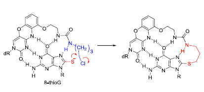
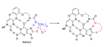
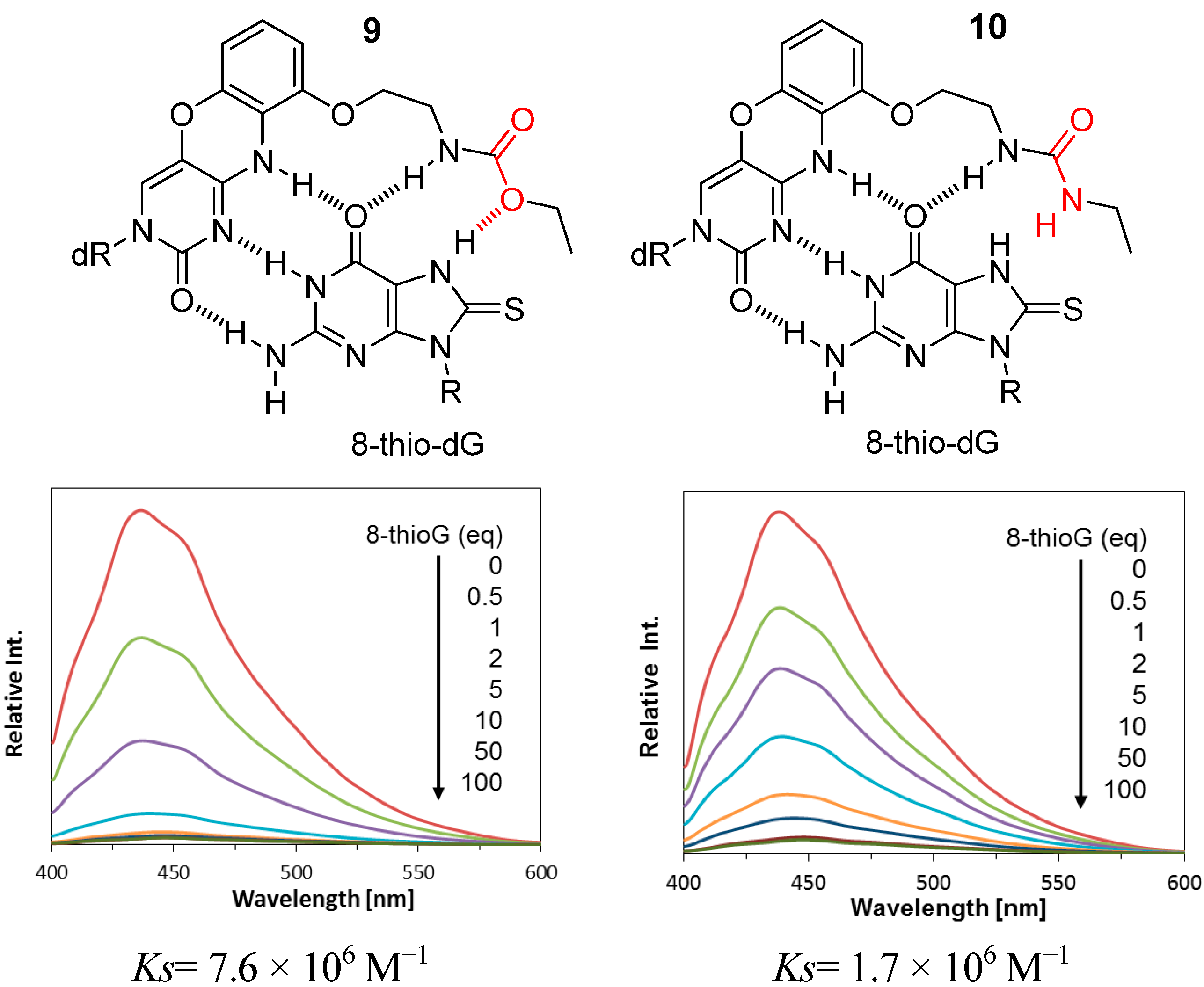
2.2. Reaction of 8-ThioG-Grasp with 8-ThioG

The reaction of the 8-thioG-grasp derivatives with triAc-8-thioG was performed at 50 °C in CH3CN in the presence of Et3N, and the reaction progress was monitored by HPLC. An equimolar mixture of 8-thioG-grasp (2b) and triAc-8-thioG formed an adduct in a nearly quantitative yield as a single product within 3 h (Figure 2).
This product was isolated, and its structure was confirmed as depicted in 7b by 1H-NMR, ESI-MS, and HMBC (see the Supporting Information). The HMBC spectrum indicated the distinct correlation between the C8 of the 8-thioguanine unit and the methylene protons next to the sulfur atom of 7b.

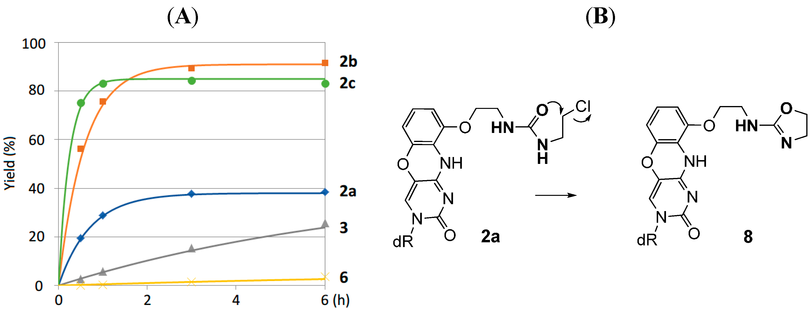
2.3. Comparison of the Reactivity between 8-ThioG-Grasp and the Control Compound

The time courses of the reaction between the 8-thioG-grasp derivatives 2a–c, 3 and triAc-8-thioG are compared in Figure 3. 2b and 2c exhibited efficient reactivity, and a relatively slow reaction was observed with 2a. In the reaction of 2a, the formation of the amino-oxazoline ring (8) as a byproduct decreased the adduct yield (Figure 3).
In contrast, the carbamate type 3 showed a significantly decreased efficiency compared with the compounds 2 with the urea-type linker. This is of great interest because 8-thioketo tautomer of 8-thioG is stable in neutral organic solvents [18] and forms more stable complexes with the carbamate type 9 than with the urea type 10 (Ks in CHCl3, 9: 7.6 × 10⁶ M−1 vs. 10: 1.7 × 10⁶ M−1) (Figure 4). Accordingly, it is reasonably explained that the 7N-H of 8-thioG is deprotonated by Et3N to form the 8-thioenolate, thereby facilitating hydrogen-bonded complexes with the urea-type compounds (2b and 2c) such as shown in Figure 1B to exhibit efficient reactivity. As the pKa value of 7N-H of 8-thioguanosine is around 8.5 (see the Supporting Information), Et3N is a suitable base for its deprotonation. No adduct was formed with N-(3-chloropropyl)-2-(pyrene-1-yl) acetamide (6), a control without a binding site, emphasizing the contribution of the hydrogen-bonded complexation of 2b and 2c for efficient reactivity. Among the urea-type 8-thioG-grasp derivatives, 2a produced the adduct in a low yield. The HPLC monitoring showed that 2a formed the byproduct 8 as a major peak, which structure was determined by H-NMR, ESI-MS and 2D-HMBC, as shown in Figure 3. The amino-oxazoline was formed by intramolecular displacement, which was faster than the intermolecular nucleophilic attack from 8-thioG. It should be emphasized that the displacement of the sulfur atom of 8-thioG in the complex with 2b or 2c is favorable compared with the intramolecular amino-oxazoline ring formation because this type of amino-oxazoline ring formation was not observed for 2b or 2c.
Figure 2. Reaction of 8-thioG-grasp with tri-Ac-8-thioG monitored by HPLC. dR and R represent 3′,5′-diOTBS-2′-deoxyribosyl and triacetyl ribosyl groups, respectively. The reaction was performed using 0.4 mM each of 2b and 8-thioG in the presence of 20 mM Et3N in CH3CN at 50 °C. HPLC conditions: column: Xbridge C8 3.5 μm, 3.0 mm × 100 mm; solvents: (A) 0.1 M TEAA buffer at pH 7.0 and (B) CH3CN, A/B = 20:80; flow rate: 0.5 mL/min; monitored by UV at 254 nm.
Molecules 20 01078 g002
Figure 3. (A) Comparison of the time courses of the reactions with 8-thioG-grasp (2a–c and 3) and the control compound 6. (B) Byproduct 8 formed via the intramolecular reaction of 2a. Product yields were obtained at the indicated time points by HPLC analysis, as described in the footnote to Figure 2. dR represents the 3′,5′-diOTBS-2′-deoxyribosyl group.
Molecules 20 01078 g003
Figure 4. Proposed complexation of 8-thioG with the carbamate or the urea type. dR and R represent 3′, 5′-diOTBS-2′-deoxyribosyl and triacetyl ribosyl groups, respectively.
Molecules 20 01078 g004

3. Experimental Section

3.1. General Information

The reagents and solvents were purchased from commercial suppliers and were used without purification. The 1H- and 13C- NMR spectra were recorded on a Bruker Avance III spectrometer. The 2D-NMR spectra were measured on a Varian Inova 500 instrument. The IR spectra were recorded on a Perkin Elmer Spectrum One FT-IR spectrometer. The ESI-HRMS spectra were measured on an Applied Biosystems Mariner Biospectrometry Workstation using neurotensin, angiotensin I, bradykinin and picolinic acid as the internal standards.

3.2. Chemistry

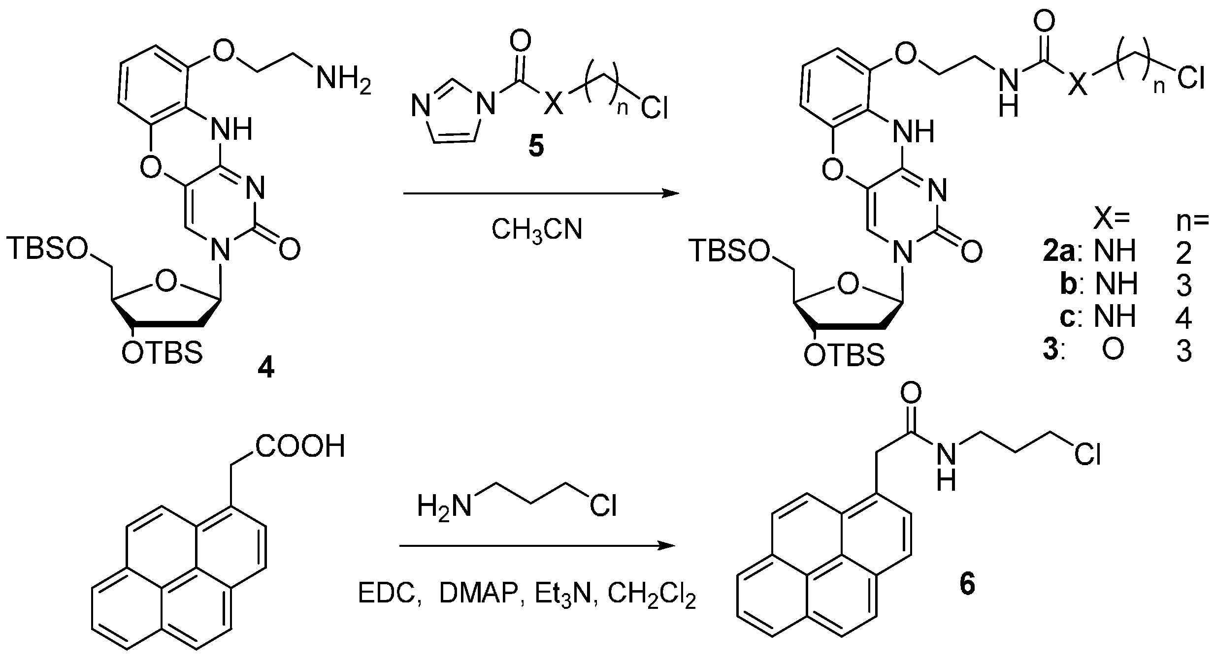
3.2.1. General Synthesis of 8-ThioG-Grasp Derivatives

1,1′-Carbonyldiimidazole (5 equiv.) was added to a solution of chloroalkylamine hydrochloride (5 equiv.) in anhydrous CH3CN (0.05 M in G-clamp unit) under an argon atmosphere. The reaction mixture was stirred at room temperature for 30 min, followed by the addition of the G-clamp unit 4 (1 equiv.) and Et3N (8 equiv.). After stirring overnight at room temperature, saturated aqueous NaHCO3 solution was added to the reaction mixture, which was extracted with CHCl3. The organic layer was washed with brine, dried over Na2SO4 and evaporated in vacuo. The resulting residue was purified by silica gel column chromatography (CH2Cl2/MeOH =100:0 to 95:5) to give 2a–c and 3 as light yellow foams.
1-(2-((3-((2R,4S,5R)-4-((tert-Butyldimethylsilyl)oxy)-5-(((tert-butyldimethylsilyl)oxy)methyl)-tetrahydrofuran-2-yl)-2-oxo-2,10-dihydro-3H-benzo[b]pyrimido[4,5-e][1,4]oxazin-9-yl)oxy)ethyl)-3-(2-chloroethyl)urea. (C2-8-ThioG-grasp, 2a): 2a was synthesized using G-clamp unit 4 and 2-chloroethylamine in 48% as a light yellow foam. 1H-NMR (400 MHz, CD3OD) δ (ppm) 7.62 (1H, s), 6.82 (1H, t, J = 8.6 Hz), 6.60 (1H, d, J = 8.6 Hz), 6.35 (1H, d, J = 8.6 Hz), 6.17 (1H, t, J = 6.4 Hz), 4.46 (1H, dt, J = 6.4, 3.4 Hz), 4.05 (2H, t, J = 4.9 Hz), 3.96–3.93 (2H, m), 3.82 (1H, dd, J = 12.1, 3.4 Hz), 3.57 (2H, t, J = 6.0 Hz), 3.54 (2H, t, J = 5.2 Hz), 3.45 (2H, t, J = 6.4 Hz), 2.33 (1H, ddd, J = 13.4, 6.1, 4.6 Hz), 2.11 (1H, dt, J = 13.4, 6.4 Hz), 0.97 (9H, s), 0.91 (9H, s,), 0.18 (3H, s), 0.16 (3H, s), 0.11 (6H, s). 13C-NMR (125 MHz, CDCl3) δ (ppm) 158.53, 152.89, 146.88, 143.06, 127.52, 124.49, 122.97, 115.06, 108.21, 107.54, 87.75, 86.00, 70.84, 69.22, 62.32, 42.09, 41.89, 39.33, 29.28, 26.04, 25.71, 18.49, 17.94, −4.58, −4.90, −5.45, −5.52. IR (cm−1): 2929, 1673, 1557, 1473. HR ESI-MS (m/z): Calcd. for [C32H53ClN5O7Si2]+: 710.3167 ([M+H]+), found 710.3176.
1-(2-((3-((2R,4S,5R)-4-((tert-Butyldimethylsilyl)oxy)-5-(((tert-butyldimethylsilyl)oxy)methyl)-tetrahydrofuran-2-yl)-2-oxo-2,10-dihydro-3H-benzo[b]pyrimido[4,5-e][1,4]oxazin-9-yl)oxy)ethyl)-3-(3-chloropropyl)urea. (C3-8-ThioG-grasp, 2b): 2b was synthesized using G-clamp unit 4 and 3-chloropropylamine in 46% as a light yellow foam. 1H-NMR (400 MHz, CD3OD) δ (ppm) 7.62 (1H, s), 6.82 (1H, t, J = 8.2 Hz), 6.60 (1H, dd, J = 8.4, 1.2 Hz), 6.35 (1H, dd, J = 8.2, 1.2 Hz), 6.17 (1H, t, J = 6.4 Hz), 4.47 (1H, dt, J = 5.7, 3.7 Hz), 4.05 (2H, t, J = 5.2 Hz), 3.95 (1H, t, J = 3.1 Hz), 3.94 (1H, t, J = 3.1 Hz), 3.82 (1H, dd, J = 12.2, 3.4 Hz), 3.57 (2H, t, J = 6.7 Hz), 3.53 (2H, t, J = 5.2 Hz), 3.27 (2H, t, J = 6.7 Hz), 2.33 (1H, ddd, J = 13.2, 6.2, 4.0 Hz), 2.12 (1H, dt, J = 13.2, 6.4 Hz), 1.92 (2H, quin, J = 6.4 Hz), 0.97 (9H, s), 0.91 (9H, s,), 0.18 (3H, s), 0.16 (3H, s), 0.11 (6H, s). 13C-NMR (125 MHz, CDCl3) δ (ppm) 158.99, 152.94, 152.67, 146.79, 143.04, 127.61, 124.38, 122.87, 115.26, 108.27, 107.74, 87.71, 85.94, 70.82, 69.45, 62.31, 42.74, 41.88, 39.39, 37.45, 33.21, 26.04, 25.71, 18.48, 17.93, −4.58, −4.90, −5.45, −5.52. IR (cm−1): 2931, 1670, 1558, 1499. HR ESI-MS (m/z): Calcd. for [C33H55ClN5O7Si2]+: 724.3323 ([M+H]+), found 724.3340.
1-(2-((3-((2R,4S,5R)-4-((tert-butyldimethylsilyl)oxy)-5-(((tert-butyldimethylsilyl)oxy)methyl)tetrahydro-furan-2-yl)-2-oxo-2,10-dihydro-3H-benzo[b]pyrimido[4,5-e][1,4]oxazin-9-yl)oxy)ethyl)-3-(4-chloro-butyl)urea. (C4-8-ThioG-grasp, 2c): 2c was synthesized using G-clamp unit 4 and 4-chlorobutylamine [19] in 82% as a light yellow foam. 1H-NMR (400 MHz, CD3OD) δ (ppm) 7.62 (1H, s), 6.81 (1H, t, J = 8.6 Hz), 6.59 (1H, d, J = 8.6 Hz), 6.24 (1H, t, J = 8.2 Hz), 6.16 (1H, t, J = 6.1 Hz), 4.46 (1H, br), 4.03 (2H, t, J = 5.2 Hz), 3.94 (2H, dd, J = 11.6, 2.4 Hz), 3.82 (1H, d, J = 9.5 Hz), 3.61 (2H, t, J = 6.4 Hz) 3.53 (2H, t, J = 6.4 Hz), 3.12 (2H, t, J = 6.7 Hz), 2.35–2.29 (1H, m), 2.11 (1H, dt, J = 13.1, 6.4 Hz), 1.88–1.73 (4H, m), 0.97 (9H, s), 0.91 (9H, s,), 0.18 (3H, s), 0.16 (3H, s), 0.11 (6H, s). 13C-NMR (125 MHz, CD3OD) δ (ppm) 161.16, 156.45, 155.65, 148.10, 144.31, 129.56, 125.02, 123.52, 109.17, 108.64, 89.33, 87.51, 72.88, 70.17, 63.73, 45.49, 42.80, 40.43, 40.32, 31.10, 28.78, 26.60, 26.25, 19.37, 18.86, −4.45, −4.65, −5.25, −5.30. IR (cm−1): 2953, 1670, 1558. HR ESI-MS (m/z): calcd. for [C34H57ClN5O7Si2]+, 738.3480 ([M+H]+); found 738.3452.
(2-((3-((2R,4S,5R)-4-((tert-Butyldimethylsilyl)oxy)-5-(((tert-butyldimethylsilyl)oxy)methyl)tetrahydro-furan-2-yl)-2-oxo-2,10-dihydro-3H-benzo[b]pyrimido[4,5-e][1,4]oxazin-9-yl)oxy)ethyl)-3-chloropropyl carbamate. (C3 (O)-8-ThioG-grasp, 3): 3 was synthesized using G-clamp unit 4 and 3-chloropropanol in 82% as a light yellow foam. 1H-NMR (400 MHz, CD3OD)   (ppm) 7.64 (1H, s), 6.83 (1H, t, J = 8.2 Hz), 6.61 (1H, dd, J = 8.2, 0.9 Hz), 6.37 (1H, dd, J = 8.2, 0.9 Hz), 6.18 (1H, t, J = 6.1 Hz), 4.47 (1H, dt, J = 5.8, 4.0 Hz), 4.19 (2H, t, J = 6.1 Hz), 4.06 (1H, t, J = 5.2 Hz), 3.96 (1H, t, J = 2.75 Hz), 3.94 (1H, t, J = 3.1 Hz), 3.83 (1H, dd, J = 12.2, 3.4 Hz), 3.63 (2H, t, J = 6.7 Hz), 3.52 (2H, t, J = 5.2 Hz), 3.27 (2H, t, J = 6.7 Hz), 2.34 (1H, ddd, J = 13.4, 6.1, 4.3 Hz), 2.12 (1H, dt, J = 13.4, 6.1 Hz), 2.06 (2H, quin, J = 6.1 Hz), 0.97 (9H, s), 0.92 (9H, s,), 0.18 (3H, s), 0.17 (3H, s), 0.11 (6H, s). 13C-NMR (125 MHz, CDCl3) δ (ppm) 156.62, 152.69, 152.02, 146.63, 143.16, 127.26, 124.14, 122.85, 115.58, 108.41, 107.15, 87.63, 86.02, 70.71, 68.38, 62.26, 41.89, 41.38, 40.39, 32.05, 26.04, 25.72, 18.48, 17.94, −4.56, −4.90, −5.46, −5.52. IR (cm−1): 2953, 1679, 1556, 1474. HR ESI-MS (m/z): calcd. for [C33H54ClN4O8Si2]+, 725.3163 ([M+H]+); found 725.3142.

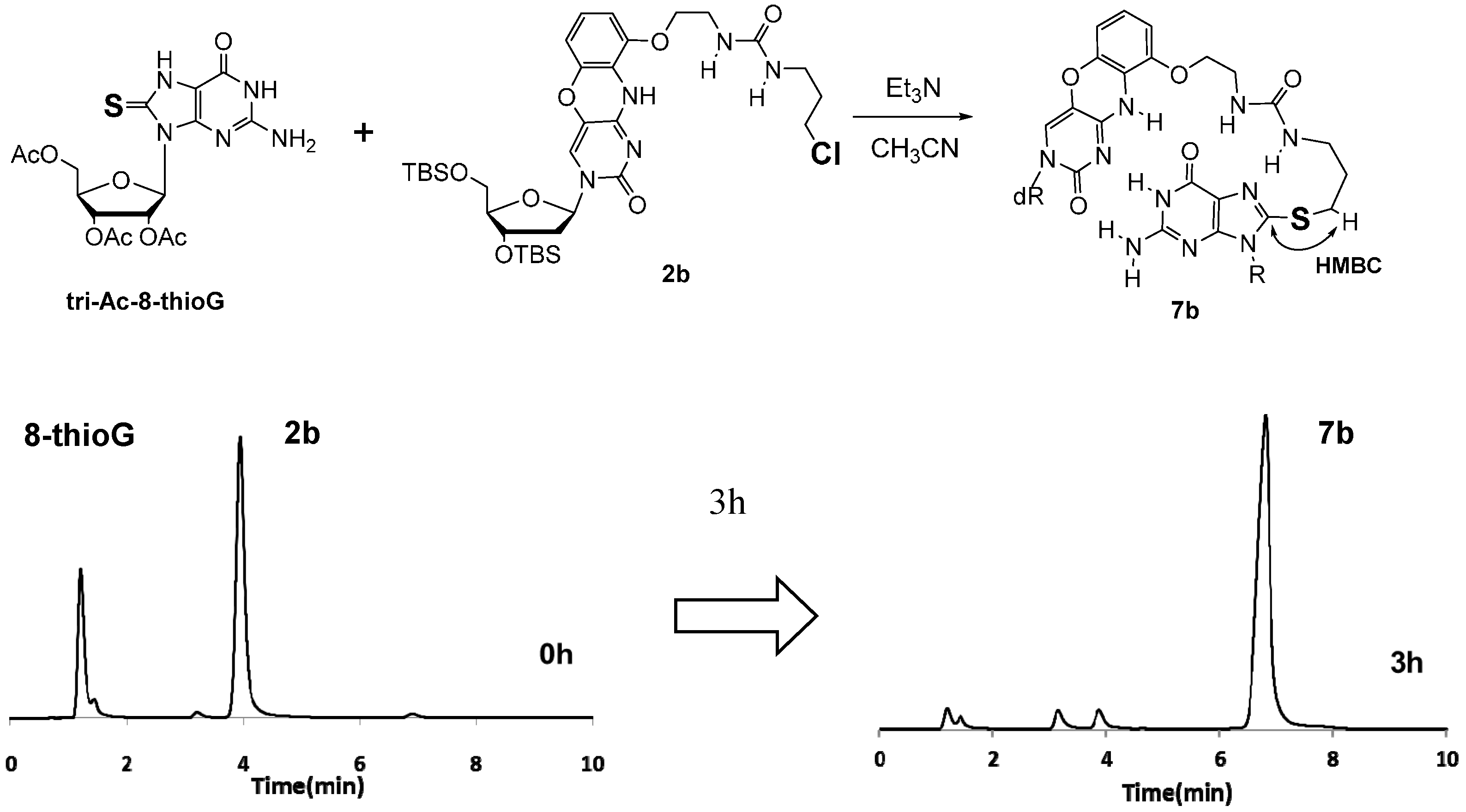
3.2.2. Synthesis of 8-ThioG Adduct 7b

Et3N (100 µL, 0.72 mmol) was added to a solution of 2b (11 mg, 0.015 mmol) and tri-Ac-8-thioG (14 mg, 0.031 mmol) in anhydrous CH3CN (1 mL) under an argon atmosphere. The reaction mixture was stirred at 50 °C for 12 h, and evaporated in vacuo. The resulting residue was purified by silica gel column chromatography (CHCl3/MeOH =70:1) to give 5b as a colorless oil (9 mg, 50%). 1H-NMR (400 MHz, CD3OD) δ (ppm) 7.51 (1H, s), 6.70 (1H, t, J = 8.2 Hz), 6.41 (1H, d, J = 8.6 Hz), 6.18 (1H, t, J = 4.9 Hz), 6.15 (1H, d, J = 6.1 Hz), 6.04–6.03 (1H, m), 5.84 (1H, d, J = 4.6 Hz), 5.77 (1H, t, J = 6.1 Hz), 4.51 (1H, dd, J = 7.6, 2.4 Hz), 4.41–4.31 (3H, m), 3.89–3.85 (4H, m), 3.76 (1H, d, J = 9.5 Hz), 3.60 (2H, br), 3.29–3.21 (4H, m), 2.29 (1H, ddd, J = 13.0, 6.5 Hz), 2.13–2.06 (1H, m), 1.96 (2H, br), 0.93 (9H, s), 0.91 (9H, s,), 0.14 (3H, s), 0.13 (3H, s), 0.09 (6H, s) 13C-NMR (125 MHz, CD3OD) δ (ppm) 172.40, 171.44, 171.20, 161.36, 160.17, 156.20, 156.01, 155.09, 155.04, 148.62, 147.36, 144.32, 129.27, 125.38, 123.73, 117.51, 115.86, 107.64, 88.03, 87.47, 81.00, 72.63, 71.79, 64.20, 42.70, 40.58, 39.76, 31.34, 29.52, 26.66, 26.34, 20.42, 19.37, 18.88, −4.29, −4.54, −5.14. IR (cm−1): 2928, 1749, 1684. HR ESI-MS (m/z): calcd. for [C49H73N10O15SSi2]+, 1129.4511 ([M+H]+); found 1129.4469.

3.2.3. N-(3-Chloropropyl)-2-(pyren-1-yl)acetamide (6)

3-Chloropropylamine hydrochloride (12 mg, 0.093 mmol), 1-(3-dimethylaminopropyl)-3-ethyl-carbodiimide hydrochloride (EDC, 17 mg, 0.089 mmol), 4,4-dimethylaminopyridine (DMAP, 1 mg 0.008 mmol) and Et3N (50 µL, 0.359 mmol) were added to a solution of 1-pyreneacetic acid (20 mg, 0.077 mmol) in anhydrous CH2Cl2 (1 mL) under an argon atmosphere. The reaction mixture was stirred at room temperature for 7 h, quenched with aqueous saturated NH4Cl solution, and extracted with AcOEt. The organic layer was washed with brine, dried over Na2SO4 and evaporated in vacuo. The resulting residue was purified by silica gel column chromatography (CH2Cl2) to give 6 as a pale yellow solid (13 mg, 50%). 1H-NMR (400 MHz, CDCl3) δ (ppm) 8.20 (2H, d, J = 7.6 Hz), 8.15 (3H, d, J = 7.9 Hz), 8.08 (1H, d, J = 9.2 Hz), 8.04 (1H, d, J = 9.2 Hz), 8.02 (1H, t, J = 7.9 Hz), 7.88 (1H, d, J = 7.9 Hz), 4.28 (2H, s), 3.31 (2H, t, J = 6.4 Hz), 3.25 (2H, q, J = 6.4 Hz), 1.78 (2H, quin, J = 6.4 Hz). 13C-NMR (125 MHz, CDCl3) δ (ppm) 171.23, 131.28, 131.16, 130.76, 129.53, 128.50, 128.16, 127.69, 127.27, 126.28, 125.60, 125.48, 125.20, 125.12, 124.62, 122.76, 42.22, 42.08, 37.28, 31.86. IR (cm−1): 3293, 1646, 1544. HR ESI-MS (m/z): calcd. for [C21H19ClNO]+, 336.1150 ([M+H]+); found 336.1161.

3.2.4. Determination of the Structure of the Byproduct 8

To a solution of 2a (70 mg, 0.099 mmol) in MeOH (4 mL) was added NaHCO3 (90 mg, 1.07 mmol) under an argon atmosphere. The reaction mixture was refluxed for 25 h, filtered, and evaporated in vacuo. The resulting residue was purified by silica gel column chromatography (CHCl3/Acetone =100:0 to 50:50) to give 8 as light yellow foams (26 mg, 39%). 1H-NMR (400 MHz, CD3OD) δ (ppm) 7.60 (1H, s), 6.81 (1H, t, J = 8.2 Hz), 6.58 (1H, d, J = 8.2 Hz), 6.35 (1H, d, J = 8.2 Hz), 6.17 (1H, t, J = 6.4 Hz), 4.47 (1H, dt, J = 6.1, 3.5 Hz), 4.35 (2H, t, J = 8.2 Hz), 4.07 (2H, J = 4.9 Hz), 3.96–3.93 (2H, m), 3.82 (1H, dd, J = 12.2, 3.1 Hz), 3.78 (2H, t, J = 8.2 Hz), 3.55 (2H, t, J = 5.2 Hz), 2.33 (1H, ddd, J = 13.3, 6.1, 4.1 Hz), 2.12 (1H, dt, J = 13.3, 6.3 Hz), 0.97 (9H, s), 0.91 (9H, s,), 0.18 (3H, s), 0.16 (3H, s), 0.11 (6H, s). 13C-NMR (125 MHz, CD3OD) δ (ppm) 164.20, 156.51, 155.66, 147.98, 144.31, 129.55, 124.95, 123.42, 117.14, 109.25, 108.51, 89.34, 87.51, 72.97, 69.64, 69.37, 63.77, 43.22, 42.77, 29.54, 26.60, 26.26, 19.36, 18.85, −4.46, −4.65, −5.26, −5.30. IR (cm−1): 2931, 1669, 1557, 1497, 1473. HR ESI-MS (m/z): calcd. for [C32H52N5O7Si2]+ , 674.3400 ([M+H]+); found 674.3438.

3.2.5. General Procedure of Reaction Monitoring by HPLC

Reaction was initiated by the addition of Et3N (20 mM) to a solution of 8-thioG-grasp derivative (0.4 mM) and triAc-8-thioG) (0.4 mM) in CH3CN at 50 °C. The reaction progress was monitored by HPLC at 0.5, 1, 3 and 5 h. Product yield with time course of reaction were obtained from reverse-phase HPLC analysis. (Column: Xbridge C8 3.5 μm, 3.0 × 100 mm; Solvent: A: 0.1 M TEAA buffer at pH 7.0, B: CH3CN, A/B= 20: 80; Flow rate: 0.5 mL/min; monitored by UV detector at 254 nm).

4. Conclusions

In this study, we designed new recognition molecules to covalently capture 8-thioguanosine based on the G-clamp skeleton by introducing chloroalkyl urea linker. 8-ThioG-grasp 2b and 2c with chloropropyl urea and chlorobutyl linker exhibited the most efficient reactivity for tri-Ac-8-thioG. It has been shown from the comparison with control compounds that the multiple hydrogen-bonded complexes contribute to the efficient reactivity. There is increasing interest in 8-thioguanosine derivatives for their biological roles in signal transduction pathways [7], the 8-thioG-grasp derivatives are expected to be a potential platform to develop specific molecules. For example, an 8-thioG-grasp derivative with a phosphate binding unit are expected to covalently trap phosphate derivatives of 8-thioguanosine and interfere their biological functions. Systematic studies are now ongoing in this line in our group, which will be reported in due course.

Supplementary Materials

Supplementary materials can be accessed at: https://www.mdpi.com/1420-3049/20/01/1078/s1.

Acknowledgments

We are grateful for support provided by a Grant-in-Aid for Challenging Exploratory Research (No. 26670004) from the Japan Society for the Promotion of Science (JSPS). S.S. and Y.F. also acknowledge the Astellas Foundation for Research on Metabolic Disorders and a Research Fellowship for Young Scientists from JSPS, respectively.

Author Contributions

Y. Fuchi and H. Obayashi performed chemistry experiments and analyzed the data. Y. Fuchi and S. Sasaki designed the capture molecules, and wrote the paper.

Conflicts of Interest

The authors declare no conflict of interest.

References

  1. Halliwell, B. Oxidative stress and cancer: Have we moved forward? Biochem. J. 2007, 401, 1–11. [Google Scholar] [CrossRef] [PubMed]
  2. Yermilov, V.; Rubio, J.; Ohshima, H. Formation of 8-nitroguanine in DNA treated with peroxynitrite in vitro and its rapid removal from DNA by depurination. FEBS Lett. 1995, 376, 207–210. [Google Scholar] [CrossRef] [PubMed]
  3. Kasai, H. Analysis of a form of oxidative DNA damage, 8-hydroxy-2′-deoxyguanosine, as a marker of cellular oxidative stress during carcinogenesis. Mutat. Res. 1997, 387, 147–163. [Google Scholar] [CrossRef] [PubMed]
  4. Maeda, H. The link between infection and cancer: Tumor vasculature, free radicals, and drug delivery to tumors via the EPR effect. Cancer Sci. 2013, 104, 779–789. [Google Scholar] [CrossRef] [PubMed]
  5. Sawa, T.; Zaki, M.H.; Okamoto, T.; Akuta, T.; Tokutomi, Y.; Kim-Mitsuyama, S.; Ihara, H.; Kobayashi, A.; Yamamoto, M.; Fujii, S.; et al. Protein S-guanylation by the biological signal 8-nitroguanosine 3′,5′-cyclic monophosphate. Nat. Chem. Biol. 2007, 3, 727–735. [Google Scholar] [CrossRef] [PubMed] [Green Version]
  6. Nishida, M.; Sawa, T.; Kitajima, N.; Ono, K.; Inoue, H.; Ihara, H.; Motohashi, H.; Yamamoto, M.; Suematsu, M.; Kurose, H.; et al. Hydrogen sulfide anion regulates redox signaling via electrophile sulfhydration. Nat. Chem. Biol. 2012, 8, 714–724. [Google Scholar] [CrossRef] [PubMed]
  7. Ida, T.; Sawa, T.; Ihara, H.; Tsuchiya, Y.; Watanabe, Y.; Kumagai, Y.; Suematsu, M.; Motohashi, H.; Fujii, S.; Matsunaga, T.; et al. Reactive cysteine persulfides and S-polythiolation regulate oxidative stress and redox signaling. Proc. Natl. Acad. Sci. USA 2014, 111, 7606–7611. [Google Scholar] [CrossRef] [PubMed]
  8. Lin, K.; Matteucci, M.D. A Cytosine analogue capable of clamp-like binding to a guanine in helical nucleic acids. J. Am. Chem. Soc. 1998, 120, 8531–8532. [Google Scholar] [CrossRef]
  9. Flanagan, W.M.; Wolf, J.J.; Olson, P.; Grant, D.; Lin, K.Y.; Wangner, R.W.; Matteucci, M.D. A cytosine analog that confers enhanced potency to antisense oligonucleotides. Proc. Natl. Acad. Sci. USA 1999, 96, 3513–3518. [Google Scholar] [CrossRef] [PubMed]
  10. Maier, M.A.; Leeds, J.M.; Balow, G.; Springer, R.H.; Bharadwaj, R.; Manoharan, M. Nuclease resistance of oligonucleotides containing the tricyclic cytosine analogues phenoxazine and 9-(2-aminoethoxy)-phenoxazine (“G-clamp”) and origins of their nuclease resistance properties. Biochemistry 2002, 41, 1323–1327. [Google Scholar] [CrossRef] [PubMed]
  11. Wilds, C.J.; Maier, M.A.; Tereshko, V.; Manoharan, M.; Egli, M. Direct observation of a cytosine analogue that forms five hydrogen bonds to guanosine: guanidino G-clamp. Angew. Chem. Int. Ed. 2002, 41, 115–117. [Google Scholar] [CrossRef]
  12. Holmes, S.C.; Arzumanov, A.A.; Gait, M.J. Steric inhibition of human immunodeficiency virus type-1 Tat-dependent trans-activation in vitro and in cells by oligonucleotides containing 2′-O-methyl G-clamp ribonucleoside analogues. Nucleic Acids Res. 2003, 31, 2759–2768. [Google Scholar] [CrossRef] [PubMed]
  13. Nakagawa, O.; Ono, S.; Li, Z.; Tsujimoto, A.; Sasaki, S. Specific fluorescent probe for 8-Oxoguanosine. Angew. Chem. Int. Ed. Engl. 2007, 119, 4584–4587. [Google Scholar] [CrossRef]
  14. Li, Z.; Nakagawa, O.; Koga, Y.; Taniguchi, Y.; Sasaki, S. Synthesis of new derivatives of 8-oxoG-clamp for better understanding the recognition mode and improvement of selective affinity. Bioorg. Med. Chem. 2010, 18, 3992–3998. [Google Scholar] [CrossRef] [PubMed]
  15. Koga, Y.; Fuchi, Y.; Nakagawa, O.; Sasaki, S. Optimization of fluorescence property of the 8-oxodGclamp derivative for better selectivity for 8-oxo-2′-deoxyguanosine. Tetrahedron 2011, 67, 6746–6752. [Google Scholar] [CrossRef]
  16. Fuchi, Y.; Sasaki, S. Efficient covalent capture of 8-nitroguanosine via a multiple hydrogen-bonded complex. Org. Lett. 2014, 16, 1760–1763. [Google Scholar] [CrossRef] [PubMed]
  17. Holmes, E.; Robins, K. Purine nucleosides. VII. Direct bromination of adenosine, deoxyadenosine, guanosine, and related purine nucleosides. J. Am. Chem. Soc. 1964, 86, 1242–1245. [Google Scholar] [CrossRef]
  18. Strassmaier, T.; Karpen, J.W. Novel N7- and N1-substituted cGMP derivatives are potent activators of cyclic nucleotide-gated channels. J. Med. Chem. 2007, 50, 4186–4194. [Google Scholar] [CrossRef] [PubMed]
  19. Song, A.; Walker, S.G.; Parker, K.A.; Sampson, N.S. Antibacterial studies of cationic polymers with alternating, random, and uniform backbones. ACS Chem. Biol. 2011, 6, 590–599. [Google Scholar] [CrossRef] [PubMed]
  • Sample Availability: Samples are available from authors.

