Article

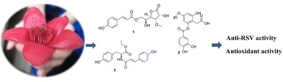
Phenolic Compounds from the Flowers of Bombax malabaricum and Their Antioxidant and Antiviral Activities

1
Institute of Traditional Chinese Medicine and Natural Products, College of Pharmacy, Jinan University, Guangzhou 510632, China
2
Shenzhen Engineering Laboratory of Lingnan Herbal Resource Development and Application, Shenzhen Institute for Drug Control, Shenzhen 518057, China
3
International Institute for Translational Chinese Medicine, Guangzhou University of Chinese Medicine, Guangzhou 510006, China
*
Authors to whom correspondence should be addressed.
These authors contributed equally to this work.
Molecules 2015, 20(11), 19947-19957; https://doi.org/10.3390/molecules201119660
Submission received: 28 August 2015 / Revised: 20 October 2015 / Accepted: 27 October 2015 / Published: 5 November 2015
(This article belongs to the Special Issue Recent Advances in Plant Phenolics)

Abstract

:
Three new phenolic compounds 13 and twenty known ones 423 were isolated from the flowers of Bombax malabaricum. Their chemical structures were elucidated by spectroscopic analyses (IR, ESI-MS, HR-ESI-MS, 1D- and 2D-NMR) and chemical reactions. The antioxidant capacities of the isolated compounds were tested using FRAP and DPPH radical-scavenging assays, and compounds 4, 6, 8, 12, as well as the new compound 2, exhibited stronger antioxidant activities than ascorbic acid. Furthermore, all of compounds were tested for their antiviral activities against RSV by the CPE reduction assay and plaque reduction assay. Compounds 4, 10, 12 possess in vitro antiviral activities, and compound 10 exhibits potent anti-RSV effects, comparable to the positive control ribavirin.

Graphical Abstract

1. Introduction

Bombax malabaricum DC. (Bombacaceae) is a very common native tree in Guangdong Province of China. In the folklore of southern China its flowers are often used as a healthy food material, which is stewed in soup with meat and cooked congee with rice [1]. This flower and four other flowers from medicinal herbs are mixed as a health care tea called “five flowers tea” [2]. According to the traditional Chinese medicine theory, the flower of B. malabaricum is sweet and cool-natured [1], and traditionally used for the treatment of diarrhea, chronic inflammation, fever, hepatitis, and contused wounds [3]. Modern pharmacology research has shown that the Bombax plant possesses many biological activities, such as antioxidant [4], anti-inflammatory [5], anticancer [6], and protection of the hepatic and cardiovascular systems [7]. Phytochemical investigations have shown that flavonoids, sesquiterpenes and phenylpropanoids are the major constituents of B. malabaricum [8,9,10].
In the present work, we found that the ethanol extract of B. malabaricum flower possessed in vitro antiviral activity against respiratory syncytial virus (RSV). Furthermore, a previous study had showed that its ethanol extract displayed good antioxidant activity [4]. As a part of our continuing study on the isolation of interesting and biologically active compounds from this plant, three new phenolic compounds and 20 known ones (Figure 1) were isolated from the ethanol extract of this plant. In this paper, the isolation and structural elucidation of the new compounds are reported. In addition, the anti-RSV activities of the isolates were evaluated by cytopathic effect (CPE) and plaque reduction assays, and their antioxidant activities were tested by using ferric-reducing antioxidant power (FRAP) and 2,2-diphenyl-1-picryhydrazyl (DPPH) radical-scavenging assays.
Figure 1. Chemical structures of 123.
Molecules 20 19660 g001

2. Results and Discussion

2.1. Identification of Compounds 123

Compound 1 was isolated as a colorless oil. Its molecular formula was determined as C16H18O8 on the basis of a HR-ESI-MS peak at m/z 361.08977 [M + Na]+ (calcd. for C16H18O8Na: 361.08939). The 1H-NMR spectrum of 1 showed the presence of four aromatic protons [δH 7.47 (2H, d, J = 8.4 Hz), 6.81 (2H, d, J = 8.4 Hz)], two trans-olefinic protons [δH 7.67 (1H, d, J = 15.9 Hz), 6.38 (1H, d, J = 15.9 Hz)] and one methoxyl group [δH 3.60 (3H, s)]. The 13C-NMR spectrum exhibited two carbonyls at δC 177.4 and 168.6, six aromatic carbons at δC 127.3, 131.4 × 2, 117.0 × 2, 161.5, one double bond at δC 147.2 and 115.2, and one methoxyl group at δC 61.1. Comparison of the 13C-NMR data of 1 with those of known compound 14 (Table S1, see Supplementary Materials) [9] showed that they were very similar, except for some differences of the chemical shifts on C-4, C-5 and C-6. These differences were about 2.0–4.7 ppm, indicating the configuration of C-4 was different from that of 14. The planar structure of 1 was verified by 1H-1H COSY, and HMBC spectra (Figure 2). The ROESY correlations between H-2 and H-3/H-5 suggested that H-2/H-3 were on the same side and H-4 was on the other side. Thus the structure of 1 was elucidated and it was named 4-epi-bombalin.
Figure 2. Key COSY and HMBC correlations of 13.
Molecules 20 19660 g002
Compound 2 was isolated as a colorless oil. The molecular formula of 2 was verified as C21H22O13 by an [M + Na]+ ion peak at m/z 505.09584 (calcd. for C21H22O13Na: 505.09526) in the HR-ESI-MS. The signals of five aromatic protons at δH 7.58 (1H, dd, J = 8.4, 2.2 Hz), 7.57 (1H, d, J = 2.2 Hz), 6.87 (1H, d, J = 8.4 Hz), 6.56 (1H, d, J = 2.2 Hz) and 6.47 (1H, d, J = 2.2 Hz), and one sugar at δH 4.89 (1H, d, J = 7.6 Hz) and 3.70–3.88 (6H, overlapped) were displayed by the 1H-NMR spectrum. Accordingly, the signals of two carbonyls at δC 177.7 and 166.6, one glucose moiety at δC 102.4, 78.2, 78.0, 75.0, 71.4 and 62.6, and two benzene rings at δC 103–159 were exhibited by the 13C-NMR spectrum. The 1H- and 13C-NMR data (Table 1) showed a number of similarities to those of 2-O-(3,4-dihydroxybenzoyl)-2,4,6-trihydroxyphenylacetic acid [11], except that 2 had an extra glucose and the chemical shifts of H-3 and H-5 at δH 6.27 and 6.18 shifted downfield by about 0.3 ppm to δH 6.56 and 6.47, suggesting the glucose was connected to C-4. And it was confirmed by the HMBC correlation between δH 4.89 (H-1′′) and δC 158.6 (C-4) (Figure 2). Acid hydrolysis of 2 afforded d-glucose (Figure S17, see Supplementary Materials), which was identified by HPLC analysis [12].
The coupling constant (7.6 Hz) of the anomeric proton indicated the glucose was β configuration. Compound 2 was thus identified as 2-O-(3,4-dihydroxybenzoyl)-2,4,6-trihydroxyphenylacetic acid 4-O-β-d-glucopyranoside.
Compound 3 was isolated as a colorless oil. The HR-ESI-MS of compound 3 exhibited a quasi-molecular ion peak at m/z 356.14900 [M + H]+ (calcd. for C20H22NO5: 356.14925) indicating the molecular formula was C20H21NO5. The IR spectrum showed the presence of NH (3357 cm−1) and carbonyl groups (1730, 1650 cm−1). The 1H-NMR spectrum showed two trans-olefinic protons at δH 7.42 (1H, d, J = 15.9 Hz) and 6.45 (1H, d, J = 15.9 Hz), two sets of aromatic protons at δH 7.38 (2H, d, J = 8.4 Hz), 6.78 (2H, d, J = 8.4 Hz) and 7.03 (2H, d, J = 8.4 Hz), 6.70 (2H, d, J = 8.4 Hz), and an ethyoxyl at δH 4.12 (2H, q, J = 7.1 Hz) and 1.19 (3H, t, J = 7.1 Hz). The 13C-NMR spectrum exhibited the presence of two carbonyl groups at δC 173.5 and 169.1, two benzene rings and one double bond in the region δC 116–161, and one methyl at δC 14.5. The structure of 3 was elucidated by the HMBC correlations from δH 4.12 (2H, q, J = 7.1 Hz, H-10) to δC 173.5 (C-9), and from δH 4.68 (1H, dd, J = 8.0, 6.4 Hz, H-8) to δC 38.1 (C-7), 128.9 (C-1), 169.1 (C-9′) and 173.5 (C-9) (Figure 2). The configration of natural amino acid was l, suggesting the configration of C-8 was S. This was confirmed by the optical rotation of 3, which was similar to that of tyrosine ( [ α ] D 27 = −36.5) [13]. Compound 3 was previously identified as a synthetic l-tyrosine derived polymer [14] and named as N-[(2E)-3-(4-hydroxyphenyl)-1-oxo-2-propen-1-yl]-l-tyrosine ethyl ester. Here it was reported for the first time as a molecule isolated from Nature.
Table 1. 1H- and 13C-NMR data of 13 (in CD3OD, J in Hz) a,b.
123
PositionδCδHPositionδCδHPositionδCδH
1177.4-1111.9-1128.9-
273.44.70 d (4.5)2159.0-2131.47.03 d (8.4)
380.24.16 dd (4.5, 3.6)3103.36.56 d (2.2)3116.46.70 d (8.4)
479.64.73 dd (8.1, 3.6)4158.6-4157.6-
574.35.35 ddd (8.1, 3.6, 3.0)5103.76.47 d (2.2)5116.46.70 d (8.4)
661.63.82 m6151.9-6131.47.03 d (8.4)
1′127.3-732.03.47 s738.12.93 dd (14.0, 8.0)
3.05 dd (14.0, 6.4)
2′131.47.47 d (8.4)8177.7-856.04.68 dd (8.0, 6.4)
3′117.06.81 d (8.4)1′121.7-9173.5-
4′161.5-2′118.17.57 d (2.2)1062.44.12 q (7.1)
5′117.06.81 d (8.4)3′146.5-1114.51.19 t (7.1)
6′131.47.47 d (8.4)4′152.6-1′127.8-
7′147.27.67 d (15.9)5′116.16.87 d (8.4)2′130.87.38 d (8.4)
8′115.26.38 d (15.9)6′124.77.58 dd (8.4, 2.2)3′116.96.78 d (8.4)
9′168.6-7′166.66.45 d (15.9)4′160.8-
3-OCH361.13.60 s1′′102.44.89 (7.6)5′116.96.78 d (8.4)
2′′75.03.446′130.87.38 d (8.4)
3′′78.03.437′142.77.42 d (15.9)
4′′71.43.408′117.96.45 d (15.9)
5′′78.23.439′169.1-
6′′62.63.88 d (12.0)
3.70 dd (12.0, 3.9)
a Signals overlapped with solvent signals; b 1, 2 was measured at 300 MHz, 3 was measured at 400 MHz.
In addition, the other 20 known phenolic compounds (Figure 1) were identified as quercetin (4) [15], isovitexin (5) [16], isoquercitrin (6) [17], apigenin-7-O-β-neohesperidoside (7) [18], rutin (8) [19], vicenin II (9) [20], kaempferol-3-O-(6″-O-E-p-coumaroyl)-β-d-glucopyranoside (10) [21], naringenin (11) [22], mangiferin (12) [23], 4-hydroxy-5-(2-oxo-1-pyrrolidinyl)-benzoic acid (13) [24], bombalin (14) [9], amurenlactone A (15) [25], eugenyl β-rutinoside (16) [26], syringin (17) [27], 3-hydroxy-1-(4-hydroxy-3,5-dimethoxyphenyl)-1-propanone (18) [28], caffeic acid (19) [29], ferulic acid (20) [30], syringic acid (21) [31], vanillic acid (22) [32] and protocatechuic acid (23) [33], respectively, by comparison of their spectroscopic data with those of previous literatures.

2.2. Antioxidant Activities

The ethanol extract of B. malabaricum flower was reported to have good antioxidant activity [4]. Among the compounds isolated from the ethanol extract of B. malabaricum flower, compounds 2, 4, 6, 8 and 12 showed potent antioxidant activities under both DPPH and FRAP assays (Table 2) [34,35]. Compound 2, a new compound from the flower of B. malabaricum, showed a strong radical scavenging activity with SC50 value of 11.3 ± 1.6 μM, while the SC50 value of ascorbic acid of 16.3 ± 0.7 μM. In addition, FRAP assay also gave the same result that this new compound possessed potent antioxidant activity. Regarding the structure of compound 2, the 3,4-dihydroxyphenyl moiety and phenolic hydroxyls were considered as the antioxidant functional groups [36]. Compounds 4, 6, 8 and 12 also exhibited potent DPPH radical-scavenging activities with SC50 values of 6.0 ± 0.3, 10.8 ± 1.2, 9.6 ± 0.7 and 14.5 ± 2.3 μM, respectively. According to the structures of these four compounds, the ring B of catechol groups may play an important role in their antioxidant activities [7], while the number of the free hydroxyl also has a positive effect on the antioxidant activities [7].
Table 2. Antioxidant activity of compounds 123.
CompoundsDPPH SC50 (μM) aFRAP Value (μM) b
1400.8 ± 25.928.8 ± 0.9
211.3 ± 1.6336.9 ± 17.0
3>500 cn.d. d
46.0 ± 0.3139.5 ± 5.0
5487.1 ± 25.618.2 ± 0.5
610.8 ± 1.2367.1 ± 23.7
7>500n.d.
89.6 ± 0.7379.6 ± 5.2
9>500n.d.
10265.4 ± 12.719.6 ± 0.6
11>500n.d.
1214.5 ± 2.3371.3 ± 14.8
13>500n.d.
14380.4 ± 22.735.8 ± 1.0
15450.8 ± 16.947.3± 1.2
16>500n.d.
17>500n.d.
18>500n.d.
1921.3 ± 0.4111.3 ± 1.5
2027.5 ± 1.3102.5 ± 3.9
21393.9 ± 11.8n.d.
22147.0 ± 8.112.3 ± 0.6
2386.1 ± 5.852.3 ± 1.2
Ascorbic acid16.3 ± 0.7417.4 ± 9.8
a SC50 is expressed as the concentration of sample needed to scavenge 50% of DPPH radical; data are represented as mean ± SD; b The FRAP value is the concentration of sample (μM) giving an absorbance increase equivalent to 1 mM Fe2+ solution; data are represented as mean ± SD; c The SC50 value of sample is higher than 500 μM; d n.d. Not detectable.

2.3. Anti-RSV Activities

In this work, we found that the ethanol extract of B. malabaricum flower possessed in vitro anti-RSV activity with an IC50 value of 50.0 μg/mL. Therefore, we investigated the in vitro anti-RSV activities of the compounds isolated from this extract. First of all, we evaluated the anti-RSV effects of the compounds with the CPE reduction assay, and found that compounds 4 [37], 10, 12 possessed this effect to a different extent. The anti-RSV activities of these three compounds were further confirmed by the plaque reduction assay which is a quantitative method. As shown in Table 3, compounds 4, 10, 12 possessed in vitro antiviral activities against RSV with IC50 values of 20.0 ± 0.6, 6.3 ± 0.2 and 40.0 ± 0.7 μM, and SI values of 12.9, >79.3, >12.5, respectively. Among the active compounds, kaempferol-3-O-(6″-O-E-p-coumaroyl)-β-d-glucopyranoside (10) showed potent anti-RSV activity comparable to the positive drug ribavirin. In our previous studies, we have found that caffeoyl acid derivatives from natural medicines had potent anti-RSV activities [36,38,39,40]. Compound 10 is a flavonoid glycoside with a cis-coumaroyl connection. These results suggest (di)hydrocinnamoyl might be the active functional groups providing potent antiviral activity against RSV. Besides, mangiferin (12), a main constituent of the flower of B. malabaricum, also demonstrated anti-RSV activity. The anti-RSV activities of compounds 10 and 12 are reported for the first time.
Table 3. Anti-RSV activity of the active compounds (n = 3).
CompoundsIC50/μM aCC50/μM bSI c
420.0 ± 0.6258.6 ± 7.912.9
106.3 ± 0.2>500.0>79.3
1240.0 ± 0.7>500.0>12.5
Ribavirin10.0 ± 1.3255.9 ± 8.225.6
a IC50 was detected by plaque reduction assay after the screening with CPE reduction assay; data are expressed as mean ± SD; b CC50 was tested by MTT assay; data were expressed as mean ± SD; c SI value equals to CC50/IC50.

3. Experimental Section

3.1. General Procedures

Melting points were determined on an X-5 micro-melting point detector (Tech, Beijing, China). Optical rotations were measured using a JASCO P-1020 polarimeter (JASCO, Hachioji-shi, Tokyo, Japan). UV spectra were recorded on a JASCO V-550 UV/VIS spectrophotometer (JASCO). IR spectra were determined using a JASCO FT/IR-480 plus spectrophotometer with KBr pellets (JASCO). NMR spectra were recorded on a Bruker AV 300 or 400 MHz spectrometer (Bruker, Faellanden, Switzerland) with TMS as internal standard. ESI-MS data were determined by a Finnigan LCQ Advantage Max mass spectrometer (Thermo Electron, Billerica, MA, USA). HR-ESI-MS data were obtained by an Agilent 6210 LC/MSD TOF mass spectrometer (Agilent, Santa Clara, CA, USA). A Dionex chromatograph was used for analytical HPLC with a P680 pump, a PDA-100 photodiode array detector, and a 5C18-MS-II column (Cosmosil, 4.6 × 250 mm, 5 μm, Nacalai Tesque, Kyoto, Japan). A Varian ProStar 210 chromatograph equipped with a Varian-306 pump, a Varian UV/VIS-152 detector, and a 5C18-MS-II column (Cosmosil, 10 × 250 mm, 5 μm, Nacalai Tesque, Kyoto, Japan) was used for preparative HPLC. D101 macroporous resin (Mitsubishi Chemical Corporation, Tokyo, Japan), Silica gel (200−300 mesh, Qingdao Marine Chemical Inc., China), Sephadex LH-20 (Pharmacia, Sweden) and ODS (Merck, Darmstadt, Germany) were used for column chromatography (CC). The precoated silica gel plates (GF254, Yantai, China) were used for Thin-layer chromatography (TLC). All reagents were purchased from Tianjin Damao Chemical Company (Tianjin, China). 3-(4,5-dimethylthiazol-2-yl)-2,5-diphenyltetrazolium bromide (MTT), l-ascorbic acid, 2,2-diphenyl-1-picrylhydrazyl (DPPH), and 2,4,6-tris(2-pyridyl)-s-triazine (TPTZ) were purchased from Sigma (St. Louis, MO, USA).

3.2. Plant Materials

The flower of Bombax malabaricum was collected in Guangzhou City, Guangdong Province of China, in May of 2010. The plant was authenticated by Prof. Guang-Xiong Zhou, College of Pharmacy, Jinan University. A voucher specimen (No. 2010051520) was deposited in the Institute of Traditional Chinese Medicine & Natural Products, Jinan University, Guangzhou, China.

3.3. Extraction and Isolation

The powdered flower of Bombax malabaricum (6.2 kg) was extracted by reflux for 3 times with 95% ethanol (20 L) The solution was evaporated in vacuo to give a residue (956.0 g) which was suspended in water and partitioned with petroleum ether and ethyl acetate, respectively. The ethyl acetate-soluble part (72.0 g) was subjected to silica gel column chromatography (10 cm × 40 cm, 200–300 mesh, 1.0 kg) eluting with chloroform/methanol (CHCl3/CH3OH, 100:0→0:100, v/v) while monitoring by TLC (CHCl3/CH3OH, 80:20, v/v) to afford seven fractions A–G. Fraction B (5.3 g) was further separated by silica gel column chromatography (2.5 × 80 cm, 200.0 g) with CHCl3/CH3OH (98:2→70:30, v/v) and Sephadex LH-20 with CHCl3/CH3OH (50/50, v/v) as eluents to yield compounds 19 (9.0 mg), 20 (10.5 mg), 21 (5.8 mg), 22 (11.2 mg) and 23 (40.5 mg). Fraction D (3.2 g) was further separated by silica gel column chromatography (2.5 × 80 cm, 180.0 g) with CHCl3/CH3OH (98:2→80:20, v/v) to yield compounds 10 (8.0 mg), 17 (8.2 mg) and 18 (25.9 mg). Fraction H (2.9 g) was purified by Sephadex LH-20 column with CHCl3/CH3OH (50:50, v/v) and preparative HPLC with CH3OH/H2O (60:40, v/v) to yield compounds 3 (10.5 mg), 4 (81.0 mg), and 13 (7.0 mg). The water-soluble part (790.0 g) was applied to a D101 macroporous resin column (20 cm × 120 cm, 10 kg) eluted with water, 10%, 30%, 60% and 95% ethanol, respectively. The 30% ethanol fraction (47.4 g) was subjected to silica gel column chromatography eluting with CHCl3/CH3OH (95:5→70:30, v/v) to afford five fractions (1–5). Fraction 2 (3.1 g) was further separated by an ODS column and a preparative HPLC to yield compounds 8 (40.1 mg), 9 (46.0 mg), 11 (450.0 mg), 12 (560.0 mg), and 16 (21.8 mg). Fraction 4 (109.6 mg) was further purified on a Sephadex LH-20 column with CH3OH and preparative HPLC with CH3OH/H2O (45:55, v/v) to yield compounds 1 (6.8 mg), 2 (7.8 mg), 14 (7.9 mg) and 15 (9.3 mg). The 60% ethanol fraction (14.2 g) was purified by ODS column chromatography and preparative HPLC (CH3OH/H2O, 50:50, v/v) to yield compounds 5 (45.0 mg), 6 (50.1 mg) and 7 (4.5 mg).

3.4. Compound Characterization

4-epi-Bombalin (1): colorless oil; [ α ] D 27 −17.9 (c = 0.26, CH3OH); UV (CH3OH) λmax (log ε): 202 (3.46), 314 (3.58) nm; IR (KBr) υmax: 3425, 1774, 1707, 1604, 1508, 1456, 1262 cm−1; 1H-NMR (300 MHz, CD3OD) and 13C-NMR (75 MHz, CD3OD) see Table 1; HR-ESI-MS (positive ion mode) m/z 361.08977 [M + Na]+ (calcd. for C16H18O8Na: 361.08939).
2-O-(3,4-Dihydroxybenzoyl)-2,4,6-trihydroxyphenylacetic acid 4-O-β-d-glucopyranoside (2): colorless oil; UV (CH3OH) λmax (log ε): 265 (3.31), 301 (3.09) nm; IR (KBr) υmax: 3446, 1541, 1456, 1077 cm−1; 1H-NMR (300 MHz, CD3OD) and 13C-NMR (75 MHz, CD3OD) see Table 1; HR-ESI-MS (positive ion mode) m/z 505.09584 [M + Na]+ (calcd. for C21H22O13Na: 505.09526).
N-[(2E)-3-(4-Hydroxyphenyl)-1-oxo-2-propen-1-yl]-l-tyrosine ethyl ester (3): colorless oil; [ α ] D 27 −31.2 (c = 0.67, CH3OH); UV (CH3OH) λmax (log ε): 228 (4.06), 310 (4.22) nm; IR (KBr) υmax: 3357, 2925, 1651, 1448, 1109, 828 cm−1; 1H-NMR (400 MHz, CD3OD) and 13C-NMR (100 MHz, CD3OD) see Table 1; HR-ESI-MS (positive ion mode) m/z 356.14900 [M + H]+ (calcd. for C20H22NO5: 356.14925).

3.5. Acid Hydrolysis and Sugar Analysis of 2

Compound 2 (3.0 mg) was dissolved in 10 mL 2N HCl and heated at 80 °C for 2 h. The mixture was evaporated to dryness, and the residue was suspended in water and partitioned with dichloromethane. The aqueous phase was concentrated in vacuum, anhydrous pyridine (1.0 mL) and l-cysteine methyl ester hydrochloride (4.0 mg) were added, and the mixture was heated at 60 °C for 1 h. After the reaction mixture was evaporated to dryness, o-tolyl isothiocyanate (10 µL) was then added, and the mixture was heated at 60 °C for 1 h. The reaction mixture was directly analyzed by an Agilent 1260 HPLC system (Agilent Technologies Inc., Santa Clara, CA, USA) equipped with a photodiode array detector and a Capcell pak C18 column (4.6 × 250 mm, 5 μm, Cosmosil, Nacalai Tesque, Kyoto, Japan) at 25 °C with isocratic elution of 25% CH3CN in 0.1% formic acid solution for 40 min at a flow rate of 0.8 mL/min. The injection volume was 10 µL and peaks were detected at 250 nm. The standards d-glucose and l-glucose were treated by the same reaction and chromatographic conditions. As a result, d-glucose from the hydrolyzate of 2 was detected by the same retention time of standard sugar derivatives.

3.6. Antioxidant Assay

FRAP assays of compounds were estimated in triplicate according to our previous report [36]. TPTZ (10 mM) was dissolved into 40 mM HCl. FRAP reagent was prepared as required by mixing 25 mL of 0.3 M acetate buffer, 2.5 mL of 10 mM TPTZ solution and 2.5 mL of 20 mM FeCl3 solution. 20 μL of sample (100 μM) and 180 μL of FRAP reagent were added to a 96-well microplate. The mixtures were vortexed for 1 min and incubated for 5 min in the dark at room temperature. And then, the absorbance was detected at 593 nm with a multi-mode detection microplate reader. FeSO4·7H2O solution at different concentrations (0.15–1.5 mM) were used to establish a calibration curve. Ascorbic acid was used as the positive group. The FRAP assay results were expressed as the concentration of sample (μM) giving an absorbance increase equivalent to 1 mM Fe2+ solution.
The antioxidant activities were also determined by the scavenging activity of stable DPPH free radicals [36]. In a 96-well microplate, 100 μL of DPPH solution (200 μM in ethanol) was added to 100 μL of the tested compound at final concentrations (0–500 μM) in ethanol. The mixtures were shaken adequately and considered to stand for 30 min in the dark. The absorbance of the mixture was detected at 517 nm with a multi-mode detection microplate reader, and ascorbic acid was used as the positive control. The scavenging capacity of DPPH was calculated in the following way: scavenging activity (%) = 100 × (Acontrol – Asample)/Acontrol, Acontrol is absorbance of control, Asample is absorbance of sample. The concentration of sample scavenged 50% of DPPH radical was defined as the SC50 value.

3.7. Cell and Virus

Human larynx epidermoid carcinoma (HEp-2, ATCC No.: CCL-23) cell and human respiratory syncytial virus Long (ATCC No.: VR-26) strain, which were purchased from Medicinal Virology Institute, Wuhan University, China. The RSV Long strain was grown and titered in HEp-2 cells. The HEp-2 cells were cultured in Dulbecco’s modified Eagle’s medium (DMEM, Gibco, Gaithersburg, MD, USA) supplemented with 10% fetal bovine serum (FBS), 2 mM l-glutamine and 100 U/mL penicillin & streptomycin solution (Sigma, St. Louis, MO, USA). The antiviral and cytotoxic assays were tested in the medium only contained 2% FBS. Ribavirin (Sigma) was used as the positive control in the anti-RSV tests. All the cells were cultured at 37 °C in a humidified atmosphere of 5% CO2 (v/v).

3.8. Anti-RSV Activities

CPE reduction assay was adopted to screen the anti-RSV activities of the isolated compounds as described in the previous reports [36,38]. First of all, the cytotoxic activities of the ethanol extract and isolates on host cells were observed under a light microscope (DP70 Olympus, Melville, NY, USA). The maximal non-cytotoxic concentration (MNCC) of the sample was defined as the maximal concentration of the sample that did not exert toxic effect (0% CPE) under microscopic monitoring. Then, the antiviral activities of the samples were tested in the beginning concentrations of their MNCCs. Briefly, 100 μL of 100 TCID50 virus suspension and a serial two-fold diluted samples were added into a 96-well microplate containing confluent cell monolayer. The medium and virus suspension without sample were added as cell and virus controls, respectively. The 96-well microplate was incubated for 3–4 days. The virus-induced CPE were observed under light microscopy in comparison with the virus control and cell control.
The samples showing anti-RSV activities in CPE assay were further determined by plaque reduction assay [38]. HEp-2 cells were inoculated in 24-well plate for 24 h. The virus suspension with 60–80 plaque forming unit (PFU) and two-fold diluted samples were added a 24-well microplate containing confluent cell monolayer. The medium and virus suspension without sample were added as the cell and virus controls, respectively. The medium was inspirited by intermittent shaking at 15 min intervals for 2 h. The cell monolayers were washed twice with PBS, and then covered with agarose overlay medium. After the agarose solidified, a serial two-fold diluted samples and controls were added to the corresponding wells, respectively. And then, the plates were incubated for 4–5 days to form RSV plaques. The cells were fixed with 10% formalin and stained by 1% crystal violet. The number of plaques was counted. In the assay, half of the maximal concentration inhibiting the RSV-induced plaque formation by the sample was defined as the IC50 value.

3.9. 3-(4,5-Dimethylthiazol-2-yl)-2,5-diphenyl tetrazolium Bromide (MTT) Assay

The cytotoxicity of the anti-RSV active compounds on HEp-2 cells was detected by MTT assay in 96-well plate (Corning, Corning, NY, USA) [38]. In brief, serial two-fold dilutions of samples were added to confluent HEp-2 cell monolayers, and the medium without the sample was used as cell control. After incubation for 72 h, medium was replaced by 30 μL of the MTT solution (Sigma), and the cells were further incubated for another 4 h to allow MTT formazan formation. Then the medium was replaced by DMSO (200 μL) in each well to dissolve the formazan crystals. The optical densities (OD) values were detected by a microplate reader (Thermo Scientific, Waltham, MA, USA) at 570 nm. Each assay was performed three times, and calculated the concentration giving 50% cytotoxic concentration (CC50). The 50% of the sample was calculated by regression analysis of the dose-response curve generated from the OD values.

4. Conclusions

In summary, three new and twenty known phenolic compounds were isolated and identified from the flower of B. malabaricum. The chemical structures of three new compounds 13 were identified by extensive spectroscopic methods and chemical reactions. All the compounds were tested for their antioxidant and antiviral activities in vitro. The results revealed that compounds 2, 4, 6, 8 and 12 had potent antioxidant activities, and compounds 4, 10, 12 possessed moderated to strong anti-RSV effects. Compound 10, a flavonoid glycoside with hydrocinnamoyl substitution, showed potent anti-RSV activity comparable to the positive control ribavirin. Compound 2, a new compound, exhibited more potent antioxidant activity than the positive control ascorbic acid. Compound 12, a main constituent in the flower of B. malabaricum, demonstrated both antiviral and antioxidant activities. Our study provides partial scientific support for the folk uses of B. malabaricum flowers.

Supplementary Materials

The 1H- and 13C-NMR data of 14, HR-ESI-MS, 1D-NMR, 2D-NMR spectra of compounds 13, as well as HPLC analysis spectrum of sugar derivatives of 2 can be accessed at: https://www.mdpi.com/1420-3049/20/11/19660/s1.

Acknowledgments

This work was supported financially by the Natural Science Foundations of China (81202429, 81273390 and 81473116), the Natural Science Foundation of Guangdong Province (No. S2013020012864) and 111 Project (No. B13038).

Author Contributions

Y.-B.Z. and P.W. fractionated the extract, isolated the compounds, elucidated structures and wrote the paper. Y.-B.Z., X.-L.Z. and C.X. performed the bioassays. G.-Q.L., W.-C.Y. and G.-C.W. helped preparing the manuscript and provided discussion. Y.-L.L. and G.-C.W. participated in its design and coordination and helped to draft the paper.

Conflicts of Interest

The authors declare no conflict of interest.

References

  1. An Editorial Committee of Flora of China. Flora of China, 1984 ed.; Science Press: Beijing, China, 1984; Volume 49, p. 106. [Google Scholar]
  2. Wang, W.P. Preparation and clinical application of dampness granules. Lishizhen Med. Mater. Med. Res. 1999, 10, 336–337. [Google Scholar]
  3. National pharmacopoeia committee of China. Pharmacopoeia of the People’s Republic of China, 2010 ed.; Chemical Industry Press: Beijing, China, 2010; p. 59. [Google Scholar]
  4. Yu, Y.G.; He, Q.T.; Yuan, K.; Xiao, X.L.; Li, X.F.; Liu, D.M.; Wu, H. In vitro antioxidant activity of Bombax malabaricum flower extracts. Pharm. Biol. 2011, 49, 569–576. [Google Scholar] [CrossRef] [PubMed]
  5. Said, A.; Aboutabl, E.A.; Nofal, S.M.; Tokuda, H.; Raslan, M. Phytoconstituents and bioctivity evaluation of Bombax ceiba L. flowers. J. Tradit. Med. 2011, 28, 55–62. [Google Scholar]
  6. Tundis, R.; Rashed, K.; Said, A.; Menichini, F.; Loizzo, M.R. In vitro cancer cell growth inhibition and antioxidant activity of Bombax ceiba (Bombacaceae) flower extracts. Nat. Prod. Commun. 2014, 9, 691–694. [Google Scholar] [PubMed]
  7. Dar, A.; Faizi, S.; Naqvi, S.; Roome, T.; Zikr-Ur-Rehman, S.; Ali, M.; Firdous, S.; Moin, S.T. Analgesic and antioxidant activity of mangiferin and its derivatives: The structure activity relationship. Biol. Pharm. Bull. 2005, 28, 596–600. [Google Scholar] [CrossRef] [PubMed]
  8. Zhang, X.H.; Zhu, H.L.; Zhang, S.W.; Yu, Q.; Xuan, L.J. Sesquiterpenoids from Bombax malabaricum. J. Nat. Prod. 2007, 70, 1526–1528. [Google Scholar] [CrossRef] [PubMed]
  9. Wu, J.; Zhang, X.H.; Zhang, S.W.; Xuan, L.J. Three novel compounds from the flowers of Bombax malabaricum. Helv. Chim. Acta 2008, 91, 136–143. [Google Scholar] [CrossRef]
  10. Qi, Y.P.; Guo, X.M.; Xia, Z.L.; Liu, J.Y. Studies on flavonoids from the roots of Gossampinus malabaria. Chin. Tradit. Herb. Drugs 2006, 37, 1786–1788. [Google Scholar]
  11. Turner, A.; Chen, S.N.; Nikolic, D.; Breemen, R.V.; Farnsworth, N.R.; Pauli, G.F. Coumaroyl iridoids and a depside from Cranberry (Vaccinium macrocarpon). J. Nat Prod. 2007, 70, 253–258. [Google Scholar] [CrossRef] [PubMed]
  12. Tanaka, T.; Nakashima, T.; Ueda, T.; Tomii, K.; Kouno, I. Facile discrimination of aldose enantiomers by reversed-phase HPLC. Chem. Pharm. Bull. 2007, 55, 899–901. [Google Scholar] [CrossRef] [PubMed]
  13. Ying, Y.Y.; Liu, C.Y.; Cheng, J.Y. Structure and optical activity of natural amino-acid. J. Beijing Inst. Petro-Chem. Technol. 1996, 4, 44–53. [Google Scholar]
  14. Schut, J.; Bolikal, D.; Khan, I.J.; Pesnell, A.; Rege, A.; Rojas, R.; Sheihet, L.; Murthy, N.S.; Kohn, J. Glass transition temperature prediction of polymers through the mass-per-flexible-bond principle. Polymer 2007, 48, 6115–6124. [Google Scholar] [CrossRef] [PubMed]
  15. Fukunaga, T.; Nishiya, K.; Kajikawa, I.; Watanabe, Y.; Suzuki, N.; Takeya, K.; Itokawa, H. Chemical studies on the constituents of Hyphear tanakae Hosokawa from different host trees. Chem. Pharm. Bull. 1988, 36, 1180–1184. [Google Scholar] [CrossRef]
  16. Rayyan, S.; Fossen, T.; Nateland, H.S.; Andersen, O.M. Isolation and identification of flavonoids, including flavone rotamers, from the herbal drug “crataegi folium cum flore” (hawthorn). Phytochem. Anal. 2005, 16, 334–341. [Google Scholar] [CrossRef] [PubMed]
  17. Takeda, Y.; Isai, N.; Masuda, T.; Honda, G.; Takaishi, Y.; Ito, M.; Otsuka, H.; Ashurmetov, O.A.; Khodzhimatov, O.K. Phlomisflavosides A and B, new flavonol bisglycosides from Phlomis spinidens. Chem. Pharm. Bull. 2001, 49, 1039–1041. [Google Scholar] [CrossRef] [PubMed]
  18. Nishibe, S.; Sakushima, A.; Noro, T.; Fukushima, S. Studies on the Chinese drug Luoshiteng (I). Xanthine oxidase inhibitors from the leaf part of Luoshiteng originating from Trachelospermum jasminoides. Shoyakugaku Zasshi 1987, 41, 116–120. [Google Scholar]
  19. Beck, M.A.; Häberlein, H. Flavonol glycosides from Eschscholtzia californica. Phytochemistry 1999, 50, 329–332. [Google Scholar] [CrossRef]
  20. El-Hagrassi, A.M.; Ali, M.M.; Osman, A.F.; Shaaban, M. Phytochemical investigation and biological studies of Bombax malabaricum flowers. Nat. Prod. Res. 2011, 25, 141–151. [Google Scholar] [CrossRef] [PubMed]
  21. Tian, Y.; Wu, J.; Zhang, S. Flavonoids from leaves of Heritiera Littoralis D. J. Chin. Pharm. Sci. 2004, 13, 214–216. [Google Scholar]
  22. Chang, E.J.; Lee, W.J.; Cho, S.H.; Choi, S.W. Proliferative effects of flavan-3-ols and propelargonidins from rhizomes of Drynaria fortunei on MCF-7 and osteoblastic cells. Arch. Pharm. Res. 2003, 26, 620–630. [Google Scholar] [CrossRef] [PubMed]
  23. Shahat, A.A.; Hassan, R.A.; Nazif, N.M.; van Miert, S.; Pieters, L.; Hammuda, F.M.; Vlietinck, A.J. Isolation of mangiferin from Bombax malabaricum and structure revision of shamimin. Planta Med. 2003, 69, 1068–1070. [Google Scholar] [PubMed]
  24. Liu, J.J.; Geng, C.A.; Liu, X.K. A new pyrrolidone derivative from Pistacia chinensis. Chin. Chem. Lett. 2008, 19, 65–67. [Google Scholar] [CrossRef]
  25. Zhou, H.Y.; Wang, D.; Cui, Z. Ferulates, amurenlactone A and amurenamide A from traditional Chinese medicine cortex Phellodendri Amurensis. J. Asian Nat. Prod. Res. 2008, 10, 409–413. [Google Scholar] [CrossRef] [PubMed]
  26. Yutaka, O.; Tsutomu, F.; Naomi, H.; Yuichi, D.; Kazuhiko, T. Tsuneyoshi, K. Biotransformation of isoeugenol and eugenol by cultured cells of Eucalyptus perriniana. Phytochemistry 1992, 31, 827–831. [Google Scholar] [CrossRef] [PubMed]
  27. Liu, R.; Yu, S.S.; Pei, Y.H. Chemical constituents from leaves of Albizia chinensis. China J. Chin. Mater. Med. 2009, 34, 2063–2066. [Google Scholar]
  28. Cao, X.; Li, C.J.; Yang, J.Z.; Wei, B.X.; Luo, Y.M.; Zhang, D.M. Study on chemical constituents from leaves of Tripterygium wilfordii. China J. Chin. Mater. Med. 2011, 36, 1028–1031. [Google Scholar]
  29. Zheng, D.; Zhang, X.Q.; Wang, Y.; Ye, W.C. Chemical constituents of the aerial parts of Blumea riparia. Chin. J. Nat. Med. 2007, 5, 421–424. [Google Scholar]
  30. Zheng, X.K.; Li, J.; Feng, W.S.; Bi, Y.F.; Ji, C.R. Two new phenylethanoid glycosides from Corallodiscus flabellate. Acta Pharm. Sin. 2003, 38, 268–271. [Google Scholar]
  31. Abbas, F.A.; Al-massarany, S.M.; Khan, S.; Al-howiriny, T.A.; Mossa, J.S.; Abourashed, E.A. Phytochemical and biological studies on Saudi Commiphora opobalsamum L. Nat. Prod. Res. 2007, 21, 383–391. [Google Scholar] [CrossRef] [PubMed]
  32. Wang, W.; Yang, C.R.; Zhang, Y.J. Phenolic constituents from the fruits of Amomum tsaoko (Zingiberaceae). Acta Bot. Yunnanica 2009, 31, 284–288. [Google Scholar] [CrossRef]
  33. OuYang, M.A.; Wang, C.Z.; Wang, S.B. Water-soluble constituents from the leaves of Ilex oblonga. J. Asian Nat. Prod. Res. 2007, 9, 399–405. [Google Scholar] [CrossRef] [PubMed]
  34. Xia, W.M.; Chen, Q.; Zhang, L.M.; Chen, S.M. Experimental study on the scavenging of active oxygen species by flavonoids. Acad. J. Sec. Mil. Med. Univ. 1997, 18, 363–365. [Google Scholar]
  35. Ma, T.X.; Shi, N.; Chen, Q.; Guo, H.J.; Wu, J.H. Comparison on the antioxidant activity of eight components from Rhodiola in vitro. Chin. Pharmacol. Bull. 2012, 28, 1224–1228. [Google Scholar]
  36. Zhang, X.L.; Guo, Y.S.; Wang, C.H.; Li, G.Q.; Xu, J.J.; Chung, H.Y.; Ye, W.C.; Li, Y.L.; Wang, G.C. Phenolic compounds from Origanum vulgare and their antioxidant and antiviral activities. Food Chem. 2014, 152, 300–306. [Google Scholar] [CrossRef] [PubMed]
  37. Kaul, T.N.; Middleton, E.; Ogra, P.L. Antiviral effect of flavonoids on human viruses. J. Med. Virol. 1985, 15, 71–79. [Google Scholar] [CrossRef] [PubMed]
  38. Xu, J.J.; Wu, X.; Li, M.M.; Li, G.Q.; Yang, Y.T.; Luo, H.J.; Huang, W.H.; Chung, H.Y.; Ye, W.C.; Wang, G.C.; Li, Y.L. Antiviral activity of polymethoxylated flavones from “Guangchenpi”, the edible and medicinal pericarps of Citrus reticulata “Chachi”. J. Agric. Food Chem. 2014, 62, 2182–2189. [Google Scholar] [CrossRef] [PubMed]
  39. Li, Y.L.; But, P.P.H.; Ooi, V.E.C. Antiviral activity and mode of action of caffeoylquinic acids from Schefflera heptaphylla (L.) Frodin. Antivir. Res. 2005, 68, 1–9. [Google Scholar] [CrossRef] [PubMed]
  40. Li, Y.L.; Leung, K.T.; Yao, F.H.; Ooi, L.S.M.; Ooi, V.E.C. Antiviral flavans from the leaves of Pithecellobibium clypearia. J. Nat. Prod. 2006, 69, 833–835. [Google Scholar] [CrossRef] [PubMed]
  • Sample Availability: Samples of the compounds 123 are available from the authors.

