Article

Hepatoprotective Triterpene Saponins from the Roots of Glycyrrhiza inflata

1
School of Pharmacy, Nanjing University of Chinese Medicine, Nanjing 210023, China
2
Jiangsu Collaborative Innovation Center of Chinese Medicinal Resources Industrialization, Nanjing 210023, China
3
Translational Medicine Research Center of Nanjing University of Chinese Medicine, Nanjing 210023, China
*
Author to whom correspondence should be addressed.
Molecules 2015, 20(4), 6273-6283; https://doi.org/10.3390/molecules20046273
Submission received: 4 March 2015 / Revised: 1 April 2015 / Accepted: 2 April 2015 / Published: 9 April 2015
(This article belongs to the Section Natural Products Chemistry)

Abstract

:
Two novel oleanane-type triterpene saponins, licorice-saponin P2 (1) and licorice-saponin Q2 (3), together with nine known compounds 2, 411, have been isolated from the water extract of the roots of Glycyrrhiza inflata. The structures of these compounds were elucidated on the basis of spectroscopic analysis, including 2D-NMR experiments (1H–1H COSY, HSQC, HMBC and ROESY). In in vitro assays, compounds 24, 6 and 11 showed significant hepatoprotective activities by lowering the ALT and AST levels in primary rat hepatocytes injured by D-galactosamine (D-GalN). In addition, compounds 24, 6, 7 and 11 were found to inhibit the activity of PLA2 with IC50 values of 6.9 μM, 3.6 μM, 16.9 μM, 27.1 μM, 32.2 μM and 9.3 μM, respectively, which might be involved in the regulation of the hepatoprotective activities observed.

1. Introduction

The genus Glycyrrhiza consists of about 30 species with a nearly global distribution, of which 18 species are found in China. Among them, three species named Glycyrrhiza uralensis, Glycyrrhiza glabra and Glycyrrhiza inflata, have been used as traditional Chinese medicine for the treatment of hepatitis, spasmodic cough, gastric ulcer, and so on. Phytochemical studies have showed that triterpenoid saponins and flavonoids were the two of major kinds of active substances of Glycyrrhiza, which have a variety of pharmacological activities, including hepatoprotective [1,2], antiviral [3], anti-inflammatory [4] and antioxidative [5] effects. Recently, we reported the chemical constituents of G. uralensis and G. glabra, as well as their cytotoxic or neuraminidase bioactivities [6,7]. As part of our ongoing research on the genus Glycyrrhiza, an extensive phytochemical investigation on the roots of G. inflata has now led to the isolation of two new oleanane-type saponins 1, 3 and nine known saponins 2, 4–11. All compounds were screened for their protective activities against D-galactosamine (D-GlaN) induced toxicity in vitro. In addition, the inhibitory activities on phospholipase A2 (PLA2) were presented. Herein, we report the isolation and structural elucidation of these saponins, along with the investigation of their protective activities.

2. Results and Discussion

The total saponin fraction of G. inflata was prepared by co-application of polyamide and macroporous resin column chromatography [7]. The resulting extract was subjected to ODS column chromatography and preparative HPLC to afford two new oleanane-type saponins 1, 3 together with nine known ones 2, 4–11. Their structures were shown in Figure 1.
Figure 1. Chemical structures of compounds 1–11.
Molecules 20 06273 g001

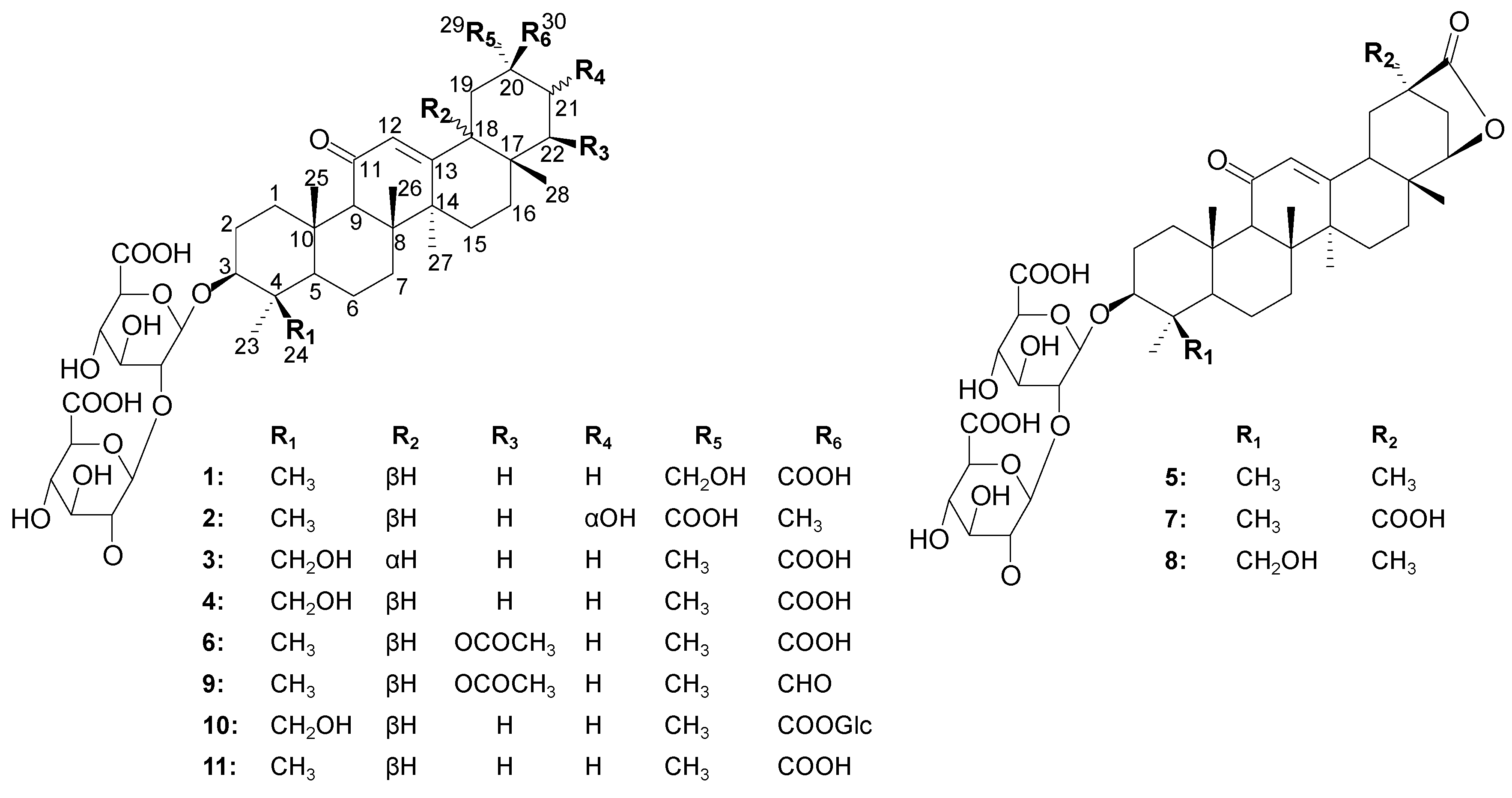
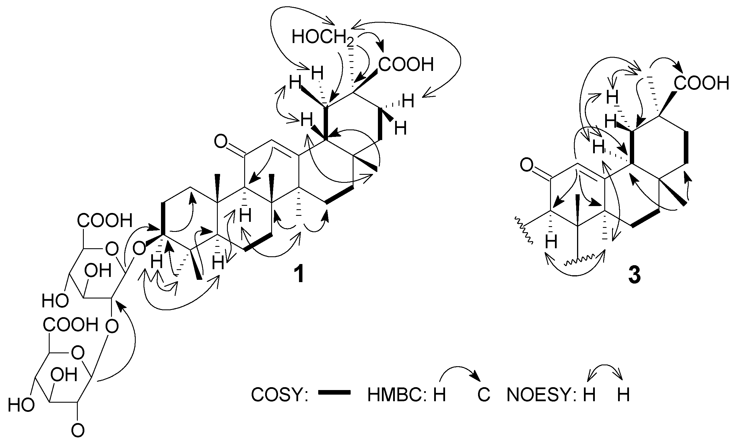
2.1. Structural Determination

Compound 1 was obtained as a white amorphous powder and showed a protonated peaks in the low-resolution positive HR-ESI-MS spectrum at m/z 861.3929 [M + Na]+ and m/z 839.4120 [M + H]+. Its molecular formula was thus determined as C42H62O17, requiring 12 degrees of unsaturation. The UV spectrum showed an absorption maximum at 250.8 nm (MeOH, log ε 4.14), indicating the presence of an α,β-unsaturated carbonyl moiety. In the 1H-NMR and 13C-NMR spectra, representative signals of eight tertiary carbons at δC 75.3–85.5, two carboxyl carbons at δC 174.8 and 174.8, and two anomeric carbons δC 105.9, δH 5.03 (1H, d, J = 8.0 Hz) and 107.3, 5.38 (1H, d, J = 7.5 Hz) suggested the presence of two monosaccharide residues. The coupling constants of the anomeric protons indicated glycosidic bonds with β-configurations [8]. Acid hydrolysis and GC comparison with authentic samples indicated the presence of D-glucuronic acid (GlcA) [9]. This deduction was further supported by the signals of two fragment ion peaks at m/z 663.3762 [M + H − C6H8O6]+ and m/z 487.3448 [M + H − 2C6H8O6]+ in the HR-ESI-MS spectrum. The glycosidic site was established unambiguously by a HMBC experiment in which a long-range correlation between H-1H 5.03) and C-3 (δC 91.3), H-1′′H 5.38) and C-2C 85.5). Thus, the carbohydrate sequence of 1 was established as 3-O-β-d-glucuronopyranosyl-(1→2)-β-d-glucuronopyranosyl. The 1H-NMR spectrum of the aglycone moiety of 1 showed signals corresponding to six tertiary methyls [δH 1.27, 1.08, 1.07, 0.85, 1.36 and 0.70, (each 3H, s)], one oxygenated methylene [δH 4.06, 3.98, (2H, d, 10.5)], one oxygenated methine [δH 3.27 (1H, dd, 4.0 and 11.5)], and one unsaturated methine [δH 5.83 (1H, s)], while the 13C NMR and DEPT spectrum displayed 30 carbon resonances, containing six methyls, ten methylenes (including one oxygenated methylene), five methines (including one oxygenated methine and one unsaturated methine), and nine quaternary carbons (including one carbonyl quaternary carbon, one unsaturated quaternary carbon and one carboxyl carbon). Therefore, compound 1 was considered to be an oleanane-type triterpene glucuronide bearing a 12(13)-double bond and a keto group at C-11. In the HMBC spectrum, correlations of δH 5.03 (H-1) to δC 91.3 (C-3) and δH 5.38 (H-1′′) to δC 85.5 (C-2) could be observed. In addition, the correlations in the HMBC spectrum from H-1 at δH 5.03, H-23 at δH 1.27 and H-24 at δH 1.08 to C-3 at δC 91.3 helped in assigning one oxygenated methine at C-3.
Detailed analysis of the above 1D-NMR data and 2D-NMR correlations indicated that 1 is an oleanane-type saponin derivative and is structurally related to the known compound licorice-saponin G2 (4). The comparison of the NMR data of 1 with those of 4 suggested that the hydroxyl group at C-24 in 4 was transposed to C-29 in 1. The HMBC correlations from δH 3.98, 4.06 (H-29) to δC 39.1 (C-19) and δC 180.2 (C-30) and the 1H–1H COSY correlations between the proton signal at δH 2.49 (H-18) and δH 1.98, 2.24 (H-19) confirmed that hydroxyl group was connected to C-29 in compound 1 (Figure 2).
The relative configuration of 1 was established on the basis of NOESY correlations of H-3 with H-1'α, H-23 and H-5, H-9 with H-5 and H-27, and H-18 with H-28β and H-19β, as well as H-29 with H-19α, H-21α which revealed that the substituent groups of C-3, H-18 were β-oriented and C-29 was α-oriented. From these spectroscopic data, compound 1 was deduced to be 3β-O-[β-d-glucuronopyranosyl-(1→2)-β-d-glucuronopyranosyl]-29-hydroxyglycyrrhetic acid, and named licorice-saponin P2. The structure of compound 1 was confirmed to be as shown in Figure 1 and the 1H- and 13C-NMR data are listed in Table 1.
Figure 2. Key COSY, NOESY and HMBC correlations of compounds 1 and 3.
Molecules 20 06273 g002
Table 1. NMR Chemical Shifts of Compounds 1–3 (C5D5N, 500 MHz).
Position123
δC multδH (J in Hz)δC multδH (J in Hz)δC multδH (J in Hz)
141.4 CH20.89 *,2.81 (m)40.5 CH21.05 *,3.00 (m)40.3 CH21.03 *,2.97(m)
228.6 CH21.84 (m),2.11 (m)27.7 CH21.98 (m),2.26 (m)27.3 CH22.12(m),2.23(m)
391.3 CH3.27 (dd,4.0,11.5)90.3 CH3.37(dd,4.0,11.5 )90.4 CH3.53(dd,4.5,11.5)
441.8 qC 40.9 qC 45.2 qC
557.3 CH0.62 (m)56.4 CH0.71 (m)56.7 CH0.87 *
619.5 CH21.18 *,1.41 (m)18.6 CH21.25 *,1.49 (m)19.1 CH21.55 *,1.72 *
734.8 CH21.12 *,1.47 (m)34.1 CH21.22 *,1.49 (m)33.9 CH21.28 *,1.54 *
847.6 qC 46.5 qC 46.4 qC
964.0 CH2.31 (s)63.1 CH2.44 (s)62.7 CH2.43(s)
1034.6 qC 38.3 qC 37.8 qC
11202.4 qC 201.1 qC 200.5 qC
12130.3 CH5.83 (s)129.7 CH5.89 (s)129.4 CH5.82(s)
13173.1 qC 171.7 qC 170.6 qC
1445.5 qC 45.0 qC 44.4 qC
1528.6 CH21.04 *,1.64 (m)27.9 CH21.05 *,1.68 (m)27.4 CH21.08 *,1.70 (m)
1628.6 CH20.90 *,2.11 (m)31.1 CH21.28 *,3.09 (m)27.3 CH20.95 *,2.10 (m)
1734.6 qC 34.1 qC 33.3 qC
1850.3 CH2.49 (m)48.2 CH2.37 (m)47.5 CH2.26 (m)
1939.1 CH21.98 *,2.24 (m)36.1 CH21.68 *,3.13 (m)40.6 CH21.59 *,2.47(m)
2053.0 qC 49.1 qC 43.4 qC
2128.6 CH21.99 *,2.23 (m)73.1 CH4.52 (m)30.5 CH21.68 *,2.18(m)
2239.8 CH21.42 *,1.61 (m)44.0 CH21.78 *,1.98 (m)36.6 CH21.38 *,1.53 *
2330.1 CH31.27 (s)29.1 CH31.35 (s)23.7 CH31.49 (s)
2418.9 CH31.08 (s)18.0 CH31.2 (s)64.2 CH23.72 (d, 12),4.40*
2518.8 CH31.07 (s)17.8 CH31.21 (s)17.4 CH31.21 (s)
2620.8 CH30.85 (s)19.9 CH31.08 (s)19.4 CH31.07 (s)
2725.6 CH31.36 (s)24.1 CH31.46 (s)24.3 CH31.40 (s)
2830.8 CH30.70 (s)30.2 CH30.93 (s)29.3 CH30.88 (s)
2972.9 CH24.06 (d, 10.5), 3.98 (d, 10.5)181.0 qC 20.6 CH31.42 (s)
30180.2 qC 22.0 CH31.44 (s)181.7 qC
1'106.7 CH5.03 (d, 8.0)105.9 CH5.09 (d, 7.5)105.3 CH5.09 (d, 8.0)
2'85.5 CH4.18 *84.8 CH4.26 *81.6 CH4.38 *
3'79.4 CH4.49 *78.6 CH4.54 *78.6 CH4.56 *
4'75.3 CH4.38 *78.2 CH4.69 *78.3 CH4.71 *
5'79.3 CH4.65 *74.2 CH4.53 *74.1 CH4.55 *
6'174.8 qC 173.8 qC 173.4 qC
1''108.1 CH5.38 (d, 7.5)107.3 CH5.45(d, 7.5)105.5 CH5.77 (d, 8.0)
2''78.5 CH4.14 *77.6 CH4.22 *76.5 CH4.31 *
3''79.4 CH4.32 *78.5 CH4.42 *78.5 CH4.42 *
4''75.3 CH4.47 *74.4 CH4.62 *78.4 CH4.63 *
5''80.1 CH4.51 *79.2 CH4.61 *73.9 CH4.62 *
6''174.8 qC 173.4 qC 173.2 qC
* where 1H-NMR signals were overlapped, chemical shift data were obtained from 2D correlations.
Compound 3 produced a protonated ion at m/z 839.4069 [M + H]+ by HR-ESI-MS, which indicated its molecular formula C42H62O17. In the 13C-NMR spectrum eight tertiary carbons at δC 70–85, two carboxyl carbons at δC 173.4 and 173.2, and two anomeric carbons δC 105.3 and 105.5 suggested the presence of two glucuronopyranosyl moieties. This conclusion was confirmed by two fragment ion peaks at m/z 663.3785 [M − C6H8O6 + H]+ and 487.3373 [M − 2C6H8O6 + H]+, as well as acid hydrolysis and GC analysis.
The NMR spectrum of 3 were very similar to that of 4, and detailed analysis revealed that the main difference could be seen in the NOESY spectrum. Comparative analysis of this spectrum showed that correlations of H-18 with H-19α and H-29 in 3 (Figure 2), rather than correlations of H-18 with H-28β in 4, were present, which indicated that H-18 of 3 was α-oriented. The other correlations of 3, including H-1β/H-25 and H-26, H-3/H-5 and H-1α, H-23/H-3 and H-1', were the same as those of 4. Accordingly, compound 3 was identified as 3β-O-[β-D-glucuronpyranosyl-(1→2)-β-D-glucuron-pyranosyl]-24-hydroxy-18α-glycyrrhetic acid, which was assigned the trivial name licorice-saponin Q2.
The known constituents, namely macedonoside A (2) [10], licorice-saponin G2 (4) [11], licorice-saponin E2 (5) [12], 22β-acetoxyglycyrrhizin (6) [13], uralsaponin D (7) [7], 24-hydroxylicorice-saponin E2 (8) [14], 22β-acetoxyglycyrrhaldehyde (9) [15], licorice-saponin A3 (10) [12] and glycyrrhizin (11) [15] were identified by comparison of their NMR data with the literature data.

2.2. Hepatoprotective Activity

All the separated compounds were assessed for their hepatoprotective activities against the increase of AST and LDH levels in primary rat hepatocytes injured by d-GalN. The maximum nontoxic concentrations of tested compounds on primary rat hepatocytes were in the range of 120–240 μM. A set of cells in culture medium treated with d-GalN was used as the model group, and in comparison to the model group, macedonoside A (2), licorice-saponin Q2 (3), licorice-saponin G2 (4), 22β-acetoxy-glycyrrhizin (6) and glycyrrhizin (11) notably lowered AST (10.3–16.5 U·L−1) and LDH (200.7–242.8 U·L−1) in the range of concentration 30–120 μM. (Table 2).
Table 2. Hepatoprotective activities of isolated saponins on AST and LDH Levels in primary cultures of rat hepatocytes injured by d-GalN a.
GroupsConcentration (μM)AST (U·L−1)LDH (U·L−1)
Control 6.9 ± 1.7157.4 ± 11.7
Model 17.1 ± 2.4 b253.5 ± 13.5 b
23014.6 ± 3.1231.0 ± 18.3 c
6013.1 ± 2.1 c213.7 ± 19.6 d
12011.2 ± 1.9 d215.9 ± 8.2 d
33013.3 ± 2.9 c224.6 ± 21.4 c
6012.9 ± 3.2 d208.6 ± 16.0 d
12010.8 ± 2.8 d200.7 ± 15.1 d
43014.8 ± 2.9237.2 ± 16.4
6013.7 ± 2.6 c232.7 ± 25.7
12012.6 ± 2.8 c224.9 ± 25.2 c
63016.5 ± 2.2242.8 ± 15.2
6014.6 ± 3.0228.7 ± 14.0 c
12013.2 ± 2.7 c221.7 ± 19.5 d
113014.1 ± 2.1 c236.3 ± 9.4 c
6013.0 ± 2.2 c219.1 ± 19.3 d
12011.6 ± 1.8 d207.8 ± 21.9 d
Silibinin Meglumine5010.3 ± 2.2 d219.1 ± 10.9 d
a n = 3, mean ± SD. Control: a set of hepatocytes maintained in culture medium. Model: a set of hepatocytes maintained in culture medium and treated only with D-GalN. b p < 0.01, compared to control group. c p < 0.05, compared to model group. d p < 0.01, compared to model group.
Comparing the activities of these saponins, compound 5 and 7 was shown to have significantly weaker hepatoprotective activities than the compound 2 and 11 owing to presence of a lactone ring at position 22(30). Compound 11 showed stronger activity than 1. That might be because an additional CH2OH group is preferable to improve the steric hindrance, thus resulting in a decrease in the bonding capacity with active targets. Interestingly, compound 3 displayed higher activity than compound 4. The reason might be that compound 3 with a 18α-H group was found to be favorable for the anti-liver injury activity. On the basis of the above analysis, it seemed that a carboxyl residue at position 29 or 30 was possibly the necessary group for hepatoprotective activity.

2.3. Enzyme Inhibition Activity

As a regulator associated with the stability of the liver cell membrane, phospholipase A2 (PLA2) is a promising target for hepatoprotective drug development [16]. To examine whether the compounds inhibit activities on PLA2, the enzyme inhibitory potency of all isolated compounds was conducted and the results were summarized in Table 3. Among these, two saponins (compounds 2 and 3) and glycyrrhizin (11) exhibited efficient inhibitory activity with IC50 value of 6.9 μM, 3.6 μM and 9.3 μM, respectively. Compounds 4, 6 and 7 showed moderate inhibitory activities with IC50 values of 16.9 μM, 27.1 μM and 32.2 μM, respectively.
What was noteworthy, is that analysis of the two assays of 1–11 showed that there was good relationship between PLA2 inhibitory activities and hepatoprotective effects, leading to the hypothesis that inhibition of PLA2 was one of the possible mechanisms of the hepatoprotective effect of licorice saponins.
Table 3. Inhibitory activities of isolated saponins on PLA2.
CompoundsIC50 (μM)
1>50
26.9 ± 0.5
33.6 ± 0.3
416.9 ± 0.3
5>50
627.1 ± 0.9
732.2 ± 0.5
8>50
9>50
10>50
119.3 ± 0.8
Diethylenetriaminepentaacetic acid1.8 ± 0.1

3. Experimental Section

3.1. General Procedures

UV-vis spectra were recorded using a UV-2401 spectrophotometer (Shimadzu, Kyoto, Japan). The 1D and 2D-NMR spectra (1H-1H COSY, NOESY, HSQC and HMBC) were obtained using an ASR-500 spectrometer (Bruker, Fällanden, Switzerland, 500 MHz for 1H and 500 MHz for 13C spectra). All compounds were dissolved in C5D5N and chemical shifts were reported in ppm (δ) relative to TMS. HR-TOF-MS was recorded on a Bruker MicroTOF-Q spectrometer. Column chromatography was performed on polyamide resin (100–200 mesh) and macroporous resin (20–40 mesh, AB-8) columns. Medium pressure liquid chromatography (MPLC) was carried out on a LISURE apparatus (Chromatography Pump E2-purifier, Lisure Science Co., Ltd, Suzhou, China) with ODS column (6.5 cm × 50 cm; 25–50 μm, Merck K GaA). Preparative HPLC was performed by Econosil C18 column (22 × 250 mm; 10 μm, Alltech, Lexington, KY, USA) on a Waters 600 HPLC instrument (Waters, Milford, MA, USA). HPLC was performed on an Agilent 1100 HPLC instrument (Agilent Technologies Inc., Palo Alto, CA, USA) connected to a UV detector, which was equipped with a Thermo C18 column (250 mm × 4.6 mm, I.D. 5 μm). GC was run on Varian CP-3800 Gas Chromatograph (VARIAN, INC., Palo Alto, CA, USA) equipped with a CP-sil 5 CB capillary column (30 m, 0.25 mm i.d., 0.25 μm) and a Saturn 2200 Mass detector. The chemical reagents were supplied by Nanjing Chemical Plant (Nanjing, China). The fluorescence value of each well in the biological activity assays was read in a ELISA plate reader (Bio-Tek Instruments, Winooski, VT, USA).

3.2. Material

The roots of Glycyrrhiza inflata were collected in Weli County, Xinjiang Uygur Autonomous Region, China, October 2013. A voucher sample (No. 20131015) was preserved in Nanjing University of Chinese Medicine, and identified by Prof. Qi-Nan Wu.

3.3. Extraction and Isolation

The roots of G. inflata (dry weight, 25 kg) were exhaustively extracted two times with boiling water (200 L × 2, each extraction lasted 2 hours). The combined solutions (about 300 L) were passed over a polyamide resin column (30 L, 100–200 mesh, 20 × 200 cm) with a flow rate of 120 mL/min, and then the effluent was chromatographed on a macroporous resin column (25 L, 20–40 mesh, 20 × 200 cm) using EtOH–H2O (70:30, 30 L, flow rate 100 mL/min) as eluent to afford the crude saponin fraction. The 70% EtOH combined elution was concentrated in vacuo. The residue (a total of about 350 g) was then subjected in four portions (90 g of residue each) to MPLC with a ODS column (800 g, 25–50 μm, 6.5 × 50 cm) using a continuous gradient of MeOH–H2O–HCOOH (50:50:1, 55:45:1, 60:40:1, 65:35:1, v/v, each 4 L) to produce four fractions (Fr. I–IV). Fr. II (about 45 g) was further subjected to MPLC chromatography on an ODS column with a gradient of MeOH–H2O–HCOOH (50:50:1, 52:48:1, 54:46:1, 56:44:1, 58:42:1, 60:40:1, each 3 L, v/v) as eluent to afford six subfractions (SFr. IIa–IIe). SFr. IIa was subjected to preparative HPLC with CH3CN–H2O–HCOOH (27:73:1, flow rate 10 mL/min) elution to give compound 2 (55 mg, tR 19.2 min). SFr. IIb was purified by preparative HPLC using CH3CN–H2O–HCOOH (27:73:1, flow rate 10 mL/min) to give compound 1 (32 mg, tR 22.6 min) and compound 10 (72 mg, tR 34.2 min). SFr. IIc was subjected to preparative HPLC with CH3CN–H2O–HCOOH (30:70:1, flow rate 10 mL/min) as eluent to afford compound 3 (38 mg, tR 22.6 min), compound 4 (205 mg, tR 29.1 min) and compound 8 (26 mg, tR 37.5 min). SFr. IId was chromatographed by preparative HPLC and eluted with CH3CN–H2O–HCOOH (32:68:1, flow rate 10 mL/min) to give compound 5 (81 mg, tR 26.1 min) and compound 7 (34 mg, tR 34.5 min). SFr. IIe was subjected to preparative HPLC with CH3CN–H2O–HCOOH (35:65:1, flow rate 10 mL/min) to produce compound 6 (87 mg, tR 30.1 min) and compound 9 (40 mg, tR 39.4 min). Separation of fraction III (about 75 g) by MPLC with ODS column chromatography yielded 11 (6.2 g) using CH3CN–H2O–HCOOH (40:6:1, flow rate 30 mL/min) as eluent.

3.4. Physical Data of New Compounds

Licorice-Saponin P2 (1)

White amorphous powder; UV (MeOH) λmax (log ε) 250.2 (4.14) nm; IR (KBr) vmax (cm−1): 3463, 2964, 1751, 1652; 1H-NMR (C5D5N, 500 MHz) and 13C-NMR (C5D5N, 125 MHz) spectral data see Table 1; HR-TOF-MS: m/z 839.4120 [M+H]+ (calcd. for C42H63O17, 839.4065).

Licorice-Saponin Q2 (2)

White amorphous powder; UV (MeOH) λmax (log ε) 250.2 (4.16) nm; IR (KBr) vmax (cm−1): 3438, 2975, 1751, 1652; 13C-NMR (C5D5N, 125 MHz) spectral data see Table 1; HR-TOF-MS: m/z 839.4069 [M+H]+ (calcd for C42H63O17, 839.4065).

3.5. Acid Hydrolysis

The configuration of the sugars of compounds 1 and 3 was determined by acid hydrolysis and GC experiments based of the literature procedure [6,9]. The specific steps were as follows: a solution of compounds 1–3 (1.0 mg each) in 1 N HCl (1 mL) was stirred at 90 °C for 2 h. After cooling, the solution was evaporated under a stream of N2. Anhydrous pyridine solutions (0.1 mL) of each residue and l-cysteine methyl ester hydrochloride (0.06 N) were mixed and warmed at 60 °C for 1 h. The trimethylsilylation reagent trimethylsilylimidazole (0.15 mL) was added, followed by warming at 60 °C for another 30 min. After drying the solution, the residue was partitioned between H2O and CH2Cl2 (1 mL, 1:1 v/v). The CH2Cl2 layer was analyzed by GC/MS. The peaks of authentic sample of D-glucuronic acid after treatment in the same way were detected at 14.23 min. The final result was to compare the retention times of monosaccharide derivatives with standard sample. The absolute configuration of sugar was confirmed to be d-glucuronic acid (d-glucuronic acid for compound 1 with retention time 14.21 min; d-glucuronic acid for compound 3 with retention time 14.22 min).

3.6. Cell Assay

Isolated rat hepatocytes were prepared from male Wistar rats by a collagenase perfusion technique as described previously [17]. The d-GalN concentration used for cell culture treatment was previously determined according to a modification of the method of Morikawa et al. [18]. The cultured cells in logarithmic growth phase were made into a single-cell suspension and seeded in 96-well plates (1 × 10⁴ cells/well) in the DMEM/F 12 with 2% FBS complete medium for 24 h at 37 °C. Then, the hepatocytes were exposed to 2 mM d-GalN for 2 h to induce hepatotoxocity. The medium with silibin meglumine (as positive drug, purity 95.6%, Hunan Xieli Pharmaceutical Co., Ltd., Zhuzhou, China) and different concentrations of test compounds was mixed in cell medium (final test compounds concentration were 30 μM, 60 μM and 120 μM, respectively), and incubated for 24 h. The obtained reacted supernatant was directly used to detect ALT and AST levels. The control group was a set of cells maintained in culture medium, while the model group was a set of cells maintained in culture medium and treated only with d-GalN. All data are expressed as the mean ± SD of at least three independent experiments as indicated. The test for the paired samples was used to determine statistical difference between parameters. These differences were considered significant for p < 0.05 or 0.01.

3.7. Assay for Inhibition against PLA2

The PLA2 inhibitory assays of compounds 1–11 and the positive drug diethylenetriaminepentaacetic acid (Purchased from Aladdin, Los Angeles, CA, USA, purity > 98.0%) were carried out according to the literature [19]. First of all, each tube was added with 1 mL fresh substrate buffer solution (pH = 8.2). After that, 50 μL tested compounds at various concentrations were placed at reaction tube and blank tube, respectively. As for control tube, 50 μL deionized water was instead. Then each tube incubated at 40 °C for 10 minutes. The reaction tube and blank tube were followed by the treatment with PLA2 enzyme (5 μL) at the concentration of 5 μg/mL. Before put them into the incubator at the temperature of 40 °C to react 30 minutes, the content of the tube should be fully blending. The optical density value of each tube was then read in an ELISA plate reader using a wavelength of 495 nm. The IC50 values were calculated from concentration-response curves by means of the GraphPad Prism 5.0 Software (San Diego, CA, USA). Each experiment was repeated three times to get the mean values.

Supplementary Materials

HRESIMS and NMR spectra of the two new compounds and the 13C-NMR data of compound 2 and 4 are available as Supplementary Materials. Supplementary materials can be accessed at: https://www.mdpi.com/1420-3049/20/04/6273/s1.

Acknowledgments

The authors greatly appreciate financial support from the National Natural Science Foundation, China (Program No. 81202881), a project funded by the Priority Academic Program Development of Jiangsu Higher Education Institutions. We also thank Qi-Nan Wu (Nanjing University of Chinese Medicine, Nanjing, China) for identification of Glycyrrhiza inflata L. and the Shanghai Institute of Organic Chemistry (Chinese Academy of Sciences, Shanghai, China) for measuring the NMR spectra.

Author Contributions

The list authors contributed to this work as follows: Y. Z. conceived and designed the experiments, J. W., S. F., and T. W. performed the experiments and analyzed the data, H. C. and C. L. wrote the paper, Y. T. and G. P. polished the paper. All authors read and approved the final manuscript.

Conflicts of Interest

The authors declare no conflict of interest.

References

  1. Shim, S.B.; Kim, N.J.; Kim, D.H. Beta-glucuronidase inhibitory activity and hepatoprotective effect of 18 beta-glycyrrhetinic acid from the rhizomes of Glycyrrhiza uralensis. Planta Med. 2000, 66, 40–43. [Google Scholar] [CrossRef] [PubMed]
  2. Wu, Y.T.; Shen, C.; Yin, J.; Yu, J.P.; Meng, Q. Azathioprine hepatotoxicity and the protective effect of liquorice and glycyrrhzic acid. Phytother. Res. 2006, 20, 640–645. [Google Scholar] [CrossRef] [PubMed]
  3. Wolkerstorfer, A.; Kurz, H.; Bachhofner, N.; Szolar, O.H. Glycyrrhizin inhibits influenza A virus uptake into the cell. Antivir. Res. 2009, 83, 171–178. [Google Scholar] [CrossRef] [PubMed]
  4. Fujisawa, Y.; Sakamoto, M.; Matsushita, M.; Fujita, T.; Nishioka, K. Glycyrrhizin inhibits the lytic pathway of complement–possible mechanism of its anti-inflammatory effect on liver cells in viral hepatitis. Micrrobiol. Immunol. 2000, 44, 799–804. [Google Scholar] [CrossRef]
  5. Fu, Y.; Chen, J.; Li, Y.J.; Zheng, Y.F.; Li, P. Antioxidant and anti-inflammatory activities of six flavonoids separated from licorice. Food Chem. 2013, 141, 1063–1073. [Google Scholar] [CrossRef] [PubMed]
  6. Wei, J.H.; Zheng, Y.F.; Li, C.Y.; Tang, Y.P.; Peng, G.P. Bioactive constituents of oleanane-type triterpene saponins from the roots of Glycyrrhiza glabra. J. Asian Nat. Prod. Res. 2014, 16, 1044–1053. [Google Scholar] [CrossRef] [PubMed]
  7. Zheng, Y.F.; Qi, L.W.; Cui, X.B.; Peng, G.P.; Peng, Y.B.; Ren, M.T.; Cheng, X.L.; Li, P. Oleanane-type Triterpene Glucuronides from the Roots of Glycyrrhiza uralensis Fischer. Planta Med. 2010, 76, 1457–1463. [Google Scholar] [CrossRef]
  8. Antonov, A.S.; Avilov, S.A.; Kalinovsky, A.I.; Anastyuk, S.D.; Dmitrenok, P.S.; Kalinin, V.I.; Taboada, S.; Bosh, A.; Avila, C.; Stonik, V.A. Triterpene Glycosides from Antarctic Sea Cucumbers. 2. Structure of Achlioniceosides A1, A2, and A3 from the Sea Cucumber Achlionice Wiolaecuspidata (=Rhipidothuria racowitzai). J. Nat. Prod. 2009, 72, 33–38. [Google Scholar]
  9. Duan, W.J.; Yang, J.Y.; Chen, L.X.; Zhang, L.J.; Jiang, Z.H.; Cai, X.D.; Zhang, X.; Qiu, F. Monoterpenes from Paeonia albiflora and their inhibitory activity on nitric oxide production by lipopolysaccharide-activated microglia. J. Nat. Prod. 2009, 72, 1579–1584. [Google Scholar] [CrossRef] [PubMed]
  10. Shibano, M.; Nukui, H.; Kita, S.; Kusano, G.; Shibata, T.; Watanabe, H.; Ohashi, H. Studies on natural medicines index compounds for HPLC Glycyrrhiza macedonica. Nat. Med. 1999, 53, 166–172. [Google Scholar]
  11. Kitagawa, I.; Zhou, J.L.; Sakagami, M.; Uchida, E.; Yoshikawa, M. Licorice-saponins F3, G2, H2, J2, and K2, five new oleanene-triterpene oligoglycosides from the root of Glycyrrhiza uralensis. Chem. Pharm Bull. 1991, 39, 244–246. [Google Scholar] [CrossRef]
  12. Kitagawa, I.; Zhou, J.L.; Sakagami, M.; Taniyama, T.; Yoshikawa, M. Licorice-saponins A3, B2, C2, D3 and E2, five new oleanene-type triterpene oligoglycosides from Chinese Glycyrrhizae Radix. Chem. Pharm Bull. 1988, 36, 3710–3713. [Google Scholar] [CrossRef]
  13. Li-Yang, J.; Nakajima, J.I.; Kimura, N.; Saitob, K.; Seo, S. Oleanane-type triterpene glycosides from Glycyrrhiza uralensis. Nat. Prod. Commun. 2007, 2, 243–248. [Google Scholar]
  14. Zhu, X.M.; Di, Y.T.; Peng, S.L.; Wang, M.K.; Ding, L.S. Chemical constituents from root of Glycyrrhiza uralensis. Chin. Tradit. Herb Drugs 2003, 34, 198–201. [Google Scholar]
  15. Zhang, R.Y.; Zhang, J.H.; Wang, M.T. Studies on the saponins from the root of Glycyrrhiza uralensis Fisch. Yao Xue Xue Bao 1986, 21, 510–515. [Google Scholar] [PubMed]
  16. Ohtsuki, K.; Abe, Y.; Shimoyama, Y. Separation of phospholipase A2 in Habu snake venom by glycyrrhizin (GL)-affinity column chromatography and identification of a GL-sensitive enzyme. Biol. Pharm Bull. 1998, 21, 574–578. [Google Scholar] [CrossRef] [PubMed]
  17. Lee, E.J.; Kim, S.R.; Kim, J.; Kim, Y.C. Hepatoprotective phenylpropanoids from Scrophularia buergeriana roots against CCl(4)-induced toxicity: action mechanism and structure-activity relationship. Planta Med. 2002, 68, 407–411. [Google Scholar] [CrossRef] [PubMed]
  18. Morikawa, T.; Ninomiya, K.; Imura, K.; Yamaguchi, T.; Akagi, Y.; Yoshikawa, M.; Hayakawa, T.; Muraoka, O. Hepatoprotective triterpenes from traditional Tibetan medicine Potentilla anserina. Phytochemistry 2014, 102, 169–81. [Google Scholar] [CrossRef] [PubMed]
  19. Liu, B.; Qi, Y.; Wang, M.; Cai, R.L.; Song, Y. A method for screening Phospolipase A2 inhibitors in vitro. Chin. Pharma Bull. 2006, 22, 890–892. [Google Scholar]
  • Sample Availability: Samples are not available from authors.

