Article

Flavonoids from Sideritis Species: Human Monoamine Oxidase (hMAO) Inhibitory Activities, Molecular Docking Studies and Crystal Structure of Xanthomicrol

1
Department of Pharmaceutical Botany, Faculty of Pharmacy, Hacettepe University, Ankara 06100, Turkey
2
Department of Biochemistry, Faculty of Pharmacy, Hacettepe University, Ankara 06100, Turkey
3
Department of Bioinformatics and Genetics, Faculty of Engineering and Natural Sciences, Cibali Campus, Kadir Has University, Fatih, Istanbul 34083, Turkey
4
Science and Technology Application and Research Center, Faculty of Pharmacy, Dicle University, Diyarbakir 21280, Turkey
5
Department of Pharmaceutical Chemistry, Faculty of Pharmacy, Dicle University, Diyarbakir 21280, Turkey
6
Department of Pharmacognosy, Faculty of Pharmacy, Near East University, Lefkoşa, Mersin-10, Turkey
*
Author to whom correspondence should be addressed.
Molecules 2015, 20(5), 7454-7473; https://doi.org/10.3390/molecules20057454
Submission received: 16 March 2015 / Revised: 9 April 2015 / Accepted: 15 April 2015 / Published: 23 April 2015

Abstract

:
The inhibitory effects of flavonoids on monoamine oxidases (MAOs) have attracted great interest since alterations in monoaminergic transmission are reported to be related to neurodegenerative diseases such as Parkinson’s and Alzheimer’s diseases and psychiatric disorders such as depression and anxiety, thus MAOs may be considered as targets for the treatment of these multi-factorial diseases. In the present study, four Sideritis flavonoids, xanthomicrol (1), isoscutellarein 7-O-[6'''-O-acetyl-β-d-allopyranosyl-(1→2)]-β-d-glucopyranoside (2), isoscutellarein 7-O-[6'''-O-acetyl-β-d-allopyranosyl-(1→2)]-6''-O-acetyl-β-d-glucopyranoside (3) and salvigenin (4) were docked computationally into the active site of the human monoamine oxidase isoforms (hMAO-A and hMAO-B) and were also investigated for their hMAO inhibitory potencies using recombinant hMAO isoenzymes. The flavonoids inhibited hMAO-A selectively and reversibly in a competitive mode. Salvigenin (4) was found to be the most potent hMAO-A inhibitor, while xanthomicrol (1) appeared as the most selective hMAO-A inhibitor. The computationally obtained results were in good agreement with the corresponding experimental values. In addition, the x-ray structure of xanthomicrol (1) has been shown. The current work warrants further preclinical studies to assess the potential of xanthomicrol (1) and salvigenin (4) as new selective and reversible hMAO-A inhibitors for the treatment of depression and anxiety.

1. Introduction

The genus Sideritis L. (Lamiaceae) comprises more than 150 species mainly distributed in countries of the Mediterranean-Macaronesian region [1]. Sideritis species , which have become very popular recently, are used as herbs either for the preparation of teas, or for their aromatic properties in local cuisines. They are found in a variety of shops, and marketed as mountain tea, dagçayi, adaçayi, yaylaçayi, malotira, té de Puerto, rabo de gato or zaharena in various Mediterranean countries [2,3,4]. Herbal teas prepared form the aerial parts of Sideritis species have also been used in traditional medicine for thousands of years in the treatment of gastrointestinal disorders such as stomach ache, indigestion and flatulence, to alleviate the symptoms of the common cold including fever, flu, sore throath, and bronchitis, and as analgesics, anticonvulsants, sedatives, tonics and diuretics [4,5,6,7]. Moreover, Sideritis species in the form of tea infusions are popularly used for improving memory function and cognitive ability [3]. The species of the genus are known to be rich in essential oils [4,8,9], diterpenes [4,10,11,12,13] and flavonoids [2,4,5,11,12,13,14,15,16].
Recently, the inhibitory effects of flavonoids on monoamine oxidases (MAO-A and MAO-B) have attracted great interest among researchers. Monoamine oxidases (MAOs) are flavoenzymes located in the outer membrane of the mitochondria that play an important role in the oxidative catabolism of endogenous and dietary amines [17,18]. MAO contains flavin adenine dinucleotide (FAD) as a co-factor and exists in two isoforms in mammals, named hMAO-A and hMAO-B [19]. MAO-A preferentially deaminates serotonin and norepinephrine, and is selectively inhibited by clorgyline, whereas MAO-B preferentially deaminates phenylethylamine and benzylamine and is selectively inhibited by L-deprenil [20,21]. Inhibitors of MAO-A are clinically used as antidepressants and anxiolytics [22,23], while MAO-B inhibitors are used in the treatment of Parkinson’s disease and in the management of symptoms associated with Alzheimer’s disease [24].
The studies on the crystal structures of MAOs and the interactions between MAOs and their ligands [24,25,26,27], have led to the design of new possible inhibitors by docking studies. Some currently used MAO inhibitors produce side effects due to a lack of selectivity towards one of the isoforms [28]. Thus, it is necessary to search for more potent, reversible and selective inhibitors of MAO-A and MAO-B. Despite the great amount of studies related to design and chemical synthesis of novel compounds as therapeutic agents or prodrugs, in recent decades identification of new compounds from natural sources or screening the novel biological activities of known natural compounds still attracts great interest.
A wide number of molecules of natural origin belonging to diverse chemical classes such as flavonoids, xanthones, proanthocyanidins, iridoids, alkaloids, curcumins, cannabinoids, etc. have been shown to inhibit human MAOs [29,30,31]. Among them, flavonoids have attracted more interest as they possess a variety of biological activities such as anti-oxidation and cardio-cerebrovascular protection besides their effects on the central nervous system. Their diverse activities give them extra advantages to be the potential multifunctional therapeutic agents for aging-related diseases [29,30]. Flavonoids are present in almost all terrestrial plants, and form part of the human diet via the consumption of vegetables, fruits, beverages and other plant-derived foods. The MAO inhibitory activities of some well-known flavonoids such as apigenin, kaempferol, quercetin, quercitrin, isoquercitrin and rutin have been previously demonstrated by in vitro and/or in vivo studies [29,30,31], however, few docking studies are available on these compounds in order to clarify the interaction of the compounds with the active site of hMAO [32].
In our previous studies, we have shown the anti-inflammatory, anti-nociceptive, anti-ulcerative antioxidant, aldose reductase and glutathione reductase activities of flavonoids isolated from various Sideritis species [6,16,33,34], in addition to the anti-inflammatory, anti-nociceptive, anti-ulcerative activities of their phenylethanoid glycosides and diterpenoids, and the anti-microbial activity of their iridoids and essential oils. Diuretic, anti-inflammatory, anti-spasmodic, antibacterial and antioxidant activities of different extracts obtained from several Sideritis species growing in Turkey were also previously demonstrated by our research group [6]. In the present work, hMAO-A and hMAO-B inhibitory activities of four Sideritis flavonoids, xanthomicrol (1), isoscutellarein 7-O-[6'''-O-acetyl-β-d-allopyranosyl-(1→2)]-β-d-glucopyranoside (2), isoscutellarein 7-O-[6'''-O-acetyl-β-d-allopyranosyl-(1→2)]-6''-O-acetyl-β-d-glucopyranoside (3), and salvigenin (4) were determined using recombinant human MAO. The molecular docking studies of these compounds were also performed to gain insights into the intermolecular interactions at the active site 3D space of both the hMAO-A and hMAO-B enzymes. In addition, the crystal structure of xanthomicrol (1), which was found to be the most selective hMAO-A inhibitor among those four flavonoids, has been described. The X-ray diffraction analysis demonstrates its exact structure with a dimeric molecule in the asymmetric unit.

2. Results and Discussion

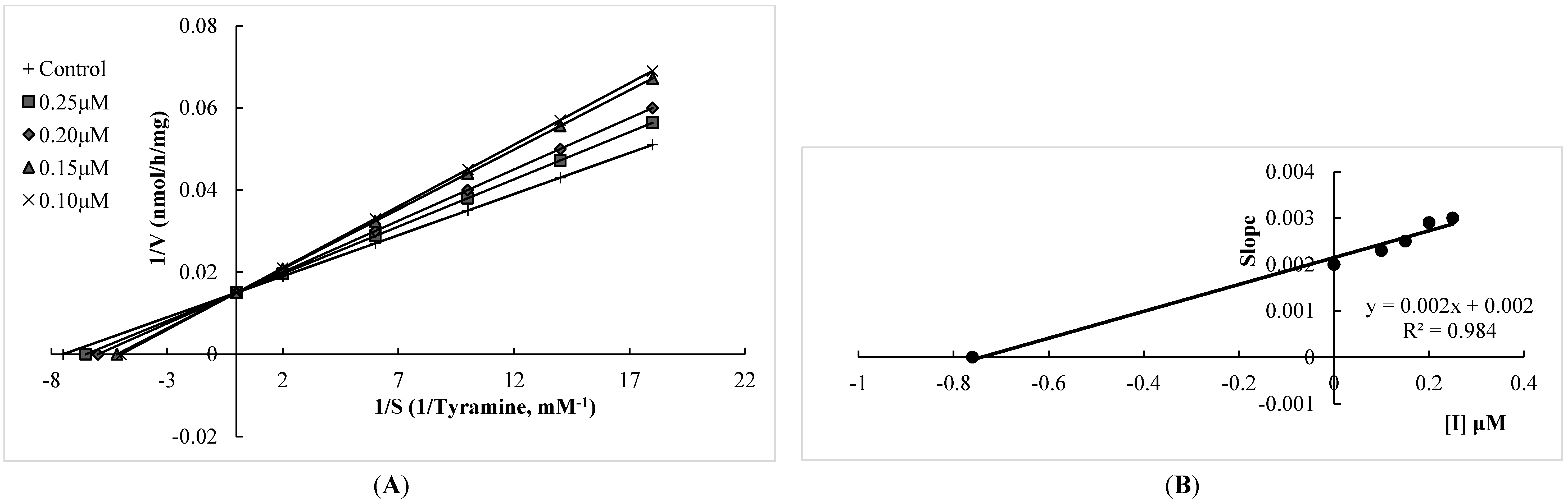
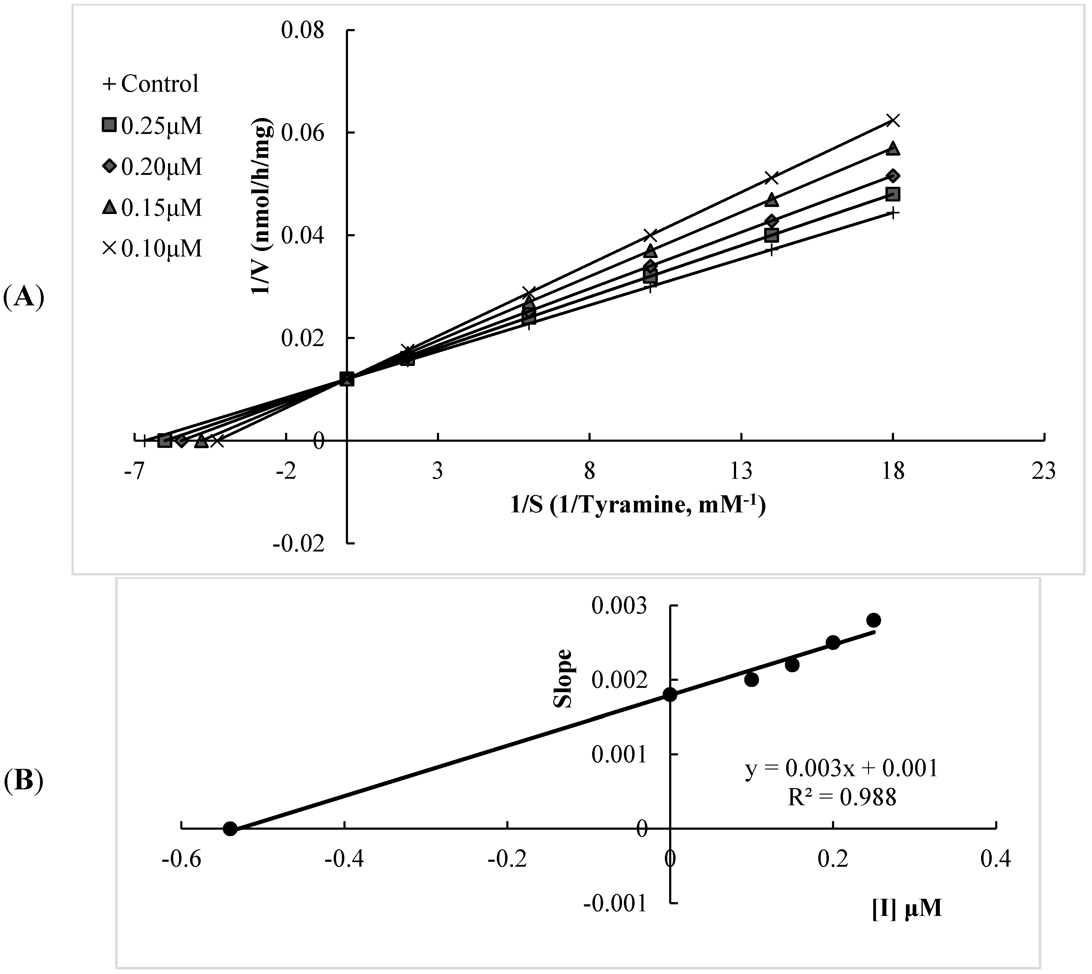
2.1. Human Monoamine Oxidase (hMAO) Inhibitory Activities

The hMAO inhibitory activities of the tested flavonoids (Figure 1) were investigated using commercially available recombinant hMAO-A and hMAO-B with tyramine as the substrate. Kinetic studies were performed in order to provide data for the interaction of four flavonoids with hMAO isoforms. The kinetic parameters Km (Michaelis–Menten constant) and Vmax (maximum rate) were determined by means of Lineweaver–Burk plots over a substrate range of 0.05–0.5 mM.
Figure 1. Structures of the four studied flavonoids: xanthomicrol (1), isoscutellarein 7-O-[6'''-O-acetyl-β-d-allopyranosyl-(1→2)]-β-d-glucopyranoside (2), isoscutellarein 7-O-[6'''-O-acetyl-β-d-allopyranosyl-(1→2)]-6''-O-acetyl-β-d-gluco-pyranoside (3) and salvigenin (4).
Molecules 20 07454 g001
The MAO-A and MAO-B inhibitory potencies of the flavonoids and two well-known MAO-A and -B inhibitors (moclobemide and selegiline, respectively) are shown in Table 1. The results showed that all of the flavonoids tested possessed a degree of selectivity for hMAO-A, with experimental selectivity index (SI) values between 0.007 and 0.086, which suggests that these compounds are highly selective towards hMAO-A. Among them, xanthomicrol (1) appears as remarkably selective, with an experimental SI of 0.007 and selectivity was better than that of moclobemide, a highly potent and selective hMAO-A inhibitor (the experimental SI of moclobemide was determined as 0.010, Table 1). All of the flavonoids tested inhibited hMAO-A in a competitive mode. The kinetic behaviors of xanthomicrol (1) and salvigenin (4), which inhibited hMAO-A potently with experimental Ki values of 0.76 ± 0.03 and 0.54 ± 0.02 μM, respectively, are presented in Figure 2 and Figure 3. Salvigenin (4) appears as the most potent hMAO-A inhibitor among the tested compounds.
MAO inhibitors can be classified as reversible or irreversible, according to their interaction with hMAO isoforms. Although irreversible MAO inhibitors such as selegiline have been extensively used as clinical drugs, this type of inhibition is reported to induce serious cardiovascular toxic effects, mainly provoked by the inhibition of the peripheral MAO-A located in gut, liver and endothelium [35,36]. Besides, the slow and variable enzyme recovery following the withdrawal of irreversible inhibitors is a disadvantage in clinical use, since the turnover rate for MAO biosynthesis in the human brain seems to require about 40 days [37]. Competitive reversible inhibitors have been previously reported to have less influence in the enzyme recovery after withdrawal, and reversible MAO-A inhibitors, such as moclobemide, have no cardiovascular effects, since the ingested tyramine is able to displace the inhibitor from the MAO active site and be metabolized in the normal way by peripheral enzyme in gut and liver [35].
Table 1. Calculated and experimentally determined Ki values of the flavonoids, selegiline and moclobemide for hMAO isoforms A and B.
CompoundsΔG for MAO-ACalculated Ki for MAO-A(μM)ΔG for MAO-BCalculated Ki for MAO-B(μM)Calculated SI **Experimental Ki for MAO-A * (μM)Experimental Ki for MAO-B * (μM)Experimental SI **Selectivity
1−7.811.89−5.7864.250.0290.76 ± 0.0399.54 ± 7.440.007MAO-A
2−4.14927.10+5.78--1100.50 ± 99.2075,600.00 ± 912.500.015MAO-A
3−3.791660.00+8.92--2800.45 ± 180.5080,500.00 ± 950.000.034MAO-A
4−8.270.867−7.423.620.2390.54 ± 0.026.27 ± 0.360.086MAO-A
Selegiline 14.90 ± 1.330.36 ± 0.0241.39MAO-B
Moclobemide 0.014 ± 0.0011.34 ± 0.030.010MAO-A
* Each value represents the mean ± SEM of three experiments; ** The selectivity index (SI) was calculated as Ki (MAO-A)/Ki (MAO-B).
Figure 2. (A) Lineweaver–Burk plot of the competitive inhibition of hMAO-A by compound xanthomicrol (1) (B) Replot of data from the Lineweaver-Burk plot. The Ki value calculated from replots of the slopes of the Lineweaver-Burk plots versus inhibitor concentrations was found as 0.76 μM.
Molecules 20 07454 g002
Figure 3. (A) Lineweaver-Burk plot of the competitive inhibition of hMAO-A by compound salvigenin (4); (B) Replot of data from the Lineweaver–Burk plot. The Ki value calculated from replots of the slopes of the Lineweaver–Burk plots versus inhibitor concentrations was found as 0.54 μM.
Molecules 20 07454 g003
Thus, new selective and reversible inhibitors of MAO-A may be useful therapeutic agents for the treatment of psychiatric diseases and Alzheimer’s and Parkinson’s diseases devoid of undesirable side-effects, such as cheese effect. All of the flavonoids inhibited hMAO-A reversibly as shown in Table 2. Significant recovery of hMAO-A activity was observed after repeated washing of xanthomicrol (1) and salvigenin (4), indicating that these flavonoids are highly reversible inhibitors of hMAO isoforms.
Table 2. Reversibility of hMAO-A inhibition with flavonoids and moclobemide *.
Compounds (0.50 μM)hMAO-A Inhibition (%)
Before WashingAfter Washing
186.49 ± 5.219.23 ± 0.75
280.22 ± 5.6610.90 ± 0.99
374.22 ± 4.8913.35 ± 1.09
489.03 ± 5.028.66 ± 0.65
Moclobemide91.22 ± 6.337.89 ± 0.52
* Each value represents the mean ± SEM of three experiment.

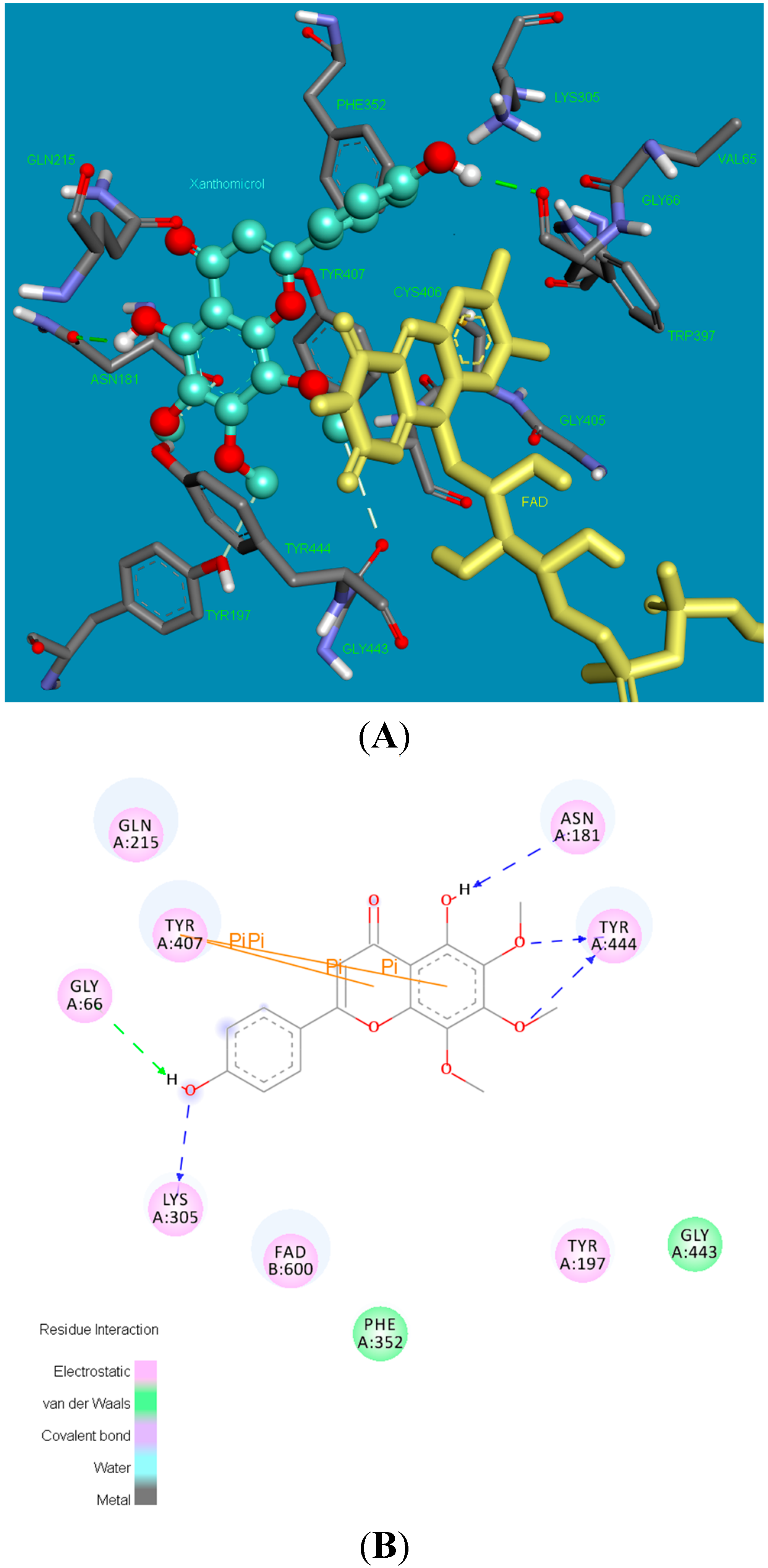
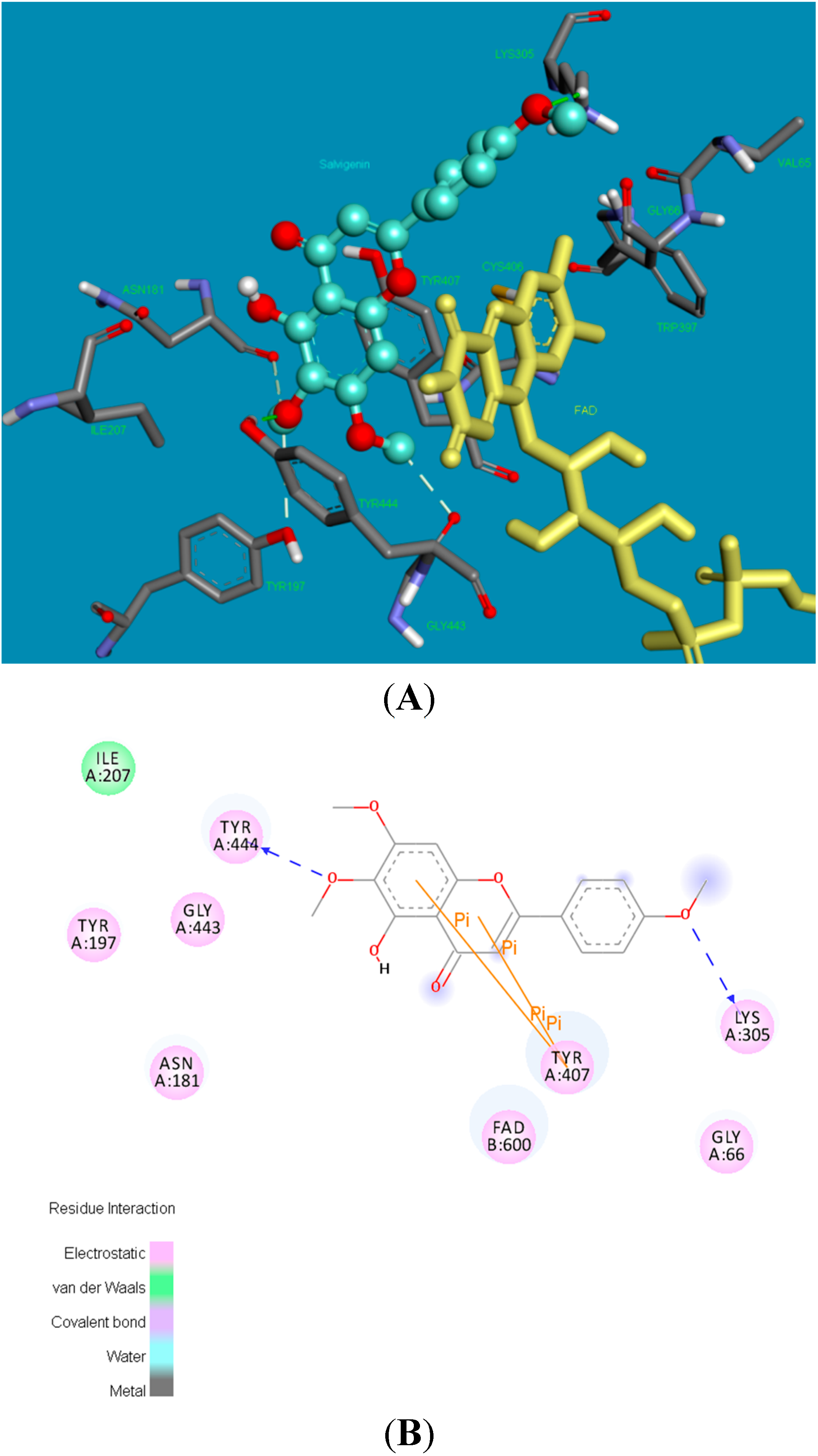
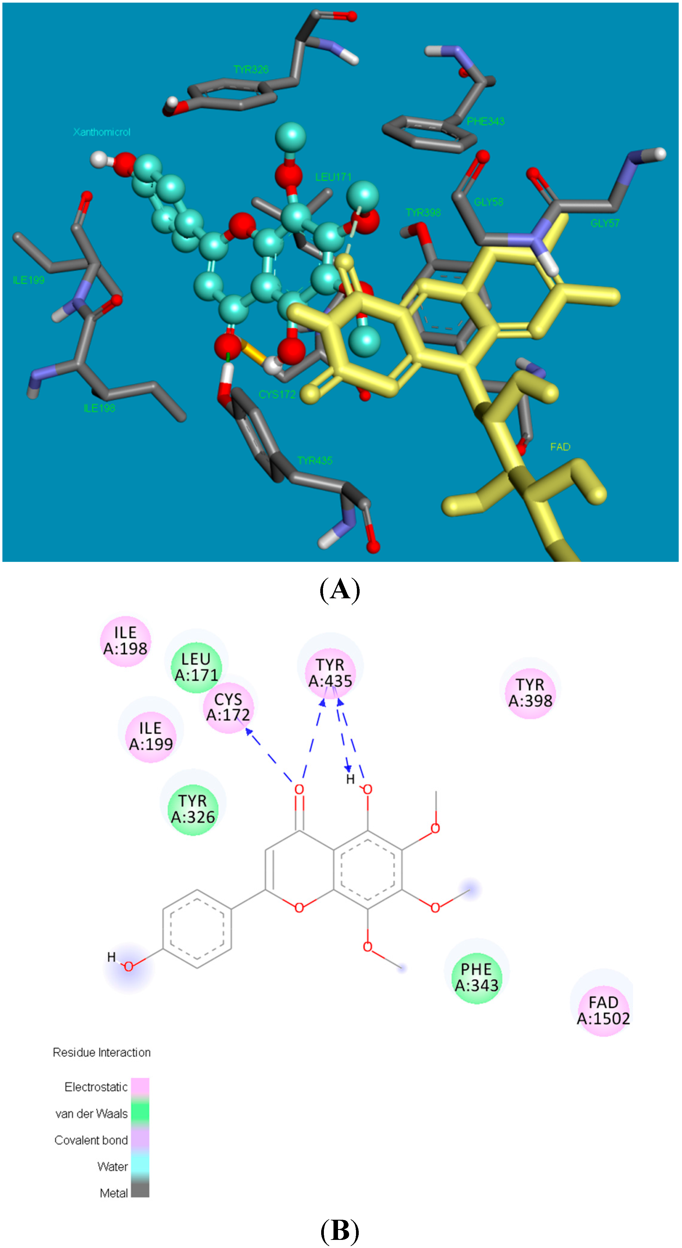
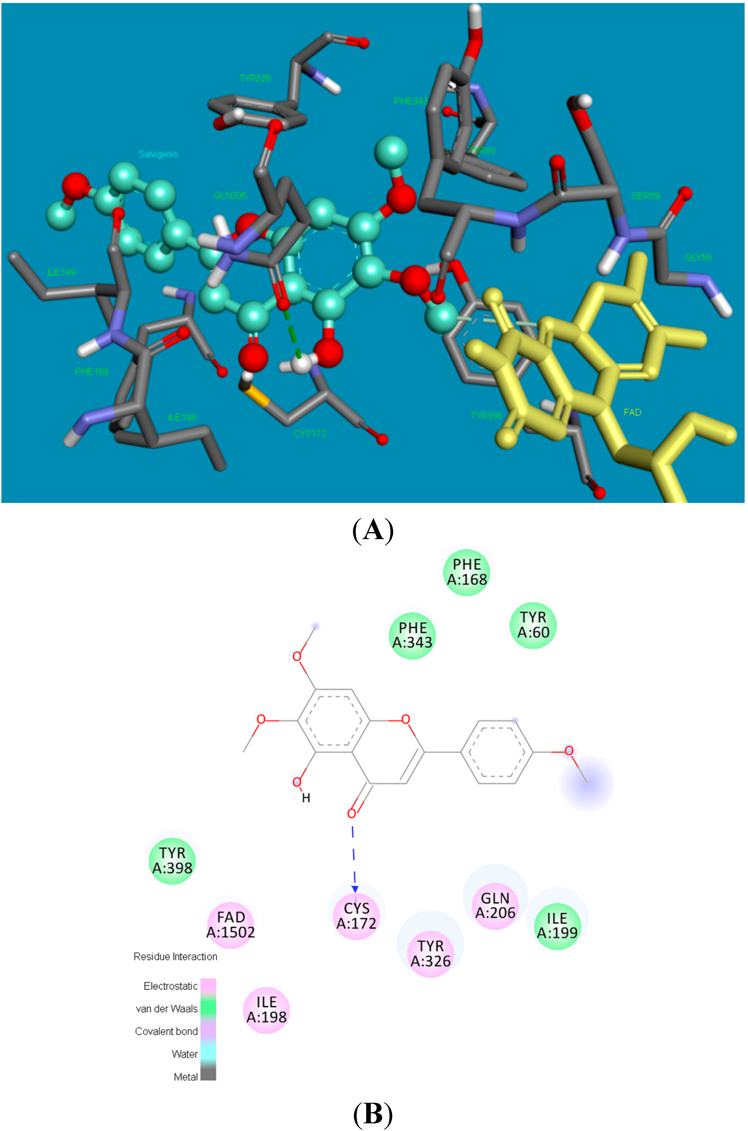
2.2. Molecular Docking Studies

Figure 4 shows the binding pose (3D and 2D) of xanthomicrol (1) in the active site of hMAO-A isoform. The coumarine ring was inserted between the TYR444 and TYR407 residues, which constitute the hydrophobic cage of the active site, forms two π-π interactions with TYR407. The following five hydrogen bonds form between moieties of the xanthomicrol (1) and the side chains of the active site amino acids of the MAO-A isoform: between the -OH and ASN181, -OCH3 and TYR444 (two), -OH and LYS305 and -OH and GLY66.
Figure 4. The 3 and 2 dimensional orientations of xanthomicrol (1) in the active site of hMAO-A. (A) 3D: Amino acid side chains are shown as sticks, the inhibitor is shown as a ball and stick (green), and the cofactor FAD is depicted as a yellow stick; (B) 2D: Purple color shows electrostatic and green color shows van der Waals atractions.
Molecules 20 07454 g004
Analysis of the optimal binding mode for the salvigenin (4) in the hMAO-A active site cavity (Figure 5) revealed that this compound is docked in the vicinity of the FAD co-factor. Salvigenin forms various electrostatic and van der Waals interactions with the active site residues lining the cavity. A hydrogen bond occurs between the methoxy group of the salvigenin (4) and the hydroxy moiety of TYR444. The other strong electrostatic interactions occur between GLY443 backbone carbonyl and methoxy groups, and backbone carbonyl of ASN181 and methoxy group of the salvigenin (4). The benzene ring of the coumarine moiety of the salvigenin (4) was sandwiched between TYR444 and TYR407 making two π-π interactions.
Figure 5. The 3 and 2 dimensional orientations of salvigenin (4) in the active site of hMAO-A. (A) 3D: Amino acid side chains are shown as sticks, the inhibitor is shown as a ball and stick (green), and the cofactor FAD is depicted as a yellow stick; (B) 2D: Purple color shows electrostatic and green color shows van der Waals atractions.
Molecules 20 07454 g005
Figure 6 shows the poses of xanthomicrol (1) in the active side of hMAO-B in 3D and 2D depictions, respectively. Four hydrogen bonds form between the carbonyl and (2 bonds) and hydroxyl (2 bonds) of xanthomicrol (1) and side chains of CYS172 and TYR435. The other interacting side chain residues of hMAO-B with xanthomicrol (1) are ILE198, ILE199, CYS172, LEU171, TYR435, TYR398, TYR326 and PHE 343.
Figure 6. The 3 and 2 dimensional orientations of xanthomicrol (1) in the active site of hMAO-B. (A) 3D: Amino acid side chains are shown as sticks, the inhibitor is shown as a ball and stick (green), and the cofactor FAD is depicted as a yellow stick; (B) 2D: Purple color shows electrostatic and green color shows van der Waals atractions.
Molecules 20 07454 g006
Figure 7 shows the binding pose of salvigenin (4) in the active site of hMAO-B isoform. The major interaction is the hydrogen bond that forms between the carbonyl group of coumarine ring and side chain residue of CYS172. The binding mode adopted by salvigenin (4) fits snugly within a cavity lined with hydrophobic amino acid residues. This hydrophobic pocket includes PHE168, TYR60, ILE199, TYR326, CYS172, ILE198, PHE343, GLN206 and TYR398 amino acids.
Figure 7. The 3 and 2 dimensional orientations of salvigenin (4) in the active site of hMAO-B. (A) 3D: Amino acid side chains are shown as sticks, the inhibitor is shown as a ball and stick (green), and the cofactor FAD is depicted as a yellow stick; (B) 2D: Purple color shows electrostatic and green color shows van der Waals atractions.
Molecules 20 07454 g007
The potency and selectivity of xanthomicrol (1) and salvigenin (4) for the hMAO enzymes can be evaluated from the above data. The detailed analysis of Figure 5, Figure 7 and Table 1 suggest that salvigenin (4) is a better inhibitor of hMAO-A than of the MAO-B isoform. The reason for that the coumarine core is stabilized by more π–π interactions as well as other various attractions in the hMAO-A active side compared to the hMAO-B isozyme (calculated Ki = 0.867 μM for hMAO-A and calculated Ki = 1.89 μM for hMAO-B). However, xanthomicrol (1) is more selective towards hMAO-A than salvigenin (4) (calculated SIs of xanthomicrol (1) and salvigenin (4) were estimated as 0.029 and 0.239, respectively).
Inhibition of MAO by flavonoids appears to be dependent on the phenyl or hydroxyphenyl ring in the 2 position as well as a double bond in the 2 and 3 position of the structure. It has been previously demonstrated that flavonoids isolated from the whole plant and fruits of Cayratia japonica showed MAO inhibitory activity as follows: flavones > flavonols > flavone glycosides > flavanonols. In this series, apigenin inhibited mouse brain MAO-A with an IC50 value of 1.17 μM. It was suggested that MAO inhibitory activity decreased with increasing number of hydroxyl groups on the B ring of the flavones. The presence of electron donating hydrophilic hydroxyl groups in the para position of the B ring has also been reported to favour the MAO-A inhibition [30,31]. Thus, it can be postulated that the p-hydroxyl group in the B ring of xanthomicrol (1) increased the selectivity of this molecule towards hMAO-A.
Monoamine oxidases are responsible for the oxidative deamination of neurotransmitters and dietary amines. Of the two isoenzymes, MAO-A has attracted considerable attention because of its ability to affect the metabolism of serotonin [23]. Depletion of serotonin levels in the central nervous system (CNS) is suggested to be involved in the pathogenesis of depressive disorders [38]. It has been reported that MAO-A activity in the brain is elevated during depression, which causes depletion of serotonin in the brain [39]. Herbal medicines, such as flavonoids, are commonly used for the treatment of depression, along with conventional antidepressants [40]. Thus, in the present study we present four flavonoids isolated from Sideritis species exhibiting highly potent hMAO inhibitory activities. Among them, salvigenin (4) was found as the most potent hMAO-A activity, whereas xanthomicrol (1) appeared as the most selective one towards the hMAO-A isoform.

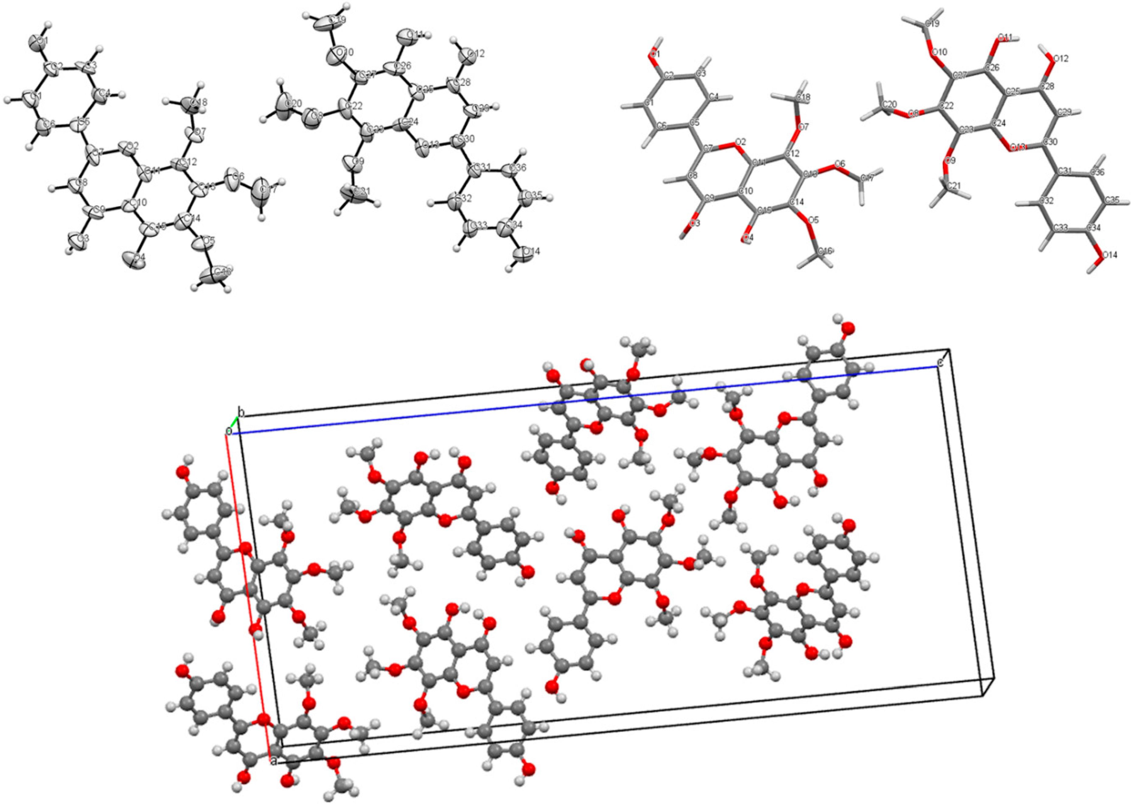
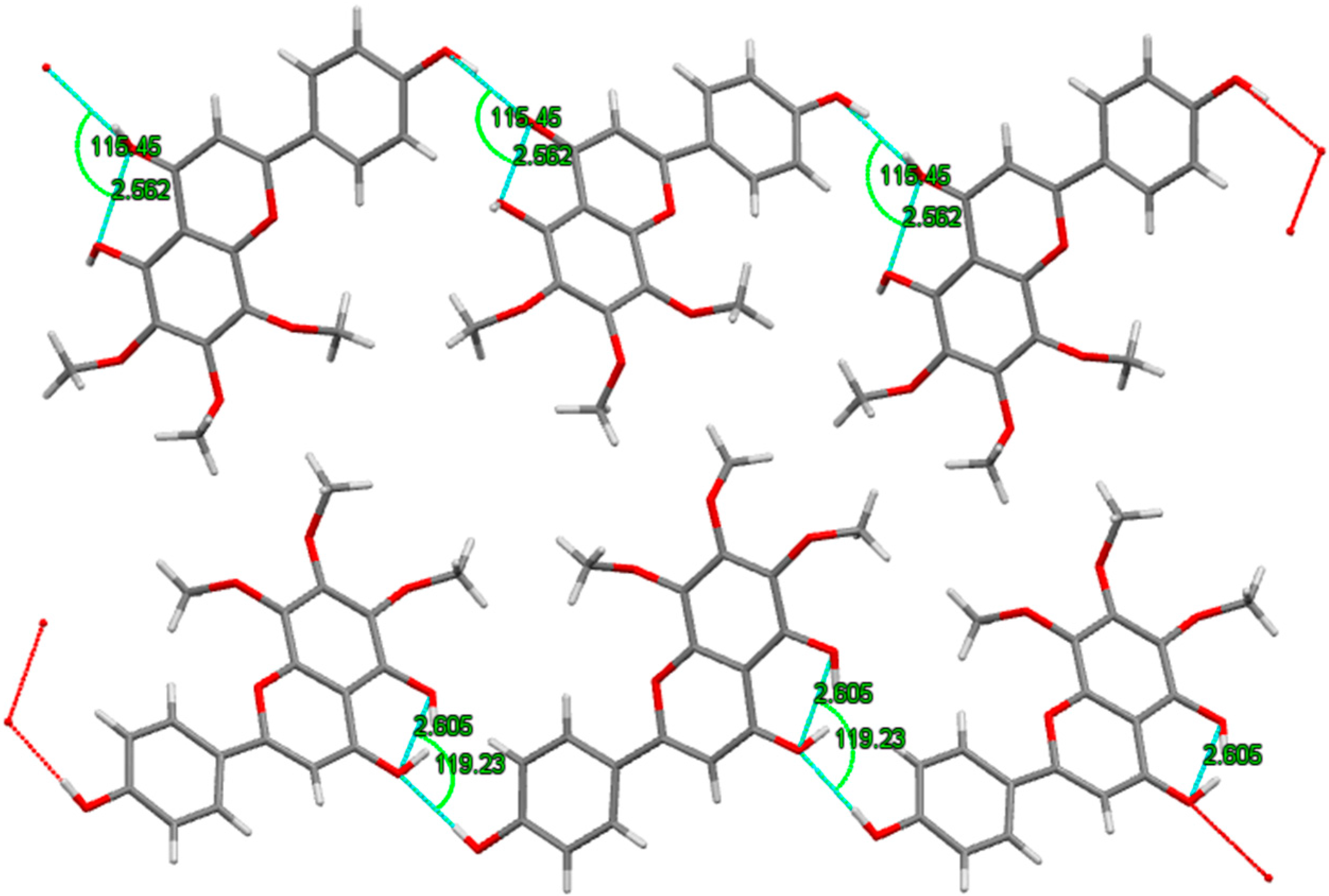
2.3. X-ray Crystallography

The crystal structure of xanthomicrol (4) is shown in Figure 8. The bonding parameters are listed in Table 3. The structure of compound was solved by direct methods using SHELXS-97 [41]. Hydrogen atoms that bonded to carbon and oxygen were positioned geometrically. The asymmetric unit shows two molecules which stand together thanks to intermolecular hydrogen bonding (Figure 9). Both of hydrogen bonds can be seen in the crystal structure. The intramolecular hydrogen bond, which appears when donor and acceptor atoms are in the same molecule, has a length of 2.562 Å. On the other hand, the distance is 2.796 Å with 119.23° angle for intermolecular hydrogen bonding. Because of the geometrical constraints, it’s probably longer in intermolecular interactions [42].
The symmetry components can also be summarized such −x, −y, 1/2+z, 1/2−x, 1/2+y, 1/2+z and 1/2+x, 1/2−y, z correspond to symmetry operations; “2-fold screw axis with direction [0, 0, 1] at 0, 0, z with screw component [0, 0, 1/2]”, “Glide plane perpendicular to [1, 0, 0] with glide component [0, 1/2, 1/2]” and “lide plane perpendicular to [0, 1, 0] with glide component [1/2, 0, 0]”, respectively.
Although synthetic xanthomicrol (1) is commercially available (Apin Chemicals and BOC Sciences), to our knowledge, this is the first report on its crystal structure. The crystal structure of salvigenin (4) has already been reported [43].
Figure 8. Crystal structure of xanthomicrol (4) showing two molecules in asymmetric unit and the crystal packing and alignment.
Molecules 20 07454 g008
Table 3. Bond angles (°) and bond distances (Å) for xanthomicrol (1).
Bond Angles (°)Bond Angles (°)Bond Angles (°)
C6 C1 C2121.3(10)C14 C13 O6120.7(11)O9 C21 H21B109.5
C6 C1 H1119.3C15 C14 O5120.4(10)H21A C21 H21B109.5
C2 C1 H1119.3C15 C14 C13120.6(11)O9 C21 H21C109.5
O1 C2 C3122.5(9)O5 C14 C13118.9(11)H21A C21 H21C109.5
O1 C2 C1117.5(10)O4 C15 C14122.1(10)H21B C21 H21C109.5
C3 C2 C1119.7(10)O4 C15 C10118.9(10)O8 C22 C27120.8(10)
C2 C3 C4120.8(10)C14 C15 C10118.9(10)O8 C22 C23118.6(10)
C2 C3 H3119.6O5 C16 H16A109.5C27 C22 C23120.3(10)
C4 C3 H3119.6O5 C16 H16B109.5C24 C23 C22120.0(10)
C5 C4 C3118.6(11)H16A C16 H16B109.5C24 C23 O9119.8(10)
C5 C4 H4120.7O5 C16 H16C109.5C22 C23 O9120.1(10)
C3 C4 H4120.7H16A C16 H16C109.5O13 C24 C23118.6(10)
C4 C5 C6121.5(11)H16B C16 H16C109.5O13 C24 C25120.1(11)
C4 C5 C7120.2(10)O6 C17 H17A109.5C23 C24 C25121.2(11)
C6 C5 C7118.3(10)O6 C17 H17B109.5C24 C25 C26117.7(10)
C1 C6 C5118.0(10)H17A C17 H17B109.5C24 C25 C28118.2(11)
C1 C6 H6 121.0O6 C17 H17C109.5C26 C25 C28124.0(10)
C5 C6 H6 121.0H17A C17 H17C109.5O11 C26 C27119.4(10)
C8 C7 O2121.5(11)H17B C17 H17C109.5O11 C26 C25120.2(10)
C8 C7 C5128.1(11)O7 C18 H18A109.5C27 C26 C25120.4(9)
O2 C7 C5110.3(9)O7 C18 H18B109.5O10 C27 C26121.9(9)
C7 C8 C9121.3(11)H18A C18 H18B109.5O10 C27 C22117.8(10)
C7 C8 H8119.4O7 C18 H18C109.5C26 C27 C22120.2(10)
C9 C8 H8119.4H18A C18 H18C109.5O12 C28 C29122.7(10)
O3 C9 C8122.3(12)H18B C18 H18C109.5O12 C28 C25119.3(11)
O3 C9 C10120.1(11)O10 C19 H19A109.5C28 C29 H29119.0 (9)
C8 C9 C10117.4(9)O10 C19 H19B109.5C29 C28 C25118.0(10)
C11 C10 C15119.6(10)H19A C19 H19B109.5C29 C30 O13120.0(10)
C11 C10 C9117.6(10)O10 C19 H19C109.5C29 C30 C31128.0(8)
C15 C10 C9122.8(10)H19A C19 H19C109.5C30 C29 C28122.0(10)
C10 C11 O2122.6(10)H19B C19 H19C109.5C30 C29 H29119.0
C10 C11 C12121.7(10)O8 C20 H20A109.5O13 C30 C31111.8(10)
O2 C11 C12115.6(9)O8 C20 H20B109.5C36 C31 C32114.9(9)
O7 C12 C13122.0(10)H20A C20 H20B109.5C36 C31 C30122.4(10)
O7 C12 C11120.5(10)O8 C20 H20C109.5C32 C31 C30122.5(10)
C13 C12 C11117.2(9)H20A C20 H20C109.5C33 C32 C31122.3(10)
Bond Distances (Å)Bond Distances (Å)Bond Distances (Å)
C1 H10.93 1C15 O41.35(1) 1C22 C271.39(2)
C1 C21.39 (1)C16 H16A0.96 1C22 O81.34(1) 1
C1 C61.37(2)C16 H16B0.96 1C23 C241.36(2)
C2 C31.36(2)C16 H16C0.96 1C23 O91.42(1) 1
C2 O11.36(1) 1C16 O51.42(1) 1C24 C251.42(2)
C3 H30.93 1C17 H17A0.96 1C24 O131.35(1) 1
C3 C41.41(1)C17 H17B0.96 1C25 C261.43(2)
C4 H40.93 1C17 H17C0.96 1C25 C281.43(2)
C4 C51.36(1)C17 O61.38(2) 1C26 C271.38(2)
C5 C61.42(2)C18 H18B0.96 1C26 O111.36(1) 1
C5 C71.49(2)C18 H18C0.96 1C27 O101.37(1) 1
C6 H60.93 1C18 O71.38(1) 1C28 C291.40(2)
C7 C81.35(2)O1 H1A0.821 1C28 O121.27(1) 1
C7 O21.37(1) 1O3 H3A0.820 1C29 H290.93 1
C8 H80.93 1O4 H4A0.819 1C29 C301.34(1)
C8 C91.41(2)C19 H19A0.96 1C30 C311.45(1)
C9 C101.47(2)C19 H19B0.96 1C30 O131.38(1) 1
C9 O31.28(1) 1C19 H19C0.96 1C31 C321.43(1)
C10 C111.37(1)C19 O101.35(1) 1C31 C361.36(2)
C10 C151.40(2)H20A0.96 1C32 H320.93 1
C11 C121.40(2)H20B0.96 1C32 C331.34(1)
C11 O21.38(1) 1H20C0.96 1C33 H330.931 1
C12 C131.37(2)C20 O81.41(1) 1C33 C341.37(1)
C12 O71.34(1) 1C21 H21A0.96 1C34 C351.38(2)
C13 C141.39(2)C21 H21C0.96 1C34 O141.37(1) 1
C13 O61.39(1) 1C21 H21B0.96 1C35 H350.93 1
C14 C151.37(2)C21 O91.45(1) 1C35 C361.37(2)
C14 O51.38(1) 1C22 C231.40(2)C36 H360.93 1
O11 H110.820 1O12 H120.822 1O14 H140.823 1
Figure 9. Hydrogen bond interactions of crystal.
Molecules 20 07454 g009
The crystal data and structure refinement for xanthomicrol (1) is summarized in Table 4.
Table 4. Crystal data and structure refinement for xanthomicrol (1).
ParameterValue
Empirical formulaC18H16O7
M344
Crystal systemOrthorhombic
Space groupPna21
a (Å)18.8736(14)
b (Å)4.1108(3)
c (Å)40.448(3)
α (°)90.00
β (°)90.00
γ (°)90.00
Volume (Å3)3138.2(4)
Z8
Dc/g·cm−31.389
Absorption Coefficient (Mo Kα) mm−10.111
Crystal size (mm)0.04 × 0.10 × 0.45
Theta range for data collection (θ Range/°)2.014° to 25.344°
Index Ranges−15 ≤ h ≤ 15, −3 ≤ k ≤ 3, −32 ≤ l ≤ 31
Reflections collected3203
Unique reflections, Rint1518 [R(int) = 0.0512]
Completeness of theta = 25.50°92%
Refinement MethodFull-matrix least-squares on F2
Data/restraints/parameters3203/1/463
Goodness of fit on F21.030
Final R indices [I > 2sigma(I)]R1 = 0.1111, wR2 = 0.0584 I > 2σ(I)
R indicesR1 = 0.1460, wR2 = 0.1314
Largest Diff. peak and hole (e·Å−3)0.210 and −0.198 e·Å−3

3. Experimental Section

3.1. Isolation of Flavonoids

Compounds 13 and 4 were previously isolated from aerial parts of S. stricta [13] and S. lycia [2], and the structures of the compounds were established by spectroscopic evidence (UV, IR, MS, NMR). Before the activity studies, structure elucidation of compounds 14 was reconfirmed by spectral (UV, MS and 1H-NMR) analysis [2,13,14,16,44].

3.2. Screening of hMAO Inhibitory Activities

3.2.1. Chemicals

hMAO-A and hMAO-B (both recombinant, expressed in baculovirus-infected BTI insect cells), R-(–)-deprenyl hydrochloride (selegiline), moclobemide, resorufin, dimethyl sulfoxide (DMSO), and other chemicals were purchased from Sigma-Aldrich (Munich, Germany). The Amplex®-Red MAO assay kit (Cell Technology Inc., Mountain View, CA, USA) contained benzylamine, p-tyramine, clorgyline, pargyline and horseradish peroxidase.

3.2.2. Determination of hMAO-A and hMAO-B Activities

The activities of hMAO-A and hMAO-B were determined using p-tyramine as common substrate and calculated as 161.50 ± 8.33 pmol/mg/min (n = 3) and 140.00 ± 7.99 pmol/mg/min (n = 3), respectively. The interactions of the synthesized compounds with hMAO isoforms were determined by a fluorimetric method described and modified previously [45,46]. The production of H2O2 catalyzed by MAO isoforms was detected using Amplex®-Red reagent, a non-fluorescent probe which reacts with H2O2 in the presence of horse radish peroxidase to produce the fluorescent product resorufin. The reaction was started by adding 200 µM Amplex Red reagent, 1 U/mL horseradish peroxidase (HRP), and p-tyramine (concentration range 0.05–0.5 mM). Control experiments were carried out by replacing the compound and reference inhibitors. The possible capacity of compounds to modify the fluorescence generated in the reaction mixture due to non enzymatic inhibition was determined by adding these compounds to solutions containing only the Amplex Red reagent in a sodium phosphate buffer. No modification was recorded. The possible capacity of compounds to inhibit HRP in the test solution was also determined. The compounds did not inhibit HRP in the experimental medium.

3.2.3. Kinetic Experiments

Synthesized compounds were dissolved in dimethyl sulfoxide, with a maximum concentration of 1% and used in the concentration range of 1.00–100.00 µM for compounds 1 and 4; 5.00–100.00 mM for compounds 2 and 3. Kinetic data corresponding the interaction of hMAO isoforms with the compounds were determined using computer-fit calculation (Prism 4.0, GraphPad SoftwareThe slopes of the Lineweavere-Burk plots were plotted versus the inhibitor concentration and the Ki values were also determined from the x-axis intercept as -Ki. Each Ki value is the representative of single determination where the correlation coefficient (R2) of the replot of the slopes versus the inhibitor concentrations was at least 0.98. SI was calculated as Ki(hMAO-A)/Ki(hMAO-B. The protein was determined according to the Bradford method [47].

3.2.4. Reversibility Experiments

Reversibility of the MAO inhibition with newly synthesized compounds was determined by centrifugation-ultrafiltration method previously described [48]. Recombinant enzymes (hMAO-A or B) were incubated with a single concentration of the synthesized compounds or the reference inhibitors in a sodium phosphate buffer (0.05 M, pH 7.4) for one hour at 37 °C. An aliquot was stored at 4 °C and used for the measurement of MAO-A and -B activities. The remaining incubated sample was placed in an Ultrafree-0.5 centrifugal tube with a 30 kDa Biomax membrane and centrifuged at 9000× g for 20 min at 4 °C. The enzyme retained in the 30 kDa membrane was re-suspended in a sodium phosphate buffer at 4 °C and centrifuged two more times. After the third centrifugation, the enzyme retained in the membrane was re-suspended in sodium phosphate buffer (300 mL) and an aliquot of this suspension was used for MAO-A and -B activity determination. Control experiments were performed simultaneously to determine the 100% MAO activity by replacing the compounds and standards with appropriate dilutions of the vehicles. The corresponding values of percent (%) MAO isoform inhibition was separately calculated for samples with and without repeated washing.

3.2.5. Molecular Docking Studies

Molecular models of the inhibitors (1,4) were built and optimized using SPARTAN 10.0 at PM3 level on iMac 3,4 GHz Intel Core i7 processor. The crystal structures of MAO-A, and MAO-B were extracted from the Protein Data Bank (PDB) [49] Their PDB codes are 2Z5X and 2V5Z for MAO-A and MAO-B respectively. Each structure was cleaned of all water molecules and inhibitors as well as all non-interacting ions were removed before being used in the docking studies. For MAO-A and MAO-B, one of the two subunits was taken as the target structure. For each protein all hydrogens were added and minimized using “Clean Geometry” toolkit, then for more complete optimization final geometry was submitted to “Prepare Macromolecule” protocol of Discovery Studio (Accelerys) assigning CHARMM force field. Missing hydrogen atoms were added based on the protonation state of the titratable residues at a pH of 7.4. Ionic strength was set to 0.145 and the dielectric constant was set to 10. Autodock Tool (ADT) interface was used for the preparation of input files of macromolecules and ligands for docking calculations. Docking was achieved using AutoDock 4.2.6 coding scripts [50] using HPxw8600-Work-Station. The detailed docking procedures are given elsewhere [51,52]. Acelrys Visualization 4.0 program was used for rendering the 2D and 3D pictures. To render visible the detailed interactions of the docked poses of the inhibitors (1,4) Acelrys Visualization 4.0 program was used.

3.3. X-ray Analysis

Single crystal X-ray diffraction patterns of xanthomicrol were collected at 293 K with Bruker APEX-II CCD diffractometer from a K780 X-ray generator applying 50 kV and 40 mA. All data were accumulated using a graphite monochromated Mo Kα (λ = 0.71073 Å) radiation. The least-squares methods was applied on the basis of all reflections with F2 > 2σ(F2) to determine the lattice parameters.

4. Conclusions

The present study provides an evidence for the isolated flavonoids from Sideritis species as new potent and selective hMAO-inhibitors. The results suggest two compounds, xanthomicrol and salvigenin as potential natural starting molecules for developing novel selective MAO-A inhibitors, for prevention and treatment of psychiatric disorders such as depression and anxiety, and also cognitive impairments in Alzheimer’s and Parkinson’s Diseases. Current study gives a clear insight in the inhibitory ability of these flavonoids towards hMAO; however, more studies are needed to clarify their efficacy in vivo. In addition, in the present study, crystal structure of xanthomicrol, which is the most selective MAO-A inhibitor tested, has been introduced for the first time.

Supplementary Data

Additional material available from Cambridge Crystallographic Data Center as Deposition No. CCDC 1051489 comprises H-atom coordinates, thermal parameters and remaining bond lengths and angles. These data can be obtained free of charge via http://www.ccdc.cam.ac.uk/conts/retrieving.html, or from the Cambridge Crystallographic Data Centre, 12 Union Road, Cambridge CB2 1EZ, UK; E-Mail: [email protected].

Acknowledgments

Present work was supported by a grant from Hacettepe University Scientific Research Projects Coordination Unit (Project No: HUAF 02T10102001). The authors would like to thank Erhan Palaska (Hacettepe University, Faculty of Pharmacy, Department of Pharmaceutical Chemistry) for the ESI-MS spectra of the compounds reported in this study.

Author Contributions

F.P.T. and G.U. conceived and designed the study. F.P.T., İ.Ç. and N.E. isolated and identified the compounds, İ.B., S.C.-Y. and G.U., performed in vitro activity studies, K.Y. performed molecular docking studies, H.T. and S.P. performed X-ray analysis, F.P.T. and G.U. interpreted the results and wrote the paper. All authors read and approved the manuscript.

Conflicts of Interest

The authors declare no conflict of interest.

References

  1. Obon de Castro, C.; Rivera-Nunez, D. Phanerogamarum Monographiae Tomus XXI: A Taxonomic Revision of the Section Sideritis (genus Sideritis) (Labiatae); J. Cramer: Berlin, Germany, 1994. [Google Scholar]
  2. Ezer, N.; Akcoş, Y. Flavonoids from Sideritis lycia. J. Hacettepe Univ. Fac. Pharm. 1995, 15, 81–87. [Google Scholar]
  3. Knörle, R. Extracts of Sideritis scardica as triple monoamine reuptake inhibitors. J. Neural Transm. 2012, 119, 1477–1482. [Google Scholar] [CrossRef] [PubMed]
  4. Gonzalez-Burgos, E.; Carretero, M.E.; Gomez-Serranillos, M.P. Sideritis spp.: Uses, chemical composition and pharmacological activities-a review. J. Ethnopharmacol. 1996, 135, 209–225. [Google Scholar]
  5. Barberan, F.A.T.; Tomas, F.; Ferreres, F. Isoscutellarein-7–0-allosyl (1–2) glucoside from Sideritis leucantha. J. Nat. Prod. 1985, 48, 28–32. [Google Scholar] [CrossRef] [PubMed]
  6. Küpeli, E.; Şahin, F.P.; Yeşilada, E.; Çalış, İ.; Ezer, N. In vivo anti-inflammatory and antinociceptive activity evaluation of phenolic compounds from Sideritis stricta. Z. Naturforschung 2007, 62c, 519–525. [Google Scholar]
  7. Baytop, T. Dağçayı. In Therapy with Medicinal plants in Turkey (Past and Present); Nobel Tip Publications: Istanbul, Turkey, 1999; p. 193. [Google Scholar]
  8. Ezer, N.; Vila, R.; Canigueral, S.; Adzet, T. Essential oil composition of four Turkish species of Sideritis. Phytochemistry 1996, 41, 203–205. [Google Scholar] [CrossRef]
  9. Baser, K.H.C. Aromatic biodiversity among the flowering plant taxa of Turkey. Pure Appl. Chem. 2002, 74, 527–545. [Google Scholar] [CrossRef]
  10. Topçu, G.; Gören, A.C.; Kılıç, T.; Yıldız, Y.K.; Tümen, G. Diterpenes from Sideritis trojana. Nat. Prod. Lett. 2002, 16, 33–37. [Google Scholar] [CrossRef] [PubMed]
  11. Sahin, F.P.; Ezer, N.; Çalış, İ. Three acylated flavone glycosides from Sideritis ozturkii Aytac & Aksoy. Phytochemistry 2004, 65, 2095–2099. [Google Scholar]
  12. Sahin, F.P.; Ezer, N.; Çalış, İ. Erratum to “three acylated flavone glycosides from Sideritis ozturkii Aytac and Aksoy” [Phytochemistry 65 (2004) 2095–2099]. Phytochemistry 2005, 66, 125. [Google Scholar]
  13. Sahin, F.P.; Ezer, N.; Calis, I. Terpenic and phenolic compounds from Sideritis stricta. Turk. J. Chem. 2006, 30, 495–504. [Google Scholar]
  14. Sezik, E.; Ezer, N. Phytochemical investigations on the plants used as folk medicine and herbal tea in Turkey I. Flavonoids of Sideritis congesta Davis et Huber-Morath. Acta Pharm. Turc. 1984, 16, 4–10. [Google Scholar]
  15. Ezer, N.; Sakar, M.K.; Rodriguez, B.; de la Torre, M.C. Flavonoid glycosides and a phenylpropanoid glycoside from Sideritis perfoliata. Int. J. Pharmacogn. 1992, 30, 61–65. [Google Scholar] [CrossRef]
  16. Güvenc, A.; Okada, Y.; Akkol, E.K.; Uman, H.; Okuyama, T.; Calis, I. Investigations of anti-inflammatory, antinociceptive, antioxidant and aldose reductase inhibitory activities of phenolic compounds from Sideritis brevibracteata. Food Chem. 2010, 118, 686–692. [Google Scholar] [CrossRef]
  17. Tipton, K.F.; Boyce, S.; O’Sullivan, J.; Davey, G.P.; Healy, J. Monoamine oxidases: Certainties and uncertainties. Curr. Med. Chem. 2004, 11, 1965–1982. [Google Scholar] [CrossRef] [PubMed]
  18. Westlund, K.N.; Denney, R.M.; Rose, R.M.; Abell, C.W. Localization of distinct monoamine oxidase A and monoamine oxidase B cell populations in human brainstem. Neuroscience 1988, 25, 439–456. [Google Scholar] [CrossRef] [PubMed]
  19. Bach, A.W.; Lan, N.C.; Johnson, D.L.; Abell, C.W.; Bembenek, M.E.; Kwan, S.W.; Seeburg, P.H.; Shih, J.C. cDNA cloning of human liver monoamine oxidase A and B: molecular basis of differences in enzymatic properties. Proc. Natl. Acad. Sci. USA 1988, 85, 4934–4938. [Google Scholar] [CrossRef] [PubMed]
  20. Johnston, J.P. Some observations upon a new inhibitor of monoamine oxidase in brain tissue. Biochem. Pharmacol. 1968, 17, 1285–1297. [Google Scholar] [CrossRef] [PubMed]
  21. Knoll, J.; Magyar, K. Some puzzling pharmacological effects of monoamine oxidase inhibitors. Adv. Biochem. Psychopharmacol. 1972, 5, 393–408. [Google Scholar] [PubMed]
  22. Pare, C.M. Unwanted effects of long-term medication in schizophrenia and depression. Pharmakop. Sych. Neuro. 1976, 9, 187–192. [Google Scholar]
  23. Shih, J.; Chen, K.; Ridd, M.J. Monoamine oxidase: From genes to behavior. Annu. Rev. Neurosci. 1999, 22, 197–217. [Google Scholar] [CrossRef] [PubMed]
  24. Binda, C.; Newton-Vinson, P.; Hubalek, F.; Edmondson, D.E.; Mattevi, A. Structure of human monoamine oxidase B, a drug target for the treatment of neurological disorders. Nat. Struct. Biol. 2002, 9, 22–26. [Google Scholar] [CrossRef] [PubMed]
  25. Binda, C.; Hubalek, F.; Li, M.; Edmondson, D.E.; Mattevi, A. Crystal structure of human monoamine oxidase B, a drug target enzyme monotopically inserted into the mitochondrial outer membrane. FEBS Lett. 2004, 564, 225–228. [Google Scholar] [CrossRef] [PubMed]
  26. Edmondson, D.E.; Binda, C.; Mattevi, A. The FAD binding sites of human monoamine oxidases A and B. Neurotoxicology 2004, 25, 63–72. [Google Scholar] [CrossRef] [PubMed]
  27. De Colibus, L.; Li, M.; Binda, C.; Lustig, A.; Edmondson, D.E.; Mattevi, A. Three-dimensional structure of human monoamine oxidase A (MAO A): Relation to the structures of rat MAO A and human MAO B. Proc. Natl. Acad. Sci. USA 2005, 102, 12684–12689. [Google Scholar] [CrossRef] [PubMed]
  28. Geldenhuys, W.J.; Darvesh, A.S.; Funk, M.O.; Van der Schyf, C.J.; Carroll, R.T. Identification of novel monoamine oxidase B inhibitors by structure-based virtual screening. Bioorg. Med. Chem. Lett. 2010, 20, 5295–5298. [Google Scholar] [CrossRef] [PubMed]
  29. Jäger, A.K.; Saaby, L. Flavonoids and the CNS. Molecules 2011, 16, 1471–1485. [Google Scholar] [CrossRef] [PubMed]
  30. Carradori, S.; D’Ascenzio, M.; Chimenti, P.; Secci, D.; Bolasco, A. Selective MAO-B inhibitors: A lesson from natural products. Mol. Divers. 2014, 18, 219–243. [Google Scholar] [CrossRef] [PubMed]
  31. Mathew, B.; Suresh, J.; Mathew, G.E.; Parasuraman, R.; Abdulla, N. Plant secondary metabolites- potent inhibitors of monoamine oxidase isoforms. Cent. Nerv. Syst. Agents Med. Chem. 2014, 14, 28–33. [Google Scholar] [CrossRef] [PubMed]
  32. Gao, L.; Fang, J.S.; Bai, X.Y.; Zhou, D.; Wang, Y.T.; Liu, A.L.; Du, G.H. In silico target fishing for the potential targets and molecular mechanisms of baicalein as an anti-parkinsonian agent: Discovery of the protective effects on NMDA receptor-mediated neurotoxicity. Chem. Biol. Drug Des. 2013, 81, 675–687. [Google Scholar] [CrossRef] [PubMed]
  33. Küpeli, E.; Sahin, F.P.; Calis, I.; Yesilada, E.; Ezer, N. Phenolic compounds of Sideritis ozturkii and their in vivo anti-inflammatory and antinociceptive activities. J. Ethnopharmacol. 2007, 112, 356–360. [Google Scholar] [CrossRef] [PubMed]
  34. Tandogan, B.; Güvenç, A.; Çalış, İ.; Ulusu, N.N. In vitro effects of compounds isolated from Sideritis brevibracteata on bovine kidney cortex glutathione reductase. Acta Biochim. Pol. 2011, 58, 471–475. [Google Scholar]
  35. Youdim, M.B.H.; Bakhle, Y.S. Monoamine oxidase: Isoforms and inhibitors in Parkinson’s disease. Br. J. Pharmacol. 2006, 147, S287–S296. [Google Scholar] [CrossRef] [PubMed]
  36. Finberg, J.P.M.; Gillman, K. Selective inhibitors of monoamine oxidase type B and the “cheese effect”. In International Review of Neurobiology, Monoamine Oxidases and Their Inhibitors; Youdim, M.B.H., Riederer, P., Eds.; Academic Press: London, UK, 2011; Volume 100, pp. 169–190. [Google Scholar]
  37. Fowler, J.S.; Volkow, N.D.; Logan, J.; Wang, G.J.; MacGregor, R.R.; Schlyer, D.; Wolf, A.P.; Pappas, N.; Alexoff, D.; Shea, C.; et al. Slow recovery of human brain MAO B after l-deprenyl (seleginine) withdrawal. Synapse 1994, 18, 86–93. [Google Scholar]
  38. Lang, U.E.; Borgwardt, S. Molecular mechanisms of depression: Per-spectives on new treatment strategies. Cell Physiol. Biochem. 2013, 31, 761–777. [Google Scholar] [CrossRef] [PubMed]
  39. Meyer, J.H.; Ginovart, N.; Boovariwala, A.; Sagrati, S.; Hussey, D.; Garcia, A.; Young, T.; Young, N.T.; Praschak-Rieder, N.; Wilson, A.A.; et al. Elevated monoamine oxidase A levels in the brain: An explanation for the monoamine imbalance of major depression. Arch. Gen. Psychiatry 2006, 63, 1209–1216. [Google Scholar]
  40. Gong, J.; Huang, J.; Ge, Q.; Chen, F.; Zhang, Y. Advanced research on the antidepressant effect of flavonoids. Curr. Opin. Complement. Altern. Med. 2014, 1, e00011. [Google Scholar]
  41. Sheldrick, G.M. A short history of SHELX. Acta Crystallogr. Sect. A 2008, 64, 112–122. [Google Scholar] [CrossRef]
  42. Zheng, J.; Kwak, K.; Chen, X.; Asbury, J.B.; Fayer, M.D. Watching Ultrafast Molecular Motions with 2D IR Chemical Exchange Spectroscopy; World Scientific Publishing: London, UK, 2005. [Google Scholar]
  43. Kanko, C.; Koukoua, G.; N’Guessan, Y.T.; Fournier, J.; Pradère, J.P.; Toupet, L. Contribution à l’étude phytochimique de Lippiamultiflora (Verbenaceae). C. R. Chim. 2004, 7, 1029–1032. [Google Scholar] [CrossRef]
  44. Ouyang, X.; Wei, L.; Pan, Y.; Huang, S.; Wang, H.; Begonia, G.B.; Ekunwe, S.I.N. Antioxidant properties and chemical constituents of ethanolic extract and its fractions of Ocimum gratissimum. Med. Chem. Res. 2013, 22, 1124–1130. [Google Scholar] [CrossRef]
  45. Chimenti, F.; Maccioni, E.; Secci, D.; Bolasco, A.; Chimenti, P.; Granese, A.; Carradori, S.; Alcaro, S.; Ortuso, F.; Yáñez, M. Synthesis, stereochemical identification, and selective inhibitory activity against human monoamine oxidase-B of 2-methylcyclohexylidene-(4-arylthiazol-2-yl) hydrazones. J. Med. Chem. 2008, 51, 4874–4880. [Google Scholar] [CrossRef] [PubMed]
  46. Yáñez, M.; Fraiz, N.; Cano, E.; Orallo, F. Inhibitory effects of cis- and trans-resveratrol on noradrenaline and 5-hydroxytryptamine uptake and on monoamine oxidase activity. Biochem. Biophys. Res. Commun. 2006, 344, 688–695. [Google Scholar] [CrossRef] [PubMed]
  47. Bradford, M.M. A rapid and sensitive method for the quantitation of microgram quantities of protein utilizing the principle of protein-dye binding. Anal. Chem. 1976, 72, 248–254. [Google Scholar]
  48. Chimenti, F.; Carradori, S.; Secci, D.; Bolasco, A.; Bizzarri, B.; Chimenti, P.; Granese, A.; Yáñez, M.; Orallo, F. Synthesis and inhibitory activity against human monoamine oxidase of N1-thiocarbamoyl-3,5-di (hetero) aryl-4,5-dihydro-(1-H)-pyrazole derivatives. Eur. J. Med. Chem. 2010, 45, 800–804. [Google Scholar] [CrossRef] [PubMed]
  49. Biological Macromolecular Resource. Available online: http://www.rcsb.org/pdb/home/ (accessed on 12 January 2015).
  50. Morris, G.M.; Huey, R.; Lindstrom, W.; Sanner, M.F.; Belew, R.K.; Goodsell, D.S.; Olson, A.J. Autodock4 and AutoDockTools4: Automated docking with selective receptor flexibility. J. Comput. Chem. 2009, 16, 2785–2791. [Google Scholar] [CrossRef]
  51. Yelekçi, K.; Büyüktürk, B.; Kayrak, N. In silico identification of novel and selective monoamine oxidase B inhibitors. J. Neural. Transm. 2013, 120, 853–858. [Google Scholar] [CrossRef] [PubMed]
  52. Gökhan-Kelekçi, N.; Şimşek, Ö.Ö.; Ercan, A.; Yelekçi, K.; Şahin, Z.S.; Işık, Ş.; Uçar, G.; Bilgin, A.A. Synthesis and molecular modeling of some novel hexahydroindazole derivatives as potent monoamine oxidase inhibitors. Bioorg. Med. Chem. 2009, 17, 6761–6772. [Google Scholar]
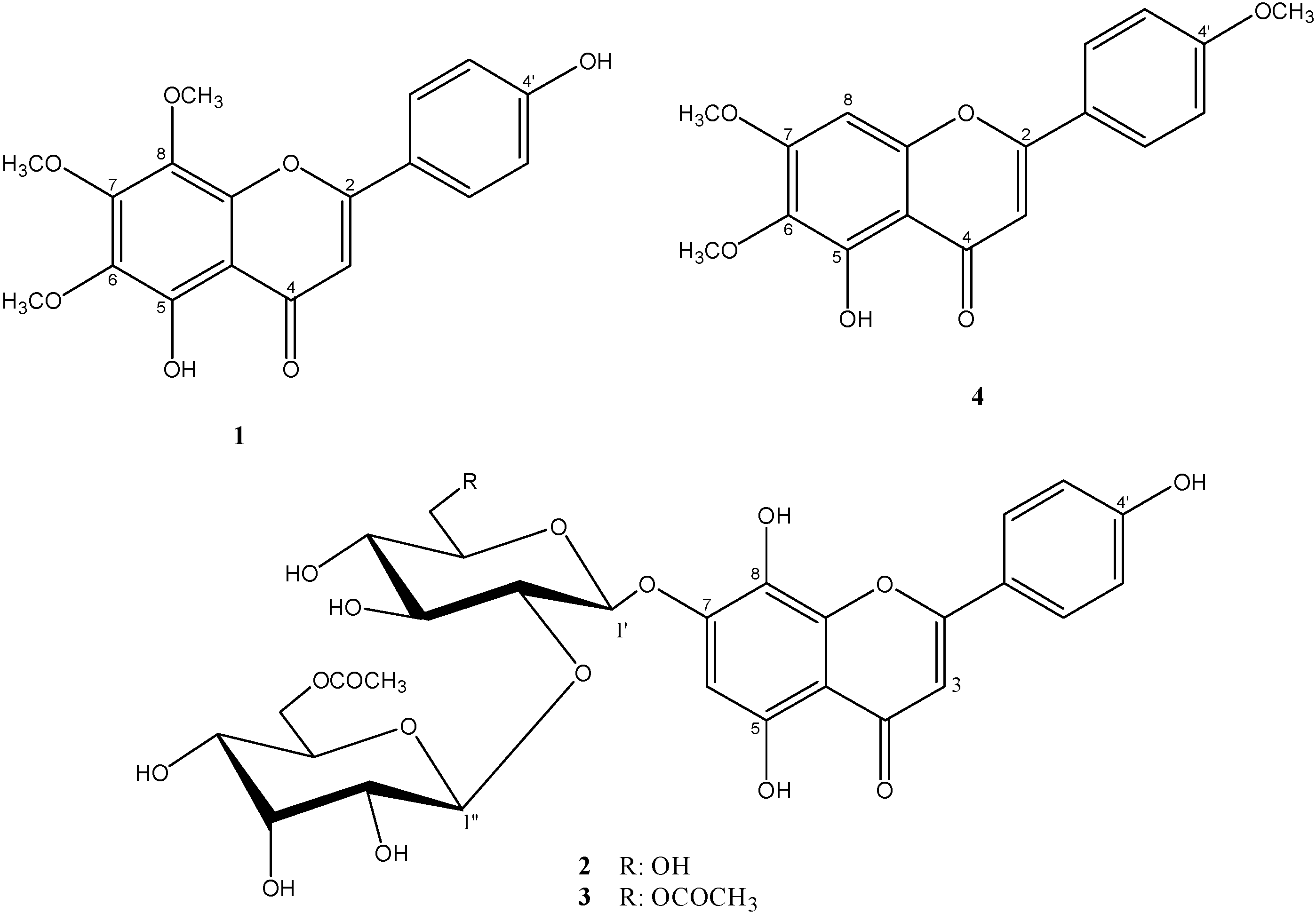
  • Sample Availability: Samples of the compounds 1 and 4 are available from the authors.

