
Mucin-Type O-Glycosylation in Invertebrates

Abstract
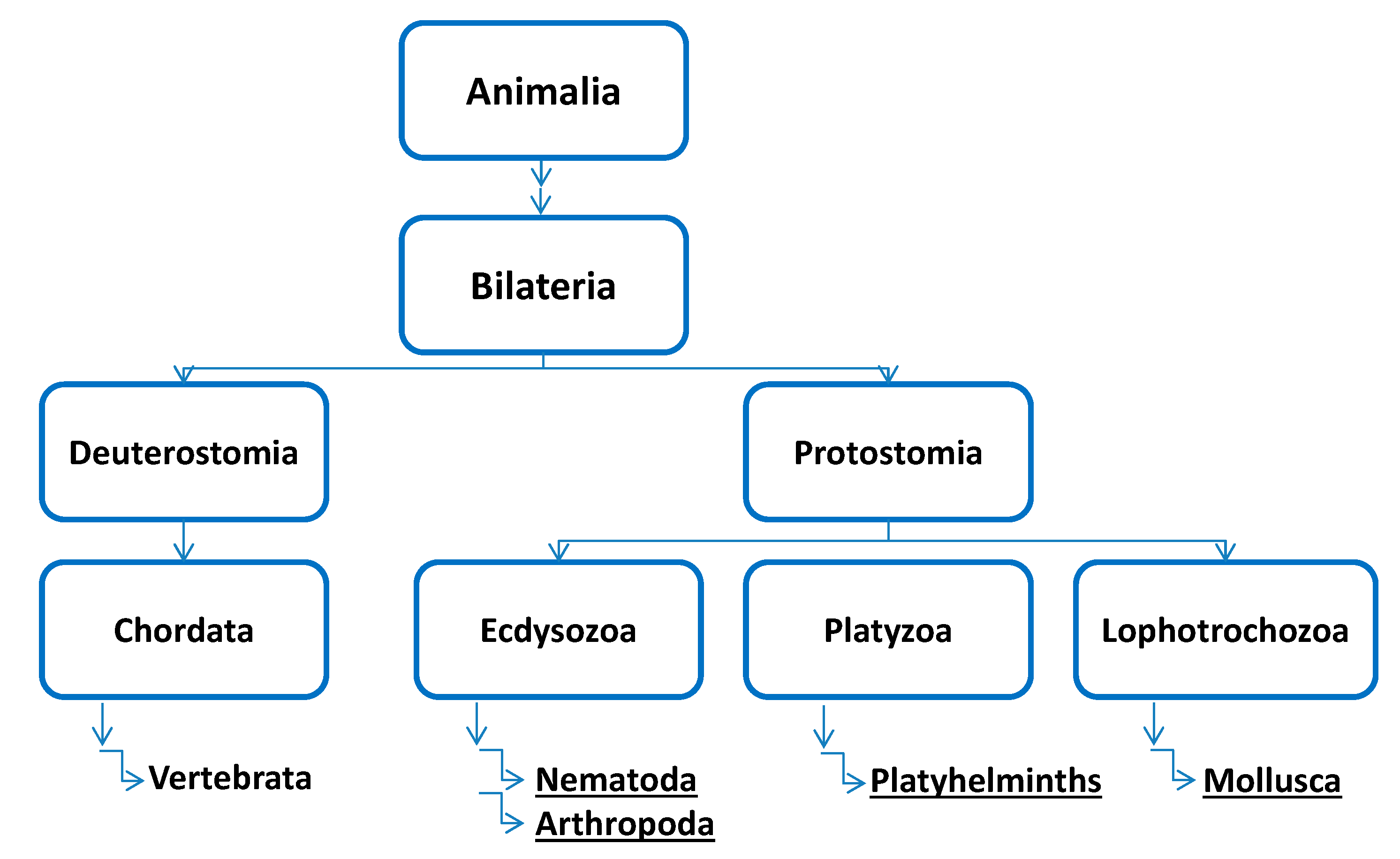
:1. Introduction

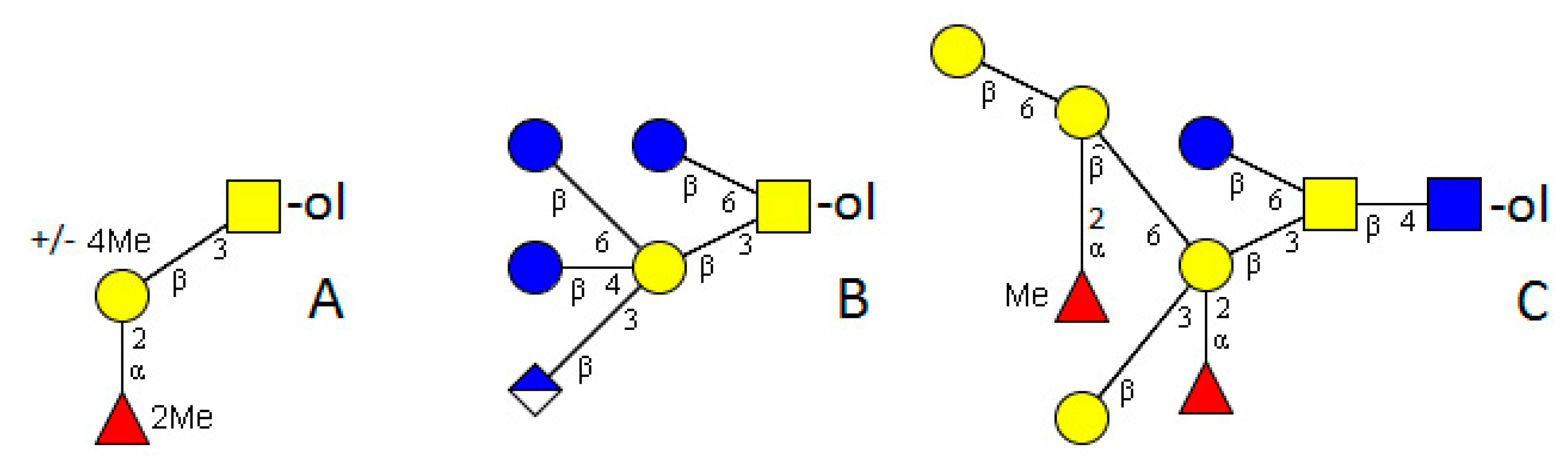
2. Nematoda

3. Arthropoda
3.1. Crustacea (Crustaceans)
3.2. Hexapoda (Insects)
3.2.1. Drosophila Melanogaster

3.2.2. Venoms
3.2.3. Insect Expression Systems

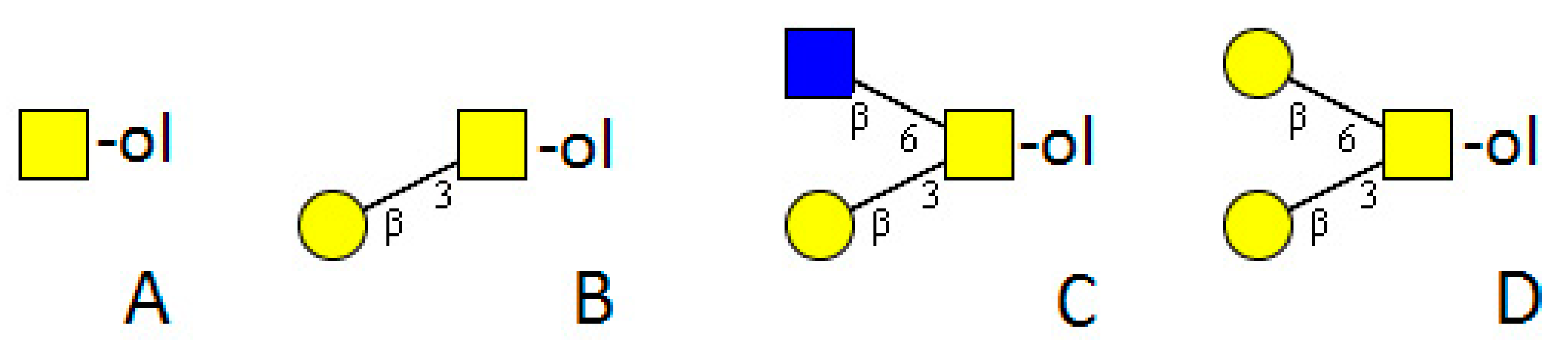
4. Platyhelminthes

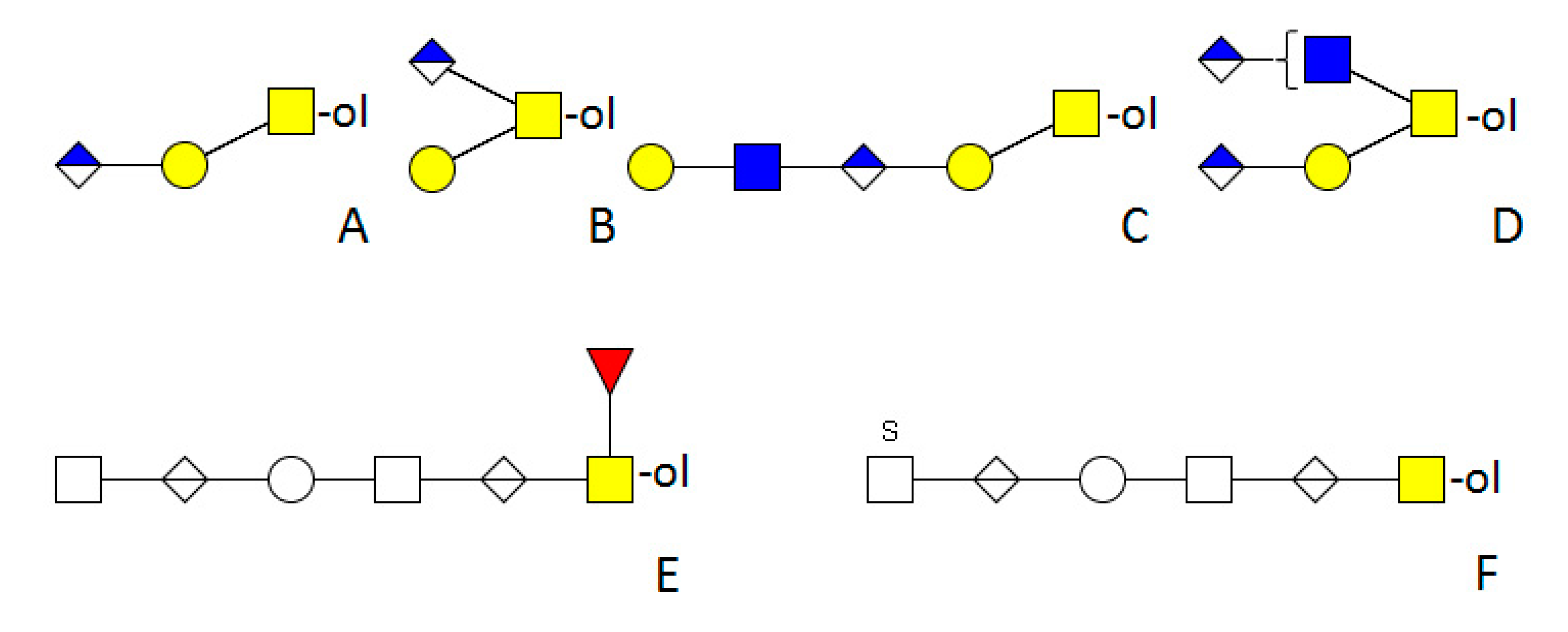
5. Mollusca
5.1. Cephalopoda
5.2. Gastropoda
5.2.1. Bivalves
5.2.2. Snails

6. Conclusions
Acknowledgments
Conflicts of Interest
References
- Van den Steen, P.; Rudd, P.M.; Dwek, R.A.; Opdenakker, G. Concepts and principles of O-linked glycosylation. Crit. Rev. Biochem. Mol. Biol. 1998, 33, 151–208. [Google Scholar] [CrossRef] [PubMed]
- Proszynski, T.J.; Simons, K.; Bagnat, M. O-glycosylation as a sorting determinant for cell surface delivery in yeast. Mol. Biol. Cell 2004, 15, 1533–1543. [Google Scholar] [CrossRef] [PubMed]
- Robledo, Y.; Marigómez, I.; Angulo, E.; Cajaraville, M.P. Glycosylation and sorting pathways of lysosomal enzymes in mussel digestive cells. Cell Tissue Res. 2006, 324, 319–333. [Google Scholar] [CrossRef] [PubMed]
- Schjoldager, K.T.B.G.; Clausen, H. Site-specific protein O-glycosylation modulates proprotein processing—Decipering specific functions of the large polypeptide GalNAc-transferase gene family. Biochim. Biophys. Acta 2012, 1820, 2079–2094. [Google Scholar] [CrossRef] [PubMed]
- Gill, D.J.; Tham, K.M.; Chia, J.; Wang, S.C.; Steentoft, C.; Clausen, H.; Bard-Chapeau, E.A.; Bard, F.A. Initiation of GalNAc-type O-glycosylation in the endoplasmic reticulum promotes cancer cell invasiveness. Proc. Natl. Acad. Sci. USA 2013, 110, E3152–E3161. [Google Scholar] [CrossRef] [PubMed]
- Radhakrishnan, P.; Dabelsteen, S.; Madsen, F.B.; Francavilla, C.; Kopp, K.L.; Steentoft, C.; Vakhrushev, S.Y.; Olsen, J.V.; Hansen, L.; Bennett, E.P.; et al. Immature truncated O-glycophenotype of cancer directly induces oncogenic features. Proc. Natl. Acad. Sci. USA 2014, 111, E4066–E4075. [Google Scholar] [CrossRef] [PubMed]
- Badirou, I.; Kurdi, M.; Legendre, P.; Rayes, J.; Bryckaert, M.; Casari, C.; Lenting, P.J.; Christophe, O.D.; Denis, C.V. In vivo analysis of the role of O-glycosylations of Von Willebrand factor. PLoS ONE 2012, 7, e37508. [Google Scholar] [CrossRef] [PubMed]
- Herzog, B.H.; Fu, J.; Xia, L. Mucin-type O-glycosylation is critical for vascular integrity. Glycobiology 2014, 24, 1237–1241. [Google Scholar] [CrossRef] [PubMed]
- Zhang, L.; Syed, Z.A.; van Dijk Härd, I.; Lim, J.M.; Wells, L.; Ten Hagen, K.G. O-Glycosylation regulates polarized secretion by modulating Tango1 stability. Proc. Natl. Acad. Sci. USA 2014, 111, 7296–7301. [Google Scholar] [CrossRef] [PubMed]
- Tran, D.T.; Ten Hagen, K.G. Mucin-type O-glycosylation during development. J. Biol. Chem. 2013, 288, 6921–6929. [Google Scholar] [CrossRef] [PubMed]
- Spiro, R.G. Protein glycosylation: nature, distribution, enzymatic formation, and disease implications of glycopeptide bonds. Glycobiology 2002, 12, 43R–56R. [Google Scholar] [CrossRef] [PubMed]
- Bennett, E.P.; Mandel, U.; Clausen, H.; Gerken, T.A.; Fritz, T.A.; Tabak, L.A. Control of mucin-type O-glycosylation: A classification of the polypeptide GalNAc-transferase gene family. Glycobiology 2012, 22, 736–756. [Google Scholar] [CrossRef] [PubMed]
- Patsos, G.; Corfield, A. O-Glycosylation: Structural Diversity and Functions. In The Sugar Code; Gabius, H.J., Ed.; Wiley-VCH: Weinheim, Germany, 2009; pp. 111–137. [Google Scholar]
- Bullen, J.W.; Balsbaugh, J.L.; Chanda, D.; Shabanowitz, J.; Hunt, D.F.; Neumann, D.; Hart, G.W. Cross-talk between two essential nutrient-sensitive enzymes. O-GlcNAc transferase (OGT) and AMP-activated protein kinase (AMPK). J. Biol. Chem. 2014, 289, 10592–10606. [Google Scholar] [PubMed]
- Live, D.; Wells, L.; Boons, G.J. Dissecting the molecular basis of the role of the O-mannosylation pathway in disease: α-Dystroglycan and forms of muscular dystrophy. ChemBioChem 2013, 14, 2392–2402. [Google Scholar] [CrossRef] [PubMed]
- Bektas, M.; Rubenstein, D.S. The role of intracellular protein O-glycosylation in cell adhesion and disease. J. Biomed. Res. 2011, 25, 227–236. [Google Scholar] [CrossRef]
- Ishio, A.; Sasamura, T.; Ayukawa, T.; Kuroda, J.; Ishikawa, H.O.; Aoyama, N.; Matsumoto, K.; Gushiken, T.; Okyjima, T.; Yamakawa, T.; Matsuno, K. O-Fucose monosaccharde of Drosophila Notch has a temperature-sensitive function and cooperates with O-glucose glycan in Notch transport and Notch signalling activation. J. Biol. Chem. 2015, 290, 505–519. [Google Scholar] [CrossRef] [PubMed]
- Takeuchi, H.; Kantharia, J.; Sethi, M.K.; Bakker, H.; Haltiwanger, R.S. Site-specific O-glucosylation of the epidermal growth factor-like (EGF) repeats of Notch: efficiency of glycosylation is affected by proper folding and amino acid sequence of individual EGF repeats. J. Biol. Chem. 2012, 287, 33934–33944. [Google Scholar] [CrossRef] [PubMed]
- Xu, C.; Ng, D.T.W. O-mannosylation: The other glycan player of ER quality control. Semin. Cell Dev. Biol. 2015, in press. [Google Scholar] [CrossRef] [PubMed]
- Nakamura, N.; Lyalin, D.; Panin, V.M. Protein O-mannosylation in animal development and physiology: From human disorders to Drosophila phenotypes. Sem. Cell Dev. Biol. 2010, 21, 622–630. [Google Scholar] [CrossRef] [PubMed]
- Brunner, A.; Kolarich, D.; Voglmeir, J.; Paschinger, K.; Wilson, I.B.H. Comparative characterisation of recombinant invertebrate and vertebrate peptide O-xylosyltransferases. Glycoconj. J. 2006, 23, 543–554. [Google Scholar] [CrossRef] [PubMed]
- Khoo, K.H.; Maizels, R.M.; Page, A.P.; Taylor, G.W.; Rendell, N.B.; Dell, A. Characterization of nematode glycoproteins: the major O-glycans of Toxocara excretory-secretory antigens are O-methylated trisaccharides. Glycobiology 1991, 2, 163–171. [Google Scholar] [CrossRef]
- Schabussova, I.; Amer, H.; van Die, I.; Kosma, P.; Maizels, R.M. O-Methylated glycans from Toxocara are specific targets for antibody binding in human and animal infections. Int. J. Parasitol. 2007, 37, 97–109. [Google Scholar] [CrossRef] [PubMed]
- Ellis, L.A.; McVay, C.S.; Probert, M.A.; Zhang, J.; Bundle, D.R.; Appleton, J.A. Terminal β-linked tyvelose creates unique epitopes in Trichinella spiralis glycan antigens. Glycobiology 1997, 7, 383–390. [Google Scholar] [CrossRef] [PubMed]
- Romarís, F.; Escalante, M.; Lorenzo, S.; Bonay, P.; Gárate, T.; Leiro, J.; Ubeira, F.M. Monoclonal antibodies raised in Btk xid mice reveal new antigenic relationships and molecular interactions among gp53 and other Trichinella glycoproteins. Mol. Biochem. Parasitol. 2002, 125, 173–183. [Google Scholar] [CrossRef]
- Borloo, J.; de Graef, J.; Peelaers, I.; Nguyen, D.L.; Mitreva, M.; Devereese, B.; Hokke, C.H.; Vercruysse, J.; Claerebout, E.; Geldhof, P. In-depth proteomic and glycomic analysis of the adult-stage Cooperia oncophora excretome/secretome. J. Proteome Res. 2013, 12, 3900–3911. [Google Scholar] [CrossRef] [PubMed]
- Guérardel, Y.; Balanzino, L.; Maes, E.; Leroy, Y.; Coddeville, B.; Oriol, R.; Strecker, G. The nematode Caenorhabditis elegans synthesizes unusual O-linked glycans: Identification of glucose-substituted mucin-type O-glycans and short chondroitin-like oligosaccharides. Biochem. J. 2001, 357, 167–182. [Google Scholar] [CrossRef] [PubMed]
- MacDonald, M.; Copeman, D.B.; Harnett, W. Do excretory-secretory products of Onchocerca gibsoni contain phosphorylcholine attached to O-type glycans? Int. J. Parasitol. 1996, 26, 1075–1080. [Google Scholar] [CrossRef]
- Hagen, F.K.; Nehrke, K. cDNA cloning and expression of a family of UDP-N-acetyl-d-galactosamine:polypeptide N-acetylgalactosaminyltransferase sequence homologs from Caenorhabditis elegans. J. Biol. Chem. 1998, 273, 8268–8277. [Google Scholar] [CrossRef] [PubMed]
- Hagen, F.; Layden, M.; Nehrke, K.; Gentile, K.; Berbach, K.; Tsao, C.C.; Forsythe, M. Mucin-type O-glycosylation in C. elegans is initiated by a family of glycosyltransferases. TIGG 2001, 13, 463–479. [Google Scholar] [CrossRef]
- Schwientek, T.; Bennett, E.P.; Flores, C.; Thacker, J.; Hollmann, M.; Reis, C.A.; Behrens, J.; Mandel, U.; Keck, B.; Schäfer, M.A.; et al. Functional conservation of subfamilies of putative UDP-N-acetylgalactosamine:polypeptide N-acetylgalactosaminyltransferases in Drosophila, Caenorhabditis elegans, and mammals: one subfamily composed of 1(2)35aa is essential in Drosophila. J. Biol. Chem. 2002, 277, 22623–22638. [Google Scholar] [CrossRef] [PubMed]
- Ju, T.; Zheng, Q.; Cummings, R.D. Identification of core 1 O-glycan T-synthase from Caenorhabditis elegans. Glycobiology 2006, 16, 947–958. [Google Scholar] [CrossRef] [PubMed]
- Nguyen, K.; van Die, I.; Grundahl, K.M.; Kawar, Z.S.; Cummings, R.D. Molecular cloning and characterization of the Caenorhabditis elegans α1-3-fucosyltransferase family. Glycobiology 2007, 17, 586–599. [Google Scholar] [CrossRef] [PubMed]
- Kawar, Z.S.; van Die, I.; Cummings, R.D. Molecular cloning and enzymatic characterization of a UDP-GalNAc:GlcNAcβR β1-4-N-acetygalactosaminyltransferase from Caenorhabditis elegans. J. Biol. Chem. 2002, 277, 34924–34932. [Google Scholar] [CrossRef] [PubMed]
- Hwang, H.Y.; Olson, S.K.; Esko, J.D.; Horvitz, H.R. Caenorhabditis elegans early embryogenesis and vulval morphogenesis require chondroitin biosynthesis. Nature 2003, 423, 439–443. [Google Scholar]
- Wang, H.; Spang, A.; Sullivan, M.A.; Hryhorenko, J.; Hagen, F.K. The terminal phase of cytokinesis in the Caenorhabditis elegans early embryo requires protein glycosylation. Mol. Biol. Cell 2005, 16, 4202–4213. [Google Scholar] [CrossRef] [PubMed]
- Rajesh, S.; Kiruthika, J.; Ponniah, A.G.; Shekhar, M.S. Identification, cloning and expression of Catechol-O-methyltransferase (COMT) gene from shrimp, Penaeus monodon and its relevance to salinity stress. Fish Shellfish Immunol. 2012, 32, 693–699. [Google Scholar] [CrossRef] [PubMed]
- Dolashka-Angelova, P.; Beltramini, M.; Dolashki, A.; Salvato, B.; Hristova, R.; Voelter, W. Carbohydrate composition of Carcinus aestuarii hemocyanin. Arch. Biochem. Biophys. 2001, 389, 153–158. [Google Scholar] [CrossRef] [PubMed]
- Katoh, T.; Tiemeyer, M. The N’s and O’s of Drosophila glycoprotein glycobiology. Glycoconj. J. 2013, 30, 57–66. [Google Scholar] [CrossRef] [PubMed]
- North, S.J.; Koles, K.; Hembd, C.; Morris, H.R.; Dell, A.; Panin, V.M.; Haslam, S.M. Glycomic studies of Drosophila melanogaster embryos. Glycoconj. J. 2006, 23, 345–354. [Google Scholar] [CrossRef] [PubMed]
- Aoki, K.; Tiemeyer, M. The glycomics of glycan glucuronylation in Drosophila melanogaster. Meth. Enzymol. 2010, 480, 297–321. [Google Scholar] [PubMed]
- Aoki, K.; Porterfield, M.; Lee, S.S.; Dong, B.; Nguyen, K.; McGlamry, K.H.; Tiemeyer, M. The diversity of O-linked glycans expressed during Drosophila melanogaster development reflects stage-and tissue-specific requirements for cell signalling. J. Biol. Chem. 2008, 283, 30385–30400. [Google Scholar] [CrossRef] [PubMed]
- Ten Hagen, K.G.; Tran, D.T. A UDP-GalNAc:polypeptide N-acetylgalactosaminyltransferase is essential for viability in Drosophila melanogaster. J. Biol. Chem. 2002, 277, 22616–22622. [Google Scholar] [CrossRef] [PubMed]
- Ten Hagen, K.G.; Tran, D.T.; Gerken, T.A.; Stein, D.S.; Zhang, Z. Functional characterization and expression analysis of members of the UDP-GalNAc:polypeptide N-acetylgalactosaminyltransferase family from Drosophila melanogaster. J. Biol. Chem. 2003, 278, 35039–35048. [Google Scholar] [CrossRef] [PubMed]
- Tian, E.; Ten Hagen, K.G. Expression of the UDP-GalNAc:polypeptide N-acetylgalactosaminyltransferase family is spatially and temporally regulated during Drosophila development. Glycobiology 2006, 16, 83–95. [Google Scholar] [CrossRef] [PubMed]
- Tian, E.; Ten Hagen, K.G. O-linked glycan expression during Drosophila development. Glycobiology 2007, 17, 820–827. [Google Scholar] [CrossRef] [PubMed]
- Tran, D.T.; Zhang, L.; Zhang, Y.; Tian, E.; Earl, L.A.; Ten Hagen, K.G. Multiple members of the UDP-GalNAc: polypeptide N-acetylgalactosaminyltransferase family are essential for viability in Drosophila. J. Biol. Chem. 2011, 287, 5243–5252. [Google Scholar] [CrossRef] [PubMed]
- Gerken, T.A.; Ten Hagen, K.G.; Jamison, O. Conservation of peptide acceptor preferences between Drosophila and mammalian polypeptide-GalNAc transferase ortholog pairs. Glycobiology 2008, 18, 861–870. [Google Scholar] [CrossRef] [PubMed]
- Müller, R.; Hülsmeier, A.J.; Altmann, F.; Ten Hagen, K.; Tiemeyer, M.; Hennet, T. Characterization of mucin-type core-1 beta1-3 galactosyltransferase homologous enzymes in Drosophila melanogaster. FEBS J. 2005, 272, 4295–4305. [Google Scholar] [CrossRef] [PubMed]
- Lin, Y.R.; Reddy, B.V.V.G.; Irvine, K.D. Requirement for a core 1 galactosyltransferase in the Drosophila nervous system. Dev. Dyn. 2008, 237, 3703–3714. [Google Scholar] [CrossRef] [PubMed]
- Rendić, D.; Sharrow, M.; Katoh, T.; Overcarsh, B.; Nguyen, K.; Kapurch, J.; Aoki, K.; Wilson, I.B.H.; Tiemeyer, M. Neural specific α3-fucosylation of N-linked glycans in the Drosophila embryo requires fucosyltransferase A and influcences developmental signalling associated with O-glycosylation. Glycobiology 2010, 20, 1353–1365. [Google Scholar] [CrossRef] [PubMed]
- Sethi, M.K.; Buettner, F.F.R.; Krylov, V.B.; Takeuchi, H.; Nifantiev, N.E.; Haltiwanger, R.S.; Gerardy-Schahn, R.; Bakker, H. Identification of glycosyltransferase 8 family members as xylosyltransferases acting on O-glucosylated Notch epidermal growth factor repeats. J. Biol. Chem. 2010, 285, 1582–1586. [Google Scholar] [CrossRef] [PubMed]
- Yano, H.; Yamamoto-Hino, M.; Abe, M.; Kuwahara, R.; Haraguchi, S.; Kusaka, I.; Awano, W.; Konoshita-Toyoda, A.; Toyoda, H.; Goto, S. Distinct functional units of the Golgi complex in Drosophila cells. Proc. Natl. Acad. Sci. USA 2005, 102, 13467–13472. [Google Scholar] [CrossRef] [PubMed]
- Tian, E.; Ten Hagen, K.G. A UDP-GalNAc:polypeptide N-acetylgalactosaminyltransferase is required for epithelial tube formation. J. Biol. Chem. 2007, 282, 606–614. [Google Scholar] [CrossRef] [PubMed]
- Zhang, L.; Zhang, Y.; Ten Hagen, K.G. A mucin-type O-glycosyltransferase modulates cell adhesion during Drosophila development. J. Biol. Chem. 2008, 283, 34076–34086. [Google Scholar] [CrossRef] [PubMed]
- Zhang, L.; Ten Hagen, K.G. Dissecting the biological role of mucin-type O-glycosylation using RNA interference in Drosophila cell culture. J. Biol. Chem. 2010, 285, 34477–34484. [Google Scholar] [CrossRef] [PubMed]
- Hemmer, W.; Focke, M.; Kolarich, D.; Wilson, I.B.H.; Altmann, F.; Wöhrl, S.; Götz, M.; Jarisch, R. Antibody binding to venom carbohydrates is a frequent cause for double positivity to honeybee and yellow jacket venom in patients with stinging-insect allergy. J. Allergy Clin. Immunol. 2001, 108, 1045–1052. [Google Scholar] [CrossRef] [PubMed]
- Maes, E.; Garénaux, E.; Strecker, G.; Leroy, Y.; Wieruszeski, J.M.; Brassart, C.; Guérardel, Y. Major O-glycans from the nest of Vespula germanica contain phospho-ethanolamin. Carbohydr. Res. 2005, 340, 1852–1858. [Google Scholar] [CrossRef] [PubMed]
- Lopez, M.; Tetaert, D.; Julinat, S.; Gazon, M.; Cerutti, M.; Verbert, A.; Delannoy, P. O-Glycosylation potential of lepidopteran insect cell lines. Biochim. Biophys. Acta 1999, 1427, 49–61. [Google Scholar] [CrossRef]
- Lopez, M.; Gazon, M.; Juliant, S.; Plancke, Y.; Leroy, Y.; Strecker, G.; Cartron, M.P.; Bailly, P.; Cerutti, M.; Verbert, A.; Delannoy, P. Characterization of a UDP-Gal:Galβ1,3GalNAc α1,4-galactosyltransferase activity in a Mamestra brassicae cell line. J. Biol. Chem. 1998, 273, 33644–33651. [Google Scholar] [CrossRef] [PubMed]
- Gaunitz, S.; Jin, C.; Nilsson, A.; Liu, J.; Karlsson, N.G.; Holgersson, J. Mucin-type proteins produced in the Trichoplusia ni and Spodoptera frugiperda insect cell lines carry novel O-glycans with phosphocholine and sulfate substitutions. Glycobiology 2013, 23, 778–796. [Google Scholar] [CrossRef] [PubMed]
- Islam, R.; Nakamura, M.; Scott, H.; Repnikova, E.; Carnahan, M.; Pandey, D.; Caster, C.; Khan, S.; Zimmermann, T.; Zoran, M.J.; Panin, V.M. The role of Drosophila cytidine monophosphate-sialic acid synthetase in the nervous system. J. Neurosci. 2013, 33, 12306–12315. [Google Scholar] [CrossRef] [PubMed]
- Koles, K.; Irvine, K.D.; Panin, V.M. Functional characterization of Drosophila sialyltransferase. J. Biol. Chem. 2004, 279, 4346–4357. [Google Scholar] [CrossRef] [PubMed]
- Freire, T.; Casaravilla, C.; Carmona, C.; Osinaga, E. Mucin-type O-glycosylation in Fasciola hepatica: Characterisation of carcinoma-associated Tn and sialyl-Tn antigens and evaluation of UDP-GalNAc:polypeptide N-acetylgalactosaminyltransferase activity. Int. J. Parasitol. 2003, 33, 47–56. [Google Scholar] [CrossRef]
- Errico, D.A.; Medeiros, A.; Míguez, M.; Casaravilla, C.; Malgor, R.; Carmona, C.; Nieto, A.; Osinaga, E. O-Glycosylation in Echinococcus granulosus: Identification and characterization of the carcinoma-associated Tn-antigen. Exp. Parasitol. 2001, 98, 100–109. [Google Scholar] [CrossRef] [PubMed]
- Casaravilla, C.; Freire, T.; Malgor, R.; Medeiros, A.; Osinaga, E.; Carmona, C. Mucin-type O-glycosylation in helminth parasites from major taxonomic groups: Evidence for widespread distribution of the Tn antigen (GalNAc-Ser/Thr) and identification of UDP-GalNAc:poloypeptide N-acetylgalactosaminyltransferase activity. J. Parasitol. 2003, 89, 709–715. [Google Scholar] [CrossRef] [PubMed]
- Freire, T.; Fernández, C.; Chalar, C.; Maizels, R.M.; Alzari, P.; Osinaga, E.; Robello, C. Characterization of a UDP-N-acetyl-d-galactosamine:polypeptide N-acetylgalactosaminyltransferase with an unusual lectin domain from the platyhelminth parasite Echinococcus granulosus. Biochem. J. 2004, 382, 501–510. [Google Scholar] [PubMed]
- Medeiros, A.; Chiribao, M.L.; Ubillos, L.; Festari, M.F.; Saldaña, J.; Tobello, C.; Domínguez, L.; Calvete, J.J.; Osinaga, E. Mucin-type O-glycosylation in Mesocestoides vogae (syn. Corti). Int. J. Parasitol. 2008, 38, 265–276. [Google Scholar] [CrossRef] [PubMed]
- Ubillos, L.; Medeiros, A.; Cancela, M.; Casaravilla, C.; Saldaña, J.; Domínguez, L.; Carmona, C.; le Pendu, J.; Osinaga, E. Characterization of the carcinoma-associated Tk antigen in helminth parasites. Exp. Parasitol. 2007, 116, 129–136. [Google Scholar] [CrossRef] [PubMed]
- Khoo, K.H.; Sarda, S.; Xu, X.; Caufield, J.P.; Mcneil, M.R.; Homans, S.W.; Morris, H.R.; Dell, A. A unique multifucosylated -3GalNAcβ1-4GlcNAcβ1-3Galα1-motif constitutes the repeating unit of the complex O-glycans derived from the cercarial glycocalyx of Schistosoma mansoni. J. Biol. Chem. 1995, 270, 17114–17123. [Google Scholar] [CrossRef] [PubMed]
- Khoo, K.H.; Chatterjee, D.; Caulfield, J.P.; Morris, H.R.; Dell, A. Structural mapping of the glycans from the egg glycoproteins of Schistosoma mansoni and Schistosoma japonicum: identification of novel core structures and terminal sequences. Glycobiology 1997, 7, 663–677. [Google Scholar] [CrossRef] [PubMed]
- Robijn, M.L.M.; Koeleman, C.A.M.; Wuhrer, M.; Royle, L.; Geyer, R.; Dwek, R.A.; Rudd, P.M.; Deelder, A.M.; Hokke, C.H. Targeted identification of a unique glycan epitope of Schistosoma mansoni egg antigens using a diagnostic antibody. Mol. Biochem. Parasitol. 2007, 151, 148–161. [Google Scholar] [CrossRef] [PubMed]
- De Walick, S.; Bexkens, M.L.; van Balkom, B.W.M.; Wu, Y.P.; Smit, C.H.; Hokke, C.H.; de Groot, P.G.; Heck, A.J.R.; Tielens, A.G.M.; van Hellemond, J.J. The proteome of the insoluble Schistosoma mansoni eggshell skeleton. J. Parasitol. 2013, 41, 523–532. [Google Scholar]
- Huang, H.H.; Tsai, P.L.; Khoo, K.H. Selective expression of different fucosylated epitopes on two distinct sets of Schistosoma mansoni cercarial O-glycans: Identification of a novel core type and Lewis X structure. Glycobiology 2001, 11, 395–406. [Google Scholar] [CrossRef] [PubMed]
- Peterson, N.A.; Hokke, C.H.; Deelder, A.M.; Yoshino, T.P. Glycotope analysis in miracidia and primary sporocysts of Schistosoma mansoni: Differential expression during the miracidium-to-sporocyst transformation. Int. J. Parasitol. 2009, 39, 1331–1344. [Google Scholar] [CrossRef] [PubMed]
- Van Roon, A.-M.M.; Aguilera, B.; Cuenca, F.; van Remoortere, A.; van der Marel, G.A.; Deelder, A.M.; Overkleeft, H.S.; Hokke, C.H. Synthesis and antibody-binding studies of a series of parasite fuco-oligosaccharides. Bioorg. Med. Chem. 2005, 13, 3553–3564. [Google Scholar] [CrossRef] [PubMed]
- Marques, E.T.A., Jr.; Ichikawa, Y.; Strand, M.; August, J.T.; Hart, G.W.; Schnaar, R.L. Fucosyltranferases in Schistosoma mansoni development. Glycobiology 2001, 11, 249–259. [Google Scholar] [CrossRef]
- Hokke, C.H.; Neeleman, A.P.; Koelman, C.A.M.; van den Eijneden, D.H. Identification of an α3-fucosyltransferase and a novel α2-fucosyltransferase in cercariae of the schistosome Trichobilharzia ocellata: Biosynthesis of the Fucα1-2Fucα1-3[Gal(NAc)β1-4]GlcNAc sequence. Glycobiology 1998, 8, 393–406. [Google Scholar] [CrossRef] [PubMed]
- Hokke, C.H.; Yazdanbakhsh, M. Schistosome glycans and innate immunity. Parasite Immunol. 2005, 27, 257–264. [Google Scholar] [CrossRef] [PubMed]
- Hokke, C.H.; Fitzpatrick, J.M.; Hoffmann, K.F. Integrating transcriptome, proteome and glycome analyses of Schistosoma biology. Trends Parasitol. 2007, 23, 165–174. [Google Scholar] [CrossRef] [PubMed]
- Thomas, P.G.; Harn, D.A., Jr. Immune biasing by helminth glycans. Cell. Microbiol. 2004, 6, 13–22. [Google Scholar] [CrossRef] [PubMed]
- Osinaga, E. Expression of cancer-associated simple mucin-type O-glycosylated antigens in parasites. IUBMB Life 2007, 59, 269–273. [Google Scholar] [CrossRef] [PubMed]
- Bui, C.T.; Shollenberger, L.M.; Paterson, Y.; Harn, D.A. Schistosoma mansoni soluble egg antigens enhance T cell responses to a newly identified HIV-1 Gag H-2b epitope. Clin. Vaccine Immunol. 2015, 22, 193–199. [Google Scholar] [CrossRef] [PubMed]
- Bui, C.T.; Shollenberger, L.M.; Paterson, Y.; Harn, D.A. Schistosoma mansoni soluble egg antigens enhance Listeria vector HIV-1 vaccine induction of cytotoxic T cells. Clin. Vaccine Immunol. 2014, 21, 1232–1239. [Google Scholar] [CrossRef] [PubMed]
- Van Remoortere, A.; Hokke, C.H.; van Dam, G.J.; van Die, I.; Deelder, A.M.; van den Eijnden, D.H. Various stages of Schistosoma express Lewisx, LacdiNAc, GalNAcβ1-4(Fucα1-3)GlcNAc and GalNAcβ1-4(Fucα1-2Fucα1-3)GlcNAc carbohydrate epitopes: detection with monoclonal antibodies that are charcterized by enzymatically synthesized neoglycoproteins. Glycobiology 2000, 10, 601–609. [Google Scholar] [CrossRef] [PubMed]
- Wammes, L.J.; Mpairwe, H.; Elliott, A.M.; Yazdanbakhsh, M. Helminth therapy or elimination: epidemiological, immunological, and clinical considerations. Lancet Infect. Dis. 2014, 14, 1150–1162. [Google Scholar] [CrossRef]
- Nakagawa, M.; Miyamoto, T.; Kusakabe, R.; Takasaki, S.; Takao, T.; Shichida, Y.; Tsuda, M. O-Glycosylation of G-protein-coupled receptor, octopus rhodopsin. Direct analysis by FAB mass spectrometry. FEBS Lett. 2001, 496, 19–24. [Google Scholar] [CrossRef]
- Kurz, S.; Jin, C.; Hykollari, A.; Gregorich, D.; Giomarelli, B.; Vasta, G.R.; Wilson, I.B.H.; Paschinger, K. Hemocytes and plasma of the Eastern oyster (Crassostrea virginica) display a diverse repertoire of sulfated and blood group A-modified N-glycans. J. Biol. Chem. 2013, 288, 24410–24428. [Google Scholar] [CrossRef] [PubMed]
- Zhang, G.; Fang, X.; Guo, X.; Li, L.; Luo, R.; Xu, F.; Yang, P.; Zhang, L.; Wang, X.; Qi, H.; et al. The oyster genome reveals stress adaptation and complexity of shell formation. Nature 2012, 490, 49–54. [Google Scholar] [CrossRef] [PubMed]
- Gerwig, G.J.; Hocking, H.G.; Stöcklin, R.; Kamerling, J.P.; Boelens, R. Glycosylation of conotoxins. Mar. Drugs 2013, 11, 623–642. [Google Scholar] [CrossRef] [PubMed]
- Hocking, H.G.; Gerwig, G.J.; Dutertre, S.; Violette, A.; Favreau, P.; Stöcklin, R.; Kamerling, J.P.; Boelens, R. Structure of the O-glycosylated conopeptide CcTx from Conus consors venom. Chem. Eur. J. 2013, 19, 870–879. [Google Scholar] [CrossRef] [PubMed]
- Geyer, H.; Wuhrer, M.; Kurokawa, T.; Geyer, R. Charcterization of keyhole limpet hemocyanin (KLH) glycans sharing a carbohydrate epitope with Schistosoma mansoni glycoconjugates. Micron 2004, 35, 105–106. [Google Scholar] [CrossRef] [PubMed]
- Lehr, T.; Frank, S.; Natsuka, S.; Geyer, H.; Beuerlein, K.; Doenhoff, M.J.; Hase, S.; Geyer, R. N-Glycosylation patterns of hemolymph glycoproteins from Biomphalaria glabrata strains expressing different susceptibility to Schistosma mansoni infection. Exp. Parasitol. 2010, 126, 592–602. [Google Scholar] [CrossRef] [PubMed]
- Yoshino, T.P.; Wu, X.J.; Gonzalez, L.A.; Hokke, C.H. Circulating Biomphalaria glabrata hemocyte subpopulations possess shared schistosome glycans and receptors capable of binding larval glycoconjugates. Exp. Parasitol. 2013, 133, 28–36. [Google Scholar] [CrossRef] [PubMed]
- Stoeva, S.; Schütz, J.; Gebauer, W.; Hundsdörfer, T.; Manz, C.; Markl, J.; Voelter, W. Primary structure and unusual arbohydrate moiety of functional unit 2-c of keyhole limpet hemocyanin (KLH). Biochim. Biophys. Acta 1999, 1435, 94–109. [Google Scholar] [CrossRef]
- Petraccioli, A.; Maio, N.; Guarino, F.M.; Scillitani, G. Seasonal variation in glycoconjugates of the pedal glandular system of the rayed Mediterranean limpet, Patella caerulea (Gastropoda: Patellidae). Zoology 2013, 116, 186–196. [Google Scholar] [CrossRef] [PubMed]
- Bürgmayr, S.; Grabher-Meier, H.; Staudacher, E. Sialic acids in gastropods. FEBS Lett. 2001, 508, 95–98. [Google Scholar] [CrossRef]
- Stepan, H.; Bleckmann, C.; Geyer, H.; Geyer, R.; Staudacher, E. Determination of 3-O- and 4-O-methylated monosaccharide constituents in snail glycans. Carbohydr. Res. 2010, 345, 1504–1507. [Google Scholar] [CrossRef] [PubMed]
- Stepan, H.; Staudacher, E. Optimization of monosaccharide determination using anthranilic acid and 1-phenyl-3-methyl-5-pyrazolone for gastropod analysis. Anal. Biochem. 2011, 418, 24–29. [Google Scholar] [CrossRef] [PubMed]
- Stepan, H.; Pabst, M.; Altmann, F.; Geyer, H.; Geyer, R.; Staudacher, E. O-Glycosylation of snails. Glycoconj. J. 2012, 29, 189–198. [Google Scholar] [CrossRef] [PubMed]
- Taus, C.; Lucini, C.; Sato, T.; Furukawa, K.; Grabherr, R.; Staudacher, E. Expression and characterization of the first snail-derived UDP-N-acetyl-α-d-galactosamine:polypeptide N-acetylgalactosaminyltransferase. Glycoconj. J. 2013, 30, 825–833. [Google Scholar] [CrossRef] [PubMed]
- Taus, C.; Windwarder, M.; Altmann, F.; Grabherr, R.; Staudacher, E. UDP-N-acetyl-α-d-galactosamine:polypeptide N-acetylgalactosaminyl-transferase from the snail Biomphalaria glabrata—Substrate specificity and preference of glycosylation sites. Glycoconj. J. 2014, 31, 661–670. [Google Scholar] [CrossRef] [PubMed]
- Van Die, I.; Cummings, R.D.; van Tetering, A.; Hokke, C.H.; Koeleman, C.A.M.; Van den Eijnden, D.H. Identification of a novel UDP-Glc:GlcNAc β1-4-glucosyltransferase in Lymnaea stagnalis that may be involved in the synthesis of complex-type oligosaccharide chains. Glycobiology 2000, 10, 263–271. [Google Scholar] [CrossRef] [PubMed]
- Staudacher, E. Methylation—An uncommon modification of glycans. Biol. Chem. 2012, 393, 675–685. [Google Scholar] [CrossRef] [PubMed]
- Wohlschlager, T.; Butschi, A.; Grassi, P.; Sutov, G.; Gauss, R.; Hauck, D.; Schmieder, S.S.; Knobel, M.; Titz, A.; Dell, A.; et al. Methylated glycans as conserved targets of animal and fungal innate defense. Proc. Natl. Acad. Sci. USA 2014, 111, E2787–E2796. [Google Scholar] [CrossRef] [PubMed]
© 2015 by the authors. Licensee MDPI, Basel, Switzerland. This article is an open access article distributed under the terms and conditions of the Creative Commons Attribution license ( http://creativecommons.org/licenses/by/4.0/).
Share and Cite
Staudacher, E. Mucin-Type O-Glycosylation in Invertebrates. Molecules 2015, 20, 10622-10640. https://doi.org/10.3390/molecules200610622
Staudacher E. Mucin-Type O-Glycosylation in Invertebrates. Molecules. 2015; 20(6):10622-10640. https://doi.org/10.3390/molecules200610622
Chicago/Turabian StyleStaudacher, Erika. 2015. "Mucin-Type O-Glycosylation in Invertebrates" Molecules 20, no. 6: 10622-10640. https://doi.org/10.3390/molecules200610622