Article

Synthesis, Structure and Antioxidant Activity of Cyclohexene-Fused Selenuranes and Related Derivatives

by
Poonam Rajesh Prasad
1,
Harkesh B. Singh
1,* and
Ray J. Butcher
2
1
Department of Chemistry, Indian Institute of Technology Bombay, Powai, Mumbai 400076, India
2
Department of Chemistry, Howard University, Washington, DC 20059, USA
*
Author to whom correspondence should be addressed.
Molecules 2015, 20(7), 12670-12685; https://doi.org/10.3390/molecules200712670
Submission received: 30 April 2015 / Revised: 30 June 2015 / Accepted: 6 July 2015 / Published: 13 July 2015
(This article belongs to the Special Issue Selenium Catalysts and Antioxidants)

Abstract

:
Synthesis, structure and antioxidant activity of new cyclohexene-fused spiroselenuranes and a spirotellurane is reported. Oxidation reactions of bis(o-formylcyclohex-1-ene)selenide/bis(2-hydroxymethylcyclohex-1-ene)selenide provide the corresponding spiroselenuranes. The glutathione peroxidase-like activity of the newly synthesized compounds has been evaluated.

Graphical Abstract

1. Introduction

The biochemistry of selenium is of considerable current interest due to the discovery of selenocysteine in a number of enzymes. The enzymes include glutathione peroxidase [1,2], iodothyronine deiodinase [3,4,5], and thioredoxin reductase etc. [6,7]. Glutathione peroxidase (GPx) functions as an antioxidant and is responsible for the destruction of harmful peroxides in various living organisms. The enzyme’s catalytic cycle involves selenol (Enz–SeH) as the active form that reduces hydroperoxides and becomes oxidized to the selenenic acid (ESeOH), which reacts with reduced glutathione (GSH) to form selenenyl sulfide adduct (ESeSG). A second molecule of glutathione then regenerates the active sites of the enzyme by attacking the selenosulfide (ESeSG) to form the oxidized glutathione (GSSG). Thus, in the overall catalytic cycle, two equivalents of glutathione are oxidized to the disulfide and water, while the hydroperoxide is reduced to the corresponding alcohol (Scheme 1) [8,9].
Scheme 1. Catalytic mechanism of GPx enzyme.
Molecules 20 12670 g009
Recently, a series of organoselenium and -tellurium compounds which exhibit promising GPx-like antioxidant activity have been reported in the literature [10,11,12,13,14,15,16]. The compounds have either Se···O/N intramolecular interaction or Se–O/N linkage. The weak intramolecular interactions (Se···N/O) play an important role in stabilizing unstable organoselenium/-tellurium compounds and modulating the GPx-like activity of enzyme mimetics [17,18,19]. The important mimetics with Se–O/Se···O include; selenide 1 [20], cyclic seleninate ester 2 [21], diselenide 3 with intramolecular interaction [22], spirodioxychalcogenuranes 45 [23] and 69 and related compounds with Se–O linkage [24,25,26,27].
Back and coworkers reported the synthesis of a series of substituted aromatic spirocycles. The methoxy-substituted selenurane (9f) proved the most effective catalyst for the reduction of hydrogen peroxide with benzyl thiol. Detailed GPx-like activity of related spirodiazaselenurane/ellurane (1013) have been evaluated by Back and coworkers [28] and Mugesh and coworkers [29,30]. Very recently, our group reported the synthesis and structure of bicyclic diacycloxy- and diazaselenuranes 1415 and related compounds (Figure 1) [31]. Although there are many reports on the synthesis, structural studies and GPx-like activities of aromatic spirodioxychalcogenuranes and spirodiazaselenurane/-tellurane, studies on the aliphatic analogues especially alicyclic are rare. In this paper, we report the synthesis, structural characterization of some new alicyclic spirodioxyselenuranes and a spirodioxytellurane.
Figure 1. Some GPx mimics.
Molecules 20 12670 g001

2. Results and Discussion
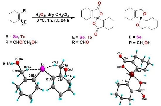
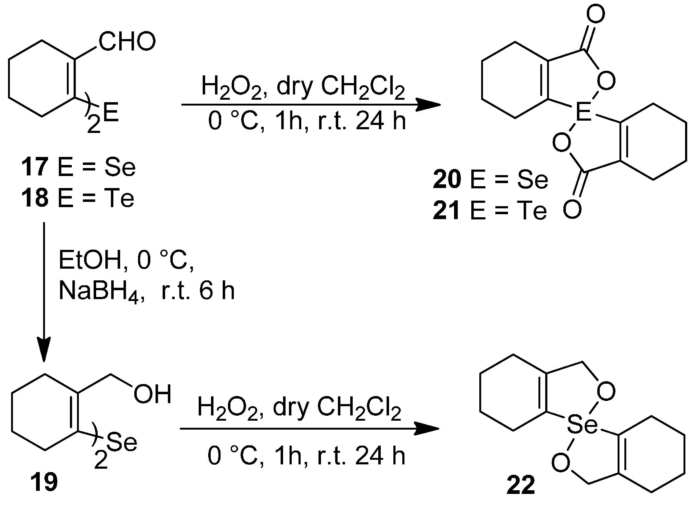
2.1. Synthesis

The precursors, di-(o-formylcyclohex-1-ene)diselenide 16 (Figure 1) and di-(o-formylcyclohex-1-ene)selenide 17 were obtained by the disodium diselenide route [32]. Di-(o-formylcyclohex-1-ene)telluride 18 was prepared by following the literature procedure [33]. Di(2-hydroxymethylcyclohex-1-ene)selenide 19 was obtained by the reduction of di-(o-formylcyclohex-1-ene)selenide 17 with NaBH4. Synthesis of spirodioxychalcogenuranes 2022 was accomplished by the oxidation of monochalcogenides 1719 with an excess of H2O2 (Scheme 2).
Scheme 2. Synthesis of selenide 19 and spirodioxychalcogenuranes (2022).
Molecules 20 12670 g010

2.2. Spectroscopy Studies

The compounds were characterized by common spectroscopic tools such as IR, 1H-, 77Se-NMR spectroscopy, mass spectrometry and single crystal X-ray diffraction studies (Supplementary Materials). The FT-IR spectrum of 19 exhibits the OH stretching frequency (C–OH) at 3298 cm−1, indicating the presence of the alcohol group. The carbonyl stretching frequency of spirodioxyselenurane 20 and -tellurane 21 were observed at (νC=O) 1683 cm−1 1663 cm−1 respectively which are in good agreement with those reported for spirodioxyselenurane 6 (1695 cm−1) and 1,1′-spirobis(3H-2,1-benzoxatellurole)-3,3′-dione [34]. For compounds 1921 the absence of the characteristic peak for the formyl group in 1H-NMR spectra confirmed the reduction or oxidation of 17. This was further corroborated by single crystal X-ray analysis (vide infra). In the 1H-NMR spectrum of compound 22, the methylene protons (–CH2–OH) are split. This is in contrast to the precursor 19, where these appeared as a singlet. It is due to the diastereotopic nature of the methylene protons. A similar pattern has been reported by Back and coworkers for 9a [35]. The 77Se-NMR spectrum of compound 22 exhibits a signal at 908 ppm. This is significantly downfield as compared to 19 (325 ppm).

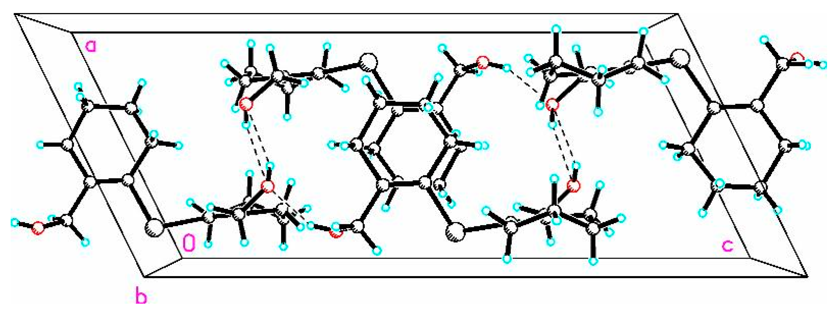
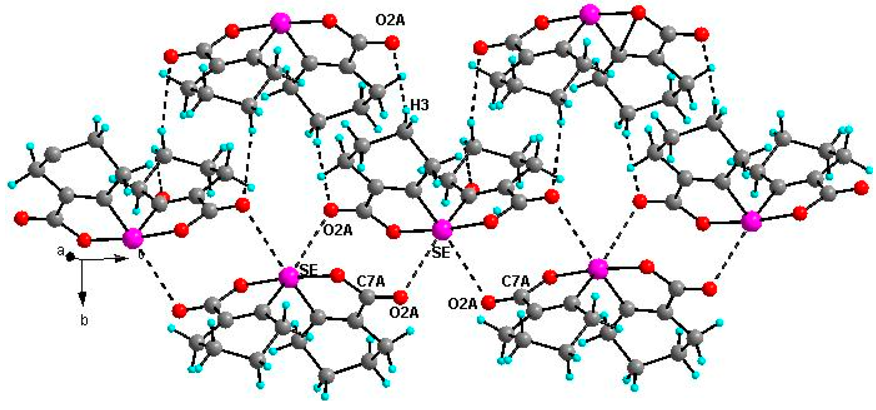
2.3. X-ray Crystallographic Studies

2.3.1. Molecular Structure of 19

The molecular structure of 19 is shown in Figure 2. The coordination geometry around the selenium atom is V-shaped with C1BA–Se–C1A bond angle being 99.10(1)°. A distinct feature of the structure is that both the hydroxyl groups are directed away from the selenium atom and there is asymmetry in the Se···OH bond distances. The Se···OH bond distances are 3.778 Å (Se–O1BA) and 4.486 Å (Se–O1A), respectively. A similar observation has been made in the case of the bis(o-formylphenyl) selenide having two formyl groups trans to the selenium [36]. Selvakumar et al. also found a similar situation in the case of a (5-(tert-butyl)-2-(phenylselanyl)-1,3-phenylene)dimethanol having two hydroxyl groups where none were coordinated to the selenium [31]. However, in di(2-hydroxybenzyl)selenide [22], out of the two OH groups, one OH group was coordinated to selenium with a bond distance of 3.008 Å. The lack of coordinating ability of the CH2OH group in 19 as well as in its aromatic analogue may be due to the poor electron donating property of the CH2OH groups and the availability of rotational degrees of freedom which could prevent the O···Se···O repulsive interaction. The compound shows hydrogen bonding interactions between the hydroxyl groups of the adjacent molecules (Figure 3).
Figure 2. Molecular structure of 19. Selected bond lengths (Å) and bond angles (°); Se–C7BA–H7BA···Se 2.710, C7BA–H7BA···Se 110.8, C7A–H7AB···Se 2.704, C7A–H7AB···Se 112.4, C1BA 1.935(5), Se–C1A 1.939(5), C1BA–Se–C1A 99.1(2), O1BA–C7BA–C6BA 111.8(4), O1A–C7A–C6A 114.2(4).
Molecules 20 12670 g002
Figure 3. The short OH···O intermolecular interactions (OH···O, 2.163 Å, C7A–O1A···H1A 106.3° and OH···O, 2.040 Å, C7A–O1A···H1BA 103.1°).
Molecules 20 12670 g003
Interestingly, in case of 19, along with Se···OH intermolecular interaction, short C–H···Se intramolecular interactions were also observed. The short interatomic distances between selenium and one of the benzylic hydrogens (C–H···Se) are 2.710 and 2.704 Å which are shorter than the reported for diselenocin [37] (2.92 Å) and sum of the van der Walls radii (2.99 Å) [38]. The C–H···Se bond angles for 19 are 110.8° and 112.4° which are good agreement with diselenocin (101.7° and 107.0°). The solid state IR (KBr) spectrum indicates Se···H–C interaction. The C–H symmetric stretching frequency (2831 cm−1) is shifted towards the lower wave number as compared with normal methylene adjacent to the electronegative atom (υ = 2855 cm−1).
To gain more information about the intramolecular Se···H interaction, density functional theory calculations have been carried out using Gaussian09 suite of programs [39]. The geometry of 19 was optimized at B3LYP/6–31+g(d) basis set. The Natural Bond Orbital (NBO) second-order perturbation energies for the C–H···Se interaction for 19 are EC-H···Se 2.49 and 1.40 kcal mol−1. It was further confirmed by Atoms in Molecules (AIM) analysis. The AIM [40,41,42] analysis was performed with AIM [43] at B3LYP/6-31+g(d) level of theory with wtbs basis set for Se and 6–31+g(d) level for remaining atoms. On carrying out AIM analysis of 19, the bond critical point was located in between C–H–Se which confirms the presence of C–H···Se intramolecular interaction (Figure 4). The values of electron density for C–H···Se were (ρ) 0.0124 and 0.0123 a.u.
Figure 4. AIM picture of 19 showing bond critical point for C–H···Se interaction.
Molecules 20 12670 g004

2.3.2. Molecular Structure of 20

Dioxyselenurane 20 crystallizes in a monoclinic crystal system with C2/c space group (Figure 5). The geometry around the central atom is distorted trigonal bipyramidal in which two Se–O bonds occupy the axial positions, two Se–C bonds and the lone pair occupy the equatorial positions. The Se–O bond length of 1.971(1) Å is slightly elongated compared to the covalent Se–O bond length (1.83 Å) and in good agreement with reported Se–O bond distance of 1.968(1) Å for 3,3′-spirobis(3-selenanaphthalide) [44] and significantly longer than that observed in 2-carboxylphenylmethyl selenoxide [45], {Se–O (1.774 Å)}. The O1A–Se–O1A bond angle is 172.99° which is deviated from the linear arrangement of 180°. Interestingly, the O1A–Se–O1A bond angle is equal to the reported bond angle for the aliphatic dioxaselenanonane 4 (172.99°) [20], and in good agreement with the reported bond angle for aromatic analogue of 3,3′-spirobis(3-selenanaphthalide) 6 (172.4°) [44]. The dihedral angle between the planes defining the two chelate rings i.e., O1A–Se–C1A–C6AC7A and Se–O1A–C7A–C6A–C1A is 75.58. The packing diagram of compound 20 shows weak intermolecular O···Se and O···H interactions with distances of 3.053(2) and 2.610(2) Å, respectively to form a two dimensional supramolecular network (Figure 6). The intermolecular O2A···H3 bond distance is 2.609(2) Å with bond angle 157.7(3)°.
Figure 5. Molecular structure of compound 20. Selected bond lengths (Å) and bond angles (°); Se–C1A 1.939(2), Se–O1A 1.970(1), Se–O1A 1.970(1), C7A–O2A 1.213(1), C1A–Se–C1A 99.27(9), O1A–Se–O1A 172.99(7).
Molecules 20 12670 g005
Figure 6. Two-dimensional packing diagram of compound 20 O2A···Se 3.053(2) Å, C7A–O2A–Se 102.8(2)°, O2A···H3 2.609(2) Å, C3A–H3–O2A 157.7(3)°.
Molecules 20 12670 g006

2.3.3. Molecular Structure of 21

Compound 21 (Figure 7) crystallizes in orthorhombic crystal system with Pbca space group. The geometry around the tellurium atom is distorted trigonal bipyramidal and is very similar to the selenium analogue 20 in which both Te–1OA and Te–O1B occupy the axial positions, while Te–C1BA, Te–C1AA and the lone pair occupy the equatorial positions. The Te–O1A and Te–O1B bond lengths are 2.114(3) and 2.082(3) Å, respectively. These distances are close to the sum of covalent radii of tellurium and oxygen (2.04 Å) [38]. The O1A–Te–O1B bond angle in 21 is 161.7(1)° which is similar to that reported for aromatic spirotellurane (161.3°) [34]. The dihedral angles between two planes i.e., Te–C1AA–C6AA–C7A–O1A and Te–O1B–C7B–C6BA–C1BA is 77.66(21). The crystal packing diagram shows intermolecular Te···O (carbonyl) interaction (Figure 8).
Figure 7. Molecular structure of compound 21. Selected bond lengths (Å) and bond angles (°); Te–C1AA 2.055(1), Te–C1BA 2.138(12), Te–O1A 2.114(3), Te–O1B 2.082(3), O2A–C7A 1.214(5), O2B–C7B 1.205(5), O1A–Te–O1B 161.71(1), C1AA–Te–C1BA 98.6(5).
Molecules 20 12670 g007
Figure 8. Two-dimensional packing diagram of compound 21. Selected bond lengths (Å) and bond angles (°); Te···O2A 2.936(35) Å, Te···O2A–C7A 126.4(29)°.
Molecules 20 12670 g008

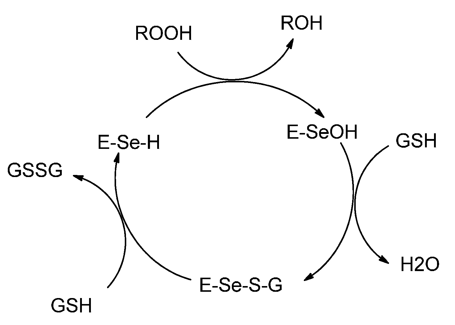
2.4. Glutathione Peroxidase-Like Activity

The glutathione peroxidase activity of the compounds was determined by the coupled reductase assay [46]. In this assay, the GPx-like activity was measured by a coupled enzyme containing nicotinamide adenine dinucleotide phosphate (NADPH) (0.01 M), glutathione (GSH) (0.05 M), catalysts (0.002 M), H2O2 (0.026 M), glutathione reductase (GR) (1.3 unit). The reduction of H2O2 by GSH was also recorded using ebselen for comparison. The decrease in NADPH concentration was monitored spectrophotometrically at 340 nm and results are summarized in Table 1. It was found that di-(o-formylcyclohex-1-ene)diselenide (16) is more efficient catalyst in comparison with ebselen, (2-phenyl-1,2-benzisoselenazol-3(2H)-one) [47], and bis(o-formylphenyl)diselenide [24]. The higher activity of 16 is probably due to the presence of stronger Se···O intramolecular interaction in comparison with bis(o-formylphenyl)diselenide. This stabilizes the selenenyl sulfide intermediate which in turn reacts with thiol to produce the disulfide. However, 17 and 19 do not show significant activity. The spirocyclic derivatives 20 and 22 where Se is in +4 oxidation state also did not show any significant activity. A similar result has been reported by Singh and co-workers [22] and Back and co-workers [23] in the case of aromatic spirocyclic derivatives. The reaction rates were similar to the control reaction rate. The GPx-like activity of compound 16 was calculated to be V0 = 49.8 ± 1.61 μM min−1 which is better than that of bis(o-formylphenyl)diselenide and standard ebselen (Table 1).
Table 1. Glutathione peroxidase-like activity of organoselenium compounds determined by coupled reductase assay.
Compound *Average (V0) μM min−1
Control10.5 ± 1.06
Ebselen [47]27.8 ± 1.99
Bis(o-formylphenyl)diselenide [24]34.3 ± 1.91
1649.8 ± 1.61
179.4 ± 0.51
196.8 ± 0.77
2016.4 ± 2.6
228.4 ± 0.24
*: The initial rates were calculated by following the depletion of NADPH by UV method at 25 °C. The reactions were carried out in 100 mM phosphate buffer, pH 7.48, with EDTA (1 mM), NADPH (0.01 M), GSH (0.05 M), catalysts (0.002 M), H2O2 (0.026 M), GR (1.3 unit).

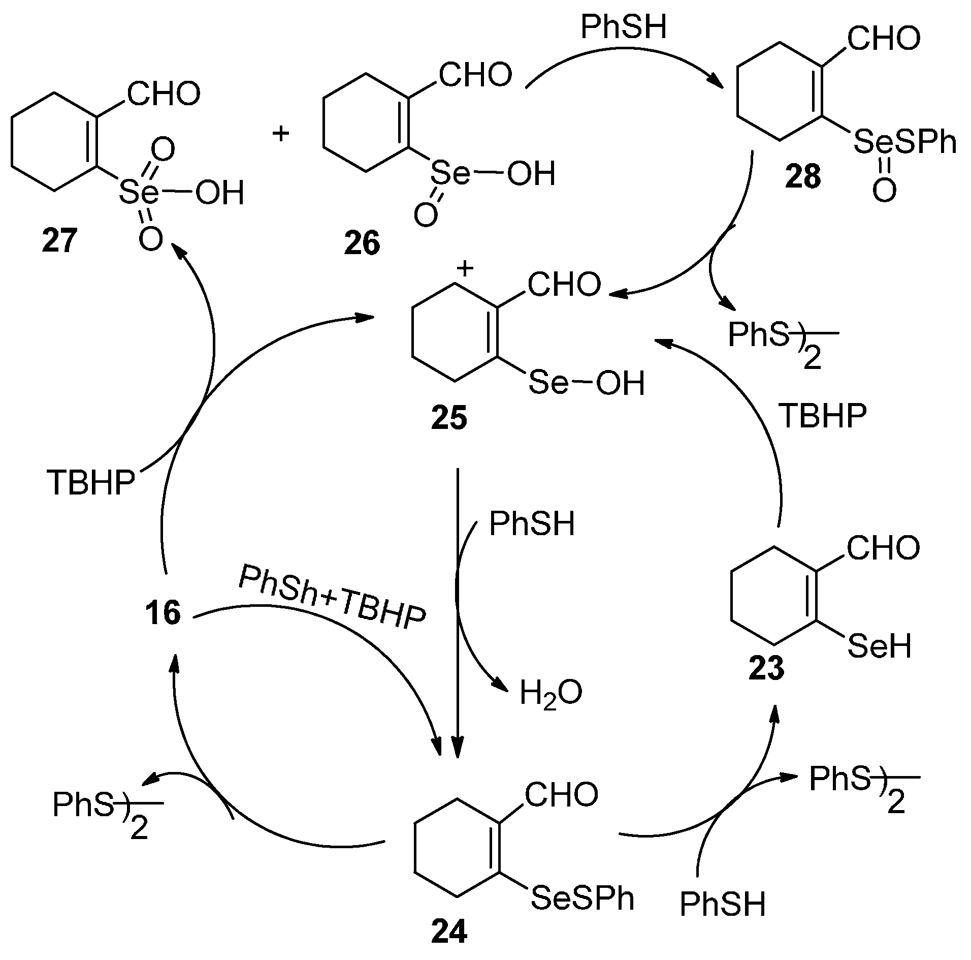
2.4.1. Mechanistic Studies on Di-(2-formylcyclohexenyl)diselenide 16

To understand the mechanism and identify the intermediates involved in the catalytic reaction, 77Se-NMR spectroscopy has been used since its chemical shift is very sensitive to the environment. The three major intermediates involved in the peroxidase reduction, i.e., RSeH, RSeOH and RSeSPh, are expected to show large differences in 77Se-NMR chemical shift values.

2.4.2. Reaction of 16 with PhSH Followed by TBHP

The reaction of di-(2-formylcyclohexenyl)diselenide 16 with 3 equivalents of PhSH (thiophenol) does not show any new peak corresponding to the formation of the expected selenenyl sulfide 24 even after 30 min. It indicates that the reactivity of PhSH towards 16 is very slow. The addition of tert-butylhydroperoxide (TBHP) to the above mixture results in the instantaneous formation of the selenenyl sulfide 24 (indicated by 77Se-NMR signal at 705 ppm). The signal is shifted downfield in comparison to the reported value of diselenide 3 (545 ppm) [22]. This significant downfield shift is due to the existence of strong Se···O intramolecular interaction in selenenyl sulfide 24 which suggests that 24 is very stable in solution and is not disproportionated to the corresponding 16.

2.4.3. Reaction of Di-(2-formylcyclohexenyl)diselenide 16 with TBHP Followed by PhSH

In the 77Se-NMR spectrum, four signals at 488, 1393, 1379, and 1338 ppm were observed for the reaction of 16 with two equivalents of TBHP. These correspond to diselenide 16, selenenic acid (RSeOH) 25, seleninic acid (RSeO2H) 26 and selenonic acid (RSeO3H) 27, respectively. Similar intermediates have been observed for the reaction of bis[2-(4,4-dimethyl-2-oxazolinyl)phenyl]diselenide [17] with H2O2. The 77Se-NMR chemical shifts are significantly downfield shifted compared with that of selenenic acid (1206 ppm), seleninic acid (1149 ppm) and selenonic (1138 ppm) formed during the reaction of (bis[2-(4,4-dimethyl-2-oxazolinyl phenyl]diselenide with an excess of H2O2 and also downfield shifted in comparison to the selenenic acid (1200 ppm) of 3 [22].
Molecules 20 12670 i001
When the reaction mixture containing TBHP was treated with seven equivalents of PhSH, peaks corresponding to the intermediates selenenic acid 25, seleninic acid 26 and selenonic acid 27 disappeared. Two new peaks at 705 and 866 ppm along with peak for 16 were observed. The peaks at 705 and 866 ppm probably correspond to the selenenyl sulfide 24 and selenoxysulfide 28, respectively. Selenoxysulfide 28 is formed by the reaction of selenenic acid 25 with PhSH. The reaction of selenenic acid with PhSH is analogous to the observation by Mugesh et al. [17] where bis[2-(4,4-dimethyl-2-oxazolinyl)phenyl]diselenide reacts rapidly with thiol to give corresponding selenoxysulfide intermediates. After the addition of 14 equivalents of PhSH, peaks corresponding to the 16 and other intermediates had disappeared except for the peak of selenenyl sulfide 24. Based on these observations and mechanisms reported for related diselenides, the following mechanism for the catalytic action of 16 (Scheme 3) is proposed.
Scheme 3. GPx activity of di-(2-formylcyclohexenal)diselenide (16).
Molecules 20 12670 g011

3. Experimental Section

3.1. General Information

All the organochalcogen derivatives were synthesized under nitrogen or argon atmosphere using standard Schlenk line techniques. Solvent were purified and dried by standard procedures and were freshly distilled prior to use [48]. All the chemicals used were reagent grade and were used as received. Melting points were recorded in capillary tubes. The NMR spectra were recorded in CDCl3 solvent. The 1H (400 MHz), 13C (100 MHz), 77Se (57.26/76.4 MHz) and 125Te (126.3 MHz) spectra were recorded on a Varian Mercury plus or Bruker 400 MHz spectrometer. Chemical shifts cited were referenced with respect to TMS for (1H and 13C) as internal standard and Me2Se (for 77Se), Me2Te (for 125Te) as external standards. Elemental analysis was performed on Carlo-Erba model 1106 and Eager 300 EA112 elemental analyzers. The IR spectra were recorded in the range 400–4000 cm−1 by using KBr pellets for solid samples on a Thermo Nicolet Avatar 320 FT-IR spectrometer. Mass spectral (MS) studies were completed by using a QTOF Micro mass spectrometer with electrospray ionization mode analysis. In the case of isotopic patterns, the value is given for the most intense peak. The UV–VIS spectra in solution for GPx activity were recorded with a JASCO, V-570 spectrometer.
The single crystal X-ray diffraction measurements for compounds were performed on Oxford Diffraction Gemini diffraction measurement device with graphite monochromated Mo Kα radiation (λ = 0.7107 Å). The structures were determined by routine heavy-atom method using SHELXS 97 [49] and refined by full-matrix least-squares with the non-hydrogen atom anisotropic and hydrogen atoms with fixed isotropic thermal parameters of 0.07 Å by means of SHELXS 97 program [50]. The structure refinement parameters for compounds 1921 are given in Table 2. CCDC-1041184 (19), CCDC-1041181 (20), CCDC-1041182 (21), contain the supplementary crystallographic data for this paper. These data can be obtained free of charge from The Cambridge Crystallographic Data Centre via www.ccdc.cam.ac.uk/data_request/cif.
Table 2. Crystal and structure refinement data for 1921.
Compound192021
Empirical formulaC14H22O2SeC56H64O16Se4C14H16O4Te
Formula weight301.281308.91375.87
Crystal systemMonoclinicMonoclinicOrthorhombic
Space groupP21/cC2/cPbca
a (Å)8.7358(12)15.0340(13)15.7046(2)
b (Å)9.1761(12)8.5925(7)8.83038(16)
c (Å)19.689(2)10.4102(8)19.1603(3)
α (°)909090
β (°)116.096(8)112.358(4))90
γ (°)909090
V3)1417.4(3)1242.69(18)2657.11(8)
Z418
D (calcd) (Mg/m3)1.4121.7481.879
Absorption coefficient (mm−1)2.6392.02717.759
Reflections collected7290113045111
Final R(F) [I > 2σ(I)] a0.05670.02370.0515
wR(F2) indices [I > 2σ(I)]0.14140.05970.1361
Data/restraints/parameters2522/17/1581963/0/872513/72/210
Goodness-of-fit on F21.0241.0991.032
a: R(Fo) = ∑||Fo| − |Fc||/∑|Fo| and wR(Fo2) = {∑[w(Fo2Fc2)2]/∑[w(Fc2)2}1/2.

3.2. Synthesis

Di(2-hydroxymethylcyclohex-1-ene)selenide 19. To a stirred solution of di-(2-formylcyclohexenyl)selenide 17 (0.106 g, 0.34 mmol) in dry methanol (5 mL) was added an excess of NaBH4 (0.051 g, 1.36 mmol) at 0 °C. The reaction mixture was stirred for 6 h. Solvent was evaporated and the residue poured into crushed ice. The product was extracted with chloroform and the combined organic layers were washed with a saturated brine solution. It was dried over sodium sulfate and concentrated to give 19 as white solid.
Molecules 20 12670 i002
Yield: 0.060 g (25%), mp 126–128 °C; 1H-NMR (400 MHz, CDCl3): δ 4.30 (s, 4H), 2.29–2.27 (dd, 8H), 1.65–1.64 (dd, 8H); 13C-NMR (CDCl3): δ 141.2 (C1), 126.7 (C2), 66.4 (C7), 34.4 (C3), 29.4 (C6), 24.4 (C4), 22.7 (C5); 77Se-NMR (CDCl3): δ 325. ES-HRMS: m/z calcd. for C14H22SeO2: 302.0785 [M]+; found: 325.0688 [M + Na]+. FT-IR υ 1019, 1429, 1443, 2875, 2855, 2926, 3298 cm−1. Anal. Calcd. for C14H22SeO2: C, 55.81; H, 7.36; found C, 56.18; H, 6.97.
Spirodioxyselenurane 20. To a stirred solution of di-(2-formylcyclohexenyl)selenide 17 (0.112 g, 0.34 mmol) in dry CH2Cl2 (10 mL) was added 30% of H2O2 (120 μL, 1.02 mmol) at 0 °C. The reaction mixture was stirred for 24 h at room temperature to give compound 20 as a white solid.
Molecules 20 12670 i003
Yield: 0.05 g (48%). mp 87–89 °C. 1H-NMR (400 MHz, CDCl3): δ (ppm) 2.57–2.50 (m, 4H), 2.39–2.38 (m, 8H), 1.86–1.67 (m, 8H); 13C-NMR (CDCl3): δ 190.3 (C7), 150.1 (C2), 140.1 (C1), 30.5 (C6), 20.5 (C4), 20.4 (C3), 20.2 (C5); 77Se-NMR (CDCl3): δ 801; ES-HRMS: m/z calcd. for C14H16SeO4: 328.0214 [M]+; found: 329.0294 [M + H]+. FT-IR (KBr) υ 740, 759, 1138, 1238, 1683, 2941cm−1. Anal. Calcd. for C14H16SeO4: C, 51.39; H, 4.93; found C, 50.84; H, 4.81.
Spirodioxytellurane 21. A solution of compound 18 (0.1 g, 0.28 mmol) in dry CH2Cl2 (5 mL) was treated with an aqueous 30% of H2O2 (99 μL, 0.86 mmol) at 0 °C. The mixture was stirred for 24 h at room temperature; the solvent was evaporated to give compound 21 as a white solid.
Molecules 20 12670 i004
Yield: 0.09 g (25%), mp 95–97 °C; 1H-NMR (400 MHz, CDCl3): δ (ppm) 1.84–1.74 (m, 8H), 2.76–2.23 (m, 8H); 13C-NMR (CDCl3): δ 190.3 (C7), 150.2 (C2), 140.2 (C1), 30.6 (C6), 20.5 (C3), 20.40 (C4), 20.2 (C5); 125Te-NMR (CDCl3): δ 1088.7. ES-HRMS: m/z calcd. for C14H16TeO4 (378.0111) [M]+; found 379.0174 [M + H]+. FT-IR (KBr) υ 702, 975, 1203, 1584, 1663, 2931cm−1. Anal. Calcd. for C14H16TeO4: C, 44.74; H, 4.29; found C, 45.02; H, 5.38.
Spirodioxyselenurane 22. To a solution of selenide 19 (0.1 g, 0.28 mmol) in dry CH2Cl2 (5 mL) was added an aqueous 30% of H2O2 (99 μL, 0.86 mmol) at 0 °C. The resultant mixture was stirred for 24 h at room temperature, the solvent was evaporated to afford 22 as a white solid.
Molecules 20 12670 i005
Yield: 0.07 g (49%). mp 82–84 °C. 1H-NMR (400 MHZ, CDCl3): δ (ppm) 4.66–4.55 (d, 4H, 2J = 12 Hz), 2.52–2.06 (m, 8H), 1.77–1.67 (m, 8H); 13C-NMR (CDCl3): δ; 144.3 (C2), 131.9 (C1), 76.1 (C7), 25.5 (C6), 25.3 (C4), 24.3 (C5), 21.5 (C3); 77Se-NMR (CDCl3): δ 908; ES-HRMS: m/z calcd. for C14H20SeO2: 300.0629, [M]+; found: 301.0705 [M + H]+. Anal. Calcd. for C14H20SeO2: C, 56.19; H, 5.74; found C, 50.60; H, 5.99.

3.3. Coupled Reductase Assay

The GPx-like activities of 1620 were measured by JASCO spectrophotometer according to the literature method using ebselen as the standard. The catalytic reaction was carried out at room temperature (25 °C) in 1 mL of the solution containing 100 mM potassium phosphate buffer, pH 7.5, 1 mM EDTA, 0.1 mM GSH, 0.25 mM of NADPH, 0.020 mM of catalyst and 0.26 mM of H2O2. The activity was followed by the decrease of NADPH on addition of H2O2 and absorption was measured at 340 nm (εmax = 6.22 × 10³ M−1 cm−1) (chemical reactions (1)–(3)). Each of the experiments was carried out in triplicate.
Molecules 20 12670 i006

4. Conclusions

Di-(2-formylcyclohexenyl)diselenide 16 showed better GPx like activity than the aromatic analogue bis(o-formylphenyl)diselenide whereas di(2-hydroxymethylcyclohex-1-ene)selenide 19 and spirodioxyselenurane 20 and 22 did not show any significant activity.

Supplementary Materials

Supplementary materials can be accessed at: https://www.mdpi.com/1420-3049/20/07/12670/s1.

Acknowledgments

H.B.S. is grateful to the Department of Science and Technology (DST), New Delhi (India), for generous funding. P.R.P. is thankful to Department of Chemistry, IIT Bombay for a teaching assistantship.

Author Contributions

All the authors have contributed equally.

Conflicts of Interest

The authors declare no conflict of interest.

References

  1. Epp, O.; Ladenstein, R.; Wendel, A. The refined structure of the selenoenzyme glutathione peroxidase at 0.2-nm resolution. Eur. J. Biochem. 1983, 133, 51–69. [Google Scholar] [CrossRef] [PubMed]
  2. Arthur, J.R.; Beckett, G.J. Roles of selenium in type I iodothyronine 5′-deiodinase and in thyroid hormone and iodine metabolism. In Selenium in Biology and Human Health; Burk, R.F., Ed.; Springer-Verlag: New York, NY, USA, 1994. [Google Scholar]
  3. Berry, M.J.; Banu, L.; Larsen, P.R. Type I iodothyronine deiodinase is a selenocysteine-containing enzyme. Nature 1991, 349, 438–440. [Google Scholar] [CrossRef] [PubMed]
  4. Behne, D.; Kyriakopoulos, A.; Meinhold, H.; Köhrle, J. Identification of type I iodothyronine 5′-deiodinase as a selenoenzyme. Biochem. Biophys. Res. Commun. 1990, 173, 1143–1149. [Google Scholar] [CrossRef]
  5. Bianco, A.C.; Salvatore, D.; Gereben, B.; Berry, M.J.; Larsen, P.R. Biochemistry, cellular and molecular biology, and physiological roles of the iodothyronine selenodeiodinases. Endocr. Rev. 2002, 23, 38–89. [Google Scholar] [CrossRef] [PubMed]
  6. Lee, S.-R.; Kim, J.-R.; Kwon, K.-S.; Yoon, H.W.; Levine, R.L.; Ginsburg, A.; Rhee, S.G. Molecular cloning and characterization of a mitochondrial selenocysteine-containing thioredoxin reductase from rat liver. J. Biol. Chem. 1999, 274, 4722–4734. [Google Scholar] [CrossRef] [PubMed]
  7. Williams, C.H., Jr.; Arscott, L.D.; Müller, S.; Lennon, B.W.; Ludwig, M.L.; Wang, P.F.; Veine, D.M.; Becker, K.; Schirmer, R.H. Thioredoxin reductase two modes of catalysis have evolved. Eur. J. Biochem. 2000, 267, 6110–6117. [Google Scholar] [CrossRef] [PubMed]
  8. Mugesh, G.; Singh, H.B. Synthetic organoselenium compounds as antioxidants: Glutathione peroxidiseactivity. Chem. Soc. Rev. 2000, 29, 347–357. [Google Scholar] [CrossRef]
  9. Mugesh, G.; du Mont, W.W.; Sies, H. Chemistry of biologically important synthetic organoseleniumcompounds. Chem. Rev. 2001, 101, 2125–2179. [Google Scholar] [CrossRef] [PubMed]
  10. Nogueira, C.W.; Rocha, J.B.T. Organoselenium and organotellurium compounds: Toxicology and pharmacology. In Organic Selenium and Tellurium Compounds; Rappoport, Z., Ed.; Wiley: Chichester, UK, 2012; Volume 3, Part II, Chapter 21. [Google Scholar]
  11. Masukawa, T. Pharmacological and toxicological aspects of inorganic and organic selenium compounds. In The Chemistry of Organoselenium and Tellurium Compounds; Patai, S., Ed.; Wiley: Chichester, UK, 1987; Volume 2, Chapter 9. [Google Scholar]
  12. Jamier, V.; Ba, L.A.; Jacob, C. Selenium- and tellurium-containing multifunctional redox agents as biochemical redox modulators with selective cytotoxicity. Chem. Eur. J. 2010, 16, 10920–10928. [Google Scholar] [CrossRef] [PubMed]
  13. Bhowmick, D.; Mugesh, G. Enzyme mimetic chemistry of organoselenium compounds. In The Chemistry of Organoselenium and Tellurium Compounds; Patai, S., Ed.; Wiley: Chichester, UK, 2013; Volume 4, Part II, Chapter 16. [Google Scholar]
  14. Bhabak, K.P.; Mugesh, G. Functional mimics of glutathione peroxidase: Bioinspired synthetic antioxidant. Acc. Chem. Res. 2010, 43, 1408–1419. [Google Scholar] [CrossRef] [PubMed]
  15. Bhuyan, B.J.; Lamani, D.S.; Mugesh, G.; Wirth, T. Current research on mimics and models of selenium-containing antioxidants. In Handbook of Chalcogen Chemistry: New Perspectives in Sulfur, Selenium and Tellurium, 2nd ed.; RSC: Cambridge, UK, 2013. [Google Scholar]
  16. Mugesh, G. Glutathione peroxidase activity of ebselen and its analogues: Some insights into the complex chemical mechanisms underlying the antioxidant activity. Curr. Chem. Biol. 2013, 7, 47–56. [Google Scholar] [CrossRef]
  17. Mugesh, G.; Panda, A.; Singh, H.B.; Punekar, N.S.; Butcher, R.J. Glutathione peroxidase-like antioxidant Activity of diaryl diselenides: A mechanistic study. J. Am. Chem. Soc. 2001, 123, 839–850. [Google Scholar] [CrossRef] [PubMed]
  18. Mukherjee, A.J.; Zade, S.S.; Singh, H.B.; Sunoj, R.B. Organoselenium chemistry: Role of Intramolecular interactions. Chem. Rev. 2010, 110, 4357–4416. [Google Scholar] [CrossRef] [PubMed]
  19. Bayse, C.A.; Pavlou, A. Tuning the activity of glutathione peroxidase mimics through intramolecular Se···N,O interactions: A DFT study incorporating solvent-assisted proton exchange (SAPE). Org. Biomol. Chem. 2011, 9, 8006–8015. [Google Scholar] [CrossRef] [PubMed]
  20. Back, T.G.; Moussa, Z.; Parvez, M. The exceptional glutathione peroxidase-like activity of di(3-hydroxypropyl) selenide and the unexpected role of a novel spirodioxaselenanonane intermediate in the catalytic cycle. Angew. Chem. Int. Ed. 2004, 43, 1268–1270. [Google Scholar] [CrossRef] [PubMed]
  21. Back, T.G.; Moussa, Z. Remarkable activity of a novel cyclic seleninate ester as a glutathione peroxidase mimetic and its facile in situ generation from allyl 3-hydroxypropyl selenide. J. Am. Chem. Soc. 2002, 124, 12104–12105. [Google Scholar] [CrossRef] [PubMed]
  22. Tripathi, S.K.; Patel, U.; Roy, D.; Sunoj, R.B.; Singh, H.B.; Wolmershäuser, G.; Butcher, R.J. o-Hydroxylmethylphenylchalcogens: Synthesis, intramolecular nonbonded chalcogen···OH interactions, and glutathione peroxidase-like activity. J. Org. Chem. 2005, 70, 9237–9247. [Google Scholar] [CrossRef] [PubMed]
  23. Back, T.G.; Kuzma, D.; Parvez, M. Aromatic derivatives and tellurium analogues of cyclic seleninate esters and spirodioxyselenuranes that act as glutathione peroxidase mimetics. J. Org. Chem. 2005, 70, 9230–9236. [Google Scholar] [CrossRef] [PubMed]
  24. Kumar, S.; Panda, S.; Singh, H.; Wolmershäuser, G.; Butcher, R. Structural aspects of some organoselenium compounds. Struct. Chem. 2007, 18, 127–132. [Google Scholar] [CrossRef]
  25. Press, D.J.; Mercier, E.A.; Kuzma, D.A.; Back, T.G. Substituent effects upon the catalytic activity of aromatic cyclic seleninate esters and spirodioxyselenuranes that act as glutathione peroxidase mimetics. J. Org. Chem. 2008, 73, 4252–4255. [Google Scholar] [CrossRef] [PubMed]
  26. Back, T.G.; Moussa, Z.J. Diselenides and allyl selenides as glutathione peroxidase mimetics. Remarkable activity of cyclic seleninates produced in situ by the oxidation of allyl ö-hydroxyalkyl selenides. J. Am. Chem. Soc. 2003, 125, 13455–13460. [Google Scholar] [CrossRef] [PubMed]
  27. Press, D.J.; McNeil, N.M.R.; Hambrook, M.; Back, T.G. Effects of methoxy substituents on the glutathione peroxidase-like activity of cyclic seleninate esters. J. Org. Chem. 2014, 79, 9394–9401. [Google Scholar] [CrossRef] [PubMed]
  28. Kuzma, D.; Parvez, M.; Back, T.G. Formation of a spirodiazaselenurane and its corresponding azaselenonium derivatives from the oxidation of 2,2′-selenobis(benzamide). Structure, properties and glutathione peroxidase activity. Org. Biomol. Chem. 2007, 5, 3213–3217. [Google Scholar] [CrossRef] [PubMed]
  29. Sarma, B.K.; Manna, D.; Minoura, M.; Mugesh, G. Synthesis, structure, spirocyclization mechanism, and glutathione peroxidase-like antioxidant activity of stable spirodiazaselenurane and spirodiazatellurane. J. Am. Chem. Soc. 2010, 132, 5364–5374. [Google Scholar] [CrossRef] [PubMed]
  30. Lamani, D.S.; Bhowmick, D.; Mugesh, G. Spirodiazaselenuranes: Synthesis, structure and antioxidant activity. Org. Biomol. Chem. 2012, 10, 7933–7943. [Google Scholar] [CrossRef] [PubMed]
  31. Selvakumar, K.; Singh, H.B.; Goel, N.; Singh, U.P.; Butcher, R.J. Synthesis and structural characterization of pincer type bicyclic diacyloxy- and diazaselenuranes. Dalton Trans. 2011, 40, 9858–9867. [Google Scholar] [CrossRef] [PubMed]
  32. Prasad, P.R.; Singh, H.B.; Butcher, R.J. Synthesis, structure and reactivity of β-chalcocyclohexenals: dichalcogenides and chalcogenides. Dalton Trans. 2015. (submitted). [Google Scholar]
  33. Minkin, V.I.; Sadekov, I.D.; Rivkin, B.B.; Zakharov, A.V.; Nivorozhkin, V.L.; Kompan, O.E.; Struchkov, Y.T. Synthesis and structure of β-tellurovinylcarbonyl compounds. J. Organomet. Chem. 1997, 536, 233–248. [Google Scholar] [CrossRef]
  34. Takaguchi, Y.; Furukawa, N. First synthesis and structural determination of 1,1′-spirobis(3H-2, 1-benzoxatellurole)-3,3′-dione ([10-Te-4(C202)]). Heteroat. Chem. 1995, 6, 481–485. [Google Scholar] [CrossRef]
  35. Press, D.J.; McNeil, N.M.R.; Rauk, A.; Back, T.G. NMR and Computational Studies of the configurational properties of spirodioxyselenuranes. Are dynamic exchange processes or temperature-dependent chemical shifts involved? J. Org.Chem. 2012, 77, 9268–9276. [Google Scholar] [CrossRef] [PubMed]
  36. Panda, A.; Menon, S.C.; Singh, H.B.; Butcher, R.J. Synthesis of some macrocycles:Bicycles from bis(o-formylphenyl) selenide: X-ray crystal structure of bis(o-formylphenyl) selenide and the first 28-membered selenium containing macrocyclic ligand. J. Organomet. Chem. 2001, 623, 87–94. [Google Scholar] [CrossRef]
  37. Iwaoka, M.; Tomoda, S. First observation of a C-H...Se “hydrogen bond”. J. Am. Chem. Soc. 1994, 116, 4463–4464. [Google Scholar] [CrossRef]
  38. Cordero, B.; Gomez, V.; Platero-Prats, A.E.; Reves, M.; Echeverria, J.; Cremades, E.; Barragan, F.; Alvarez, S. Covalent radii revisited. Dalton Trans. 2008, 21, 2832–2838. [Google Scholar] [CrossRef] [PubMed]
  39. Frisch, M.J.; Trucks, G.W.; Schlegel, H.B.; Scuseria, G.E.; Robb, M.A.; Cheeseman, J.R.; Scalmani, G.; Barone, V.; Mennucci, B.; Petersson, G.A.; et al. Gaussian 09; Revision D.01; Gaussian, Inc.: Wallingford, CT, USA, 2009. [Google Scholar]
  40. Bader, R.F.W. Atoms in Molecules: A Quantum Theory; Oxford University Press: New York, NY, USA, 1990. [Google Scholar]
  41. Popelier, P. Atoms in molecules: An introduction; Pearson: Harlow, UK, 2000. [Google Scholar]
  42. Matta, C.F.; Boyd, R.J. The Quantum Theory of Atoms in Molecules; Wiley-VCH: Weinheim, Germany, 2007. [Google Scholar]
  43. Biegler-Konig, F.; Schonbohm, J.; Bayles, D. Software news and updates AIM2000-a program to analyze and visualize atoms in molecules. J. Comput. Chem. 2001, 22, 545–559. [Google Scholar]
  44. Dahlén, B.; Lindgren, B. Formatin and crystal structure of 3,3′-spirobi(3-selenaphthalide). Acta Chem. Scand. 1993, 27, 2218–2220. [Google Scholar] [CrossRef]
  45. Dahlen, B. The molecular structure of o-carboxyphenyl methyl sulphoxide and o-carboxyphenyl methyl selenium oxide. Acta Crystallogr. B 1973, 29, 595–602. [Google Scholar] [CrossRef]
  46. Wilson, S.R.; Zucker, P.A.; Huang, R.R.; Spector, A. Development of synthetic compounds with glotathione peroxidase activity. J. Am. Chem. Soc. 1989, 111, 5936–5939. [Google Scholar] [CrossRef]
  47. Müller, A.; Cadenas, E.; Graf, P.; Sies, H. A novel biologically active seleno-organic compound-1: Glutathione peroxidase-like activity in vitro and antioxidant capacity of PZ 51 (Ebselen). Biochem. Pharmacol. 1984, 33, 3235–3239. [Google Scholar] [CrossRef]
  48. Perrin, D.D.; Armargo, W.L.F.; Perrin, D.R. Purification of Laboratory Chemicals, 4th ed.; Pergamon Press: Oxford, UK, 1996. [Google Scholar]
  49. Sheldrick, G.M. SHELXS-97, Program for Crystal Structure Solution University of Göttingen; University of Göttingen: Gottingen, Germany, 1990. [Google Scholar]
  50. Sheldrick, G.M. SHELXL-97, Program for Crystal Structure Refinement; University of Göttingen: Gottingen, Germany, 1997. [Google Scholar]
  • Sample Availability: Samples of the compounds not available from the authors.

