Article

Molecular Structures of the Products of a Diphosphonate Ester Building Block with Lewis Bases

1
Microscale Science Institute, Weifang University, Weifang 261061, China
2
New Materials and Function Coordination Chemistry Lab, Qingdao University of Science & Technology, Qingdao 266042, China
*
Author to whom correspondence should be addressed.
Molecules 2015, 20(8), 14435-14450; https://doi.org/10.3390/molecules200814435
Submission received: 29 December 2014 / Revised: 18 June 2015 / Accepted: 23 June 2015 / Published: 7 August 2015
(This article belongs to the Section Organic Chemistry)

Abstract

:
By treating a suitable Wittig reagent under acid conditions, the phosphonate ester 1,4-bimethylenebenzene phosphonate ethyl ester (H2[BBPE], 1) was obtained. As a building block, compound 1 has been reacted with the Lewis-base N,N-dimethylpiperazine, ammonia and NaOH yielded compounds 24. The crystal structures show that a 1D chain forming a tubular channel is constructed through hydrogen bonds in 1; hydrogen bonds form two 1D chains with left-hand and right-hand helixes and form 3D networks in compound 2; 1D hydrogen-bond chains are further connected together to afford a 3D network architecture in compound 3; the phosphonate is coordinated by two Na atoms which present different coordination environments in compound 4. Additionally, the relationships between the structure and fluorescence of the four compounds in the solid state and in different solvents have also been studied at room temperature.

1. Introduction

Recently, metal phosphonates have attracted a great deal of research interest as a new class of inorganic-organic materials due to their structural diversity and many potential and practical applications in the fields of nonlinear optics, catalysis, gas storage, ion exchangers and so on [1,2,3,4,5,6,7]. In all these cases, phosphonates play an important role because of their three oxygen donor atoms [8,9,10]. Accordingly, by modifying different organic functional groups, the complexing ability of phosphonates and the corresponding properties of the resulting materials will be changed [11,12,13,14,15,16,17,18,19]. On the other hand, multifunctional phosphonates, such as bisphosphonic acids, aminophosphonic acids and carboxyphosphonic acids, have been proved as good candidates for their potential to form extended hydrogen-bonded assemblies which could be utilized for the preparation of one-, two-, and three-dimensional hydrogen-bonded networks and microporous solids [20,21,22,23,24,25,26,27]. For instance, Yuan et al. combined 1-aminoethylidene diphosphonic acid (AEDPH4) and 1-aminopropane-1,1,3 triphosphonic acid (APTPH6) with 2,2′-bipyridyl-like ligands to build up higher-dimensional supramolecular architectures [3]. Amir et al. produced supramolecular isomerism and isomorphism using the structures of 1,4-butanebisphosphonic acid and its organic ammonium salts [2]. In general, single crystals of metal phosphonates are difficult to obtain because of their low solubility, so investigations on the structure of soluble salts of phosphonates are valuable in this field. Sparked by this thought, we designed and synthesized the diphosphonate ester of 1,4-bimethylenebenzene bisphosphonate ethyl ester (H2[BBPEH2], 1, where BBPEH2 = [O(OC2H5)(HO)PCH2C6H5CH2P(OH) (OC2H5)O]2−} as a bidentate ligand, as shown in Scheme 1. Furthermore, by using a general solution method and room temperature, 1 acted as a building block that easily reacted with organic and inorganic bases, and the formed compounds displayed better solubility in organic solvents or water. We thus obtained compounds 24 as shown in Scheme 2. As expected, the single crystal structure of two self-assembly acid-base hydrogen-bonded supramolecular architectures and one organic-inorganic hybrid coordination polymer were obtained. Fluorescence spectra of 14 in the solid-state, in methanol and water solutions have been determined. In this paper, we wish to report the syntheses, crystal structures and fluorescence properties of these four compounds.
Scheme 1. Synthesis of compound 1.
Molecules 20 14435 g006
Scheme 2. Synthesis of compounds 24.
Molecules 20 14435 g007

2. Results and Discussion

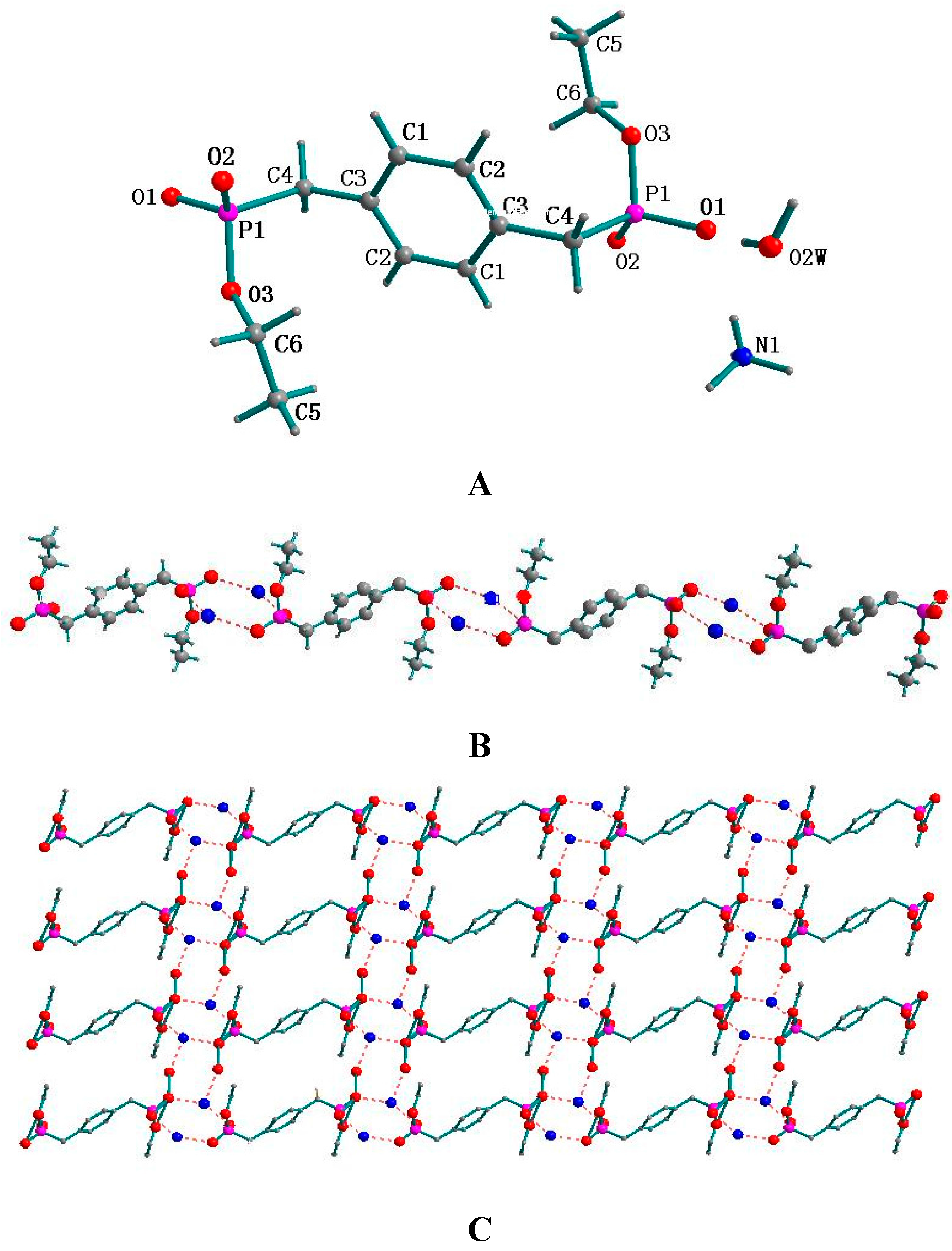
2.1. Supramolecular Structure of 1

As depicted in Figure 1A, the unit of 1 contains two independent H2[BBPEH2] molecules. In each molecule, there exists a 2-fold symmetric axis, which lies in the plane of the phenyl ring and passes through the two middle points of the C5-C5 and C5-C5 bonds. The P-O bond lengths are in the range of 1.5215(16) and 1.581(2) Å, which match those found in the similar structures [13]. In the unit lattice, each pair of adjacent H2[BBPEH2] molecules are connected each other by two O2-H1···O2 hydrogen bonds and then, a 1D zigzag chain forming a tubular channel is created, just as shown in Figure 1B, with the diameter of the channel being 2.384 Å.
Figure 1. (A) Molecular structure of 1 with atomic numbering and (B) the molecular arrangement by the hydrogen bonds between adjacent H2[BBPEH2] molecules.
Molecules 20 14435 g001

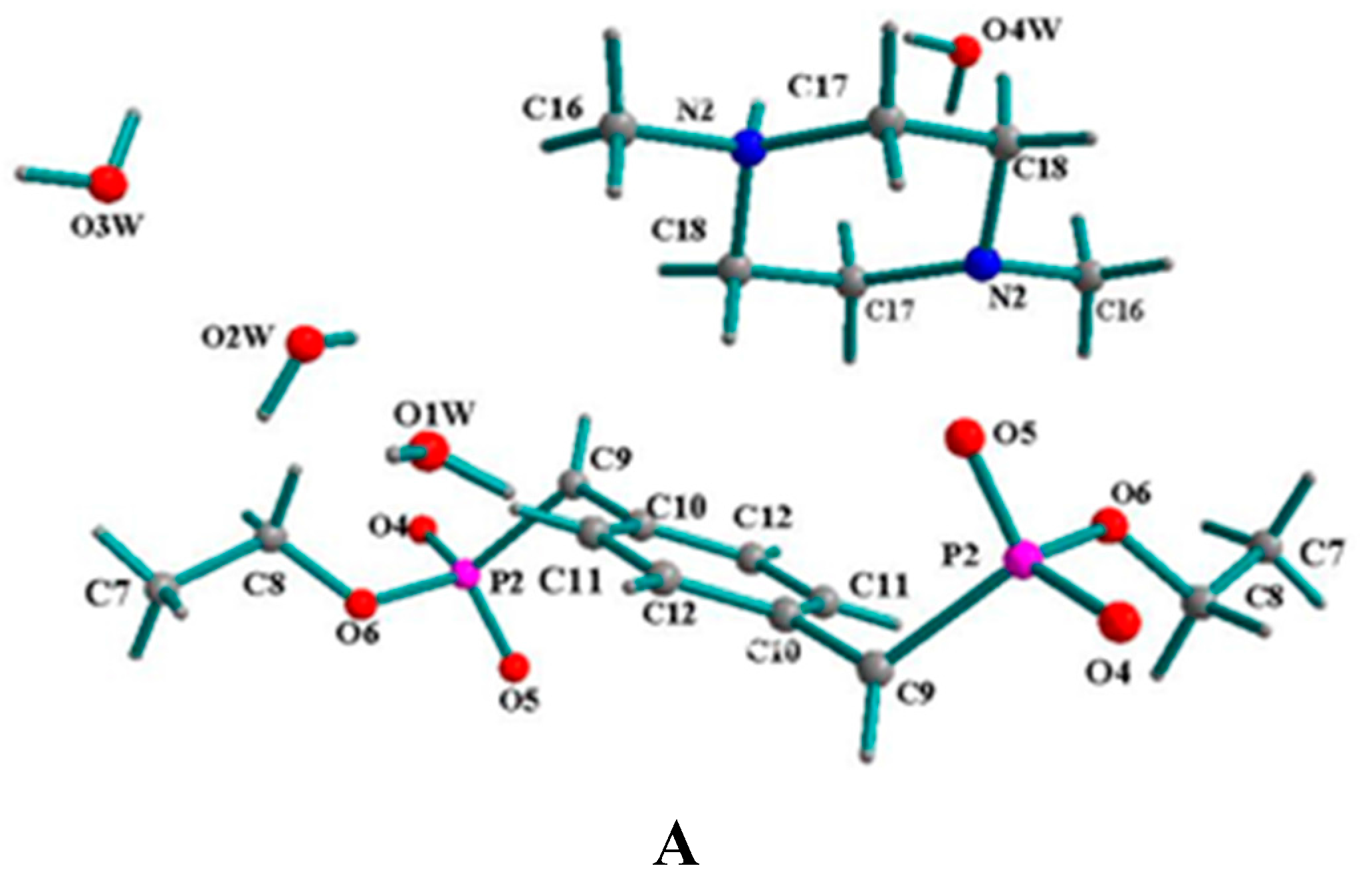
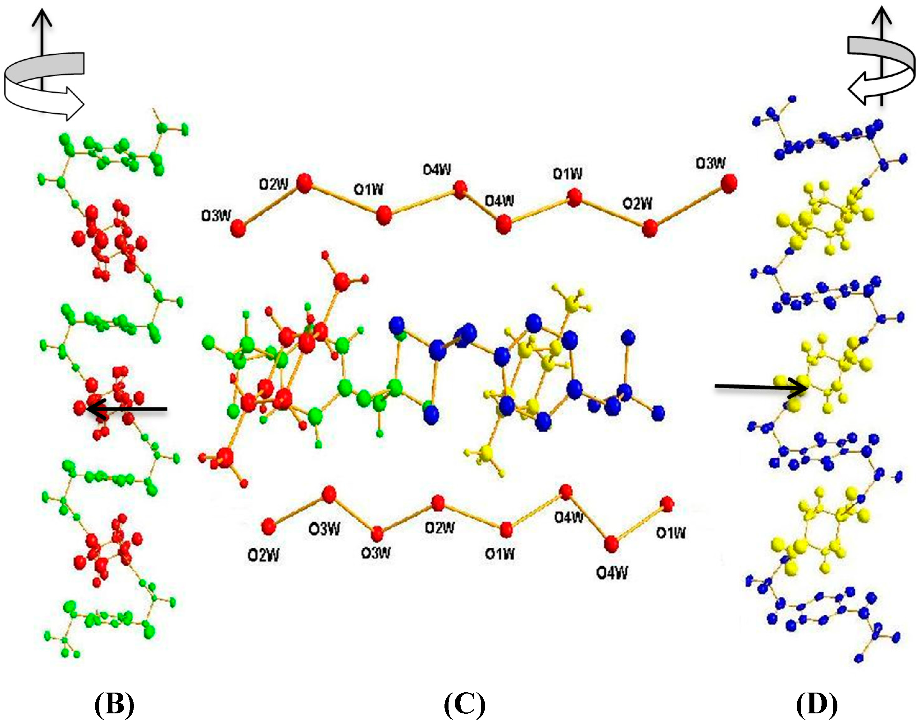
2.2. Supramolecular Structure of 2

The molecular structure of 2 with atomic numbering is shown in Figure 2A. A unit cell of compound 2 contains of two [BBPEH2]2− anions, two protonated N,N-dimethylpiperazine cations and eight lattice water molecules, which are connected together through hydrogen bonds and electrostatic effects. Each H2[BBPEH2] molecule has lost two protons and transferred them to the N,N-dimethylpiperazine nitrogen atoms and form a [BBPEH2]2− anion. Both N,N-dimethylpiperazine nitrogen atoms are protonated. In compound 2, there are two types of anion-cation groups with different symmetric equivalence. For one type of anion-cation group, the P1=O1 bond length is 1.444(2) Å, and the P1-O2 and P1-O3 bond lengths are 1.675(2) and 1.689(2) Å, respectively. The [BBPEH2]2− anions and protonated N,N-dimethylpiperazines are connected each other by N1-H1···O2 hydrogen bonds to form a 1D helical chain, which obeys the right-handed rule along an axis (named 1D-R, shown as green-red in Figure 2B). For the other anion-cation group, the P2=O5 bond length is 1.404 (2) Å, and the P2-O4 and P1-O2 bond lengths are 1.463(2) and 1.693(2) Å, respectively. The [BBPEH2]2− anions and protonated N,N-dimethylpiperazines are joined each other through N2-H2···O5 hydrogen bonds to afford a 1D helical chain, which obeys the left-handed rule along an axis (named 1D-L, shown as blue-yellow in Figure 2D). Four lattice water molecules play important roles in connecting the 1D chains and further constructing 3D networks (see Figure 2C). The O(2W)-H···O(1) and O(3W)-H···O(2) hydrogen bonds join the two water molecules [H2O2W and H2O3W] with the adjacent 1D-R helix chains, respectively, while at the same time the O(4W)-H···O(5) and O(1W)-H···O(4) hydrogen bonds join the other two water molecules [H2O4W and H2O1W] with the adjacent 1D-L helix chains, respectively. On the other hand, four water molecules are linked one by one through O-H···O hydrogen bonds to form a “water cluster chain”, with the O-O distances in the water cluster ranging between 2.654 and 3.076 Å and the O-H···O bond angles being from 81.49 to 134.63° (see Figure 2C). Finally, the water cluster chains connect all 1D chains together to construct 3D networks in the unit lattice. This 3D networks are further consolidated by some C-H···π interactions (see Table 1)
Figure 2. (A) Molecular structure of 2 with atomic numbering, (B) 1D-R right-handed zigzag chain shown as green-red, (C) 3D supramolcular architecture of 2 constructed by water chains and two different symmetry equivalent anion-cation groups and (D) 1D-L left-handed zigzag chain shown as blue-yellow.
Molecules 20 14435 g002aMolecules 20 14435 g002b
Table 1. Selected C-H···π Interactions in Compounds 2, 3 and 4.
CompoundC-H···πSymmetry CodeC···π
Centroid-Centroid (Å)
∠C-H···π (°)
2C(14)-H(14B)···Cg(3)x, −1 + y, z3.640149.55
C(14)-H(14B)···Cg(3)1− x, 1 − y, −z3.640149.55
C(17)-H(17B)···Cg(4)x, y, z3.695136.52
C(17)-H(17B)···Cg(4)x, 1 − y, 1 − z3.695136.52
3C(5)-H(5A)···Cg(1)x, −½ + y, ½ − z4.044164.92
C(5)-H(5A)···Cg(1)x, −½ − y, ½ + z4.044164.92
4C(2)-H(2B)···Cg(1)x, y, z3.792145.37
C(2)-H(2B)···Cg(1)x, −y, −z3.792145.37
In 2, Cg(3) denotes phenyl ring containing C(4)~C(6); Cg(4) denotes phenyl ring containing C(10)~C(12); in 3, Cg(1) denotes phenyl ring containing C(1)~C(3); in 4, Cg(1) denotes phenyl ring containing C(4)~C(6).

2.3. Supramolecular Structure of 3

The molecular structure of 3 with atomic numbering scheme is shown in Figure 3A. A unit cell of 3 contains two [BBPEH2]2−, four protonated ammonia and four water molecules, which are connected together through hydrogen bonds and electrostatic effects. Just as in 2, each H2[BBPEH2] molecule has lost two protons and transferred the protons to two ammonia molecules forming a [BBPEH2]2− anion. The nitrogen atoms of ammonia are protonated. The cation-anion ratio is 2:1. As shown in Figure 3B, N(1)-H(1)···O(1) (2.8331 Å) and N(1)-H(6)···O(2) (2.7781 Å) hydrogen bonds firstly join the [BBPEH2]2− anions to form a 1D chain, and then, in the unit lattice, all of the 1D chains are linked together forming the 2D array of hydrogen bonds (see Figure 3C) and afford a 3D network architecture through supramolecular interactions (see Figure 3D). For instance, as shown in Figure 3C, two 1D chains are linked together through N-H···O hydrogen bond s. The other 1D chains denoted are joined by the lattice water molecule through O-H···O hydrogen bonds. Further, through N-H···O hydrogen bond, the lattice water molecule and the protonated ammonia molecule are connected, which ultimately make the four 1D chains to build up a 3D network architecture. Most important, by observing the connecting mode among the oxygen and nitrogen atoms in protonated ammonia (see Figure 3E), one can find two six-membered rings and four five-membered rings.
Figure 3. (A) Molecular structure of 3 with atomic numbering; (B) 1D chain joined through N1-H1···O1 and N1-H6···O2 hydrogen bonds; (C) 2D supramolecular network viewing along the c axis in the unit cell; (D) view of the 3D architecture and (E) stable connection mode among atoms of oxygen and nitrogen.
Molecules 20 14435 g003aMolecules 20 14435 g003b
The cluster aggregation is controlled by a “vertex-connected” and “edge-sharing” association mode [28]. The six-membered rings adopt a “puckered-boat” confirmation. It is evident that these stable five-membered rings and six-membered rings help to strengthen the connection among the above four 1D chains. In the solid state, the C(5)-H(5A)···Cg(1) and C(5)-H(5A)···Cg(1) supramolecular interactions (Table 1) help to stabilize the 3D networks.

2.4. Supramolecular Structure of 4

The molecular structure of compound 4 with atomic numbering scheme is shown in Figure 4A. The X-ray crystal structural analysis indicates that the repeat unit of compound 4 contains one [BBPEH2]2− anion, two sodium cations, and six water molecules. Although the coordination modes can be described as a distorted octahedral geometry, there are two types of Na atoms with different modes. Take Na2 and Na1 cation for instance. Na2 is connected with the Na1 cation through three bridged oxygen atoms (O3W, O3W and O2W) in one direction, whereas in the opposite direction, the Na2 cation is connected with another sodium cation of Na1 only through one bridged oxygen atom of the O4W atom from H2O4W. Each of these Na2 cations is connected with two sodium cations, so for the Na2 cation, its coordination mode can be described by four coordinate oxygen atoms being bridged with two other sodium cations and two coordinated oxygen atoms from two [BBPEH2]2− anions. As seen from Figure 4A, for the Na1 cation, its coordination mode is similar to that of Na2, and apart from four bridged oxygen atoms, two coordinated O1W oxygen atoms come from two water molecules but not from two [BBPEH2]2− anions as in Na2. As shown in Figure 4A, since each [BBPEH2]2− anion is a bidentate ligand, coordinated to two sodium cations Na2 with its two apical O2 oxygen atoms. The Na-O distances are in the 2.413–2.525Å range, which is comparable to that reported for other Na-O bonds [about 2.455 Å] [29]. It is noticeable that, for the same [BBPEH2]2− anion, its two coordinated sodium atoms [Na2] adopt the same mode to connect with another two pairs of sodium atoms [Na1], but the two connecting directions are opposite, just as shown in Figure 4B. All the above coordination generates a 3D network coordination polymer. Two types of C-H···π interactions (see Table 1) further consolidate the 3D networks in the lattice.
Figure 4. (A) Molecule structure of 4 with atomic numbering scheme; (B) 2D network in the unit cell and (C) 3D network coordination polymer.
Molecules 20 14435 g004

2.5. Fluorescence Properties

In order to understand the effect of solvents on the crystal structure, the fluorescence spectra for the compounds 14 in the solid-state, in methanol solution and in water solution at 298 K, upon excitation at 356 nm, have been determined and their fluorescence spectra are shown in Figure 5.
Figure 5. (A) The solid-state emission spectra (ex = 356 nm) for 1–4; (B) The emission spectra (ex = 356 nm) for 1–4 in methanol solution and (C) The emission spectra (ex = 356 nm) for 14 in water.
Molecules 20 14435 g005
Seen from Figure 5A, in the solid-state, 1 exhibits an emission peak at 426 nm. Under the same experimental conditions, 2, 3 and 4 exhibit emission peaks at 423, 420 and 428 nm, respectively, which are very similar to that of 1. These emissions can be tentatively ascribed to an intraligand transition. Based on these similarities, one can deduce that the electron transition modes in 2, 3 and 4 are very close to that in 1 [30], although the building blocks of [BBPEH2]2− have been connected to form a 3D network architecture through hydrogen bonds (in 2 and 3 ) or coordination bonds (in 4). On the other hand, compound 4 has the strongest fluorescence intensity among 14, suggesting that the structure of coordination environment with a Na atom is better for the transfer of electrons than the others. Although 2 and 3 are both supramolecular compounds constructed with the same [BBPEH2]2− building block and different organic bases, the fluorescence intensity of 3 is much stronger than that of 2, which implies that the type of organic base and the type of hydrogen bond in supramolecular compounds have a direct influence on the electron transitions.
From Figure 5B and Figure 5C, for compound 1, the emission peak in methanol solution is at about 421 nm, which is very near to that in the solid state, indicating the electronic transition mode in methanol solution is the same as that in the solid-state and the supramolecular skeleton of 1 has not been destroyed in the methanol solution. On the other hand, in water solution, no evident emission peak is observed, which suggests that the supramolecular skeleton of 1 in water solution may have collapsed. For 2, the emission peaks in methanol solution and water solution are both at about 583 nm, which is a 160 nm red shift compared with that in the solid-state, which implies that the supramolecular architecture of 2 has been completely degraded down by the solvents and the electronic transition mode in the solution is different from that in the solid-state. For 3, in the 400–600 nm band no emission peak observed in methanol solution, while in water solution, there is an emission peak at 423 nm, which differs from that in the solid-state by only 5 nm. For 4, the emission peak in methanol solution is at about 584 nm, which is a 164 nm red shift compared with that in the solid-state, while in water solution, the emission peak is at 419 nm, which is almost the same with that observed in the solid-state. The phenomena in 3 and 4 reveal that for 3 and 4, water is better than methanol to maintain the molecular construction, which results in the electronic transition modes in the water solution being the same as those in the solid-state. In methanol solution, the fluorescence spectra of 2 and 4 are red-shifted about 160 nm, indicating that the solid-state skeleton of 2 and 4 has been destroyed, however, their methanol solution still has intense fluorescence. The fluorescence quantum yields of 2 and 4 were measured in methanol at room temperature, using rhodamine B as a standard. Compounds 2 and 4 possessed relative quantum yields of 0.18 and 0.14, respectively, while for 3, after its supramolecular skeleton is destriyed, no evident emission peak is observed in the 400–600 nm band in the methanol solution of 3.
Comparing the fluorescence spectra in solution with those in the solid-state, one can find that: (1) the coordination bond is powerful to transfer electrons; (2) solvents have remarkable influences on maintaining the skeletons. Sometimes, the constructs can remain intact in the solvents, such as 3 and 4 in water solution and 1 in methanol solution, and sometimes, solvents will destroy the skeletons, such as 2 in water and methanol solutions.

3. Experimental Section

3.1. Materials and Methods

All chemicals were of analytical reagent grade and used directly without further purification. Elemental analyses for carbon, hydrogen and nitrogen were performed by a Perkin-Elmer 240 °C Elemental instrument (Waltham, MA, USA). Infrared spectra (4000–400 cm−1) were recorded with KBr optics on a Nicolet AVATAR 360 FTIR spectrophotometer (Waltham, MA, USA). The photoluminescence spectra were examined with a HITACHI F-4500 fluorescence spectrophotometer (Tokyo, Japan) equipped with a xenon lamp as the excitation source. Single crystals of this series compounds were obtained in high yield. As a result, all emission data were tested, using single crystals without any impurities as starting materials.

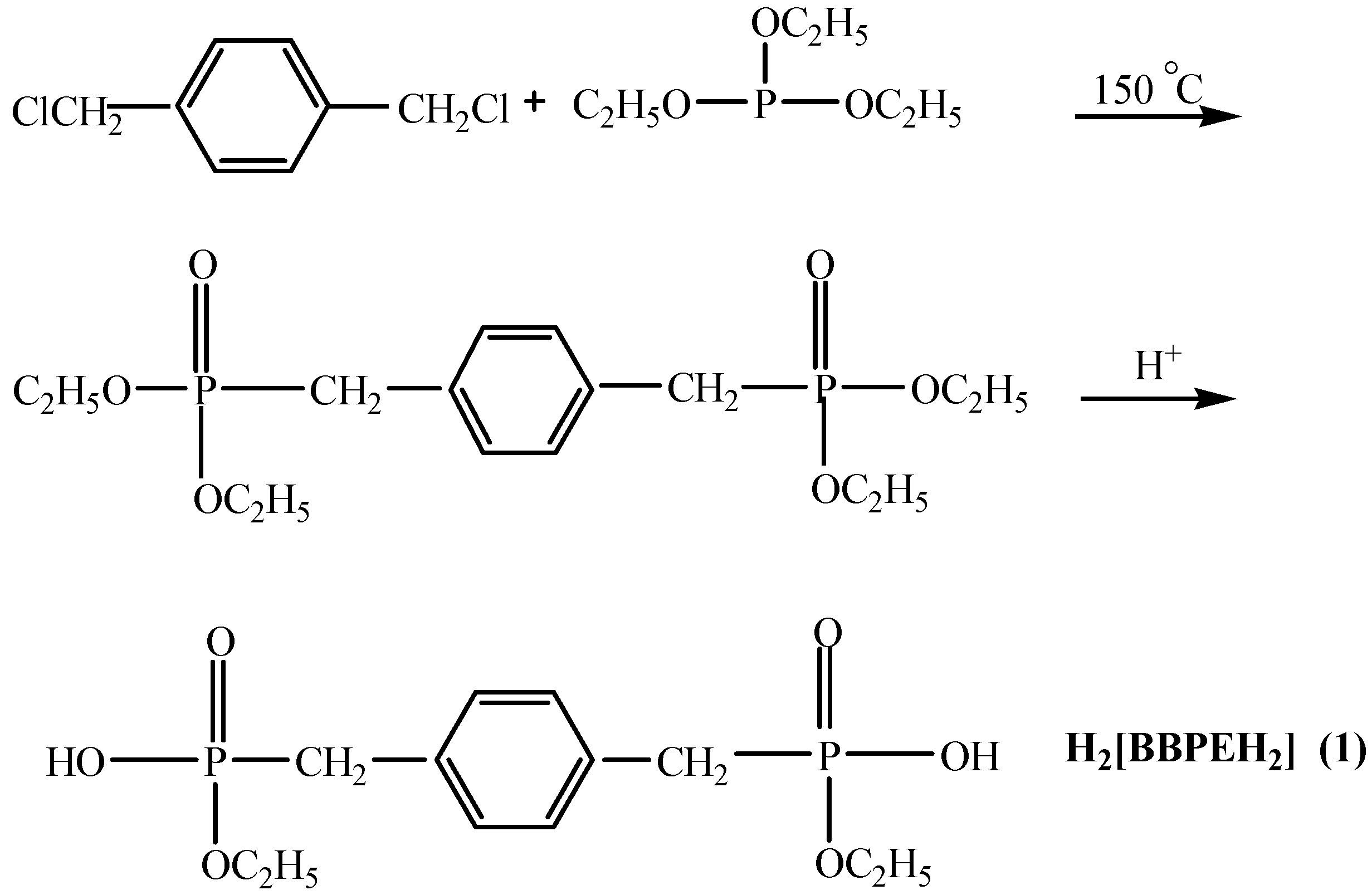
3.2. Synthesis of H2[BBPEH2] (1)

The reaction path is shown in Scheme 1. To a toluene solution of 1,4-bis(chloromethyl)benzene (17.5 g, 0.1 mol) was added dropwise triethyl phosphite (33.2 g, 0.2 mol) and the mixture was stirred at 150 °C. Two h later, the reaction mixture was cooled to room temperature. Removal of solvent from the reaction mixture in vacuo afforded the corresponding product as a white solid [31] which was further washed with petroleum ether, and finally hydrolyzed in sulfuric acid (5 mol/L) at 40 °C with stirring (yield 86%). The colorless block crystals of 1,4-bismethylenebenzene bisphosphonate ethyl ester (H2[BBPEH2]) were recrystallized from a mixed solvent of ethanol and distilled water (1:1) one week later. Elemental analysis (%), Calcd. for C12H16O6P2: C, 45.28; H, 5.07. Found: C, 45.45; H, 4.89. As the building block, 1 was reacted with the Lewis bases N,N-dimethylpiperazine, ammonia and sodium hydroxide. H2[BBPEH2], has changed from a tetrabasic acid into a binary acid and then, further deprotonated to become the [BBPEH2]2− anion.

3.3. Synthesis of [BBPEH2]2−·[Protonated N,N-dimethylpiperazine]2+·4H2O (2)

Compound 1 (644 mg, 2.0 mmol) was dissolved in the mixture of water (10.0 mL) and ethanol (10.0 mL) with stirring and then put into a straight glass tube. N,N-dimethylpiperazine (0.27 mL, 2.0 mmol) was carefully layered onto it. Colorless block crystals were observed after one week. Elemental analysis (%), Calcd. for C36H80N4O20P4: C, 42.67; H, 7.96; N, 5.53. Found: C, 42.80; H, 7.85; N, 5.72.

3.4. Synthesis of [BBPEH2]2−·[NH4]22+·2H2O (3)

The same procedure as for 2 was used. Ammonia (0.160 mL, 4.0 mmol) was used in place of N,N-dimethylpiperazine and colorless block crystals were obtained after one week. Elemental analysis (%), Calcd. for C12H30N2O8P2: C, 36.72; H, 7.71; N, 7.14. Found: C, 36.90; H, 6.89; N, 7.30.

3.5. Synthesis of {[BBPEH2]·[Na2(H2O)6]} (4)

Compound 1 (644 mg, 2.0 mmol) and sodium hydroxide (160 mg, 4.0 mmol) were dissolved in a mixture of water (10.0 mL) and ethanol (10.0 mL) with stirring. Upon slow evaporation of the solvents at room temperature, colorless block single crystals suitable for X-ray analysis were obtained after one week. Elemental analysis (%), Calcd. for C6H9NaO6P: C, 31.17; H, 3.93. Found: C, 32.12; H, 3.73.

3.6. Single Crystal X-ray Diffraction Studies

The diffraction data for 14 were collected on a Enraf-Nonius CAD-4 diffractometer with graphite-monchromated Mo-Kα radiation [λ = 0.71073 Å, T = 293(2) K] by ω-scan mode. The structures were solved by direct methods and refined by least squares on F2 by using the SHELXTL [32] software package. All non-hydrogen atoms were anisotropically refined. The hydrogen atom positions were fixed geometrically at calculated distances and allowed to ride on the parent carbon atoms. The molecular graphics were plotted using SHELXTL. Atomic scattering factors and anomalous dispersion corrections were taken from International Tables for X-ray Crystallography [33]. Further detailed information regarding the crystallographic data and structural analysis for 14 is listed in Table 2. Table 3 lists the hydrogen bonds in the compounds 13. All the bond distances and bond angles of the four compounds are in the normal ranges. Mercury (version 3.0, CCDC, Cambridge, UK) and Dimond 3.0 software (CRYSTAL IMPACT, Bonn, Germany) were used to visualize the structures. Some C-H···π interactions in compounds 2, 3 and 4 are listed in Table 1.
Table 2. Crystal Data and Structure Refinement Summary for Compounds 14.
Empirical FormulaC6H8O3PC18H40N2O10P2C6 H15NO4PC12H30Na2O12P2
M159.09506.46196.16474.28
crystal size/mm0.24 × 0.20 × 0.160.30 × 0.18 × 0.140.22 × 0.20 × 0.180.24 × 0.22 × 0.18
T/K293(2)293(2)293(2)293(2)
λ0.710730.710730.710730.71073
crystal systemmonocliniccentriccentriccentric
space groupC2P-1P-1Pnma
a/Å13.431(3)9.3520(19)13.079(3)8.2010(16)
b/Å8.8309(18)10.458(2)9.1170(18)28.502(6)
c7.7810(16)15.736(3)8.9340(18)9.7120(19)
α/deg9081.56(3)89.88(3)90
β/deg123.98(3)82.84(3)107.82(3)90
γ/deg9063.46(3)90.02(3)90
V3765.3(3)1358.9(5)1014.2(4)2270.1(8)
Z4244
ρ calcd/g.cm−31.3811.2381.2851.352
F (000)332544420952
μ/mm−10.3040.2080.2520.280
θ range/deg2.94 to 26.981.31 to 29.311.64 to 26.971.43 to 25.00
completeness to θ98.7%74.9%97.2%99.6%
range of h, k, l−17 ≤ h ≤ 14,
−11 ≤ k ≤ 11,
0 ≤ l ≤ 9
0 ≤ h ≤ 11,
−11 ≤ k ≤ 12,
−18 ≤ l ≤ 18
−15 ≤ h ≤ 15,
−10 ≤ k ≤ 10,
0 ≤ l ≤ 10
0 ≤ h ≤ 9,
−33 ≤ k ≤ 0,
0 ≤ l ≤ 11
reflections collected/unique1773/8816013/55854426/21502035/2035
Rint0.03340.12210.06910.0000
data/restraints/parameters881/1/605585/0/2972150/2/1342035/0/133
GOF on F21.1441.0271.0861.065
final R indices [I > 2σ (I)]R1 = 0.0504
wR2 = 0.1373
R1 = 0.0574
wR2 = 0.1688
R1 = 0.0374
wR2 = 0.0994
R1 = 0.0962
wR2 = 0.2405
R indices (all data)R1 = 0.0545
wR2 = 0.1416
R1 = 0.0954
wR2 = 0.1946
R1 = 0.0496
wR2 = 0.1069
R1 = 0.1459
wR2 = 0.2891
peak, hole/e.Å−30.600 and −0.3580.584 and −0.6100.350 and −0.3781.396 and −1.108
Table 3. Selected Hydrogen Bond Lengths (Å) and Angles (°) for Compounds 13.
CompoundD-H···ASymmetry CodeD···A (Å)∠D-H···A (°)
1O(2)-H(1)···O(2)x, y, −z2.5114165.95
2N(1)-H(1)···O(2)x, −1 + y, z2.6265175.86
O(1w)-H(11W)···O(4W)−1 + x, y, z3.0756126.99
O(2W)-H(12W)···O(1)−1 + x, y, z2.8627155.48
O(3W)-H(13W)···O(2)2 − x, 1 − y, −z3.0145164.35
O(4W)-H(14W)···O(5)1 − x, 1 − y, 1 − z2.9162172.15
O(1W)-H(21W)···O(4)x, −1 + y, z2.8216164.14
3N (1)-H(1)···O(1)x, ½ − y, ½ + z2.8338168.93
O(2W)-H(3)···O(2)x, ½ − y, ½ + z2.7913171.10
N(1)-H(5)···O(2W)1 − x, −1/2 + y, ½ − z2.8304162.13
N(1)-H(6)···O(2)1 − x, ½ + y, ½ − z2.7767169.00

4. Conclusions

By using 1 as the building block, hydrogen-bonded supramolecules and coordinated polymers have been obtained by reaction with Lewis bases. X-ray single crystal diffraction shows that the differences in the structures of the in/organic bases lead to organophosphonic ester supramolecular and coordination polymer structures ranging from 1D helix chains to 3D networks. Solid state fluorescence spectra show that the fluorescence intensities of 14 are very different from each other, which indicate that the different types of base in the compounds have direct effects on the fluorescence intensity. Fluorescence spectra in solutions indicate that solvents have remarkable influencs on maintaining the integrity of the molecular skeletons. In a larger sense, compound 1 with multiple phosphonate groups can be used to construct higher-dimensional and stable supramolecular architectures extending other alkalis because of its good solubility. In additional, they can be used as models to design new metal phosphonates and explore the variety of structural possibilities in forming organic-inorganic hybrid materials and metal-organic frameworks. Work is currently underway to synthesize metal organophosphonic esters and study the correlation factors that affect the structures and properties.

Supplementary Materials

The crystallographic file in cif format for 14 compounds has been deposited with CCDC number 710324–710327. These data can be obtained free of charge via http://www.ccdc.cam.ac.uk/conts/retrieving.html. Supplementary materials can be accessed at: https://www.mdpi.com/1420-3049/20/08/14435/s1.

Acknowledgments

This work was supported by the Natural Science Foundation of Shandong Province, China. (No. Z2007B01 and Y2007B14).

Author Contributions

Yufeng Li and Fangfang Jian designed research; Yufeng Li performed research and analyzed the data; Yufeng Li and Fangfang Jian wrote the paper. All authors read and approved the final manuscript.

Conflicts of Interest

The authors declare no conflict of interest.

References

  1. Fu, R.B.; Hu, S.M.; Wu, X.T. Mix-Solvothermal Syntheses, Characterization, and Properties of New Zinc Diphosphonates with Zn-O-P Chains, Layers, and 3D Frameworks. Cryst. Growth Des. 2007, 7, 1134–1144. [Google Scholar]
  2. Mahmoudkhani, A.H.; Langer, V. Supramolecular Isomerism and Isomorphism in the Structures of 1,4-Butanebisphosphonic Acid and Its Organic Ammonium Salts. Cryst. Growth Des. 2002, 2, 21–25. [Google Scholar]
  3. Wu, S.M.; Chen, S.P.; Li, M. Self-assembly of organic acid-base compounds from 2-D layered network to 3-D supramolecular framework: Synthesis, structure and photoluminescence. CrystEngComm. 2007, 9, 907–914. [Google Scholar]
  4. Ortiz-Avila, C.Y.; Bhardwaj, C.; Clearfield, A. Zirconium Polyimine Phosphonates, a New Class of Remarkable Complexing Agents. Inorg. Chem. 1994, 33, 2499–2500. [Google Scholar]
  5. TianHua, Z.; Zhang-Zhen, H.; Xiang, X. Ligand Geometry Directed Polar Cobalt(II) Phosphonate Displaying Weak Ferromagnetism. Cryst. Growth Des. 2013, 13, 838–843. [Google Scholar]
  6. Kumar, T.S.; Yang, T.; Mishra, S. 5′-Phosphate and 5′-phosphonate ester derivatives of (N)-methanocarba adenosine with in vivo cardioprotective activity. J. Med. Chem. 2013, 56, 902–914. [Google Scholar] [CrossRef] [PubMed]
  7. Ferreira, C.L.; Holley, I.; Bensimon, C. Pharmacokinetic Modulation of Radiolabeled Chelates Facilitated by Phosphonate Ester Coordinating Groups. Mol. Pharmaceutics. 2012, 9, 2180–2186. [Google Scholar] [CrossRef]
  8. Tolis, E.I.; Helliwell, M.; Langley, S. Synthesis and characterization of iron(III) phosphonate cage complexes. Angew. Chem. Int. Ed. 2003, 42, 3804–3808. [Google Scholar] [CrossRef] [PubMed]
  9. Harvey, H.G.; Teat, S.J.; Tang, C.C. Synthesis and Characterisation of Three Novel Cation-Containing (NH4+/(CHNH3+) H3N7/NH3+C2H4NH3+). Inorg. Chem. 2003, 42, 2428–2439. [Google Scholar] [CrossRef]
  10. Yue, Q.; Yang, J.; Li, G.H. Homochiral Porous Lanthanide Phosphonates with 1D Triple-Strand Helical Chains: Synthesis, Photoluminescence, and Adsorption Properties. Inorg. Chem. 2006, 45, 4431–4439. [Google Scholar] [CrossRef] [PubMed]
  11. Alberti, G.; Brunet, E.; Dionigi, C. Shaping solid-state supramolecular cavities: Chemically induced accordionlike movement of γ-zirconium phosphate containing polyethylenoxide pillars. Angew. Chem. Int. Ed. 1999, 38, 3351–3353. [Google Scholar] [CrossRef]
  12. Tang, S.F.; Song, J.L.; Li, X.L. Novel Luminescent Lanthanide(III) Diphosphonates with Rarely Observed Topology. Cryst. Growth Des. 2007, 7, 360–366. [Google Scholar] [CrossRef]
  13. Mao, J.G.; Wang, Z.K.; Clearfield, A. New Lead Inorganic-Organic Hybrid Microporous and Layered Materials: Synthesis, Properties, and Crystal Structures. Inorg. Chem. 2002, 41, 6106–6111. [Google Scholar] [CrossRef] [PubMed]
  14. Jiang, J.G.; Wang, Z.K.; Clearfiels, A. Synthesis, Characterization, and Crystal Structures of Two New Divalent Metal Complexes of N,N′-Bis(phosphonomethyl)-1,10-diaza-18-crown-6:  A Hydrogen-Bonded 1D Array and a 3D Network with a Large Channel. Inorg. Chem. 2002, 41, 3713–3720. [Google Scholar]
  15. Yang, B.P.; Mao, J.G. New Types of Metal Squarato-phosphonates:  Condensation of Aminodiphosphonate with Squaric Acid under Hydrothermal Conditions. Inorg. Chem. 2005, 44, 566–571. [Google Scholar] [CrossRef] [PubMed]
  16. Liu, B.; Yin, P.; Yi, X.Y. Template- and pH-Directed Assembly of Diruthenium Diphosphonates with Different Topologies and Oxidation States. Inorg. Chem. 2006, 45, 4205–4213. [Google Scholar] [CrossRef] [PubMed]
  17. Chandrasekhar, V.; Azhakar, R.; Bickley, J.F. Influence of O-H···OP Hydrogen Bonding on the Supramolecular Architectures of Phosphorus-Based Hydrazones:  Alternate Right- and Left-Handed Fused Helical Chains Based on O-H···OP Hydrogen Bonds in the Crystal Structure of C6H5P(O)[N(CH3)NCHC6H4-p-OH]2. Cryst. Growth Des. 2006, 6, 910–914. [Google Scholar] [CrossRef]
  18. Bakhmutova, E.V.; Ouyang, X.; Medvedev, D.G. Cobalt Phosphonates:  An Unusual Polymeric Cobalt Phosphonate Containing a Clathrated Phosphonate Anion and a Layered Bisphosphonate. Inorg. Chem. 2003, 42, 7046–7051. [Google Scholar] [CrossRef] [PubMed]
  19. Tang, S.F.; Song, J.L.; Mao, J.G. Syntheses, Crystal Structures, and Characterizations of a Series of New Layered Lanthanide Carboxylate-Phosphonates. Eur. J. Inorg. Chem. 2006, 10, 2011–2019. [Google Scholar] [CrossRef]
  20. Serre, C.; Ferey, G. Hybrid Open Frameworks. 8. Hydrothermal Synthesis, Crystal Structure, and Thermal Behavior of the First Three-Dimensional Titanium(IV) Diphosphonate with an Open Structure:  Ti3O2(H2O)2(O3P-(CH2)-PO3)2·(H2O)2, or MIL-22. Inorg. Chem. 1999, 38, 5370–5373. [Google Scholar] [CrossRef]
  21. Serpaggi, S.; Ferey, G. Hybrid open frameworks (MIL-n). Part 6 Hydrothermal synthesis and X-ray powder ab initio structure determination of MIL-11, a series of lanthanide organodiphosphonates with three-dimensional networks, LnIIIH[O3P(CH2)nPO3] (n = 1–3). J. Mater. Chem. 1998, 8, 2749–2755. [Google Scholar] [CrossRef]
  22. Neeraj, S.; Natarajan, S.; Rao, C.N.R. Amine Phosphates as Intermediates in the Formation of Open-Framework Structures. Angew. Chem. Int. Ed. 1999, 38, 3480–3483. [Google Scholar] [CrossRef]
  23. Choudhuty, A.; Neeraj, S.; Natarajan, S. An Unusual Open-Framework Cobalt(II) Phosphate with a Channel Structure That Exhibits Structural and Magnetic Transitions. Angew. Chem. Int. Ed. 2000, 39, 3091–3093. [Google Scholar] [CrossRef]
  24. Calin, N.; Sevov, S.C. Novel Mixed-Valence Heteropolyoxometalates:  A Molybdenum Diphosphonate Anion [MoV7MoVIO16(O3PPhPO3H)4]3− and Its One- and Two-Dimensional Assemblies. Inorg. Chem. 2003, 42, 7304–7308. [Google Scholar] [CrossRef] [PubMed]
  25. Ushak, S.; Spodine, E.; Fur, E.L. Two New Hybrid Organic/Inorganic Copper(II)-Oxovanadate(V) Diphosphonates: [Cu2(phen)2(O3PCH2PO3) (V2O5)(H2O)]·H2O and [Cu2(phen)2(O3P(CH2)3PO3)(V2O5)]·C3H8. Synthesis, Structure, and Magnetic Properties. Inorg. Chem. 2006, 45, 5393–5398. [Google Scholar] [CrossRef] [PubMed]
  26. Dumas, E.; Sassoye, C.; Smith, K.D. Synthesis and Characterization of [Mo7O16(O3PCH2PO3)3]8−: A Mixed-Valent Polyoxomolybdenum Diphosphonate Anion with Octahedrally and Tetrahedrally Coordinated Molybdenum. Inorg. Chem. 2002, 41, 4029–4032. [Google Scholar] [CrossRef] [PubMed]
  27. Sharma, C.V.K.; Hessheimer, A.J.; Clearfield, A. Novel materials based on self-assembly of organophosphonic acids. Polyhedron 2001, 20, 2095–2104. [Google Scholar] [CrossRef]
  28. Rosa, C.; Berta, C.; Nuria, F.H. Supramolecular Aggregation of Hexameric Water Clusters into a 2D Water Polymer Containing (H2O)18 Holes. Crys. Growth Design 2006, 6, 629–631. [Google Scholar]
  29. Djamal, D.; Roger, B.; Donald, C.C. Schroeter and Vossen's red salt revealed. New J. Chem. 2002, 26, 614–616. [Google Scholar]
  30. Song, J.L.; Zhao, H.H.; Mao, J.G.; Dunbar, K.R. New Types of Layered and Pillared Layered Metal Carboxylate-Phosphonates Based on the 4,4′-Bipyridine Ligand. Chem. Mater. 2004, 16, 1884–1889. [Google Scholar] [CrossRef]
  31. Ge, G.Z.; Miao, Y.X. Application of wittig reaction and the modified method in fluorescent whitener syntheses. Textile Auxiliaries 2006, 23, 10–11. [Google Scholar]
  32. Sheldrick, G.M. SHELXTL, v5 Reference Manual; Siemens Analytical X-Ray Systems: Madison, WI, USA, 1997. [Google Scholar]
  33. Wilson, A.J. International Table for X-Ray Crystallography; Kluwer Academic: Dordrecht, The Netherlands, 1992; Volume C: Tables 6.1.1.4, 500–502 and 4.2.6.8, 219–222, respectively. [Google Scholar]
  • Sample Availability: Samples of the compounds 14 are available from the authors.

