Article

New Alcamide and Anti-oxidant Activity of Pilosocereus gounellei A. Weber ex K. Schum. Bly. ex Rowl. (Cactaceae)

by
Jéssica K. S. Maciel
1,†,
Otemberg S. Chaves
2,†,
Severino G. Brito Filho
2,†,
Yanna C. F. Teles
1,†,
Marianne G. Fernandes
2,†,
Temilce S. Assis
1,3,†,
Pedro Dantas Fernandes
4,†,
Alberício Pereira De Andrade
4,†,
Leonardo P. Felix
4,†,
Tania M. S. Silva
5,†,
Nathalia S. M. Ramos
5,†,
Girliane R. Silva
5,† and
Maria De Fátima Vanderlei De Souza
1,2,*
1
Post-Graduation Program in Development and Technological Innovation in Medicines, Health Science Center, Federal University of Paraiba, Campus I, João Pessoa, PB, 58051-900, Brazil
2
Post-Graduation Program in Bioactive Natural and Synthetic Products, Health Science Center, Federal University of Paraiba, Campus I, João Pessoa, PB, 58051-900 Brazil
3
Health Science Centre, Physiology and Pathology Department, Federal University of Paraiba, Campus I, Cidade Universitária—João Pessoa, PB, 58059-900, Brazil
4
Department of Agroecology and Agriculture, Center of Agricultural and Environmental Sciences, University of Paraiba State, 351 Baraúnas Street, Campina Grande, PB, 58429-500, Brazil
5
Postgraduate Program in Development and Technological Innovation in Medicines, Department of Molecular Sciences, Rural Federal University of Pernambuco, Campus Dois Irmãos, Recife, PE, 52171-900, Brazil
*
Author to whom correspondence should be addressed.
These authors contributed equally to this work.
Molecules 2016, 21(1), 11; https://doi.org/10.3390/molecules21010011
Submission received: 19 October 2015 / Revised: 8 December 2015 / Accepted: 10 December 2015 / Published: 22 December 2015
(This article belongs to the Section Natural Products Chemistry)

Abstract

:
The Cactaceae family is composed by 124 genera and about 1438 species. Pilosocereus gounellei, popularly known in Brazil as xique-xique, is used in folk medicine to treat prostate inflammation, gastrointestinal and urinary diseases. The pioneering phytochemical study of P. gounellei was performed using column chromatography and HPLC, resulting in the isolation of 10 substances: pinostrobin (1), β-sitosterol (2), a mixture of sitosterol 3-O-β-d-glucopyranoside/stigmasterol 3-O-β-d-glucopyranoside (3a/3b), 132-hydroxyphaeophytin a (4), phaeophytin a (5), a mixture of β-sitosterol and stigmasterol (6a/6b), kaempferol (7), quercetin (8), 7′-ethoxy-trans-feruloyltyramine (mariannein, 9) and trans-feruloyl tyramine (10). Compound 9 is reported for the first time in the literature. The structural characterization of the compounds was performed by analyses of 1-D and 2-D NMR data. In addition, a phenolic and flavonol total content assay was carried out, and the anti-oxidant potential of P. gounellei was demonstrated.

1. Introduction

Cactaceae is a family belonging to the order Caryophyllales with 124 genera and about 1438 species [1] distributed throughout American territory in tropical and temperate dry regions with wide occurrence in Mexico and Brazil [2]. The family is remarkable due to the evolution of several adaptations for aridity, its species showing outstanding diversity of growth forms [3].
Caatinga is a dry eco-region located in northeastern Brazil considered the third diversity center of Cactaceae species, where the family plays an important role, mainly due to the use of its species in human and animal foods [4,5]. Furthermore, many species are used in traditional medicine, for example Nopalea cochenillifera is used as an anti-inflammatory, diuretic and hypoglycaemic [6] agent and Cereus jamacaru is used to treat ulcers and bronchitis [7].
Previous phytochemical studies on Cactaceae species have reported the presence of flavonoids, such as quercetin, rutin and kaempferol, as well as the anti-oxidant activity of several species, e.g., Opuntia monocantha, Opuntia ficus-indica, Cereus jamacaru, Pilosocereus pachycladus and Pilosocereus arrabidae [8,9]. In plants, this activity is related to the presence of phenolic molecules such as flavonoids, tannins, phenolic acids, anthocyanins and others [10]. The anti-oxidant activity of phenolics is justified by the presence of conjugated double bonds, which provide resonance, and the hydroxyl groups on an aromatic ring [11], which can chelate metals, eliminate free radicals by donating hydrogen, oxidize and induce enzymes to interact and stabilize free radicals [12,13].
The occurrence of nitrogen compounds such as β-phenethylamines, tetrahydroisoquinolines and their derivatives in this family is also well reported [14,15,16]. Three alkaloids have been isolated from the Neobuxbaumia genus: salsolidine, carnegine and anhalidine [17].
Pilosocereus gounellei A. Weber ex K. Schum. Bly. ex Rowl, popularly known as xique-xique, is an endemic species from the Caatinga region and its cladodes are used by local people as food, cooked or baked to produce flour, cakes and pastries [18,19]. In folk medicine, its roots are used to treat urinary infections and prostate inflammation. Its cladodes are used to treat gastritis [5,20,21].
This work describes the first isolation of secondary metabolites of P. gounellei and the evaluation of the anti-oxidant activity of its extracts and methanol fraction. Anti-oxidant activity of Cactaceae species has been reported previously, justifying our investigation of P. gounellei’s anti-oxidant potential [8,9].

2. Results and Discussion

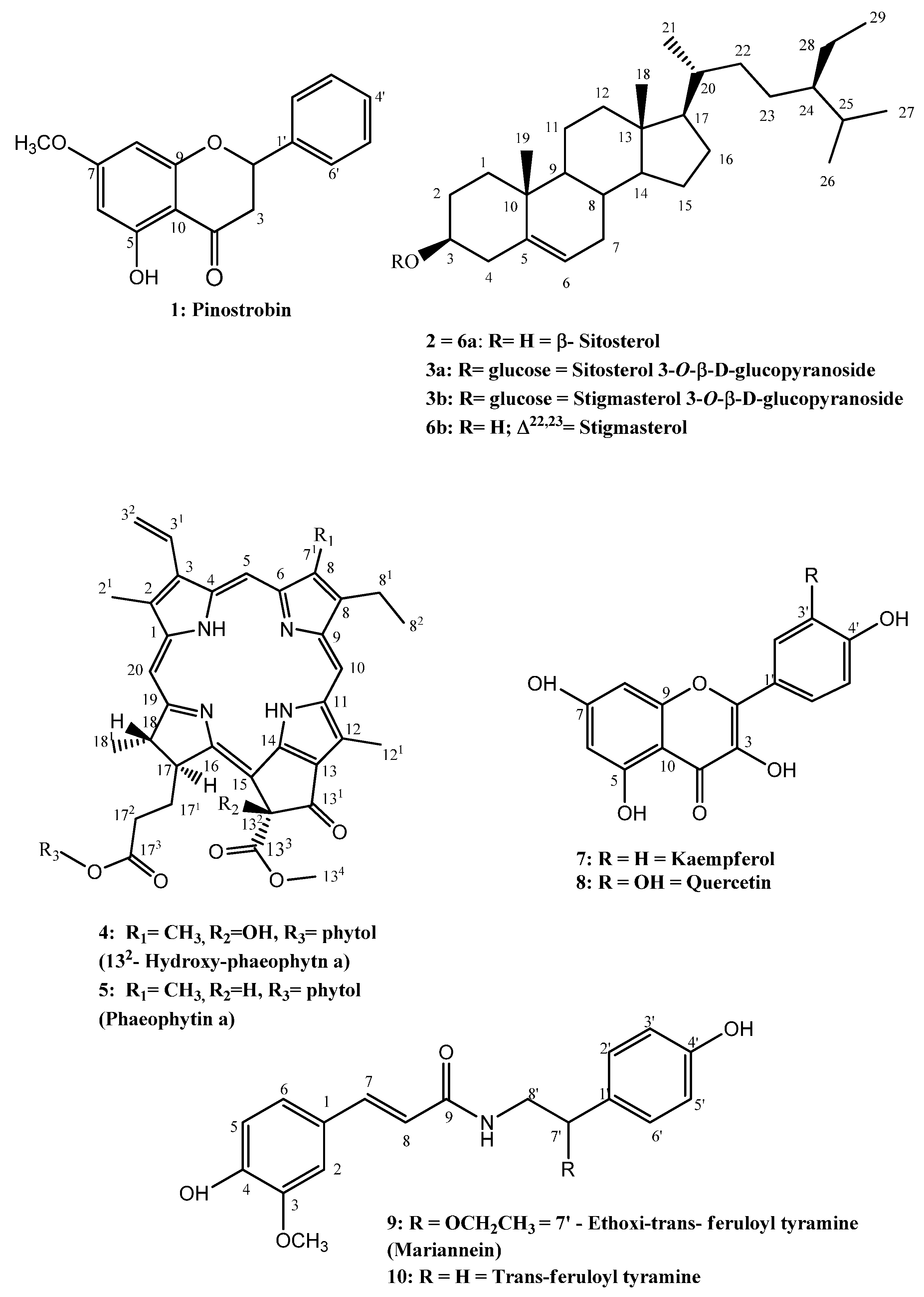
2.1. Identification of Isolated Compounds

Chromatographic procedures led to the isolation of 10 compounds from P. gounellei (Figure 1 and Figure 2). The isolated compounds were identified by analysis of their 1-D and 2-D NMR data and comparisons with the literature. Compound 1 was isolated as colourless crystals soluble in chloroform and it was identified as the flavonoid pinostrobin [22]. Compounds 2, 3a/3b and 6a/6b were identified as steroids: β-sitosterol, the mixture of sitosterol 3-O-β-d-glucopyranoside/stigmasterol, 3-O-β-d-glucopyranoside [23] and sitosterol/stigmasterol [24], respectively. These phytosteroids are widespread in plants, being important components of vegetable cell walls and membranes. In addition, they are known as anti-inflammatory agents and precursors of vitamin D [25].
Compounds 4 and 5 were isolated as dark green amorphous solids. The 1H- and 13C-NMR indicated that the compounds are chlorophyll derivatives. When compared with the literature data, they were identified as 132-hydroxyphaeophytin a (4) [26] and phaeophytin a (5) [27]. These porphyrinic compounds are derived from chlorophyll a and are widely present in the vegetable kingdom [28]. The substances 7 and 8 were isolated as a yellow amorphous powder and were identified as the flavonoids kaempferol (7) [29] and quercetin (8) [30]. Flavonoids have great relevance in the pharmaceutical field, displaying anti-oxidant, anti-inflammatory and antimicrobial activities [31], and have been previously reported from several species of the genera Opuntia and Pilosocereus [8].
Figure 1. Compounds isolated from Pilosocereus gounellei.
Molecules 21 00011 g001
The 1H-NMR spectrum of compound 9 (Figure 2) showed two doublets (δH 7.11 and δH 6.75) integrating for two protons each, suggesting the presence of a para-substituted ring. Two doublets in δH 6.79 (H-5), coupling ortho, and δH 7.11 (H-2), coupling meta with H-6 and a double doublet at δH 6.98 (H-6) coupling ortho and meta with H-5 and H-2, respectively, indicated the presence of an AMX ring system. The presence of a methoxy group in one aromatic ring was shown by a singlet at δH 3.79 (3H). The presence of the coumaroyl and tyramine units was suggested by the following signals: a pair of doublets at δH 6.52 (H-8) and δH 7.31 (H-7), both with J = 16 Hz, characteristic of trans olefin protons; an interesting triplet at δH 8.00, referring to a proton bonded to N [32]; two double doublets at δH 3.29 (H-8a′) and 3.25 (H-8b′) referring to the methylene carbon adjacent to N and one triplet on δH 4.27 (H-7′), attributed to a proton on the oximethinic carbon [33]. Other relevant signals in the 1H-NMR spectra of 9 were two signals at δH 3.27 (q) and δH 1.07 (t) attributed to an ethoxy group attached to an oximethinic carbon whose proton was found at δH 4.27 (t) [33].
Figure 2. Compound 9 HMBC (3J and 2J) and COSY correlations.
Molecules 21 00011 g002
The 13C-NMR spectrum showed signals for 20 carbons, supporting the information provided by the 1H-NMR spectrum about the presence of the coumaroyl and tyramine portions of compound 9. The coumaroyl portion was defined by the signals at δC 165.8 (amide α,β-unsaturated carbonyl) and two signals at δC 139.46 and δC 119.01 (the α,β-unsaturated carbons C-7 and C-8). The tyramine portion was confirmed by the signals at δC 45.61 (CH2, C-8′) and an oximethinic carbon δC 79.6 (CH, C-7′), indicating the position the ethoxy group is attached to [32]. The presence of one ethoxy group in compound 9 was reinforced by the signals at δC 63.51 (CH2, C-1′′) and δC 15.42 (CH3, C-2′′). The 13C-NMR spectrum confirmed the presence of two aromatic rings, with δC 128.07 (C-2′/6′), δC 115.34 (C-3′/5′) of one AA′BB′ system and δC 110.9 (C-2), δC 115.83 (C-5) and δC 121.82 (C-6) of the AMX system, showing one methoxy group at δC 55.75 (H3CO–C-3) [32,34].
The absence of a correlation for proton δH 8.00 (t, N-H) with any carbon in the HMQC spectrum, and the correlation shown by the proton at δH 8.00 (t, 1H) and the carbon at δC 165.8 (2J) in the HMBC spectrum demonstrated that the compound possesses an amide carbonyl [32,33,34], being a trans-feruloyl derivative [32,33,34]. The HMBC spectrum showed correlations (3J) that confirmed the presence of the coumaroyl moiety: H-7 with C-2, C-6 and C-9; H-8 with C-1; H-5 with C-3 and C-1. Other correlations in the HMBC spectrum suggest the presence of the ethoxy group and also the trans-feruloyl tyramine portion: H-1′′ with C-7′ and H-8′ with C-9 and C-1′. These data allowed identification of compound 9 as 7′-ethoxy-trans-feruloyl- tyramine, reported herein for the first time. The COSY spectrum supported the proposed structure by showing correlations between hydrogen (N-H) δH 8.00 (t) with H-8′; H-8′ and H-7′; H-1′′ and H-2′′ (Table 1). The optical rotation of 9 was found to be −10° (0.01; MeOH) establishing the S(−) absolute configuration at the C-7 chiral center [33].
Table 1. 13C-NMR (100 MHz), 1H-NMR (400 MHz), HMQC and HMBC data of 7’-ethoxy-trans-feruloyltyramine (9) (DMSO-d6, δ).
PositionHMQCHMBC
δC, TypeδH (J in Hz)3J (H C)2J (H C)
1126.62 C-
2110.9 C-H7.11, d (1.2)4, 6, 73
3148.03 C-
4148.46 C-
5115.83 C-H6.79, dd (8.0, 0.72)1, 34, 6
6121.82 C-H6.98, dd (8.0, 1.2)2, 4, 75
7139.46 C-H7.31, d (16.0)2, 6 ,91, 8
8119.01 C-H6.52, d (16.0)1 9
9165.87 C-
1’130.78 C-
2’/6’128.07 C-H7.12, dd (8.0, 1.2)2’, 6’, 4’, 7’3’, 5’
3’/5’115.34 C-H6.75, dd (8.0, 1.2)1’, 3’, 5’4’
4’157.08 C-
7’79.64 C-H4.27, t (5.0)1’’, 2’, 6’8’
8’45.61 C-H23.29 dd, (13.0, 5.9)
3.25, dd (13.0, 5.9)
1’7’
1’’63.51 C-H23.27, q (6.9) 2’’
2’’15.42 C-H31.07, t (6.9) 1’’
OMe-355.56 C-H33.79, s3
N-H-8.0, t (5.0) 9
The molecular formula of new compound 9 was established as C20H23NO5 from a pseudomolecular ion peak at m/z 358.1706 [M + H]+ (calcd. C20H24NO5, 358.1648) indicating the formula C20H23NO5 for the new compound. The corresponding loss of the ethoxy radical fragment was seen in EIMS [M + H], FTMS as 312.17 (C18H18NO4) and the loss of ethanol was seen in EIMS [M + H] as 310.17, confirming the proposed structure of compound 9. The hypotheses that compound 9 is an artefact was eliminated based on previous studies that described the isolation of trans-feruloyltyramine and its derivatives by several different extraction methods [33,35]. Liang et al. performed the extraction of eight nitrogenated substances from Portulaca oleracea using microwave irradiation and different solvents such as dichloromethane, ethyl acetate, methanol, ethanol, ethanol 70%, ethanol 30% and water. The eight isolated substances, including N-feruloylnormetanephrine and the N-trans-feruloyltyramine, were extracted with all the tested solvents, showing that those solvents did not promote the formation of different radicals or artefacts [35].
Compound 10 was obtained as a pale amorphous solid and its spectra showed a structure similar to compound 9, differing in the absence of signals related to the ethoxy group. Comparisons of the spectral data of compound 10 with compound 9 and literature data allowed identification of compound 10 as trans-feruloyltyramine, previously isolated from other plant species [32,33,34]. Compound 10 was shown to possess action against weeds, improvement of seed germination [34] and anti-inflammatory activity by inhibiting COX enzymes [36].

2.2. Total Phenolic, Total Flavone and Flavonol Contents and Anti-Oxidant Activity of Extracts and Methanolic Fraction from P. gounellei

The values of total phenolic, flavone and flavonol contents and anti-oxidant activity (DPPH and ABTS) of ethanol extracts of stems, roots, flowers, fruits and methanol fraction are shown in Table 2. The fruit extract showed the best antiradical activity in the ABTS test (IC50 = 10.4 ± 0.24) and the flower extract showed the lowest anti-oxidant activity (IC50 = 76.9 ± 0.61); the cladode extract showed a slightly higher activity (IC50 = 62.4 ± 0.44) than the flower extract. The scavenging activity of free radicals of the roots extract showed an activity (IC50 = 41.6 ± 1.06) similar to the methanol fraction (IC50 = 40.9 ± 0.69). Thus, we can classify the activity of extracts and methanol fraction of P. gounellei (Pg) as follows: Pg fruits > MeOH fraction = Pg roots > Pg cladodes > Pg flowers. In the DPPH test, the fruit extract showed the best anti-oxidant activity (IC50 = 11.3 ± 0.12), followed by the root extract (IC50 = 102.1 ± 1.49). The methanol fraction (IC50 = 130.1 ± 3.02) showed similar activity to the cladode extract (IC50 = 136.0 ± 3.48) and the least potent was the flower extract (IC50 = 194.3 ± 2.33).
Table 2. Total phenolic, total flavones and flavonols contents and DPPH and ABTS free radical scavenging activity.
SamplesTotal Phenolics Contents (mg GAE/g ± SEM) aTotal Flavones and Flavonols Contents (mg QE/g ± SEM) bFree-Radical Scavenging Activity (IC50) c
DPPH (μg/mL ± SEM)ABTS˙+ (μg/mL ± SEM)
Pg Cladodes45.1 ± 2.50 112.625 ± 1.08 1136.0 ± 3.48 162.4 ± 0.44 1
Pg Methanol Fraction59.0 ± 2.39 213.667 ± 0.833 1130.1 ± 3.02 140.9 ± 0.69 2
Pg Roots61.1 ± 1.03 24.920 ± 0.550 2102.1 ± 1.49 241.6 ± 1.06 2
Pg Flower43.5 ± 2.16 18.460 ± 0.550 3194.3 ± 2.33 376.9 ± 0.61 3
Pg Fruits127.9 ±1.67 32.417± 0.417 411.3 ± 0.12 410.4 ± 0.24 4
Ascorbic acid--3.6 ± 0.06 5-
Trolox---5.0 ± 0.25 5
All values are mean ± S.E.M (n = 3); a GAE = Gallic acid equivalent per gram of sample; b QE = Equivalent quercetin per gram of sample; c Value defined as the concentration of sample that scavenged 50% of the DPPH or ABTS˙+. There are no significant differences among values marked with the same superscript numbers in individual columns.
A correlation has been shown between anti-oxidant activity and total phenolic content in natural products, especially between extracts with the two highest values. The fruit extract showed a greater amount of phenolics (127.9 ± 1.67 mg GAE/g) and also showed the greater anti-oxidant activity in DPPH (IC50 = 11.3 ± 0.12) and ABTS (IC50 = 10.4 ± 0.24) test. The flower extract presented the lowest anti-oxidant activity in DPPH (IC50 = 194.3 ± 2.33) and ABTS (IC50 = 76.9 ± 0.61) tests as well as the lowest phenolic content (43.5 ± 2.16). When analysing the other extracts and methanol phase of the plant, a sequence of linear correlations between total phenolic content and radical scavenging activity was not observed.
Among all tested samples, only the fruit extract has sufficient anti-oxidant activity to be considered for nutraceutical use. According to the anti-oxidant activity index (AAI), all extracts and phases of P. gounellei showed an index lower than 0.5, thus presenting poor activity. Surprisingly, the fruit extract presented an AAI value of 2.01, which is considered as very strong.
Many studies have reported the relationship between total phenolics assay results and anti-oxidant activity [37]; our study confirms these findings. The relationships between the total phenolic content and the antiradical activity of DPPH (1/EC50), and antiradical activity of ABTS˙+ (1/EC50) are shown in Figure 3. The Pearson correlation coefficients (r) of these plots were approximately 0.943 for the DPPH assay and 0.944 for the ABTS assay. This result suggests that 94% of the anti-oxidant capacity of extracts and methanol fraction from P. gounellei is due to the contribution of phenolic compounds. It is interesting to mention that there was no inverse correlation between the total phenolic content and the anti-oxidant activity by the DPPH and ABTS methods and flavones/flavonols content, at least when comparing the extract with the highest anti-oxidant activity, the extract from the fruits. Therefore, it is possible that the anti-oxidant activity may be attributed to other phenolic compounds than flavonols and/or flavones.
Figure 3. Correlation among the total phenolic content and the antiradical activity DPPH and antiradical activity ABTS˙+ of ethanol extracts and methanol fraction of P. gounellei.
Molecules 21 00011 g003

3. Experimental Section

3.1. General Procedures

Column chromatography separations (CC) were performed on glass columns packed with silica gel 60 (Merck, Nottingham, UK) 7734 (0.063–0.2 mm particles, 70–230 mesh), flash silica (0.04–0.0063 mm particles, 230–400 mesh), Amberlite XAD or Sephadex LH-20. Thin layer chromatography (TLC) was performed on silica gel PF254 plates (Merck, Nottingham, UK). For HPLC experiments, a high-performance liquid chromatograph (HPLC–DAD, equipped with a binary pump (LC-6AD), and diode array detector SPD-M20A (Shimadzu, Kyoto, Japan) and Rheodyne injector (7125) with a 20 μL loop for the analytical column and 500 μL for the semipreparative column were used. An analytical Luna C-18 100A column (250 mm × 4.6 mm × 5 µm, Phenomenex, Torrance, CA, USA), semipreparative Luna C-18 column (250 mm × 21.2 mm × 5 µm, Phenomenex) and Millipore filter membranes (0.45 µm Supelco®, Bellefonte, PA, USA) were used.
The solvents used in the chromatographic procedures were p.a. grade: n-hexane, dichloromethane, chloroform and ethyl acetate. Methanol HPLC grade (Tedia®, Rio de Janeiro, Brazil). Water was obtained from a Millipore® MilliQ system (Millipore, São Paulo, Brazil).
To evaluate the total phenols, total flavones and flavonols content and anti-oxidant activity, the following reagents were used: Folin-Ciocalteu, gallic acid, Trolox (6-hydroxy-2,5,7,8-tetramethylchroman-2-carboxylic acid 97%), ABTS (2,2′-azinobis-(3-ethylbenzothiazoline-6-sulfonic acid) diammonium salt 98%) (Sigma-Aldrich, Sternheim, Germany), ethanol, methanol, sodium carbonate, potassium persulfate, aluminium chloride (Vetec, Rio de Janeiro, Brazil) and Milli-Q water. Samples were solubilized in an ultrasonic bath, 3.5 L (Unic 1600A, Unique, São Leopoldo, Brazil). Readings were performed on Asys HiTech UVM 340 apparatus (São Paulo, Brazil).
Isolated compounds were identified using 1-D and 2-D NMR analysis acquired on the following spectrometers: Varian Oxford (200 MHz), Varian (500 MHz) (Varian, Palo Alto, CA, USA) and Avance III (Bruker, Coventry, UK) using deuterated solvents. The high-resolution mass spectra were obtained using LC-HRMS analysis performed on an Accela 600 HPLC system combined with an Exactive (Orbitrap) mass spectrometer from Thermo Fisher Scientific (Bremen, Germany). EIMS was obtained with a Shimadzu QP-2000 spectrometer (Kyoto, Japan). The [ α ] D 25 25 °C was determined using a MCP 200 polarimeter (Anton Paar, Saint Laurent, QC, Canada).

3.2. Botanical Material

Pilosocereus gounellei was collected in Boa Vista City-PB (Brazil) in November 2010. The plant was identified by Prof. Dr. Leonardo Person Felix (CCA/UFPB) and a voucher specimen (15437) was deposited in the Herbarium Prof. Jaime Coelho Morais of the Agricultural Sciences Center (CCA/UFPB).

3.3. Extraction and Isolation

The botanical material (cladodes) was dried in an oven with circulating air at 40 °C and ground using a mechanical mill, yielding 5.18 kg of powder, which was macerated with 10 L of EtOH at room temperature, for 72 h. The extraction solution was concentrated in a rotary evaporator at 40 °C, yielding 237.13 g of crude ethanolic extract (CCEE). CCEE (10 g) was submitted to vacuum liquid chromatography (VLC) using silica gel and eluted with hexane (Hex), chloroform (CHCl3) and methanol (MeOH) to obtain the corresponding fractions. The chloroform fraction (8.0 g) was chromatographed in a silica column using solvents of increasing polarity: Hex, dichloromethane (CH2Cl2) and MeOH. The fractions Hex–CH2Cl2 (3:7), CH2Cl2 and CH2Cl2–MeOH (9:1) were combined and chromatographed using the same method yielding 188 fractions that were analysed using TLC. The fractions 35/38 (8.00 mg), after recrystallization, gave pure colourless crystals of compound 1. The fractions 54/59 (3.51 g) contained compound 2 and the fractions 146/150 (2.72 g) afforded a white powdery precipitate of compound 3. CCEE (5.00 g) was dissolved in CHCl3:H2O (1:1) and separated using a separating funnel, yielding an aqueous fraction and a chloroform fraction. A portion of the chloroform fraction (2.0 g) was subjected to successive column chromatography on silica gel, following the methodology previously described, resulting in the isolation of compound 4 (20.50 mg).
CCEE (131.84 g) was submitted to column chromatography using Amberlite XAD as the stationary phase and eluted with H2O, MeOH, Hex, ethyl acetate (EtOAc) and acetone. The hexane fraction from XAD was chromatographed using VLC on silica gel 60 with hexane, CH2Cl2 and MeOH. The hexane fraction was chromatographed on flash silica column yielding the subfractions 28/33 (92.30 mg) that were purified using preparative TLC, eluted with Hex–EtOAc (80:20), to give compound 5 (10.20 mg).
The Hex–CH2Cl2 (1:1) fraction was chromatographed on silica gel resulting in fractions 13/15 (white crystals), corresponding to 6 (3.51 g). The methanolic fraction from XAD (9.00 g) was chromatographed on Sephadex eluted with MeOH–CH2Cl2 (7:3) resulting in 33 fractions. Fraction 12 yielded compound 7 (15.00 mg) and fraction 14 yielded compound 8 (12.00 mg), both as yellow powders.
The roots were dried in an oven at 40 °C and ground in a mechanical mill, yielding 1.37 kg of powder that was macerated with 10 L of EtOH at room temperature, for 72 h. The obtained solution was concentrated in a rotary evaporator at 40 °C, resulting in 27.00 g of root crude ethanol extract (RCEE).
To isolate nitrogen-containing compounds, the method described by Souza and Silva [38] was used. The acidified chloroform fraction (ACF, 3.38 g) was chromatographed on a silica column eluted with hexane, EtOAc and MeOH, yielding 159 fractions. The combined fractions 88/114 (580.80 mg) were analysed by HPLC-DAD using a semipreparative column at room temperature. As the mobile phase, Milli-Q water and MeOH were used gradient-wise, and the concentration of the MeOH was increased from 50 to 100% in a 20-min run. The chromatogram showed three peaks, and the one at higher retention time was found to be the major component; thus, it was isolated and identified as compound 9 (8.00 mg).
RCEE (10.00 g) was solubilized in H2O and yielded 5.00 g of precipitate. A sample (2.50 g) of it was dissolved in MeOH–CHCl3 (1:1) and submitted to filtration on Sephadex LH-20 with MeOH–CHCl3 (1:1), resulting in 10 fractions. The subfraction 9/10 (10.00 mg) was proved to be pure by TLC and identified as 10. The remaining 2.50 g of the precipitate was chromatographed on silica gel 60 column and eluted with hexane, EtOAc and MeOH, yielding the pure fractions 5/7 which corresponded to compound 3.

3.4. Spectral Data

Pinostrobin (5-hydroxy-7-methoxy-flavanone, 1): colorless crystals, molecular formula (C16H14O2) 1H-NMR (500 MHz, CDCl3): 11.99 (1 H, s, 5-OH), 7.45 ( 5 H, m, H-2’, H-3’, H-4’ e H-5’), 6.06 (1H, d, J = 2.5 Hz, H-6), 6.05 (1H, d, J = 2.5, H-8), 5.41 (1H, dd, J = 13.00 and 3.00 Hz, H-2), 3.79 (3H, s, 7-OMe), 3.07 (1H, dd,17.00Hz e 13.00, H-3), 2.82 (1H, dd, 17.00Hz, 3.00 Hz, H-3). 13C-NMR (125 MHz, CDCl3): 79.20 (C-2), 43.41 (C-3), 195.73 (C-4), 162.79 (C-5), 95.20 (C-6), 167.80 (C-7), 94.30 (C-8), 164.17 (C-9), 138.39 (C-1’), 126.19 (C2’/C-6’), 128.9 (C-3’, C-4’/C-5’), 55.69 (OCH3).
β-Sitosterol (2): 1H-NMR and 13C-NMR data were consistent with the literature [24].
Sitosterol 3-O-β-d-glucopyranoside/stigmasterol 3-O-β-d-glucopyranoside (3): 1H-NMR and 13C-NMR data were consistent with the literature [24].
132-Hydroxy-(132-S)-pheophytin a (4): amorphous green powder 1H-NMR and 13C-NMR data were consistent with the literature [26].
Phaeophytin a (5): morphous green powder, 1H-NMR and 13C-NMR data were consistent with the literature [27].
β-Sitosterol and stigmasterol (6): 1H-NMR and 13C-NMR data were consistent with the literature [24].
Kaempferol (3,4′,5,7-Tetrahydroxyflavone, 7): yellow amorphous powder, 1H-NMR and 13C-NMR data were consistent with the literature [29].
Quercetin (3,3′,4′,5,7-Pentahydroxyflavone, 8): yellow amorphous powder, 1H-NMR and 13C-NMR data were consistent with the literature [30].
7’-Ethoxy-trans-feruloyltyramine (9): a pale amorphous solid, molecular formula (C20H23NO5), 1H-NMR (δ, 400 MHz, DMSO-d6) and 13C-NMR (δ, 100 MHz, DMSO-d6): see Table 1.
trans-Feruloyltyramine (10): pale amorphous solid, molecular formula (C18H19NO4), 1H-NMR (δ, 400 MHz, DMSO-d6), 7.11 (1H, sl, H-2), 6.78 (1H, d, J = 8.0Hz, H-5), 6.98 (1H, d, J = 8.0 Hz, H-7), 7.30 (1H, d, J = 16Hz, H-7), 6.43, (1H, d, J = 15.8 Hz, H-8), 7.00 (2H, d, J = 8.0, H-2’/H-6’), 6.68 (2H, d, J = 8.0 Hz, H-3’/H-5’), 2.64 (2H, t, H-7’), 3.33 (2H, m, H-8’), 3.79 (3H, s, OCH3), 8.01 (1H, t, NH). 13C-NMR (δ, 100 MHz, DMSO-d6) 126.93 (C-1), 111.12 (C-2), 148.37 (C-3), 148.75 (C-4), 116.18 (C-5), 122,09 (C-6), 139.56 (C-7), 119.53 (C-8), 128.19 (C-1’), 130.02 (C-2’/C-6’), 115.67 (C-3’/C-5’), 156.18 (C-4’), 34.27 (C-7’), 41.27 (C-8’), 56.07 (OCH3).

3.5. Total Phenol Content Assay

The total phenolics content was evaluated by the method of Gulcin et al., [39] with some modifications, using the Folin-Ciocalteu reagent and gallic acid as a positive control. Samples of cladodes, roots, flowers and fruit and the methanolic fraction from P. gounellei, from a stock solution of 5 mg/mL, solubilized in EtOH, were transferred to a 1.0 mL Eppendorf tube by adding 20.0 µL of Folin-Ciocalteu reagent, stirring for 1 min. Then Na2CO3 (60.0 µL, 15%) was added to the mixture and stirred for 30 s. Finally, distilled water (900 μL) was added to give a final concentration of 100 μg/mL. After 2 h, the absorbance of the samples was measured at 760 nm. The concentration of the phenolic compounds was determined as equivalent milligram of gallic acid per gram of sample (mg GAE/g), from the calibration curve constructed with gallic acid standard (2.5 to 15.0 µg/mL), considering the average standard error (SEM).

3.6. Total Flavones and Flavonols Content Assay

The flavones and total flavonols content were determined adapting the methodology described by Mihai et al. [40]. Stock solutions (1.0 mg/mL) of extracts from cladodes, roots, flowers and fruits and methanolic fraction from P. gounellei were prepared. Each sample solution (400 µL) and methanolic solution of aluminium chloride (200 µL, 2%) were added in a volumetric flask. The final volume was adjusted with the same solvent to 10 mL. Reaction occurred for 30 min in the dark. The reading was performed at a wavelength of 425 nm. The analysis was evaluated in triplicate and the total flavones and flavonols content was determined from the calibration curve constructed with straight line equation of quercetin solutions (1.0 to 40.0 µg/mL) and expressed in equivalent milligrams of quercetin by gram of extract (mg QE/g), considering the average standard error (SEM).

3.7. DPPH˙ Radical Scavenging Activity Assay

The anti-oxidant activity of ethanolic extracts of the cladodes, roots, flowers and methanol fraction from P. gounellei was performed against the free radical DPPH following the methodology of Silva et al. [41]. Stock solutions were prepared from the extracts and methanol fraction at several concentrations (0.10 to 5.0 mg/mL). Through preliminary analysis, appropriate quantities of stock solutions of the samples and 450 µL of the solution of DPPH˙ (23.6 mg/mL in EtOH) were transferred to 0.5 mL Eppendorf tubes and the volume was completed with EtOH, following homogenization. Samples were sonicated for 30 min and the amount of DPPH˙ was recorded on a UV-vis device at a wavelength of 517 nm in a 96-well plate. Ascorbic acid was used as a positive control and all concentrations were tested in triplicate.
The percentage scavenging activity (% SA) was calculated from the equation:
%   SA = 100 × ( A b s control A b s sample ) A b s control
where Abscontrol is the absorbance of the control containing only the ethanol solution of DPPH, and Abssample is the absorbance of the radical in the presence of the sample or standard ascorbic acid. The anti-oxidant activity index (AAI) was calculated according to Scherer and Godoy [42] as follows:
AAI   =   final concentration of DPPH ( μ g · mL 1 ) IC 50 ( μ g · mL 1 )
AAI values below 0.5 indicate low anti-oxidant activity, values between 0.5 and 1.0 indicate moderate activity, values between 1.0 and 2.0 indicate strong activity and AAI values above 2.0 indicate very strong anti-oxidant activity.

3.8. Determination of Anti-oxidant Activity against the Radical Cation ABTS˙+

The determination of anti-oxidant activity from extracts of cladodes, roots, flowers, fruits and methanolic fraction from P. gounellei against the radical cation ABTS˙+ was carried out following the methodology described by Re [43] using Trolox as the standard compound. The starting concentrations of the solutions of the samples were 0.1–1.0 mg/mL, with the addition of 450 µL of the radical ABTS˙+ solution to give final concentrations of 2.5–100.0 μg/mL samples. Samples were protected from light and sonicated for 6 min. Absorbance of the samples and the positive control were measured at a wavelength of 734 nm using a microplate of 96 wells. Each concentration was tested in triplicate. The percentage of free radical scavenging activity of ABTS˙+ was calculated by the equation:
%   SA = 100 × ( A b s control A b s sample ) A b s control
where Abscontrol is the absorbance of the control containing only the ethanol solution of ABTS+ and Abssample is the absorbance of the radical in the presence of the sample or standard ascorbic acid.
The antiradical efficiency was established using linear regression analysis and the 95% confidence interval (p < 0.05) obtained using the statistical program GraphPad Prism 5.0. The results were expressed as EC50 ± SEM (sample concentration required to eliminate 50% of the DPPH radicals available, plus or minus the SEM).

3.9. Statistical Analyses

The results are expressed as the mean ± standard error of the mean (SEM). Analysis of variance (ANOVA one-way and Tukey’s post hoc test) were used to evaluate the differences of the means between groups. The antiradical efficiency was established using linear regression analysis. Pearson correlation coefficients (r) were used to express correlations and confidence interval of 95% (p < 0.05) obtained using the statistical program GraphPad Prism 5.0 (GraphPad Software Inc., San Diego, CA, USA). The results were expressed by sample concentration required to eliminate 50% of the DPPH radicals available, plus or minus the SEM (EC50 ± SEM).

4. Conclusions

The phytochemical study of Pilosocereus gounellei led to the isolation and identification of 10 compounds: pinostrobin, β-sitosterol, a mixture of β-sitosterol/stigmasterol, 132-hydroxy-phaeophytin a, phaeophytin a, sitosterol 3-O-β-d-glucopyranoside/stigmasterol 3-O-β-d-gluco-pyranoside, kaempferol, quercetin, the new substance 7′-ethoxy-trans-feruloyltyramine and trans-feruloyltyramine. The evaluation of anti-oxidant activity from P. gounellei demonstrated that the fruit ethanol extract possesses excellent anti-oxidant activity, mainly because of the presence of phenolic compounds reported in the genus and the Cactaceae family.

Acknowledgments

The authors are grateful to INSA/MCT/CNPq (No. 562730/2010-9), CAPES and FACEPE (PRONEM APQ-0741-1.06/14) for financial support and to UFPB for technical assistance.

Author Contributions

Jéssica K. S. Maciel, Otemberg S. Chaves, Severino G. Brito Filho, Yanna C. F. Teles, Marianne G. Fernandes (in memoriam) and Maria de Fátima Vanderlei de Souza carried out the isolation and identification of compounds. Pedro Dantas Fernandes, Alberício Pereira de Andrade and Leonardo P. Felix collected and identified the plant. Temilce S. Assis carried out the antioxidant assay. Tania M. S. Silva, Nathalia S. M. Ramos and Girliane R. Silva carried out liquid cromatography and DCR.

Conflicts of Interest

The authors declare no conflict of interest.

References

  1. Hunt, D.; Taylor, N.P.; Charles, G. The New Cactus Lexicon: Descriptions and Illustrations of the Cactus Family; DH Books: Milborne Port, UK, 2006. [Google Scholar]
  2. Wallace, R.S. Molecular systematic study of the cactaceae: Using chloroplast DNA variation to elucidate cactus phylogeny. Bradleya 1995, 13, 1–12. [Google Scholar]
  3. Hernández-Hernández, T.; Hernández, H.M.; De-Nova, J.A.; Puente, R.; Eguiarte, L.E.; Magallón, S. Phylogenetic relationships and evolution of growth form in Cactaceae (Caryophyllales, Eudicotyledoneae). Am. J. Bot. 2011, 98, 44–61. [Google Scholar] [CrossRef] [PubMed]
  4. Ribeiro, E.M.S.; Meiado, M.V.; Leal, I.R. The role of clonal and sexual spread in cacti species dominance at the Brazilian Caatinga. Gaia 2015, 9, 27–33. [Google Scholar]
  5. Lucena, C.M.; Costa, G.M.; Sousa, R.F.; Carvalho, T.K.N.; Marreiros, N.A.; Alves, C.A.B.; Pereira, D.D.; Lucena, R.F.P. Conhecimento local sobre cactáceas em comunidades rurais na mesorregião do sertão da Paraíba (Nordeste, Brasil). Biotemas 2012, 25, 281–291. [Google Scholar] [CrossRef]
  6. Necchi, R.M.M.; Alves, I.A.; Alves, S.H.; Manfron, M.P. In vitro antimicrobial activity, total polyphenols and flavonoids contents of Nopalea cochenillifera (L.) Salm-Dyck (Cactaceae). J. Res. Pharm. 2012, 2, 1–7. [Google Scholar]
  7. Davet, A.; Virtuoso, S.; Dias, J.F.G.; Miguel, M.D.; Oliveira, A.B.; Miguel, O.G. Atividade antibacteriana de Cereus jamacaru DC, Cactaceae. Rev. Bras. Farmacogn. 2009, 19, 561–564. [Google Scholar] [CrossRef]
  8. Gonçalves, A.S.M.; Peixe, R.G.; Sato, A.; Muzitano, M.F.; de Souza, R.O.M.A.; de Barros Machado, T.; Amaral, A.C.F.; Moura, M.R.L.; Simas, N.K.; Leal, I.C.R. Pilosocereus arrabidae (Byles & Rowley) of the Grumari Sandbank, RJ, Brazil: Physical, chemical characterizations and antioxidant activities correlated to detection of flavonoids. Food Res. Int. 2015, 70, 110–117. [Google Scholar]
  9. Nasciemnto, U.T.; Moura, N.P.; Vasconcelos, M.A.S.; Maciel, I.S.M.; Albuquerque, U.P. Chemical characterization of native wild plants of dry seasonal forest of the semi-arid region of northeastern Brazil. Food Res. Int. 2011, 44, 2112–2119. [Google Scholar] [CrossRef]
  10. Neill, S.O.; Gould, K.S.; Kilmartin, P.A.; Mitchell, K.A.; Markham, K.R. Antioxidant activities of red versus green leaves in Elatostema rugosum. Plant Cell Environ. 2002, 25, 539–547. [Google Scholar] [CrossRef]
  11. Pascoal, A.; Rodrigues, S.; Teixeira, A.; Feás, X.; Estevinho, L.M. Biological activities of commercial bee pollens: Antimicrobial, antimutagenic, antioxidant and anti-inflammatory. Food chem. Toxicol. 2014, 63, 233–239. [Google Scholar] [CrossRef] [PubMed]
  12. Bouhlel, I.; Limem, I.; Skandrani, I.; Nefatti, A.; Ghedira, K.; Dijoux-Franca, M.G.; Leila, C.G. Assessment of isorhamnetin 3-O-neohesperidoside from Acacia salicina: Protective effects toward oxidation damage and genotoxicity induced by aflatoxin B1 and nifuroxazide. J. Appl. Toxicol. 2010, 30, 551–558. [Google Scholar] [CrossRef] [PubMed]
  13. Bouhlel, I.; Skandrani, I.; Nefatti, A.; Valenti, K.; Ghedira, K.; Mariotte, A.M.; Hininger-favier, I.; Laporte, F.; Dijoux-Franca, M.G.; Chekir-Ghedira, L. Antigenotoxic and antioxidant activities of isorhamnetin 3-O-neohesperidoside from Acacia salicina. Drug Chem. Toxicol. 2010, 32, 258–267. [Google Scholar] [CrossRef] [PubMed]
  14. Starha, R.; Chybidziurova, A.; Lacny, Z. Alkaloids of the genus Turbinicarpus (Cactaceae). Biochem. Syst. Ecol. 1999, 27, 839–841. [Google Scholar] [CrossRef]
  15. Ordaz, C.; Ferrigni, N.R.; Mclaughlin, J.L. Dehydroheliamine, A trace alkaloid from the saguaro, Carneglea gigantea (Cactaceae). Phitochemistry 1983, 22, 2101–2102. [Google Scholar] [CrossRef]
  16. Starha, R. Alkaloids from the Cactus Genus Gymnocalycium (Cactaceae)-II. Biochem. Syst. Ecol. 1997, 25, 363–364. [Google Scholar] [CrossRef]
  17. Ortiz, C.M.F.; Dávila, P.; Portilla, L.B.H. Alkaloids from Neobuxbaumia species (Cactaceae). Biochem. Syst. Ecol. 2003, 31, 581–585. [Google Scholar] [CrossRef]
  18. Almeida, C.A.; Figueirêdo, R.M.F.; Queiroz, A.J.M.; Oliveira, F.M.N. Características físicas e químicas da polpa de xiquexique. Rev. Ciênc. Agron. 2007, 38, 440–443. [Google Scholar]
  19. Lucena, C.M.; Lucena, R.F.P.; Costa, G.M.; Carvalho, T.K.N.; Costa, G.G.S.; Alves, R.R.N.; Pereira, D.D.; Ribeiro, J.E.S.; Alves, C.A.B.; Quirino, C.G.M.; et al. Use and knowledge of Cactaceae in Northeastern Brazil. J. Ethnobiol. Ethnomed. 2013, 9, 62. [Google Scholar] [CrossRef] [PubMed]
  20. Roque, A.A.; Rocha, R.M.; Loiola, M.I.B. Uso e diversidade de plantas medicinais da Caatinga na comunidade rural de Laginhas, município de Caicó, Rio Grande do Norte (nordeste do Brasil). Rev. Bras. Plantas Med. 2010, 12, 31–42. [Google Scholar] [CrossRef]
  21. Agra, M.F.; Silva, K.N.; Basílio, I.J.L.D.; Freitas, P.F.; Barbosa-Filho, J.M. Survey of medicinal plants used in the region Northeast of Brazil. Rev. Bras. Farmacogn. 2008, 18, 472–508. [Google Scholar] [CrossRef]
  22. Murillo, M.C.A.; Suarez, L.E.C.; Salamanca, J.A.C. Actividad insecticida sobre Spodoptera frugiperda (Lepidóptera: Noctuidae) de los compuestos aislados de la parte aérea de Piper septuplinervium (Miq.) C. DC. y las inflorescencias de Piper subtomentosum Trel. & Yunck. (Piperaceae). Quim. Nova 2014, 37, 442–446. [Google Scholar]
  23. Mclaughlin, J.L.; Rogers, L.L.; Anderson, J.E. The use of biological assys to evaluate botanicals. Drug. Inf. J. 1998, 32, 513–524. [Google Scholar]
  24. Kojima, H.; Sato, N.; Hatano, A.; Ogura, H. Sterol glucosides from Prunellavulgaris. Phytochemistry 1990, 29, 2351–2355. [Google Scholar] [CrossRef]
  25. Dannhardt, G.; Kiefer, W. Cyclooxygenase inhibitors-current status and future prospects. Eur. J. Med. Chem. 2001, 36, 109–126. [Google Scholar] [CrossRef]
  26. Teles, Y.C.F.; Gomes, R.A.; Oliveira, M.S.; Lucena, K.L.; Nascimento, J.S.; Agra, M.F.; Igoli, J.O.; Gray, A.I.; Souza, M.F.V. Phytochemical investigation of Wissadula periplocifolia (L.) C. Presl and evaluation of its antibacterial activity. Quim. Nova 2014, 37, 1491–1495. [Google Scholar]
  27. Chaves, O.S.; Gomes, R.A.; Tomaz, A.C.; Fernandes, M.G.; Graças Mendes, L.J.R.; Agra, M.F.; Braga, V.A.; Souza, M.F.V. Secondary Metabolites from Sida rhombifolia L. (Malvaceae) and the Vasorelaxant Activity of Cryptolepinone. Molecules 2013, 18, 2769–2777. [Google Scholar] [CrossRef] [PubMed]
  28. Brito-Filho, S.G. Feofitinas e Esteróides glicosilados de Turnera subulata Sm.(TURNERACEAE); Dissertação (Mestrado) Programa de Pós-Graduação em Produtos Sintéticos e Bioativos, Universidade Federal da Paraíba: João Pessoa, Brazil, 2011; p. 90. [Google Scholar]
  29. Costa, D.A.; Matias, W.N.; Lima, I.O.; Xavier, A.L.; Costa, V.B.M.; Diniz, M.F.F.M.; Agra, M.F.; Batista, L.M.; Souza, M.F.V. First secondary metabolites from Herissantia. crispa L (Brizicky) and the toxicity activity against Artemia. salina Leach. Quim. Nova 2009, 32, 48–50. [Google Scholar] [CrossRef]
  30. Gomes, R.A.; Maciel, J.K.S.; Agra, M.F.; Souza, M.F.V.; Falcão-Silva, V.S.; Siqueira-Junior, J.P. Phenolic compounds from Sidastrum micranthum (A. St.-Hil.) fryxell and evaluation of acacetin and 7,4'-Di-O-methylisoscutellarein as motulator of bacterial drug resistence. Quim. Nova 2011, 34, 1385–1388. [Google Scholar] [CrossRef]
  31. Williams, R.J.; Spencer, J.R.; Rice-Evans, C. Flavonoids: Antioxidants or signaling molecules? Free Radic. Biol. Med. 2004, 36, 838–849. [Google Scholar] [CrossRef] [PubMed]
  32. Efdi, M.; Ohguchi, Y.A.; Akao, Y.; Nozawa, Y.; Korestsu, M.; Ishihara, H.M. N-TransFeruloyltyramine as a Melain Biosynthesis Inhibitor. Biol. Pharm. Bull. 2007, 30, 1972–1974. [Google Scholar] [CrossRef] [PubMed]
  33. Greca, M.D.; Previtera, L.; Purcaro, R.; Zarrelli, A. Cinnamic acid amides and lignanamides from Aptenia cordifolia. Tetrahedron 2006, 62, 2877–2882. [Google Scholar]
  34. Cavalcante, J.M.S.; Nogueira, T.B.S.S.; Tomaz, A.C.A.; Silva, D.A.; Agra, M.F.; Souza, M.F.V.; Carvalho, P.R.C.; Ramos, S.R.; Nascimento, S.C.; Gonçalves-Silva, T. Steroidal and phenolic compounds from Sidastrum paniculatum Fryxell and evaluation of cytotoxic and anti-inflammatory activities. Quim. Nova 2010, 33, 846–849. [Google Scholar] [CrossRef]
  35. Liang, X.; Tian, J.; Li, L.; Gao, J.; Zhang, Q.; Gao, P.; Song, S. Rapid determination of eight bioactive alkaloids in Portulacaoleracea L. by the optimal microwave extraction combined with positive-negative on version multiple reaction monitor (+/−MRM) technology. Talanta 2014, 120, 167–172. [Google Scholar] [CrossRef] [PubMed]
  36. Park, J.B. Isolation and Characterization of N-Feruloyltyramine as the P-selectine Expression Supressor from Garlic (Allium sativum). J. Agric. Food Chem. 2009, 57, 8868–8872. [Google Scholar] [CrossRef] [PubMed]
  37. Oliveira, M.F.; Pinheiro, L.S.; Pereira, C.K.S.; Matias, W.N.; Gomes, R.A.; Chaves, O.S.; Souza, M.F.V.; Almeida, R.N.; Assis, T.S. Total phenolic content and antioxidant activity the some Malvaceae species. Antioxidants 2012, 1, 33–43. [Google Scholar] [CrossRef]
  38. Silva, D.A.; Silva, T.M.S.; Claudio, A.; Costa, D.A.; Cavalcante, J.M.S.; Matias, W.N.; Braz Filho, R.; Souza, M.F.V. Constituintes químicos e atividade antioxidante de Sida galheirensis. Quim. Nova 2006, 29, 1250–1254. [Google Scholar] [CrossRef]
  39. Gulcin, I.; Sat, I.G.; Beydemir, S.; Elmastas, M.; Kufrevioglu, O.I. Comparison of antioxidant activity of clove (Eugenia caryophylata Thunb) buds and lavender (Lavandula stoechas L.). Food Chem. 2004, 87, 393–400. [Google Scholar] [CrossRef]
  40. MihaI, C.M.; Mărghitas, L.A.; Dezmirean, D.S.; Chirilă, F.; Moritz, R.F.; Schlüns, H. Interactions among flavonoids of propolis affect antibacterial activity against the honeybee pathogen Paenibacillus larvae. J. Invertebr. Pathol. 2012, 110, 68–72. [Google Scholar] [CrossRef] [PubMed]
  41. Silva, E.M.S.; Freitas, B.M.; Santos, F.A.R. Chemical Composition and Free Radical Scavenging Activity of Pollen loads from Stingless bee Melipona subnitida Ducke. J. Food Compost. Anal. 2006, 19, 507–511. [Google Scholar] [CrossRef]
  42. Scherer, R.; Godoy, H.T. Antioxidant activity index (AAI) by 2,2-diphenyl-1-picrylhydrazyl method. Food Chem. 2009, 112, 654–658. [Google Scholar] [CrossRef]
  43. Re, R.; Pelegrini, N.; Proteggente, A.; Pannala, A.; Yang, M.; Riceevans, C. Antioxidant activity applying an improved ABTS radical cátion decolorization assay. Free Radic. Biol. Med. 1999, 26, 1231–1237. [Google Scholar] [CrossRef]
  • Sample Availability: Samples of the compounds 110 are available from the authors.

