Article

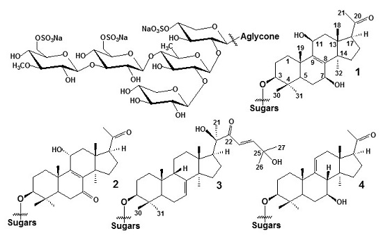
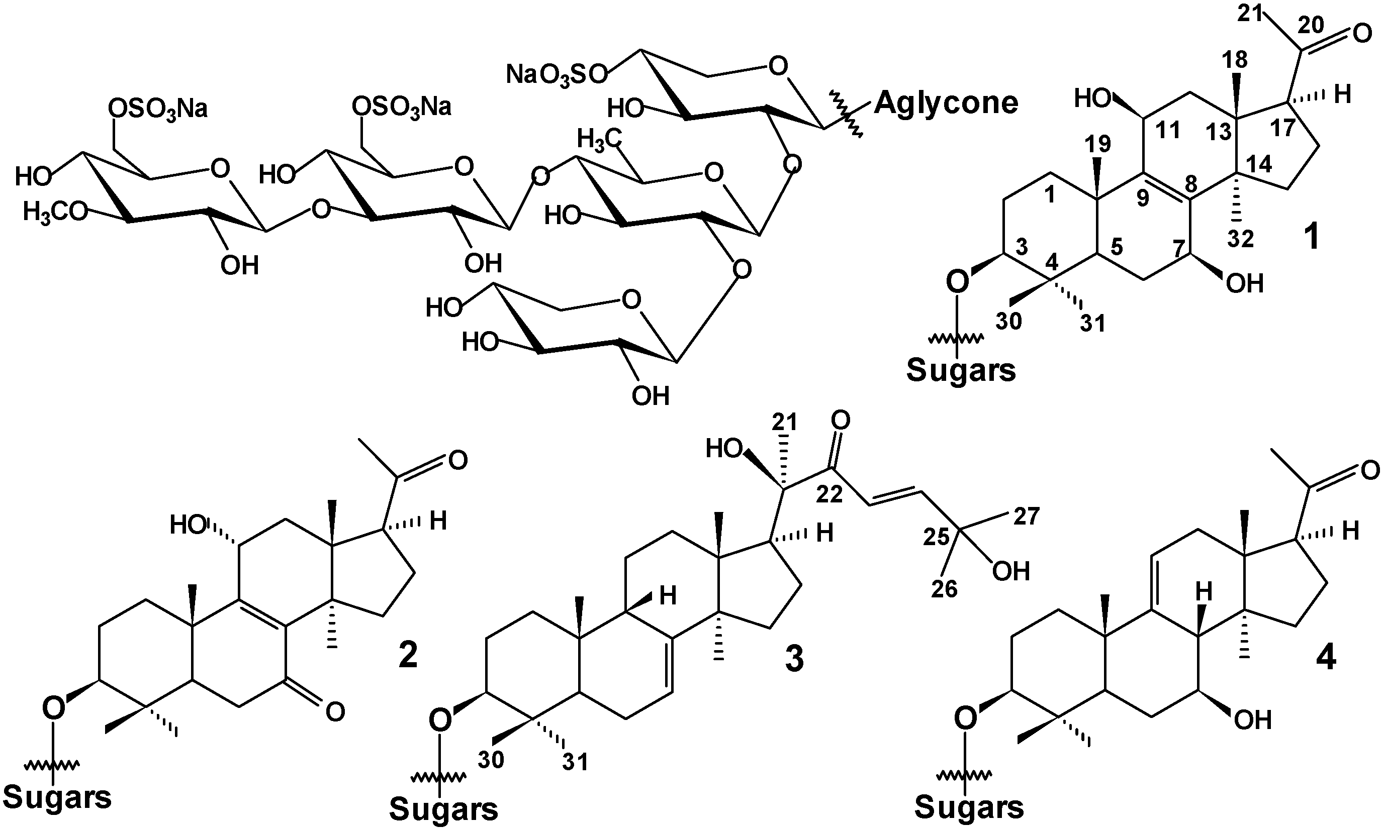
Structures and Biogenesis of Fallaxosides D4, D5, D6 and D7, Trisulfated Non-Holostane Triterpene Glycosides from the Sea Cucumber Cucumaria fallax

by
Alexandra S. Silchenko
1,
Anatoly I. Kalinovsky
1,
Sergey A. Avilov
1,
Pelageya V. Andryjaschenko
1,
Pavel S. Dmitrenok
1,
Vladimir I. Kalinin
1,
Ekaterina A. Chingizova
1,
Kirill V. Minin
2 and
Valentin A. Stonik
1,*
1
G.B. Elyakov Pacific Institute of Bioorganic Chemistry, Far Eastern Branch of the Russian Academy of Sciences, Pr. 100-letya Vladivostoka 159, 690022 Vladivostok, Russia
2
P.P. Shirshov Institute of Oceanology of the Russian Academy of Sciences, Nakhimovsky Pr., 36, 117997 Moscow, Russia
*
Author to whom correspondence should be addressed.
Molecules 2016, 21(7), 939; https://doi.org/10.3390/molecules21070939
Submission received: 24 June 2016 / Revised: 15 July 2016 / Accepted: 15 July 2016 / Published: 20 July 2016
(This article belongs to the Collection Triterpenes and Triterpenoids)

Abstract

:
Four new trisulfated triterpene glycosides, fallaxosides D4 (1), D5 (2), D6 (3) and D7 (4) have been isolated from the sea cucumber Cucumaria fallax (Cucumariidae, Dendrochirotida). The structures of the glycosides have been elucidated by 2D NMR spectroscopy and HRESIMS. All the glycosides have the lanostane aglycones of a rare non-holostane type with 7(8)-, 8(9)- or 9(11)-double bonds, one or two hydroxyl groups occupying unusual positions in the polycyclic nucleus and shortened or normal side chains. The pentasaccharide carbohydrate moieties of 14 have three sulfate groups. The cytotoxic activity of glycosides 14 against the ascite form of mouse Ehrlich carcinoma cells and mouse spleen lymphocytes and hemolytic activity against mouse erythrocytes have been studied.

Graphical Abstract

1. Introduction

Triterpene glycosides are characteristic metabolites of sea cucumbers (class Holothurioidea, phylum Echinodermata). Majority of them contain holostane type aglycones (lanostane derivatives with 18(20)-lactone) [1], but rare non-holostane aglycones, i.e., lanostane derivatives have not 18(20)-lactone [2,3,4,5,6,7].
Recently we have started studies on the Far-Eastern sea cucumber Cucumaria fallax [6] which contains exclusively non-holostane oligoglycosides having unusual double bond positions and uncommon sites of oxidation in their aglycone moieties. Herein we report the isolation of four new trisulfated glycosides, fallaxosides D4–D7 (compounds 14) with earlier unknown aglycones and their structures, established by analysis of 1H-, 13C-NMR and 2D NMR (1H−1H COSY, 1D TOCSY, HMBC, HSQC, ROESY) spectra and confirmed by HRESI mass spectrometry. The biogenesis of these unusual metabolites is also discussed.

2. Results and Discussion

The sea cucumbers were extracted with 70% ethanol under reflux during 5 h. The concentrated extract was sequentially submitted to the column chromatography on Polychrom-1 (powdered Teflon) in H2O→50% ethanol in order to eliminate salts and polar impurities and on Si gel using CHCl3/EtOH/H2O (100:125:25 and 100:150:50) as mobile phases to obtain the fraction containing polar trisulfated pentaosides (glycosides belonging to the group A7). Further separation of the fraction by HPLC on a semi-preparative reversed phase column using MeOH/H2O/NH4OAc (1 M water solution) as mobile phase in ratio 60/39/1 gave the subfractions A7I–A7V. Each of the subfractions was rechromatographed using HPLC. The HPLC of subfraction A7I with the same solvent system in ratio of 35/64/1 gave fallaxoside D4 (1) and fallaxoside D5 (2). The HPLC of subfraction A7II using the solvent system in ratio of 50/49/1 followed by 45/54/1 and 47/51/2 gave fallaxoside D7 (4). The HPLC of subfraction A7V with the same solvents in ratio of 58/41/1 gave fallaxoside D6 (3).
The presence five characteristic doublet signals at δH 4.76–5.22 (1H, d, J = 6.9–8.4 Hz), correlated by HSQC spectra with the signals of anomeric carbons at δC 102.0–105.1 in the 1H-NMR spectra of the carbohydrate chains of fallaxosides D4 (1), D5 (2), D6 (3) and D7 (4) (Scheme 1) and known fallaxosides D1 and D2 [7] were indicative of a pentasaccharide chain and β-configurations of glycosidic bonds. The positions of all the interglycosidic linkages and the place of linkage of the carbohydrate chain to an aglycone were deduced by the analysis of the ROESY and HMBC spectra of the carbohydrate parts of the glycosides (Table 1). Indeed, the cross-peaks between H-1 of the first monosaccharide residue (xylose) and H-3 (C-3) of an aglycone; H-1 of the second mono- saccharide residue (quinovose) and H-2 (C-2) of the first monosaccaharide residue (xylose); H-1 of the third monosaccharide residue (glucose) and H-4 (C-4) of the second monosaccharide residue (quinovose); H-1 of the fourth monosaccharide residue (3-O-methylglucose) and H-3 (C-3) of the third monosaccharide unit (glucose); H-1 of the fifth monosaccharide residue (xylose) and H-2 (C-2) of the second monosaccharide residue (quinovose) were observed. The δC values characteristic of α- and β-shifting effects of sulfate groups were observed for C-4 (δC 76.1) and C-5 (δC 64.0) of the first xylose residue, for C-6 (δC 67.3) and C-5 (δC 74.8) of the glucose residue and C-6 (δC 67.0) and C-5 (δC 75.5) of terminal 3-O-methylglucose residue. These data indicated the presence of a pentasaccharide carbohydrate chain with three sulfate groups.
The 13C-NMR spectra of the carbohydrate parts of compounds 14 (Table 1) were identical to each other and coincided with those of cucumariosides of the group A7 isolated first from Cucumaria japonica [8] and with the corresponding spectra of fallaxosides D1 and D2 having the same carbohydrate chain [6]. The structure of such a carbohydrate chain was previously elucidated by desulfation whereby a known desulfated derivative was obtained. This derivative was obtained from a monosulfated glycoside where very detailed chemical evidence of the sugar sequence was obtained by a variety of methods including specific enzymatic hydrolysis, periodate oxidation and Smith degradation, etc. All the obtained progenins were characterized by 13C-NMR [8]
The NMR spectra of the aglycone part of fallaxoside D4 (1) revealed the presence of 24 carbon atoms (Table 2), including in six methylenes, five methines and six methyl groups as well as seven quaternary carbon signals, that corresponded to the 22,23,24,25,26,27-hexanorlanostane type of aglycones previously found in glycosides of C. fallax [6]. The resonances of two olefinic carbons with δC 139.9 (C-8) and 142.9 (C-9) were assigned to the tetrasubstituted double bond. The HMBC correlations between H-32 (δH 1.00, 3H, s) and C-8 and between H-11 (δH 4.81, 1H, brd, J = 6.8 Hz) and C-9 as well as between H-19 (δH 1.57, 3H, s) and C-9 confirmed the 8(9)-double bond position. The signal at δC 211.8 indicated the presence of a keto group. The signals at δH 2.83 (1H, t, J = 8.8 Hz, H-17) and δC 58.9 (C-17) were assigned to a methine group adjacent to a ketone group. The HMBC correlations from H-17 and H-21 (δH 2.14, 3H, s) to the carbon at δC 211.8 (C-20) allowed the positioning of the group at C-20. There were two downshifted resonances in the 13C-NMR spectrum of 1 at δC 67.7 (C-7) and δC 65.2 (C-11) corresponding to the oxygen bearing allylic type methines suggesting the presence of two hydroxyls. Their positions at C-7 and C-11 were corroborated by the correlations from H-6b (δH 1.99, 1H, m) to C-7 and from H-12b (δH 2.27, 1H, d, J = 14.2 Hz) to C-11 in the HMBC spectrum as well as ROESY correlations between H-1(2.47 m) and H-11 and between H-5 (1.07 dd, 2.7, 15.6 Hz) and H-7. Seeing that absolute configuration at C-5 with α-orientation of hydrogen, as well as configurations of C-10, C-13, C-14, and C-17 stereocenters in the sea cucumber triterpene glycosides were established earlier [9], the H-5–H-7 H-15α–H7 and H-32–H-7 NOESY correlations are indicative of β-orientation of hydroxyl group at C-7. The configuration of the C-11 stereocenter in 1 was proposed by analysis of the ROESY spectrum and MM2 optimized models of 1. The β-orientation of hydroxyl group at C-11 was proposed, based on the comparison of MM2 optimized models of aglycones with α- and β-oriented hydroxyls. The observed in the ROESY spectrum of 1 correlations from H-11 to H-1 and vice versa would be realized for both 11-α and 11β-hydroxyls. However, the correlations from H-11 with methyl groups H-19 and H-18 should be the realized in the ROESY spectrum at 11α-hydroxy orientation. Nevertheless, these correlations were absent in the ROESY spectrum of 1. Hence the β-orientation of the hydroxyl group at C-11 is the most probable.
The HR ESI MS (negative and positive ion modes) of fallaxoside D4 (1) exhibited ion peaks [M3Na − H2O − Na] at m/z 1403.3543 (calc. 1403.3572), [M3Na − H2O − 2Na]2− at m/z 690.1841 (calc. 690.1840) and [M3Na − H2O − 3Na]3− at m/z 452.4596 (calc. 452.4596). The molecular formula of 1 was deduced as C53H83O35S3Na3 based on the HR ESI MS and 13C-NMR spectroscopic data. However, the ions observed in the mass spectra corresponded to the loss of water upon ionization of fallaxoside D4 (1), presumably due to the lability of the dihydroxy-ene-fragment in the rings B and C of its aglycone. The structure of the carbohydrate chain of 1 was also confirmed by fragment ion peaks at m/z 929, corresponding to a monodesulfated carbohydrate chain [MeGlcOSO3Na + GlcOSO3Na + Qui + Xyl + XylOSO3Na − Na2SO4] or [M3Na − Na2SO4 − agl (C24H37O3)]. Other fragment ions at m/z 827.2 corresponded to a didesulfated carbohydrate chain [MeGlcOSO3Na + GlcOSO3Na + Qui + Xyl + XylOSO3Na − Na2SO4 − NaSO3 + H] or [M3Na − Na2SO4 − NaSO3 − agl (C24H37O3) + H]; at m/z 797.1 [M3Na − Na2SO4 − agl (C24H37O3) − Xyl + H]; at m/z 695.2 [M3Na − Na2SO4 − NaSO3 − agl (C24H37O3) − Xyl + 2H]; at m/z 563.1 [M3Na − Na2SO4 − NaSO3 − agl (C24H37O3) − 2Xyl + 3H]; at m/z 519 [M3Na − Na2SO4 − agl (C24H37O3) − 2Xyl − Qui + H]; at m/z 417.1 [M3Na − Na2SO4 − NaSO3 − agl (C24H37O3) − 2Xyl − Qui + 2H], observed in the ESI MS/MS spectra of 1.
Taken together all these data indicate that fallaxoside D4 (1) is 22,23,24,25,26,27-hexanor-3β-O-{6-O-sodium sulfate-3-O-methyl-β-d-glucopyranosyl-(1→3)-6-O-sodium sulfate-β-d-glucopyranosyl-(1→4)-[β-d-xylopyranosyl-(1→2)]-β-d-quinovopyranosyl-(1→2)-4-O-sodium sulfate-β-d-xylopyranosyl}-7β,11β-dihydroxy-20-ketolanost-8-ene.
The structure of the aglycone moiety of fallaxoside D5 (2) was proved to be similar and NMR spectra also confirmed the 22,23,24,25,26,27-hexanorlanostane skeleton system of its aglycone. The difference in the spectra (Table 3) was in the presence of an additional signal of a ketone group at δC 201.4 instead of one of hydroxyl group signals in the 13C-NMR spectrum.
The signals of the quaternary olefinic carbons indicated the presence of 8(9)-double bond and were downshifted (δC 140.0 and 163.1) when compared with those in the 13C-NMR spectrum of 1, suggesting that the ketone group adjoins the double bond. Additionally, the signals at δH 2.56 (1H, m, H-6a) and δH 2.50 (1H, m, H-6b) demonstrated the formation of a spin coupled system in the COSY spectrum with the signal of H-5 (1H, dd, J = 3.9, 13.5 Hz) only and were also noticeably downshifted. These data indicated the closeness of these atoms to the ketone group that was positioned at C-7. The HMBC correlation from both H2-6 to C-7 (δC 201.4) and C-10 (δC 40.6) confirmed the ketone position. Hence, the hydroxyl group at C-7 of the aglycone of 1 was substituted with a ketone group in the aglycone of 2. The signal at δC 64.1 (C-11) in the 13C-NMR spectrum of 2, correlated by the HSQC with the resonance at δH 4.70 (1H, dd, J = 5.4, 8.9 Hz, H-11) indicated the presence of oxygen bearing methine group. The HMBC correlations from both H-12 (δH 2.58, 1H, dd J = 8.8, 13.1 Hz, H-12a) and (δH 2.40, 1H, dd J = 5.1, 13.4 Hz, H-12b) to C-11 corroborated the attachment of hydroxyl group to C-11.
The orientation of hydroxyl group was proposed as α based on the ROESY spectrum. Clear NOE correlations from H-11 to both β-oriented methyl groups Me-18 and Me-19 were observed. Hence the hydroxyl groups at C-11 are opposite oriented in fallaxosides D4 (1) and D5 (2) when compared each other. The HRESIMS (negative ion mode) of fallaxoside D5 (2) exhibited quasi-molecular ion peaks at m/z 1419.3502 (calc. 1419.3521) [M3Na − Na], at m/z 698.1806 (calc. 698.1815) [M3Na − 2Na]2− and at m/z 457.7911 (calc. 457.7912) [M3Na − 3Na]3−. This and 13C-NMR spectroscopic data allowed the determination of the molecular formula of 2 as C53H81O35S3Na3. The presence of sulfate groups and sequence of monosaccharide units in the carbohydrate chain of 2 was confirmed by fragmentary ions at m/z 1299.4 [M3Na − Na − HSO4Na], 1179.4 [M3Na − Na − 2HSO4Na], 911.1 [M3Na − Na2SO4 − agl + H]; 877.3 [M3Na − Na − MeGlcOSO3Na − GlcOSO3Na +H]; 797.1 [M3Na − Na2SO4 − agl − Xyl + H]; 745.3 [M3Na − Na − MeGlcOSO3Na − GlcOSO3Na − Xyl + 2H]; 665.1 [M3Na − Na2SO4 − agl − 2Xyl + 2H]; 599.3 [M3Na − Na − MeGlcOSO3Na − GlcOSO3Na − Qui − Xyl + H]; 519 [M3Na − Na2SO4 − agl − 2Xyl − Qui + H] detected in the ESI MS/MS (negative ion mode) spectrum. All these data indicate that fallaxoside D5 (2) is 22,23,24,25,26, 27-hexanor-3β-O-{6-O-sodium sulfate-3-O-methyl-β-d-glucopyranosyl-(1→3)-6-O-sodium sulfate-β-d-glucopyranosyl-(1→4)-[β-d-xylopyranosyl-(1→2)]-β-d-quinovopyranosyl-(1→2)-4-O-sodium sulfate-β-d-xylopyranosyl}-11α-hydroxy-7,20-diketolanost-8-ene.
The 13C-NMR spectrum of the aglycone part of fallaxoside D6 (3) revealed the presence of 30 carbons indicating the presence of an aglycone with normal non-shortened side chain (Table 4). The absence of a γ-lactone was deduced from the absence of the signal at ≈ δC 176 and contemporary presence of the resonances of Me-18 (δC 24.8, C-18, δH 1.28, 3H, s, H-18) in the 13C- and 1H-NMR spectra, respectively, indicating the aglycone of fallaxoside D6 (3) to be of the lanostane type without a lactone, in contrast with aglycones of the majority of sea cucumber glycosides. The resonances of an olefinic methine group at δC 122.2 (C-7) and δH 5.59 (1H, m, H-7) in NMR spectra as well as of an olefinic quaternary carbon at δC 149.7 (C-8) in the 13C-NMR spectrum were assigned to the 7(8)-double bond typical of many sea cucumber glycosides [1,9]. The presence of an isolated H-5/H2-6/H-7/H-9/H-11/H-12 spin system in the COSY spectrum of 3 as well as the HMBC correlation between H-32 (δH 1.03, 3H, s) and C-8 confirmed the double bond position. The signal at δC 205.3 and resonances of two olefinic tertiary carbons at δC 119.9 and 156.5 in the 13C-NMR spectrum indicated the presence of a keto group and an additional double bond, correspondingly. Extensive analysis of the COSY, HSQC and HMBC spectra of 3 revealed these functionalities could be positioned only in the side chain of the aglycone. Actually the multiplicity and the coupling constants of olefinic protons at δH 7.41 (1H, d, J = 15.5 Hz, H-23) and δH 7.40 (1H, d, J = 15.5 Hz, H-24) were indicative of a Z-configured 23(24)-double bond vicinal on the one hand to the keto group (C-22) and from the other hand to the quaternary oxygen bearing carbon at δC 70.5 (C-25). The presence of the resonances of two heminal methyl groups at δH1.47 (3H, s, H-26) and δH 1.48 (3H, s, H-27) in the 1H-NMR spectrum correlated by HSQC spectrum with corresponding carbons resonances at δC 29.3 (C-26) and δC 29.2 (C-27) as well as the HMBC correlations from H-26 and H-27 to C-24 and C-25 and from H-23 and H-24 to C-22 and C-25 confirmed the structure of side chain having 22-keto- and 25-hydroxyl groups and 23Z,24-double bond. The correlations observed in the ROESY spectrum of 3 (Table 4) corroborated the configurations of stereocenters C-3, C-5, C-10, C-13, C-14, C-17 as well as (20R) configuration (NOE between H-16 and H-23 and vice versa) established earlier for similar aglycone of frondoside C [10]. A ROESY correlations of a signal at 2.28 brd, J = 12.8 Hz, H-9 with signals of methyl group at C-10 and C-14 established the rare 9β-H configuration in 3 characteristic of 7(8)-unsaturated sea cucumber glycosides [1,9,11]. Thus, these data allowed us to determine all the structural and stereochemical features of this unusual aglycone.
The HRESIMS (negative ion mode) of fallaxoside D6 (3) exhibited pseudomolecular ion peaks [M3Na − 2Na]2− at m/z 740.2279 (calc. 740.2284) and [M3Na − 3Na]3− at m/z 485.8224 (calc. 485.8225). This and 13C-NMR spectroscopic data allowed the determination of the molecular formula of 3 as C59H93O35S3Na3. The presence of sulfate groups and sequence of monosaccharide units in the carbohydrate chain of 3 was confirmed by the fragmentary ion peaks at m/z 1383.4 [M3Na − NaHSO4]; 827.2 [M3Na − Na2SO4 − NaSO3 − agl + H]; 797.1 [M3Na − Na2SO4 − agl − Xyl + H]; 695.2 [M3Na − Na2SO4 − NaSO3 − agl − Xyl + 2H]; 563.1 [M3Na − Na2SO4 − NaSO3 − agl − 2Xyl + 3H]; 519 [M3Na − Na2SO4 − agl − 2Xyl − Qui + H] detected in the ESI MS/MS (negative ion mode) spectrum.
All these data indicate that fallaxoside D6 (3) is 3β-O-{6-O-sodium sulfate-3-O-methyl-β-d-glucopyranosyl-(1→3)-6-O-sodium sulfate-β-d-glucopyranosyl-(1→4)-[β-d-xylo-pyranosyl-(1→2)]-β-d-quinovopyranosyl-(1→2)-4-O-sodium sulfate-β-d-xylopyranosyl}-22-keto-25-hydroxylanosta-7,23Z-diene.
The structure of the aglycone moiety of fallaxoside D7 (4) was found by extensive NMR spectroscopy (Table 5) to be similar to that of 12, indicating the presence of a 22,23,24,25,26,27-hexanorlanostane aglycone with the keto group at C-20 (δC 212.5). The double bond was positioned as 9(11) according to the signal of the quaternary olefinic carbon at δC 147.6 (C-9) and olefinic methine resonances at δC 116.0 (C-11) in the 13C-NMR spectrum and δH 5.27 (1H, m, H-11) in the 1H-NMR spectrum. The long range COSY correlation H-8/H-11 as well as the characteristic correlation from Me-19 (δH 1.00, 3H, s, H-19) to C-9 confirmed the double bond position. The signal at δC 71.6 (C-7) in the 13C-NMR spectrum of 4, correlated by the HSQC with the resonance at δH 3.83 (1H, m, H-7) indicated the attachment of hydroxyl group to this carbon. Its position as C-7 was confirmed by the COSY spectrum, where the signals of spin coupled system H-5/H2-6/H-7/H-8 were observed.
The orientation of hydroxyl group was proposed as β based on the ROESY spectrum. Clear NOE correlations from H-7 to both α-oriented H-5 (δH 0.86, 1H, brd, J = 13.7 Hz) and Me-32 (δH 0.99, 3H, s) and vice versa were observed. (8S)-Configuration was proposed based on the correlations between H-8 and the both Me-18 and Me-19, observed in the ROESY spectrum of 4.
The HR ESI MS (negative ion mode) of fallaxoside D7 (4) exhibited ion peaks [M3Na − 2Na]2− at m/z 691.1917 (calc. 691.1918) and [M3Na − 3Na]3− at m/z 453.1320 (calc. 453.1315). This and 13C-NMR spectroscopic data allowed the determination of the molecular formula of 4 as C53H83O34S3Na3. The identity of the carbohydrate chain structure of fallaxoside D7 (4) to those of 13 confirmed by the presence of the fragmentary ion peaks with the same m/z: 797.1, 695.2, 665.1, 563.1, 519.0 and 417.1 in the ESI MS/MS (negative ion mode) spectrum of 4.
On the base of all the above discussed data, the structure of fallaxoside D7 (4) was established to be 22,23,24,25,26,27-hexanor-3β-O-{6-O-sodium sulfate-3-O-methyl-β-d-glucopyranosyl-(1→3)-6-O-sodium sulfate-β-d-glucopyranosyl-(1→4)-[-β-d-xylopyranosyl-(1→2)]-β-d-quinovopyranosyl-(1→2)-4-O-sodium sulfate-β-d-xylopyranosyl}-7β-hydroxy-20-ketolanost-9(11)-ene.
The cytotoxic activities of fallaxosides D4 (1), D5 (2), D6 (3) and D7 (4) against mouse spleen lymphocytes and ascite form of mouse Ehrlich carcinoma cells along with hemolytic activity against mouse erythrocytes were studied. None of these glycosides were active in these tests at the dosage studied (IC50 > 100 µM/mL). This could be explained by the absence of the 18(20)-lactone moiety that is essential for the membranolytic action of sea cucumber glycosides and by the presence of three sulfate groups that decreased the membranolytic activity of the glycosides [12].
It is most probable that holostane type glycosides play a role in chemical defense against predators because of their strong membranolytic activities and not only as regulators of oocyte maturation. Non-holostane glycosides may play only a regulatory role and are evolutionary precursors of holostane glycosides [12].
In summary, four novel triterpene glycosides 14 have been isolated from the sea cucumber Cucumaria fallax along with the series of other uncommon glycosides [6]. All of these compounds contain new non-holostane aglycones with some unprecedented structural features. It is obvious that the isolated glycosides, including the earlier studied fallaxoside C2 and D2 (having a 7-keto-8(9)-en fragment), C1 and D1 (having a 7,11-diketo-8(9)-en fragment) [6], D4 (1) (with a 7,11-dihydroxy-8(9)-en fragment), D5 (2) (with a 7-keto-11-hydroxy-8(9)-en fragment) and D7 (4) (with a 7-hydroxy-9(11)-en fragment), are biogenetically related with each other. They have oxygen-containing functional groups (ketones or hydroxyls) at the same C-7 and/or C-7 and C-11 positions of aglycones. Another unusual structural feature of many isolated glycosides includes the presence of a 8(9)-double bond in the majority of glycosides isolated from this species. This suggests that in C. fallax, the protosterol cation, formed from squalene, is cyclized not only into 9(11)-unsaturated parkeol or into 9β-H-lanosta-7,24-dienol, as shown earlier in other sea cucumbers [13], but also into lanosta-8(9),24-diene-3β-ol (lanosterol).

3. Experimental Section

3.1. General Experimental Procedures

Melting points were determined with a Kofler-Thermogenerate apparatus (Leica VMTG, Wien, Austria). Specific rotation was measured on a 343 Polarimeter (Perkin-Elmer Corporation, Waltham, MA, USA). NMR spectra were recorded on an Advance III-700 spectrometer (Bruker Bio Spin, Rheinstetten, Germany) at 700.13 MHz/176.04 MHz (1H/13C) in C5D5N/D2O (4/1) with TMS as an internal reference (δ = 0). The ESIMS (negative ion mode) were recorded using an Agilent 6510 Q-TOF apparatus (Agilent Corporation, Palo Alto, CA, USA), 50% MeOH was used as the solvent, sample concentration 0.01 mg/mL. HPLC was performed using an Agilent 1100 chromatograph equipped with a differential refractometer on a Ascentis RP Amide (10 × 250 mm, 5 µm) column (Supelco, Bellefonte, PA, USA).

3.2. Animal Material

The samples of Cucumaria fallax (Cucumariidae, Dendrochirotida) were collected during the 41-st scientific cruise of the research vessel Akademik Oparin in the Pacific Ocean near Black Brothers Islands, Kurile Islands (46°23′9′′ N, 150°46′25′′ E) on 20 July, 2011 using Sigsbee trawl from the depth of 150 m (collector Kirill Minin, P.P. Shyrshov Institute of Oceanology of the Russian Academy of Sciences, Moscow, Russia). The sea cucumber taxonomic identification was carried out by Dr. Vadim G. Stepanov (Kamchatka Department of the Institute of Geography, Far East Division of the Russian Academy of Sciences, Petropavlosk-Kamchatsky, Russia), the voucher specimen is deposited in collection of Kamchatka Department of the Institute of Geography.

3.3. Extraction and Isolation

The sea cucumbers were minced and extracted with 70% ethanol under reflux during 5 h. The concentrated in vacuo ethanolic extract of C. fallax (28.2 g of dry wt after ethanol extraction) was sequentially submitted to column chromatography on Polychrom-1 (powdered Teflon) in H2O→50% ethanol and on Si gel using CHCl3/EtOH/H2O (100:125:25 and 100:150:50) as mobile phases to obtain the fraction A7 (165 mg). Further separation of the fraction by HPLC on a semi-preparative Supelco Ascentis RP-Amide (10 × 250 mm) reverse phase column using MeOH/H2O/NH4OAc (1 M water solution) as mobile phase in ratio 60/39/1 gave the subfractions A7I–A7V, each of them was subsequently rechromatographed. The HPLC of subfraction A7I with the same solvent system in ratio of 35/64/1 gave 3.5 mg of fallaxoside D4 (1) and 3.5 mg of fallaxoside D5 (2). The HPLC of subfraction A7II using the solvent system in ratio of 50/49/1 followed by 45/54/1 and 47/51/2 gave 1.8 mg of fallaxoside D7 (4). The HPLC of subfraction A7V with the same solvents in ratio of 58/41/1 gave 7.2 mg of fallaxoside D6 (3).
Fallaxoside D4 (1): Colorless powder; MP: 212–214 °C; [ α ] D 20 −2 (c 0.1, 50% MeOH); NMR: See Table 1 and Table 2; HRESIMS (−) m/z 1403.3543 (calc. for C53H83O35S3Na3, 1403.3572) [M3Na − H2O − Na], m/z 690.1841 (calc. 690.1840) [M3Na − H2O − 2Na]2−, 452.4596 (calc. 452.4596) [M3Na − H2O − 3Na]3−; ESI MS/MS (−) m/z 929 [M3Na − Na2SO4 − agl (C24H37O3)]; 827.2 [M3Na − Na2SO4 − NaSO3 − agl (C24H37O3) + H]; 797.1 [M3Na − Na2SO4 − agl (C24H37O3) − Xyl + H]; 695.2 [M3Na − Na2SO4 − NaSO3 − agl (C24H37O3) − Xyl + 2H]; 563.1 [M3Na − Na2SO4 − NaSO3 − agl (C24H37O3) − 2Xyl + 3H]; 519 [M3Na − Na2SO4 − agl (C24H37O3) − 2Xyl − Qui + H]; 417.1 [M3Na − Na2SO4 − NaSO3 − agl (C24H37O3) − 2Xyl − Qui + 2H].
Fallaxoside D5 (2): Colorless powder; MP: 206–208 °C; [ α ] D 20 –3 (c 0.1, 50% MeOH); NMR: see Table 1 and Table 3; HRESIMS (−) m/z 1419.3502 (calc. for C53H81O35S3Na3, 1419.3521) [M3Na − Na], 698.1806 (calc. 698.1815) [M3Na − 2Na]2−, 457.7911 (calc. 457.7912) [M3Na − 3Na]3−; ESI MS/MS (−) m/z 1299.4 [M3Na − Na − HSO4Na], 1179.4 [M3Na − Na − 2HSO4Na], 911.1 [M3Na − Na2SO4 − agl + H]; 877.3 [M3Na − Na − MeGlcOSO3Na − GlcOSO3Na +H]; 797.1 [M3Na − Na2SO4 − agl − Xyl + H]; 745.3 [M3Na − Na − MeGlcOSO3Na − GlcOSO3Na − Xyl + 2H]; 665.1 [M3Na − Na2SO4 − agl − 2Xyl + 2H]; 599.3 [M3Na − Na − MeGlcOSO3Na − GlcOSO3Na − Qui − Xyl + H]; 519 [M3Na − Na2SO4 − agl − 2Xyl − Qui + H].
Fallaxoside D6 (3): Colorless powder; MP: 225–227 °C; [ α ] D 20 –8 (c 0.1, 50% MeOH); NMR: see Table 1 and Table 4; HRESIMS (−) m/z 740.2279 (calc. for C59H93O35S3Na3, 740.2284) [M3Na − 2Na]2−, 485.8224 (calc. 485.8225) [M3Na − 3Na]3−; ESI MS/MS (−) m/z 1383.4 [M3Na − NaHSO4]; 827.2 [M3Na − Na2SO4 − NaSO3 − agl + H]; 797.1 [M3Na − Na2SO4 − agl − Xyl + H]; 695.2 [M3Na − Na2SO4 − NaSO3 − agl − Xyl + 2H]; 563.1 [M3Na − Na2SO4 − NaSO3 − agl − 2Xyl + 3H]; 519 [M3Na − Na2SO4 − agl − 2Xyl − Qui + H].
Fallaxoside D7 (4): Colorless powder; MP: 210–212 °C; [ α ] D 20 –5 (c 0.1, 50% MeOH); NMR: See Table 1 and Table 5; HRESIMS (−) m/z 691.1917 (calc. for C53H83O34S3Na3, 691.1918) [M3Na − 2Na]2−, 453.1320 (calc. 453.1315) [M3Na − 3Na]3−; ESI MS/MS (−) m/z 797.1 [M3Na − Na2SO4 − agl − Xyl + H]; 695.2 [M3Na − Na2SO4 − NaSO3 − agl − Xyl + 2H]; 665.1 [M3Na − Na2SO4 − agl − 2Xyl + 2H]; 563.1 [M3Na − Na2SO4 − NaSO3 − agl − 2Xyl + 3H], 519.0 [M3Na − Na2SO4 − agl − 2Xyl − Qui + H]; 417.1 [M3Na − Na2SO4 − NaSO3 − agl − 2Xyl − Qui + 2H].

3.4. Cell Culture

Splenocytes from CD-1 line mice were used. The spleen were isolated from mice and centrifuged (450× g) for 5 min. The splenocytes were washed three times and resuspended in phosphate-buffered saline, pH 7.2–7.4 at final concentration 2–5 × 10⁶ cells/mL.

3.5. Cytotoxicity Activity

Water solutions (10 µL), containing different concentrations (0.12–100.00 µM) of the tested substances and 90 µL of the cell suspension were added to the wells of a 96-well plate and incubated for 1 h at 37 °C. After the incubation, 10 µL of the cell suspension were added to 10 µL of trypan blue solution (0.4% in PBS) and placed on a slide. After 1–5 min incubation the amount of alive and dead cells were calculated with an Imager A1 optical microscope (Carl Zeiss, Oberkochen, Germany) using the AxioVision (Carl Zeiss) 4.7.1 software. The cytotoxic activity of the substances was calculated as the ratio of the dead cells to general cells amount. ED50 was calculated with SigmaPlot 10.0 software.

3.6. Hemolytic Activity

Blood was taken from a CD-1 mouse. The erythrocytes were washed three times with 0.9% NaCl, centrifuged (450× g) on a LABOFUGE 400R centrifuge (Heraeus, Hanau, Germany) for 5 min [14] followed by resuspension in phosphate-buffered saline (PBS), pH 7.2–7.4. Erythrocytes were used at a concentration providing an optical density of 1.5 at 700 nm for a non-hemolyzed sample. Twenty µL of a water solution of test substance with a fixed concentration (0.12–100.00 µM) were added to a well of a 96-well plate containing 180 mL of the erythrocyte suspension and incubated for 1 h at 37 °C. The plates were centrifuged (900× g) on a LMC-3000 laboratory centrifuge (Biosan, Riga, Latvia) for 10 min as proposed in [15]. Ten µL of the supernatant were placed on special microplate with a plate bottom for determination of the optical density on a Multiskan FC spectrophotometer (Termo Fisher Scientific, Waltham, MA, USA) at λ = 570 nm. ED50 values were calculated using the SigmaPlot 3.0 software. Triton X-100 (Biolot, Saint Petersburg, Russian Federation) at a concentration of 1%, causing 100% cell hemolysis was used as positive control. The erythrocyte suspension in phosphate-buffered saline, pH 7.2–7.4 (PBS) with 20 µL of the solvent without a tested compound was used as negative control.

Acknowledgments

The authors are very appreciative to the Grant of the Russian Science Foundation No. 16-14-10131 for partial financial support and Vadim G. Stepanov (Kamchatka Department of the Institute of Geography, Far East Division of the Russian Academy of Sciences, Petropavlosk-Kamchatsky, Russia) for taxonomic identification of sea cucumbers.

Author Contributions

A.S.S., V.A.S. and V.I.K. wrote the paper; A.S.S., S.A.A. and P.V.A. conceived, designed and performed the experiments concerning isolation of the glycosides; A.S.S. and A.I.K. analized the NMR data; P.S.D. performed the mass-spectrometry experiments and analized their results; E.A.C. performed the bioassay; K.V.M. carriedf out serach and collection of biological materials and performed primary extraction.

Conflicts of Interest

The authors declare no conflict of interest.

References

  1. Stonik, V.A.; Kalinin, V.I.; Avilov, S.A. Toxins from the sea cucumbers (Holothuroids): Chemical structures, properties, taxonomic distribution, biosynthesis and evolution. J. Nat. Toxins 1999, 8, 235–248. [Google Scholar] [PubMed]
  2. Kalinin, V.I.; Kalinovskii, A.I.; Stonik, V.A. Onekotanogenin-A new triterpene genin from the holothurian Psolus fabricii. Chem. Nat. Comp. 1987, 23, 560–563. [Google Scholar] [CrossRef]
  3. Kalinin, V.I.; Silchenko, A.S.; Avilov, S.A.; Stonik, V.A.; Smirnov, A.V. Sea Cucumbers Triterpene Glycosides, the Recent Progress in Structural Elucidation and Chemotaxonomy. Phytochem. Rev. 2005, 4, 221–236. [Google Scholar] [CrossRef]
  4. Silchenko, A.S.; Avilov, S.A.; Kalinovsky, A.I.; Dmitrenok, P.S.; Kalinin, V.I.; Morre, J.; Deinzer, M.L.; Woodward, C.; Collin, P.D. Glycosides from the North Atlantic sea cucumber Cucumaria frondosa V-Structures of five new minor trisulfated triterpene oligoglycosides, frondosides A7-1, A7-3, A7-4, and isofrondoside C. Can. J. Chem. 2007, 85, 626–636. [Google Scholar] [CrossRef]
  5. Silchenko, A.S.; Kalinonovsky, A.I.; Avilov, S.A.; Andryjashenko, P.V.; Dmitrenok, P.S.; Kalinin, V.I.; Stonik, V.A. 3β-O-Glycosylated 16β-acetoxy-9β-H-lanosta-7,24-diene-3β,18,20β-triol, an intermediate metabolite from the sea cucumber Eupentacta fraudatrix and its biosynthetic significance. Biochem. Syst. Ecol. 2012, 44, 53–60. [Google Scholar] [CrossRef]
  6. Silchenko, A.S.; Kalinovsky, A.I.; Avilov, S.A.; Andryjaschenko, P.V.; Dmitrenok, P.S.; Kalinin, V.I.; Martyyas, E.A.; Minin, K.V. Fallaxosides C1, C2, D1 and D2, Unusual Olygosulfated Triterpene Glycosides from the Sea Cucumber Cucumaria fallax (Cucumariidae, Dendrochirotida) and taxonomic status of this animal. Nat. Prod. Commun. 2016, 11, 939–945. [Google Scholar]
  7. Miyamoto, T.; Togawa, K.; Higuchi, R.; Komori, T.; Sasaki, T. Structures of four new triterpenoid oligoglycosides: Ds-penaustrosides A, B, C and D from the sea cucumber Pentacta australis. J. Nat. Prod. 1992, 55, 940–946. [Google Scholar] [CrossRef] [PubMed]
  8. Drozdova, O.A.; Avilov, S.A.; Kalinovskii, A.I.; Stonik, V.A.; Mil'grom, Y.M.; Rashkes, Y.V. Trisulfated glycosides from the holothurian Cucumaria japonica. Chem. Nat. Comp. 1993, 29, 309–313. [Google Scholar] [CrossRef]
  9. Ilyin, S.G.; Reshetnyak, M.V.; Afyatullov, S.S.; Stonik, V.A.; Elyakov, G.B. Crystallic and molecular structure of diacetate of holost-8(9)-en-3α,16β-diol. Proc. USSR Acad. Sci. 1985, 284, 356–359. [Google Scholar]
  10. Avilov, S.A.; Drozdova, O.A.; Kalinin, V.I.; Kalinovsky, A.I.; Stonik, V.A.; Gudimova, E.N.; Riguera, R.; Jimenez, C. Frondoside C. A new nonholostane triterpene glycoside from the sea cucumber Cucumaria frondosa: Structure and cytotoxicity of its desulfated derivative. Can. J. Chem. 1998, 76, 137–141. [Google Scholar]
  11. Stonik, V.A. Some terpenoid and steroid derivatives from echinoderms and sponges. Pure Appl. Chem. 1986, 58, 423–436. [Google Scholar] [CrossRef]
  12. Kalinin, V.I.; Aminin, D.L.; Avilov, S.A.; Silchenko, A.S.; Stonik, V.A. Triterpene glycosides from sea cucucmbers (Holothurioidae, Echinodermata), biological activities and functions. In Studies in Natural Product Chemistry (Bioactive Natural Products); Atta-ur-Rahman, Ed.; Elsevier Science Publisher: Amsterdam, The Netherland, 2008; Volume 35, pp. 135–196. [Google Scholar]
  13. Makarieva, T.N.; Stonik, V.A.; Kapustina, I.I.; Boguslavsky, V.M.; Dmitrenok, P.S.; Kalinin, V.I.; Cordeiro, M.L.; Djerassi, C. Biogenetic studies on marine lipids. 42. Biosynthetic studies of steroid and triterpenoid metabolites in the sea cucumber Eupentacta fraudatrix. Steroids 1993, 58, 508–517. [Google Scholar] [CrossRef]
  14. Prokofieva, N.G.; Likhatskaya, G.N.; Volkova, O.V.; Anisimov, M.M.; Kiseleva, M.I.; Ilyin, S.G.; Budina, T.A.; Pokhilo, N.D. The action of betulafolientetraol on the erythrocytes and model membranes. Biol. Membr. 1992, 9, 954–960. [Google Scholar]
  15. Taniyama, S.; Arakawa, O.; Terada, M.; Nishio, S.; Takatani, T.; Mahmud, Y.; Noguchi, T. Ostreopsis sp., a possible origin of palytoxin (PTX) in parrotfish Scarus ovifrons. Toxicon 2003, 42, 29–33. [Google Scholar] [CrossRef]
  • Sample Availability: Samples of the compounds are available from the authors.
Scheme 1. Structures of the glycosides: 1—fallaxoside D4; 2—fallaxoside D5; 3—fallaxoside D6; 4—fallaxoside D7.
Molecules 21 00939 sch001
Table 1. NMR Spectroscopic data (700 MHz, C5D5N/D2O (4/1 v/v)) of the carbohydrate moieties of 14.
AtomδC mult. a,b,cδH mult. (J in Hz) dHMBCROESY
Xyl1 (1→C-3)
1104.7 CH4.76 d (6.9)C: 3H-3, H-3, 5 Xyl1
281.3 CH3.97 t (8.6)C: 1 Qui2; C: 1, 3 Xyl1H-1 Qui2; H-4 Xyl1
375.2 CH4.31 t (8.6)C: 2, 4 Xyl1H-1, 5 Xyl1
476.1 CH4.98 dd (8.6, 13.8)C: 3 Xyl1H-2 Xyl1
564.0 CH24.76 d (11.2)C: 1, 3 Xyl1
3.87 dd (8.6, 11.2) H-1, 3 Xyl1
Qui2 (1→2Xyl1)
1102.0 CH5.20 d (7.8)C: 2 Xyl1H-2 Xyl1; H-3, 5 Qui2
282.4 CH3.92 t (8.6)C: 1 Xyl5; C: 1, 3 Qui2H-1 Xyl5; H-4 Qui2
375.2 CH3.98 t (8.6)C: 2, 4 Qui2H-5 Qui2
486.3 CH3.43 t (8.6)C: 1 Glc3; C: 3, 5, 6 Qui2H-1 Glc3; H-2, 6 Qui2
570.8 CH3.56 dd (6.0, 9.5) H-1, 3, 6 Qui2
617.8 CH31.55 d (6.0)C: 4, 5 Qui2H-4, 5 Qui2
Glc3 (1→4Qui2)
1103.9 CH4.78 d (7.8)C: 4 Qui2H-4 Qui2; H-5 Glc3
273.4 CH3.81 mC: 1, 3 Glc3
386.5 CH4.13 t (8.6)C: 1 MeGlc4; C: 2, 4 Glc3H-1 MeGlc4; H-1 Glc3
469.1 CH3.80 t (8.6)C: 5, 6 Glc3
574.8 CH4.08 m H-1 Glc3
667.3 CH24.94 d (11.2)
4.59 dd (6.9, 11.2)C: 5 Glc3H-4 Glc3
MeGlc4 (1→3Glc3)
1104.7 CH5.15 d (8.4)C: 3 Glc3H-3 Glc3; H-3, 5 MeGlc4
274.3 CH3.78 t (8.4)C: 1 MeGlc4H-4 MeGlc4
386.3 CH3.64 t (9.3)C: 2, 4 MeGlc4, OMeH-1, 5 MeGlc4
469.8 CH4.01 mC: 3, 5 MeGlc4H-2, 6 MeGlc4
575.5 CH4.01 mC: 4, 6 MeGlc4H-1, 3 MeGlc4
667.0 CH24.92 brd (11.0)C: 4, 5 MeGlc4
4.75 brd (8.4)C: 5 MeGlc4
OMe60.4 CH33.80 sC: 3 MeGlc4
Xyl5 (1→2Qui2)
1105.1 CH5.22 d (7.6)C: 2 Qui2H-2 Qui2; H-3, 5 Xyl5
274.8 CH3.91 t (7.6)C: 1, 3 Xyl5
376.3 CH4.07 t (8.4)C: 2, 4 Xyl5H-1, 5 Xyl5
470.1 CH4.05 mC: 3 Xyl5H-2 Xyl5
566.4 CH24.28 dd (5.1, 11.8)C: 1, 4 Xyl5
3.66 t (9.3)C: 1, 3, 4 Xyl5H-1, 3 Xyl5
a Recorded at 176.04 MHz; b Bold = interglycosidic positions; c Italic = sulfate position; d Multiplicity by 1D TOCSY.
Table 2. NMR Spectroscopic data (700 MHz, C5D5N/D2O (4/1 v/v)) of the aglycone moiety of fallaxoside D4 (1).
PositionδC Mult. aδH Mult. (J in Hz)HMBCROESY
134.9 CH22.47 m H-11, H-19
1.32 mC: 18H-3, H-5, H-11
226.6 CH21.90 m
1.79 m H-19, H-30
388.6 CH3.13 dd (4.7, 12.2)C: 30, 31, C: 1 Xyl1H-1, H-5, H-31, H-1 Xyl1
439.5 C
551.4 CH1.07 dd (2.7, 15.6)C: 4, 19, 30H-1, H-3, H-7, H-31
630.8 CH22.35 m H-31
1.99 mC: 5, 7, 10H-19, H-30
767.7 CH4.55 t (8.1)C: 8, 9H-5, H-15, H-32
8139.9 C
9142.9 C
1038.3 C
1165.2 CH4.81 brd (6.8)C: 8, 9, 13H-1
1242.8 CH22.53 dd (7.5, 14.2)C: 13, 18H-17, H-32
2.27 d (14.2)C: 9, 11, 13, 14, 18H-18, H-21
1345.2 C
1451.5 C
1532.0 CH22.71 brd (10.8) βC: 14, 32H-18
1.60 brt (10.8) α H-7, H-32
1622.6 CH22.41 m H-18
1.68 m H-32
1758.9 CH2.83 t (8.8)C: 12, 13, 16, 18, 20, 21H-12, H-21, H-32
1819.7 CH31.23 sC: 12, 13, 14, 17H-12, H-15, H-16, H-19, H-21
1922.2 CH31.57 sC: 1, 5, 9, 10H-1, H-2, H-5, H-6, H-18, H-30
20211.8 C
2131.3 CH32.14 sC: 17, 20
3016.4 CH30.99 sC: 3, 4, 5, 31H-2, H-6, H-19, H-31
3127.9 CH31.15 sC: 3, 4, 5, 30H-3, H-5, H-6, H-30
3226.1 CH31.00 sC: 8, 13, 14, 15H-7, H-12, H-15, H-16, H-17
a Recorded at 176.04 MHz.
Table 3. NMR Spectroscopic data (700 MHz, C5D5N/D2O (4/1 v/v)) of the aglycone moiety of fallaxoside D5 (2).
PositionδC Mult. aδH Mult. (J in Hz)HMBCROESY
134.1 CH22.29 m H-3, H-5
2.01 mC: 10H-11
226.5 CH22.00 mC: 3H-19
1.77 m H-19, H-30
387.8 CH3.13 dd (4.0; 11.6)C: 4, 30, C: 1 Xyl1H-1, H-5, H-31, H-1 Xyl1
439.5 C
550.5 CH1.83 dd (3.9; 13.5)C: 4, 6, 10, 19, 30, 31H-1, H-3, H-31
636.9 CH22.56 mC: 5, 7, 10
2.50 mC: 5, 7, 10H-19, H-30
7201.4 C
8140.0 C
9163.1 C
1040.6 C
1164.1 CH4.70 dd (5.4; 8.9)C: 8, 9, 10, 13H-1, H-18, H-19
1243.5 CH22.58 dd (8.8; 13.1)C: 9, 11, 13, 18H-18, H-21
2.40 dd (5.1; 13.4)C: 11, 13, 17, 18H-17, H-32
1348.5 C
1448.7 C
1533.4 CH22.30 mC: 14, 17, 32H-32
1.86 m H-18
1622.1 CH22.27 m H-18
1.68 m H-32
1758.6 CH2.91 t (9.0)C: 12, 13, 16, 18, 20H-12, H-21, H-32
1818.6 CH30.58 sC: 12, 13, 14, 17H-12, H-15, H-16, H-19, H-21
1919.8 CH31.10 sC: 1, 5, 9, 10H-2, H-6, H-18
20211.7 C
2131.2 CH32.15 sC: 17, 20H-17, H-18
3016.0 CH31.01 sC: 3, 4, 5, 31H-2, H-6
3127.0 CH31.05 sC: 3, 4, 5, 30H-3, H-5, H-6
3225.0 CH31.40 sC: 8, 13, 14, 15H-12, H-15, H-17
a Recorded at 176.04 MHz.
Table 4. NMR spectrosopic data (700 MHz, C5D5N/D2O (4/1 v/v)) of the aglycone moiety of fallaxoside D6 (3).
PositionδC Mult. aδH Mult. (J in Hz)HMBCROESY
135.5 CH21.32 m H-11
1.27 m
227.0 CH21.95 m
1.78 m H-19, H-30
388.9 CH3.14 dd (4.1, 11.6)C: 30, C: 1 Xyl1H-5, H-31, H-1 Xyl1
439.5 C
549.7 CH0.84 brd (12.7)C: 1, 4, 30H-1, H-3, H-31
623.1 CH21.92 m H-31
1.83 m H-19, H-30
7122.2 CH5.59 mC: 9, 14H-15, H-32
8149.7 C
948.1 CH2.28 brd (12.8) H-18, H-19
1035.6 C
1122.8 CH21.63 m
1.38 m H-32
1234.8 CH21.96 m H-17, H-21
1.74 m H-18
1345.3 C
1452.9 C
1533.4 CH21.59 m H-18
1.50 m H-7, H-32
1622.2 CH21.93 m H-23
1.54 m H-17, H-32
1752.8 CH2.39 t (8.8)C: 14, 16, 18H-21, H-32
1824.8 CH31.28 sC: 12, 14, 17H-9, H-12, H-16, H-21
1924.5 CH30.93 sC: 1, 5, 9, 10H-2, H-6, H-9
2080.5 C
2124.5 CH31.62 sC: 17, 20, 22H-12, H-17, H-18
22205.3 C
23119.9 C7.41 d (15.5)C: 22, 24, 25H-17, H-21, H-26, H-27, H-16
24156.5 C7.40 d (15.5)C: 22, 23, 25
2570.5 C
2629.3 CH31.47 sC: 24, 25, 27
2729.2 CH31.48 sC: 24, 25, 26
3017.4 CH31.02 sC: 3, 4, 5, 31H-2, H-6
3128.7 CH31.17 sC: 3, 4, 5, 30H-3, H-5, H-6
3230.7 CH31.03 sC: 8, 13, 14, 15H-7, H-11, H-12, H-15, H-17
a Recorded at 176.04 MHz.
Table 5. NMR spectrosopic data (700 MHz, C5D5N/D2O (4/1 v/v)) of Aglycone Moiety of fallaxoside D7 (4).
PositionδC Mult. aδH Mult. (J in Hz)HMBCROESY
136.4 CH21.60 m H-11, H-19
1.24 m H-3, H-5, H-11
226.7 CH22.00 m
1.77 m H-19, H-30
388.5 CH3.10 dd (4.1; 11.8) H-1, H-5, H-31, H-1 Xyl1
439.5 C
549.3 CH0.86 brd (13.7) H-1, H-3, H-7, H-31
631.8 CH22.15 m H-31
1.79 m
771.6 CH3.83 m H-5, H-32
849.8 CH2.38 m H-15, H-18, H-19
9147.6 C
1038.9 C
11116.0 CH5.27 mC: 10, 13H-1
1235.8 CH22.34 m H-32
1.94 m H-18
1346.8 C
1447.3 C
1536.6 CH22.05 m H-32
1.94 m
1622.5 CH22.31 m H-18
1.62 m H-32
1759.3 CH2.92 t (9.0)C: 13, 18H-12, H-32
1816.4 CH30.59 sC: 12, 13, 17H-8, H-12, H-19
1921.8 CH31.00 sC: 5, 9, 10H-1, H-2, H-8, H-18
20212.5 C
2131.2 CH32.18 sC: 17, 20
3016.6 CH30.94 sC: 3, 4, 5, 31H-2, H-31
3127.9 CH31.14 sC: 3, 4, 5, 30H-3, H-5, H-6, H-30
3218.3 CH30.99 sC: 8, 13, 15H-7, H-15, H-16, H-17
a Recorded at 176.04 MHz

