Article

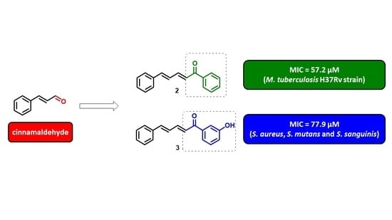
Antibacterial and Antitubercular Activities of Cinnamylideneacetophenones

by
Carlos R. Polaquini
1,†,
Guilherme S. Torrezan
1,†,
Vanessa R. Santos
2,
Ana C. Nazaré
1,
Débora L. Campos
3,
Laíza A. Almeida
1,
Isabel C. Silva
3,
Henrique Ferreira
4,
Fernando R. Pavan
3,
Cristiane Duque
2 and
Luis O. Regasini
1,*
1
Laboratory of Green and Medicinal Chemistry, Department of Chemistry and Environmental Sciences, Institute of Biosciences, Humanities and Exact Sciences, São Paulo State University (Unesp), São José do Rio Preto, SP 15054-000, Brazil
2
Department of Pediatric Dentistry and Public Health, School of Dentistry, São Paulo State University (Unesp), Araçatuba, SP 16015-050, Brazil
3
Department of Biological Sciences, School of Pharmaceutical Sciences, São Paulo State University (Unesp), Araraquara, SP 14800-903, Brazil
4
Department of Biochemistry and Microbiology, Institute of Biosciences, São Paulo State University (Unesp), Rio Claro, SP 13506-900, Brazil
*
Author to whom correspondence should be addressed.
These two authors contributed equally to this work.
Molecules 2017, 22(10), 1685; https://doi.org/10.3390/molecules22101685
Submission received: 30 August 2017 / Revised: 2 October 2017 / Accepted: 3 October 2017 / Published: 10 October 2017
(This article belongs to the Special Issue Emerging Drug Discovery Approaches against Infectious Diseases)

Abstract

:
Cinnamaldehyde is a natural product with broad spectrum of antibacterial activity. In this work, it was used as a template for design and synthesis of a series of 17 cinnamylideneacetophenones. Phenolic compounds 3 and 4 exhibited MIC (minimum inhibitory concentration) and MBC (minimum bactericidal concentration) values of 77.9 to 312 µM against Staphylococcus aureus, Streptococcus mutans, and Streptococcus sanguinis. Compounds 2, 7, 10, and 18 presented potent effects against Mycobacterium tuberculosis (57.2 µM ≤ MIC ≤ 70.9 µM). Hydrophilic effects caused by substituents on ring B increased antibacterial activity against Gram-positive species. Thus, log Po/w were calculated by using high-performance liquid chromatography-photodiode array detection (HPLC-PDA) analyses, and cinnamylideneacetophenones presented values ranging from 2.5 to 4.1. In addition, the effects of 3 and 4 were evaluated on pulmonary cells, indicating their moderate toxicity (46.3 µM ≤ IC50 ≤ 96.7 µM) when compared with doxorubicin. Bioactive compounds were subjected to in silico prediction of pharmacokinetic properties, and did not violate Lipinski’s and Veber’s rules, corroborating their potential bioavailability by an oral route.

Graphical Abstract

1. Introduction

Cinnamaldehyde (1) is the main component in cinnamon bark oil (50–70%), and it is responsible for its spicy and sweet taste [1,2]. Structurally, cinnamaldehyde is a natural simple phenylpropanoid, bearing an α,β-unsaturated aromatic aldehyde. The synthetic applications of 1 are vast and related to its abundance and low economic costs, and have led to a collection of compounds with high chemodiversity [3].
The spectrum of antibacterial activity of 1 has been extensively reported and includes effects against Gram-positive, Gram-negative, and mycobacteria species, such as Escherichia coli [4], Helicobacter pylori [5], Pseudomonas aeruginosa [6], Salmonella enterica [7], Staphylococcus aureus [8], Mycobacterium avium [9], Mycobacterium bovis, and Mycobacterium tuberculosis [10]. Moreover, 1 demonstrated selectivity for pathogenic species in comparison to commensal intestinal microbiota [11,12]. The mode of action of 1 involves bacterial multi-targets, including membrane disruption [13], cell division inhibition [14], and reactive-oxygen species induction [15].
In vitro and in vivo toxicity of 1 have been clearly elucidated due to its application as a food additive [16,17]. In this context, 1 was classified as GRAS (generally recognized as safe) by the FDA (Food and Drug Administration) [18]. On the other hand, cinnamaldehyde possess an aliphatic aldoxyl group, which has undesirable functionality due to its high reactivity [19]. Thus, we designed a series of cinnamylideneacetophenones with the replacement of the aldoxyl by an acetophenone moiety. The resonance of an additional benzene ring leads to compounds that are chemically more stable than cinnamaldehyde, allowing insertion of versatile substituents to derive preliminary structure-activity relationships (Scheme 1).
In our ongoing search for new antibacterials from natural product-based synthetic compounds, we synthesized a series of 17 cinnamylideneacetophenones (218). Their antibacterial and antitubercular activities were evaluated against Gram-positive and Gram-negative species, as well as M. tuberculosis. The two most active compounds (3 and 4) were investigated regarding their selective toxicity on eukaryotic cells by using two human pulmonary cell lineages (MRC-5 and A549). In addition, hydrophilicity of all compounds was measured by HPLC-PDA experiments, which enabled calculation of partition coefficients (log Po/w). In silico investigations using the Molinspiration online property calculation toolkit contributed to the prediction of absorption, distribution, metabolism and excretion (ADME) properties of selected bioactive cinnamylideneacetophenones.

2. Results and Discussion

2.1. Chemistry

Synthesis of cinnamylideneacetophenones 218 was achieved by Claisen-Schmidt condensation, using protocol reported by Weldon and co-authors, with minor modifications [20,21,22] (Scheme 2).
Reactions between cinnamaldehyde and corresponding acetophenone were performed under basic catalysis, at room temperature, resulting in yields of 33–98% (Table 1). Cinnamylideneacetophenones exhibited substitutions at para- and meta-positions on ring B by electron-donating groups (OH, NH2, OMe, OCH2O, and Me) and electron-withdrawing groups (F, Cl, Br, NO2, and CF3). In general, a clear and unambiguous relationship between the stereoelectronic effect of substituents on ring B and reaction yields cannot be established.
The structure of compounds was identified by their melting point, UV-Vis, 1H-NMR, and 13C-NMR spectra analyses and was compared with previous data [20,21,22,23]. Out of 17 compounds, compound 9 was identified as a new chemical entity. UV-Vis spectra performed on HPLC-PDA experiments indicated λmax values ranging from 343 to 361 nm, corroborating the presence of an α,β,γ,δ-unsaturated ketone system in cinnamylideneacetophenone structures. For all compounds, NMR data, including chemical shifts, integrations, multiplicities, and coupling constants, corresponded with the proposed structures. Cinnamylideneacetophenones were analyzed by HPLC-PDA and their purity was measured through peak area values, which ranged from 96.1 to 99.8%. Spectra and chromatograms were presented as supplementary material.

2.2. Antibacterial, Antitubercular Activity, and Structure-Activity Relationship (SAR)

For antibacterial evaluations, three Gram-positive and two Gram-negative species were used, including Staphylococcus aureus (ATCC 14458), Streptococcus mutans (ATCC 25175), Streptococcus sanguinis (ATCC 10557), Pseudomonas aeruginosa (ATCC 15442) and Escherichia coli (ATCC 10536). The antibacterial potency was demonstrated by MIC (minimum inhibitory concentration) and MBC (minimum bactericidal concentration) values. MIC and MBC were determined by broth microdilution method, in 96-well microtiter plates, following the criteria previously described by the National Committee for Clinical Laboratory Standards [24]. Penicillin, vancomycin, and chlorhexidine were used as positive controls. All cinnamylideneacetophenones were assayed in concentrations ranging from 3.9 to 500 µM.
For antitubercular assays, Mycobacterium tuberculosis H37Rv (ATCC 27294) was used as a reference strain. Antitubercular potency was expressed as MIC values, which were established by protocol reported by Palomino and co-author, with minor modifications [25,26]. Isoniazid was used as the reference antitubercular drug. Compounds 118 were evaluated in concentrations ranging from 1 to 200 µM (Table 2).
Cinnamaldehyde (1) displayed MIC > 500 µM against S. aureus, S. mutans, S. sanguinis, P. aeuruginosa, and E. coli. These results corroborated other investigations, which have described MIC values higher than 500 µM. Domadia and collaborators reported MIC values of 1000 mg/L (7566 µM) against E. coli [14]. Ferro and co-authors described MIC values of 0.25 mg/mL (1891 µM) against the S. aureus strains [27]. On the other hand, our result against M. tuberculosis (MIC > 200 µM) was opposite to that described by Ochoa and colleagues, who reported MIC value 3.12 µg/mL (23 µM) [10].
We used unsubstituted cinnamylideneacetophenone (2) as a framework to investigate the relationship between substitutions on ring B and antibacterial/antitubercular activities. Firstly, 2 was compared with phenolic compounds 3 and 4, which suggested the hydroxyl group improved antibacterial activity, mainly against Gram-positive species. Compound 3 demonstrated MIC and MBC values of 77.9 and 156 µM against S. aureus, S. mutans and S. sanguinis, respectively. Compound 4 displayed similar antibacterial potency with MIC and MBC values ranging from 77.9 to 312 µM. On the other hand, the hydroxyl group reduced antitubercular activity, because unsubstituted compound 2 (MIC = 57.2 µM) was more potent than phenolics 3 (MIC = 88.8 µM) and 4 (MIC = 81.9 µM). Also, aminoderivative 5 exhibited activity against S. mutans (MIC = MBC = 313 µM) and was not able to act against S. aureus, S. sanguinis, nor M. tuberculosis. These results indicate that electron-donating and hydrophilic groups interfere with bioactivity, and weak acidic compounds 3 and 4 were more active than basic compound 5.
Second, comparisons were made to confirm the importance of hydrophilic, acidic, and electron-donating groups for bioactivity. Methoxylated compounds 6 and 7 were less active than their hydroxylated congeners 3 and 4 against S. aureus, S. mutans, and S. sanguinis, with MIC values ≥ 296 µM. 3,4-Methylenedioxylated compound 8, which is closely similar to 6 and 7, was inactive with MIC values > 500 µM. Vanillyl compound 9, which possess meta-methoxyl and para-hydroxyl groups, was active effect against S. aureus (MIC = MBC = 279 µM).
Third, comparisons were made to establish if hydrophilicity or electron donation is pivotal for bioactivity. Cinnamylideneacetophenones carrying hydrophobic and electron-withdrawing substituents (F, Cl, Br, CF3, and NO2) (1016) were inactive against S. aureus, S. mutans, and S. sanguinis. In addition, methylated compounds 17 and 18, which are compounds substituted by hydrophobic and electron-donating groups, were also inactive. Thus, the electronic effect was not crucial for antibacterial activity, but hydrophilicity increased it. In contrast, antitubercular assays indicated para-fluorinated and para-methylated compounds 10 (MIC = 67.4 µM) and 18 (MIC = 70.9 µM), respectively, were active against M. tuberculosis. This comparison suggested hydrophobic groups reduced the antibacterial activity and the electronic nature of the substituent was irrelevant for antitubercular activity.
Fourth, we evaluated the effect of the position of the substituent on bioactivity. For pairs of regioisomers 6/7 and 17/18, the position of the methoxyl and methyl groups, respectively, were essential for antitubercular activity. Para-substituted compounds 7 and 18 presented MIC values of 66.6 and 70.9 µM, respectively, against M. tuberculosis, while their respective meta-substituted compounds were inactive (MIC > 200 µM). The position of hydroxyl and nitro substituents of regioisomer pairs 3/4 and 15/16 did not interfere with antitubercular activity.
Interestingly, cinnamylideneacetophenones that were bioactive against Gram-positive species were not able to inhibit Gram-negative species growth, such as P. aeruginosa and E. coli, therefore, they exhibited reduced spectrum. Gram negative bacteria demonstrate a complex barrier system, which makes them less susceptible to action of antibacterial agents. As part of this system, the cell wall is mainly responsible for impermeability due to the presence of an external envelope, composed of lipopolysaccharides, phospholipids, and proteins [28,29].

2.3. Determination of Partition Coefficient Log Po/w

Preliminary structure-activity relationship (SAR) indicated the essentiality of the hydrophilicity for antibacterial and antitubercular activities. Thus, we calculated the partition coefficient (n-octanol/water) (log Po/w) by using the HPLC-PDA method suggested by Organisation for Economic Co-operation and Development (OECD) protocols [30]. Log Po/w values of the cinnamylideneacetophenones 218 are summarized in Table 2. The reference compounds, aniline, phenol, acetophenone, benzoic acid, benzene, toluene, benzophenone, and thymol, were used to construct a log K versus log Po/w curve. The capacity factor (log K) of the cinnamylideneacetophenones was determined from retention times. Anti-Staphylococcus and anti-Streptococcus compounds exhibited log Po/w values ranging from 2.5 to 3.3. Among these, compounds 3 and 4 were the most potent, with log Po/w values of 2.7 and 2.6, respectively. The four most active anti-Mycobacterium compounds (MIC ≤ 70.9 µM) presented log Po/w values ranging from 3.2 to 3.5. Highly hydrophobic compounds displayed log Po/w > 3.5 (1114 and 17) and were not able to act against S. aureus, S. mutans, S. sanguinis, and M. tuberculosis.

2.4. Evaluation of Cytotoxicity and Determination of Selectivity Index

The two most potent compounds (3 and 4) were investigated for their selective toxicity against eukaryotic cells. Human lung fibroblast (MRC-5, ATCC, CCL-171) and human lung adenocarcinoma cells (A549, ATCC, CCL-185) were used for cytotoxicity evaluations. The choice for pulmonary cells was based on their importance as host site infection of S. aureus and M. tuberculosis [26,31]. The cytotoxic assays were performed using protocol described by our group [32]. Compounds 3 and 4 displayed similar effects against both human cells, demonstrating values of IC50 (concentration able to inhibit 50% of cell growth) ranging from 46.3 to 96.7 µM (Table 3). These compounds were moderately toxic against pulmonary cells when compared with doxorubicin which exhibited IC50 values of 0.7 and 1.8 µM against MRC-5 and A549, respectively. In order to compare the effect towards prokaryotic cells versus eukaryotic cells, the selectivity indexes (SI) of 3 and 4 were calculated as the ratio of cytotoxicity to antimicrobial activities (SI = IC50 eukaryotic cell/MIC prokaryotic cells) (Table 3). The effectiveness of a compound is indicated by SI ≥ 10. However, SI ≤ 1 indicates toxicity of that compound for eukaryotic cells and low selectivity for the prokaryotic cells. Despite 3 and 4 demonstrating promising antibacterial and antitubercular activities, these compounds showed SI values ranging from 0.5 to 1.2.
Antimicrobial agents with in vitro toxicity against human cells should not be ignored. Although SI values are relevant as a general orientation, this ratio cannot be used as the unique parameter to decide if a compound can continue to preclinical trials [33,34]. In this context, in vivo toxicity trials are highly indicated to capture the potential of 3 and 4 as valuable molecules.
The low selectivity of the cinnamylideneacetophenones 3 and 4 can be correlated with their phenolic nature. Studies report the ability of phenolic compounds to act on widespread radical processes shared by eukaryotic and prokaryotic cells [35]. We hypothesized this behavior may be responsible by simultaneous activity toward bacteria and pulmonary cells. Therefore, we evaluated the radical scavenging ability of 3 and 4 using DPPH (2,2-diphenyl-1-picrylhydrazyl) protocol [36,37]. Compounds 3 and 4 were evaluated at 1600, 800, 400, and 200 µM and exhibited all percentages of DPPH scavenging ability below 8% (Figure 1), suggesting their low efficacy on radical processes. Altogether, low selectivity of 3 and 4 cannot be strongly related to radical processes. Quercetin and gallic acid were used as positive controls due to their phenolic nature and potent radical scavenging ability [38,39]. These compounds were tested in the same concentrations used for compounds 3 and 4, demonstrating percentages of DPPH scavenging ability ≥95%.

2.5. In Silico Prediction of ADME of Cinnamylideneacetophenones

Several reasons contribute to the attrition of drug candidates, including pharmacokinetic difficulties, insufficient efficacy, and adverse reactions in animal and humans. In the development phase of oral drugs, nearly 30% of drug candidates fail due to poor ADME properties [40]. Bioactive cinnamylideneacetophenones were also subjected to pharmacokinetic filters. An in silico study was performed for prediction of ADME properties using the Molinspiration property explorer toolkits [41]. In Table 4, we summarize the computed molecular properties of the selected compounds, such as absorption (%ABS), topological polar surface area (TPSA), lipophilicity (milog P), molecular weight (MW), number of hydrogen bond donors (HBD), number of hydrogen bond acceptors (HBA), violations of Lipinski’s rule of five (Ro5), and number of rotatable bonds (NROTB).
TPSA has been used as descriptor for characterizing absorption and passive transportation properties through biological membranes, allowing a good prediction of transport of candidate drugs in the intestines and through the blood-brain barrier (BBB). TPSA is sum of van der Waals surface areas of electronegative atoms (oxygen and nitrogen). Erlt and co-authors described TPSA values useful for prediction of drug transport properties [42]. Compounds with TPSA values in the range ≤150 Å2 have good intestinal absorption and ≤90 Å have BBB penetration. All selected compounds exhibited TPSA values ≤46.53 Å2. The four more potent antitubercular cinnamylideneacetophenones (2, 7, 10, and 18) displayed TPSA values ranging from 17.07 to 26.30 Å2, suggesting their efficient BBB penetration, which is a crucial property to combat tuberculous meningitis [43]. TPSA was used to calculate the percentage of absorption (%ABS) according to the equation: %ABS = 109 − 0.345 × TPSA. All selected compounds demonstrated high %ABS, ranging from 92.9 to 100. The three most potent anti-Mycobacterium compounds (2, 10, and 18) exhibited a %ABS value of 100, which is relevant for their oral absorption, as well as for penetration through the highly hydrophobic mycolic acid layer of M. tuberculosis [44].
In order to achieve the optimum properties intended for oral administration, Lipinski and collaborators established a series of rules for drug candidates such as: (a) a log P less than 5, (b) a molecular weight less than 500 Da, (c) less than five hydrogen bond donors (sum of OH and NH groups), and (d) less than 10 hydrogen bond acceptors (sum of oxygen and nitrogen) [45]. Selected cinnamylideneacetophenones did not violate Lipinski’s parameters, corroborating their oral drug likeness properties.
Among pharmacokinetic difficulties to drug candidates, low oral bioavailability has been considered a central reason for interrupting further drug development phases. In this context, Veber and collaborators suggested a filter for drug candidates, containing two essential criteria for oral bioavailability in rats: (a) TPSA equal or less than 140 Å2 and (b) ten or fewer rotatable bonds [46]. All selected compounds exhibited NROTB ≤5 and TPSA values ≤46.53 Å2, making them potentially bioavailable by an oral route.

3. Materials and Methods

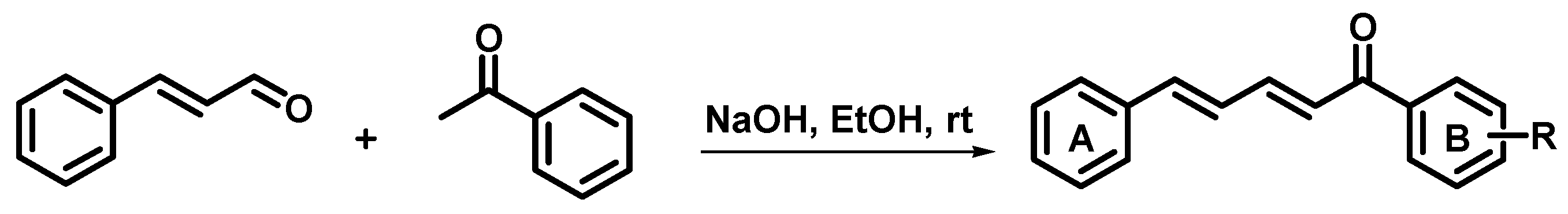
3.1. Synthesis of Cinnamylideneacetophenones

All starting reagents were purchased from Sigma-Aldrich. Compounds were synthesized by Claisen-Schmidt condensation, according to the method reported by Weldon and co-authors, with minor modifications [20,21,22]. Cinnamaldehyde (6 mmol) was added to solution of respective acetophenone (5 mmol) in ethanol (5 mL). The solution was stirred at room temperature for 30 min, followed by dropwise addition of 5 mL ethanolic solution of NaOH (1.0 M). The progress of reaction was checked by thin-layer chromatography (TLC) analyses. Reaction medium was poured into crushed ice. Precipitated crude products were filtered, washed with cold water, and dried at room temperature. Soluble crude products were partitioned with ethyl acetate (3 × 25 mL). Organic phase was concentrated under reduced pressure. Compounds 2, 7, 8 and 107 were recrystallized in ethanol. Compounds 36, 9 and 18 were recrystallized in mixtures of hexane and chloroform.

3.2. Antibacterial Activity of Cinnamylideneacetophenones

Microorganisms used in this study were Staphylococcus aureus (ATCC 14458), Streptococcus mutans (ATCC 25175), Streptococcus sanguinis (ATCC 10557), Pseudomonas aeruginosa (ATCC 15442), and Escherichia coli (ATCC 10536). MIC and MBC values were determined by broth microdilution method, in 96-well microtiter plates, following the criteria previously described by the National Committee for Clinical Laboratory Standards [24]. Bacterial cell cultures in Brain Heart Infusion broth (BHI) at the mid-log phase were harvested by centrifugation for 10 min at 3000× g. Supernatant was discarded and the pellet resuspended in Mueller-Hinton broth (Difco Laboratories, Detroit, MI, USA). The final bacterial suspension was 5–10 × 105 CFU (colony forming units) per mL. Compounds were diluted in sterile deionized water in concentrations ranging from 500 to 3.9 µM. Bacterial suspension was inoculated in each well. The plates were incubated at 37 °C for 24 h in 5% CO2. Thus, 15 µL of 0.01% resazurin staining was added to each well and incubated for 4 h to determine cell viability. Wells corresponding to MIC and at least three previous wells were homogenized, serially diluted, and plated on Mueller-Hinton agar to determine MBC. The plates were incubated at 37 °C for 24 h in 5% CO2. Number of colonies forming units per mL (CFU/mL) was determined. The MBC was considered when the compounds killed more than 99% of the tested bacterial culture. Penicillin G, vancomycin, chlorhexidine, and culture medium were used as positive and negative controls, respectively. Three independent assays were performed.

3.3. Antitubercular Activity of Cinnamylideneacetophenones

The antitubercular activity of compounds was determined by the REMA (Resazurin Microtiter Assay) method [25,26]. Stock solutions of compounds were prepared in DMSO (dimethyl sulfoxide) and diluted in Middlebrook 7H9 broth (Difco) supplemented with oleic acid, albumin, dextrose, and catalase (OADC), to obtain the final concentration range from 100 to 1 µM. Suspension of Mycobacterium tuberculosis H37Rv strain (ATCC 27294) was cultured in Middlebrook 7H9 broth supplemented with OADC. When the culture obtained a turbidity of McFarland standard No. 1, it was adjusted by 3–5 × 10⁵ CFU per mL. Thus, 100 µL of inoculum were added to each well in a 96-well microplate with 100 µL of the compound solution. Plates were incubated for 7 days at 37 °C and 5% CO2. Resazurin was added (30 µL of 0.01%). The fluorescence of the wells was measured after 24 h on Cytation 3 (BioTek, Winooski, VT, USA) equipment. MIC value was defined as the lowest concentration resulting in 90% inhibition of growth of M. tuberculosis. Isoniazid and culture medium were used as positive and negative controls, respectively. Three independent assays were performed.

3.4. Partition Coefficient (log Po/w) of Cinnamylideneacetophenones

Values of log Po/w were determined using the HPLC method according to OECD-Guidelines for the Testing of Chemicals [30], on Shimadzu (Kyoto, Japan) HPLC-PDA (model CBM 20-A) equipped with a UV-Vis detector (model SPD-20A), binary pumping system (model LC-20AD), solvent degasser (model DGU-20A3r), and a Phenomenex Gemini C-18 column (Torrance, CA, USA) (250 mm × 4.6 mm; 5 µm; 110 Å). Analysis was performed on isocratic mode in MeOH:H2O (3:1) and 1.0 mL/min. Injected volume of sample was 20.0 µL and the wavelength in the detector was adjusted according to λmax of each compound. Reference compounds phenol, acetophenone, benzoic acid, toluene, and thymol were used to construct the curve log K × log Po/w. Capacity factor of the cinnamylideneacetophenones was determined from their retention times and interpolated in linearity curve log K × log Po/w. Experiments were performed in duplicate.

3.5. Toxic Effect on Lung Cells of Selected Cinnamylideneacetophenones

Cytotoxicity of compounds was evaluated according to Silva and colleagues [32]. The cell lineages used were MRC-5 (human lung fibroblast cell line—ATCC, CCL-171) and A549 (human lung adenocarcinoma epithelial cell line—ATCC, CCL-185). Cells were routinely maintained in complete medium supplemented with 10% fetal bovine serum (FBS) plus amphotericin B (2 mg/L) and gentamicin (50 mg/L) at 37 °C in a humidified 5% CO2 atmosphere. For cytotoxicity assay, 2.5 × 10⁴ cells/mL were seeded into a 96-well cell culture plate with a total volume of 100 µL for 24 h. Cells were treated with compounds at concentrations ranging from 100 to 1 µM. After 24 h incubation, medium was removed, and 50 µL of resazurin (0.01% w/v) was added to each well, and the plates were incubated at 37 °C for 3 h. Fluorescence of wells was measured on a Synergy H1 microplate reader (BioTek, Winooski, VT, USA) in which were used excitation (530 nm) and emission (590 nm) filters. The IC50 values were calculated from a calibration curve by regression analysis. Doxorubicin and untreated cells constituted the positive and negative controls, respectively. Three independents assays were performed.

3.6. DPPH Scavenging Ability of Selected Cinnamylideneacetophenones

Radical scavenging activity of compounds 3 and 4 was determined using 2,2-diphenyl-1-picrylhydrazyl (DPPH) [36,37]. Aliquots of 10 µL of 3, 4, and positive controls (quercetin and gallic acid) at concentrations 1600, 800, 400, and 200 μM (in EtOH) were individually added in 190 µL DPPH (100 μM). The mixtures were incubated in the dark at 25 °C for 30 min. Remaining DPPH was determined spectroscopically at 490 nm by blanking against absolute ethanol. Radical scavenging ability was expressed by using mean values obtained from triplicates as the percentage of radicals reduced by calculating the following equation: DPPH scavenging % = [1 − (Asample/A) × 100], where A is test absorbance without sample (only EtOH and DPPH) and Asample is test absorbance with compounds.

4. Conclusions

In summary, a series of cinnamylideneacetophenones containing several substituents on ring B were designed and synthesized as part of our ongoing search for antibacterial and antitubercular compounds based on natural products. Five compounds (36 and 9) exhibited potent antibacterial activity against Gram-positive species. Seven compounds (24, 7, 9, 10 and 18) were able to inhibit M. tuberculosis growth. Preliminary SAR investigations suggested hydrophilicity is the central parameter for antibacterial and antitubercular activities. Furthermore, our results corroborated the potential of cinnamaldehyde as a privileged starting material and template for synthetic collection of hits; it did not violate Lipinski’s and Veber’s boundaries.

Supplementary Materials

Supplementary materials are available online.

Acknowledgments

The authors gratefully acknowledge financial support from the Coordination for the Improvement of Higher Education Personnel (CAPES), Brazilian Council for Scientific and Technological Development (CNPq) (Grants 471129/2013-5 and 306251/2016-7), and the São Paulo Research Foundation (FAPESP) (Grant 2014/18330-0). LAA thanks FAPESP research fellowship (2016/06401-5).

Author Contributions

L.O.R., C.D., F.R.P., and H.F. conceived and designed the experiments; C.R.P., G.S.T., V.R.S., A.C.N., D.L.C., L.A.A., and I.C.S. performed the experiments; everybody contributed to data analysis.

Conflicts of Interest

The authors declare no conflict of interest.

References

  1. Al-bayati, F.A.; Mohammed, M.J. Isolation, identification, and purification of cinnamaldehyde from Cinnamomum zeylanicum bark oil. An antibacterial study. Pharm Biol. 2011, 47, 61–66. [Google Scholar] [CrossRef]
  2. Fabra, M.J.; Mayorga, J.L.C.; Randazzo, W.; Lagarón, J.M.; Rubio, A.L.; Aznar, R.; Sánches, G. Efficacy of cinnamaldehyde against enteric viruses and its activity after incorporation into biodegradable multilayer systems of interest in food packaging. Food Environ. Virol. 2016, 8, 125–132. [Google Scholar] [CrossRef] [PubMed]
  3. Adabiardakani, A.; Mohammad, H.H.K.; Kargar, H. Cinnamaldehyde schiff base derivatives: A short review. World Appl. Program. 2012, 2, 472–476. [Google Scholar]
  4. Visvalingam, J.; Palaniappan, K.; Holley, R.A. In vitro enhancement of antibiotic susceptibility of drug resistant Escherichia coli by cinnamaldehyde. Food Control. 2017, 79, 288–291. [Google Scholar] [CrossRef]
  5. Ali, S.M.; Khan, A.A.; Ahmed, I.; Musaddiq, M.; Ahmed, K.S.; Polasa, H.; Rao, L.V.; Habibullah, C.M.; Sechi, L.A.; Ahmed, N. Antimicrobial activities of eugenol and cinnamaldehyde against the human gastric pathogen Helicobacter pylori. Ann. Clin. Microbiol. Antimicrob. 2005, 4, 1–7. [Google Scholar] [CrossRef] [PubMed]
  6. Utchariyakiat, I.; Surassmo, S.; Jaturanpinyo, M.; Khuntayaporn, P.; Chomnawang, M.T. Efficacy of cinnamon bark oil and cinnamaldehyde on anti-multidrug resistant Pseudomonas aeruginosa and the synergistic effects in combination with other antimicrobial agents. BMC Complement. Altern. Med. 2016, 16, 1–7. [Google Scholar] [CrossRef] [PubMed]
  7. Manu, D.; Mendonca, A.F.; Daraba, A.; Dickson, J.S.; Sebranek, J.; Shaw, A.; Wang, F.; White, S. Antimicrobial efficacy of cinnamaldehyde against Escherichia coli O157:H7 and Salmonella enterica in carrot juice and mixed berry juice held at 4 °C and 12 °C. Foodborne Pathog. Dis. 2017, 14, 302–307. [Google Scholar] [CrossRef] [PubMed]
  8. Shi, C.; Zhang, X.; Zhao, X.; Meng, R.; Liu, Z.; Chen, X.; Guo, N. Synergistic interactions of nisin in combination with cinnamaldehyde against Staphylococcus aureus in pasteurized milk. Food Control 2017, 71, 10–16. [Google Scholar] [CrossRef]
  9. Wong, S.Y.Y.; Grant, I.R.; Friedman, M.; Elliott, C.T.; Situ, C. Antibacterial activities of naturally occurring compounds against Mycobacterium avium subsp. paratuberculosis. Appl. Environ. Microbiol. 2008, 74, 5986–5990. [Google Scholar] [CrossRef] [PubMed]
  10. Ochoa, S.A.; Moorillón, G.V.N.; Torres, L.E.S.; García, M.V.; Ramírez, B.E.S.; Valdez, L.M.R.; Chavira, B.E.R. Quantitative structure-activity relationship of molecules constituent of different essential oils with antimycobacterial activity against Mycobacterium tuberculosis and Mycobacterium bovis. BMC Complement. Altern. Med. 2015, 15, 1–11. [Google Scholar] [CrossRef]
  11. Si, W.; Gong, J.; Tsao, R.; Zhou, T.; Yu, H.; Poppe, C.; Johnson, R.; Du, Z. Antimicrobial activity of essential oils and structurally related synthetic food additives towards selected pathogenic and beneficial gut bacteria. J. Appl. Microbiol. 2006, 100, 296–305. [Google Scholar] [CrossRef] [PubMed]
  12. Tiihonen, K.; Kettunem, H.; Bento, M.H.L.; Saarinen, M.; Lahtinen, S.; Ouwehand, A.C.; Schulze, H.; Rautonen, N. The effect of feeding essential oils on broiler performance and gut microbiota. Br. Poult. Sci. 2010, 51, 381–392. [Google Scholar] [CrossRef] [PubMed]
  13. Shen, S.; Zhang, T.; Yuan, Y.; Lin, S.; Xu, J.; Ye, H. Effects of cinnamaldehyde on Escherichia coli and Staphylococcus aureus membrane. Food Control 2015, 47, 196–202. [Google Scholar] [CrossRef]
  14. Domadia, P.; Swarup, S.; Bhunia, A.; Sivaraman, J.; Dasgupta, D. Inhibition of bacterial cell division protein FtsZ by cinnamaldehyde. Biochem. Pharmacol. 2007, 74, 831–840. [Google Scholar] [CrossRef] [PubMed]
  15. Hu, L.B.; Zhou, W.; Yang, J.D.; Chen, J.; Yin, Y.F.; Shi, Z.Q. Cinnamaldehyde induces PCD-like death of Microcystis. aeruginosa via reactive oxygen species. Water Air Soil Pollut. 2011, 217, 105–113. [Google Scholar] [CrossRef]
  16. Adams, T.B.; Cohen, S.M.; Douli, J.; Feron, V.J.; Goodman, J.I.; Marnett, L.J.; Munro, I.C.; Portoghese, P.S.; Smith, R.L.; Waddell, W.J.; et al. The FEMA GRAS assessment of cinnamyl derivatives used as flavor ingredients. Food Chem. Toxicol. 2004, 42, 157–185. [Google Scholar] [CrossRef] [PubMed]
  17. Bickers, D.; Calow, P.; Greim, H.; Hanifin, J.M.; Rogers, A.E.; Saurat, J.H.; Sipes, I.G.; Smith, R.L.; Tagami, H. A toxicologic and dermatologic assessment of cinnamyl alcohol, cinnamaldehyde and cinnamic acid when used as fragrance ingredients. Food Chem. Toxicol. 2005, 43, 799–836. [Google Scholar] [CrossRef] [PubMed]
  18. Venkitanarayanan, K.; Kollanoor-Johny, A.; Darre, M.J.; Donoghue, A.M.; Donoghue, D.J. Use of plant-derived antimicrobials for improving the safety of poultry products. Poult. Sci. 2013, 92, 493–501. [Google Scholar] [CrossRef] [PubMed]
  19. LoPachin, R.M.; Gavin, T. Molecular mechanisms of aldehyde toxicity: a chemical perspective. Chem. Res. Toxicol. 2014, 27, 1081–1091. [Google Scholar] [CrossRef] [PubMed]
  20. Correa, R.; Fenner, B.P.; Buzzi, F.C.; Filho, F.V.C.; Nunes, R.J. Antinociceptive activity and preliminary structure-activity relationship of chalcone-like compounds. Z. Naturforsch. C J. Biosci. 2008, 63, 830–836. [Google Scholar] [CrossRef]
  21. Weldon, D.J.; Saulsbury, M.D.; Goh, J.; Rowland, L.; Campbell, P.; Robinson, L.; Miller, C.; Christian, J.; Amis, L.; Taylor, N.; et al. One-pot synthesis of cinnamylideneacetophenones and their in vitro cytotoxicity in breast cancer cells. Bioorg. Med. Chem. Lett. 2014, 24, 3381–3384. [Google Scholar] [CrossRef] [PubMed]
  22. Bhat, I.; Kumar, A. Synthesis and anti-inflammatory activity of some novel 1,5 benzodiazepine derivatives. Asian J. Pharm. Clin. Res. 2016, 9, 63–66. [Google Scholar]
  23. Jin, H.; Xiang, L.; Wen, F.; Tao, K.; Liu, Q.; Hou, T. Improved synthesis of chalconoid-like compounds under ultrasound irradiation. Ultrason. Sonochem. 2008, 15, 681–683. [Google Scholar] [CrossRef] [PubMed]
  24. Wayne, P.A.; NCCLS: National Committee for Clinical Laboratory Standards. Methods for Dilution Antimicrobial Susceptibility Tests for Bacteria that Grow Aerobically; Approved Standard, 5th ed.; CLSI Document M7-A5; National Committee for Clinical Laboratory Standards: Wayne, PA, USA, 2002. [Google Scholar]
  25. Palomino, J.C.; Martin, A.; Camacho, M.; Guerra, H.; Swings, J.; Portaels, F. Resazurin microtiter assay plate: Simple and inexpensive method for detection of drug resistance in Mycobacterium tuberculosis. Antimicrob. Agents Chemother. 2002, 46, 2720–2722. [Google Scholar] [CrossRef] [PubMed]
  26. Fernandes, G.F.S.; Souza, P.C.; Marino, L.B.; Chegaev, K.; Guglielmo, S.; Lazzarato, L.; Fruttero, R.; Chung, M.C.; Pavan, F.R.; Santos, J.L. Synthesis and biological activity of furoxan derivatives against Mycobacterium tuberculosis. Eur. J. Med. Chem. 2016, 123, 523–531. [Google Scholar] [CrossRef] [PubMed]
  27. Ferro, T.A.; Araujo, J.M.; Pinto, B.L.S.; Santos, J.S.; Souza, E.B.; Silva, B.L.R.; Colares, V.L.P.; Novais, T.M.G.; Filho, C.M.B.; Calixto, J.B.; et al. Cinnamaldehyde inhibits Staphylococcus aureus virulence factors and protects against infection in a Galleria mellonella model. Front. Microbiol. 2016, 7, 1–10. [Google Scholar] [CrossRef] [PubMed]
  28. Denyer, S.P.; Maillard, J.Y. Cellular impermeability and uptake of biocides and antibiotics in Gram-negative bacteria. J. Appl. Microbiol. 2002, 92, 35S–45S. [Google Scholar] [CrossRef] [PubMed]
  29. Zgurskaya, H.I.; López, C.A.; Gnanakaran, S. Permeability barrier of Gram-negative cell envelopes and approaches to bypass it. ACS Infect. Dis. 2015, 1, 512–522. [Google Scholar] [CrossRef] [PubMed]
  30. OECD. Test No. 117: Partition coefficient (n-octanol/water), HPLC method. In OECD Guidelines for the Testing Chemicals; OECD Publishing: Paris, France, 2004; pp. 1–11. [Google Scholar]
  31. Tong, S.Y.C.; Davis, J.S.; Eichenberger, E.; Holland, T.L.; Fowler, V.G.J. Staphylococcus aureus infections: Epidemiology, pathophysiology, clinical manifestations, and management. Clin. Microbiol. Rev. 2015, 28, 603–661. [Google Scholar] [CrossRef] [PubMed]
  32. Silva, I.C.; Polaquini, C.R.; Regasini, L.O.; Ferreira, H.; Pavan, F.R. Evaluation of cytotoxic, apoptotic, mutagenic, and chemopreventive activities of semi-synthetic esters of gallic acid. Food Chem. Toxicol. 2017, 105, 300–307. [Google Scholar] [CrossRef] [PubMed]
  33. Vieira, P.B.; Silva, N.L.F.; Silva, G.N.S.; Silva, D.B.; Lopes, N.P.; Gnoatto, S.C.; Silva, M.V.; Macedo, A.J.; Batista, J.; Tasca, T. Caatinga plants: Natural and semi-synthetic compounds potentially active against Trichomonas vaginalis. Bioorg. Med. Chem. Lett. 2016, 26, 2229–2236. [Google Scholar] [CrossRef] [PubMed]
  34. Mohammed, R.S.; Zeid, A.A.; Hawary, S.S.; Sleem, A.A.; Ashour, W.E. Flavonoid constituents, cytotoxic and antioxidant activities of Gleditsia. triacanthos L. leaves. Saudi J. Biol. Sci. 2014, 21, 547–553. [Google Scholar] [CrossRef] [PubMed]
  35. Li, C.; Sun, P.; Yu, H.; Na, Z.; Wang, J. Scavenging ability of dendritic PAMAM bridged hindered phenolic antioxidants towards DPPH˙ and ROO˙ free radicals. RSC Adv. 2017, 7, 1869–1876. [Google Scholar] [CrossRef]
  36. Regasini, L.O.; Vellosa, J.C.R.; Silva, D.H.S.; Furlan, M.; Oliveira, O.M.M.; Khalil, N.M.; Brunetti, I.L.; Young, M.C.M.; Barreiro, E.J.; Bolzani, V.S. Flavonols from Pterogyne. nitens and their evaluation as myeloperoxidase inhibitors. Phytochemistry 2008, 69, 1739–1744. [Google Scholar] [CrossRef] [PubMed]
  37. Nazaré, A.C.; Faria, C.M.; Chiari, B.G.; Petrônio, M.S.; Regasini, L.O.; Silva, D.H.S.; Corrêa, M.A.; Isaac, V.L.; Fonseca, L.M.; Ximenes, V.F. Ethyl ferulate, a component with anti-inflammatory properties for emulsion-based creams. Molecules 2014, 19, 8124–8139. [Google Scholar] [CrossRef] [PubMed]
  38. Ghribia, L.; Ghouilaa, H.; Omrib, A.; Besbesb, M.; Janneta, H.B. Antioxidant and anti-acetylcholinesterase activities of extracts and secondary metabolites from Acacia cyanophylla. Asian Pac. J. Trop Biomed. 2014, 4, S417–S423. [Google Scholar] [CrossRef] [PubMed]
  39. Kumar, S.; Sandhir, R.; Ojha, S. Evaluation of antioxidant activity and total phenol in different varieties of Lantana camara leaves. BMC Res. Notes 2014, 7, 1–9. [Google Scholar] [CrossRef] [PubMed]
  40. Polkam, N.; Ramaswamy, V.R.; Rayam, P.; Allaka, T.R.; Anantaraju, H.S.; Dharmarajan, S.; Perumal, Y.; Gandamalla, D.; Yellu, N.R.; Balasubramanian, S.; et al. Synthesis, molecular properties prediction and anticancer, antioxidant evaluation of new edaravone derivatives. Bioorg. Med. Chem. Lett. 2016, 26, 2562–2568. [Google Scholar] [CrossRef] [PubMed]
  41. Molinspiration Cheminformatics. Available online: www.molinspiration.com (accessed on 20 July 2017).
  42. Ertl, P.; Rohde, B.; Selzer, P. Fast calculation of molecular polar surface area as a sum of fragment-based contributions and its application to the prediction of drug transport properties. J. Med. Chem. 2000, 43, 3714–3717. [Google Scholar] [CrossRef] [PubMed]
  43. Rock, R.B.; Olin, M.; Baker, C.A.; Molitor, T.W.; Peterson, P.K. Central nervous system tuberculosis: Pathogenesis and clinical aspects. Clin. Microbiol. Rev. 2008, 21, 243–261. [Google Scholar] [CrossRef] [PubMed]
  44. Glickman, M.S.; Cox, J.S.; Jacobs, W.R. A novel mycolic acid cyclopropane synthetase is required for cording, persistence, and virulence of Mycobacterium tuberculosis. Mol. Cell 2000, 5, 717–727. [Google Scholar] [CrossRef]
  45. Lipinski, C.A. Lead-and drug-like compounds: The rule-of-five revolution. Drug Discov. Today Technol. 2004, 1, 337–341. [Google Scholar] [CrossRef] [PubMed]
  46. Veber, D.F.; Johnson, S.R.; Cheng, H.Y.; Smith, B.R.; Ward, K.W.; Kopple, K.D. Molecular properties that influence the oral bioavailability of drug candidates. J. Med. Chem. 2002, 45, 2615–2623. [Google Scholar] [CrossRef] [PubMed]
Sample Availability: Samples of the compounds are not available.
Scheme 1. Design of cinnamylideneacetophenones.
Molecules 22 01685 sch001
Scheme 2. Synthesis of cinnamylideneacetophenones.
Molecules 22 01685 sch002
Figure 1. Radical scavenging ability of selected cinnamylideneacetophenones. DPPH = 2,2-diphenyl-1-picrylhydrazyl.
Molecules 22 01685 g001
Table 1. Yields of cinnamylideneacetophenones.
EntryNameRYield (%)
2(2E,4E)-1,5-diphenylpenta-2,4-dien-1-oneH33
3(2E,4E)-1-(3-hydroxyphenyl)-5-phenylpenta-2,4-dien-1-onem-OH91
4(2E,4E)-1-(4-hydroxyphenyl)-5-phenylpenta-2,4-dien-1-onep-OH82
5(2E,4E)-1-(3-aminophenyl)-5-phenylpenta-2,4-dien-1-onem-NH235
6(2E,4E)-1-(3-methoxyphenyl)-5-phenylpenta-2,4-dien-1-onem-OMe78
7(2E,4E)-1-(4-methoxyphenyl)-5-phenylpenta-2,4-dien-1-onep-OMe35
8(2E,4E)-1-(3,4-methylenedioxy)-5-phenylpenta-2,4-dien-1-one-OCH2O-83
9(2E,4E)-1-(4-hydroxy-3-methoxyphenyl)-5-phenylpenta-2,4-dien-1-onem-OMe,p-OH98
10(2E,4E)-1-(4-fluorophenyl)-5-phenylpenta-2,4-dien-1-onep-F71
11(2E,4E)-1-(4-chlorophenyl)-5-phenylpenta-2,4-dien-1-onep-Cl72
12(2E,4E)-1-(4-bromophenyl)-5-phenylpenta-2,4-dien-1-onep-Br86
13(2E,4E)-1-(3,4-dichlorophenyl)-5-phenylpenta-2,4-dien-1-onem,p-diCl79
14(2E,4E)-1-(3-(trifluoromethyl)phenyl)-5-phenylpenta-2,4-dien-1-onem-CF388
15(2E,4E)-1-(3-nitrophenyl)-5-phenylpenta-2,4-dien-1-onem-NO285
16(2E,4E)-1-(4-nitrophenyl)-5-phenylpenta-2,4-dien-1-onep-NO284
17(2E,4E)-1-(3-methylphenyl)-5-phenylpenta-2,4-dien-1-onem-Me49
18(2E,4E)-1-(4-methylphenyl)-5-phenylpenta-2,4-dien-1-onep-Me53
Table 2. Antibacterial and antitubercular activities and log Po/w of cinnamylideneacetophenones.
Molecules 22 01685 i001
EntryRSa *Sm *Ss *Pa *Ec *Mt *log Po/w
MICMBCMICMBCMICMBCMICMICMIC
1->500->500->500->500>500>200-
2H>500->500->500->500>50057.23.2
3m-OH77.915677.915677.9156>500>50088.82.7
4p-OH77.977.9156312156312>500>50081.92.6
5m-NH2>500-313313>500->500>500>2002.5
6m-OMe296296>500->500->500>500>2003.3
7p-OMe>500->500->500->500>50066.63.2
8-OCH2O->500->500->500->500>500>2003.2
9m-OMe,p-OH279279>500->500->500>50083.52.7
10p-F>500->500->500->500>50067.43.3
11p-Cl>500->500->500->500>500>2003.7
12p-Br>500->500->500->500>500>2003.8
13m,p-diCl>500->500->500->500>500>2004.1
14m-CF3>500->500->500->500>500>2003.7
15m-NO2>500->500->500->500>500>2003.3
16p-NO2>500->500->500->500>500>2003.2
17m-Me>500->500->500->500>500>2003.6
18p-Me>500->500->500->500>50070.93.5
PEN-----0.40.4-118--
VAN-3.36.8--3.33.3-53.8--
CHX---2.44.74.79.519.4---
ISO---------0.3-
* MIC = minimum inhibitory concentration; MBC = minimum bactericidal concentration. MIC and MBC in µM. Sa = Staphylococcus aureus; Sm = Streptococcus mutans; Ss = Streptococcus sanguinis; Pa = Pseudomonas aeruginosa; Ec = Escherichia coli; Mt = Mycobacterium tuberculosis; PEN = penicillin; VAN = vancomycin; CHX = chlorhexidine; ISO = isoniazid; - not determined.
Table 3. Toxicity against human lung cells and selectivity index of selected cinnamylideneacetophenones.
EntryMRC-5SIA549SI
IC50 *SaMtIC50 *SaMt
346.30.60.596.71.21.2
453.10.70.677.51.00.9
doxorubicin0.7--1.8--
* IC50 in µM.
Table 4. Lipinski’s and Veber’s parameters of selected cinnamylideneacetophenones.
Entry%ABSTPSA (Å2)milog PMWHBDHBALipinski’s ViolationsNROBTVeber’s Violations
Ro5--≤5.00<500≤5≤101--
Vb-≤140-----≤100
110017.072.48132.1610020
210017.074.33234.3010040
396.137.303.83250.3021040
496.137.303.85250.3021040
594.143.093.38249.3122040
699.926.304.36264.3220050
799.926.304.39263.3220050
992.946.533.67280.3231050
1010017.074.49252.2910040
1810017.074.78248.3210040
%ABS = percent absorption; TPSA = topological polar surface area; milog P = lipophilicity; MW = molecular weight; HBD = number of hydrogen bond donors; HBA = number of hydrogen bond acceptors; NROBT = number of rotatable bonds; Ro5 = Boundaries of Lipinski’s rules; Vb = Boundaries of Veber’s rules.

