A Highly Selective and Sensitive Fluorescent Turn-Off Probe for Cu2+ Based on a Guanidine Derivative


Department of Applied Chemistry, College of Science, Northeast Agricultural University, Harbin 150030, China
*
Author to whom correspondence should be addressed.
Molecules 2017, 22(10), 1741; https://doi.org/10.3390/molecules22101741
Submission received: 16 September 2017
/
Accepted: 13 October 2017
/
Published: 16 October 2017
Abstract
:A new highly selective and sensitive fluorescent probe for Cu2+, N-n-butyl-4-(1′-cyclooctene-1′,3′,6′-triazole)-1,8-naphthalimide (L), was synthesized and evaluated. The structure of compound L was characterized via IR, 1H-NMR, 13C-NMR and HRMS. The fluorescent probe was quenched by Cu2+ with a 1:1 binding ratio and behaved as a “turn-off” sensor. An efficient and sensitive spectrofluorometric method was developed for detecting and estimating trace levels of Cu2+ in EtOH/H2O. The ligand exhibited excitation and emission maxima at 447 and 518 nm, respectively. The equilibrium binding constant of the ligand with Cu2+ was 1.57 × 10⁴ M−1, as calculated using the Stern-Volmer equation. Ligand L is stable and can be used to detect Cu2+ in the range of pH from 7 to 12. The sensor responded to Cu2+ rapidly and a large number of coexisting ions showed almost no obvious interference with the detection.
1. Introduction
Fluorescent sensors for chemical species used in biological and environmental detection are currently an attractive field of research [1,2,3]. In particular, pollution by heavy metal ions has attracted much attention because these metal ions not only play important roles in various physiological processes but also have distinctly toxic impact on human health [4]. It is highly desired to detect these in relevant systems. The importance of metal ion sensing can never be overestimated because of their widespread distribution in environmental systems and biological processes [5]. Copper is a significant metal pollutant because of its widespread application and is also an essential trace element in biological systems. Cu2+ is present in various biological processes, as the third most abundant transition metal ion in the human body. In addition, its homeostasis is critical for the metabolism and development of living organisms [6,7,8]. For example, Cu2+ plays a critical role as a catalytic cofactor for a variety of metalloenzymes, including superoxide dismutase, cytochrome oxidase and tyrosinase [9,10]. The misregulation or excess accumulation of Cu2+ could cause serious diseases [11]. Its concentration in the neuronal cytoplasm may contribute to the etiology of Alzheimer’s or Parkinson’s disease [12,13,14]. However, exposure to high levels of Cu2+ will cause gastrointestinal disturbance and even liver or kidney damage. The detection of Cu2+ is meaningful for both disease diagnosis and environmental monitoring [15]. The normal average concentration of Cu2+ in blood is in the range of 15.7–23.6 μM [16]. The U.S. Environmental Protection Agency (EPA) set a limit for Cu2+ in drinking water of 20 μM [17]. The detection of Cu2+ levels in water requires an efficient, selective and highly sensitive chemosensor [18,19].
Numerous methods to detect metal ions have been developed, such as atomic absorption spectrometry [20], inductively coupled plasma atomic emission spectroscopy [21], visual detection [22,23], chemiluminescence detection [24] and electrochemical techniques [25,26]. However, these methods have disadvantages, such as expensive instrumentation, complicated sample preparation and tedious operating procedures, resulting in the need for other sensitive, simple detection methods for copper ions. Fluorescent molecules offer many advantages over other systems in this area as a result of their emissive behavior, easy signal detection, sensitivity, selectivity and low cost [27,28]. The development of the fluorescence method has been demonstrated to be promising because of its advantages of wide applicability, ease of manipulation, high selectivity, sensitivity, rapidity, nondestructive methodology and direct visual perception [29,30]. Thus, a number of high-performance sensors have been developed to detect metal ions [31]. Cu2+ is a typical ion with a pronounced quenching effect on fluorophores via mechanisms that are inherent to the paramagnetic species [32,33,34,35].
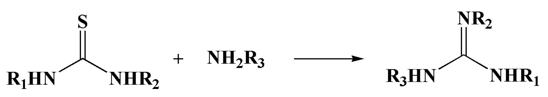
Many excellent chemosensors have been reported for Cu2+ in the past few years. However, many of these sensors only exhibit sensitive and selective Cu2+ detection in pure organic systems (Table 1) [36,37,38]. Hence, we designed and synthesized a new fluorescent sensor, N-n-butyl-4-(1′-cyclooctene-1′,3′,6′-triazole)-1,8-naphthalimide, based on the transformation of thiourea into guanidine derivatives, as shown in Scheme 1. This novel compound has a highly sensitive fluorescent response toward Cu2+ in mixed aqueous solutions.
2. Results and Discussion
2.1. Synthesis and Characterization
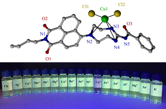
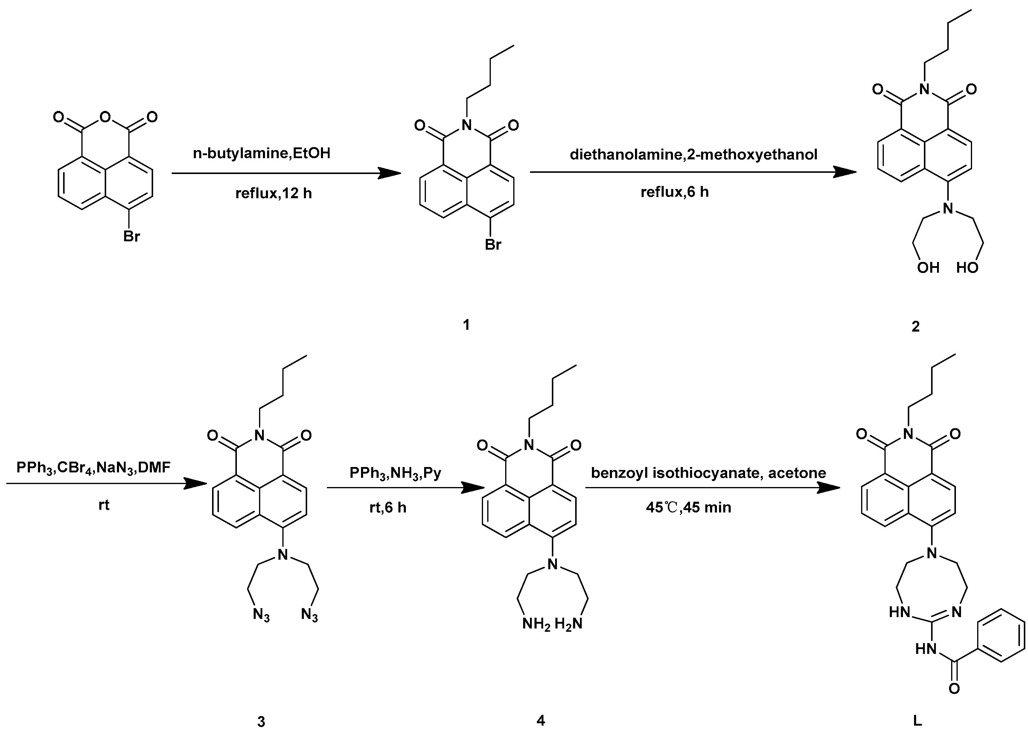
Compound L was synthesized through the route outlined in Scheme 2. The structure modification was focused on the C-4 position. A dihydroxyethylamino moiety was introduced to replace the Br atom, and the subsequent reaction modified the -OH group to N3; compound 4 was obtained in a 67% yield from the amination. Thiourea is easily transformed into guanidine; however, the conversion from thiourea to guanidine has attracted little attention as a potential fluorogenic reaction because most thiourea derivatives have the same color or fluorescence as their guanidine analogues [39]. Initially, we envisioned that a similar molecular recognition/reactivity motif might be incorporated into fluorophores to serve as a ratiometric fluorescent chemodosimeter for the selective detection of Hg2+, as reported in the literature [40], because compound 4 has two free amino groups. The expected thiourea derivative was not achieved; however, the product transformed into the inner guanidine ring because compound 4 contained a primary amine subunit. Compound L was obtained through intramolecular cyclization with a recognition/reactivity motif that could be incorporated into fluorophores so that metal ion addition led to a variation of the fluorescence.
Compound L was obtained with a yield of 57% in the final reaction, and its structure was characterized by 1H-NMR, 13C-NMR and HRMS. As evidenced by the signals at 6.57–8.42 ppm in the 1H-NMR spectrum, the number of Ar-H is 10, which indicated that benzoyl isothiocyanate had reacted with compound 4. The signals at δ = 9.02 ppm indicated the existence of benzamide, and the signals at δ = 1.26 ppm showed that -NH2 was substituted. Furthermore, the molecular ion peak in the HRMS-ESI ([M + H]+, 484.2332 m/z) spectrum was consistent with the theoretically calculated molecular mass of compound L.
2.2. Spectral Characteristics
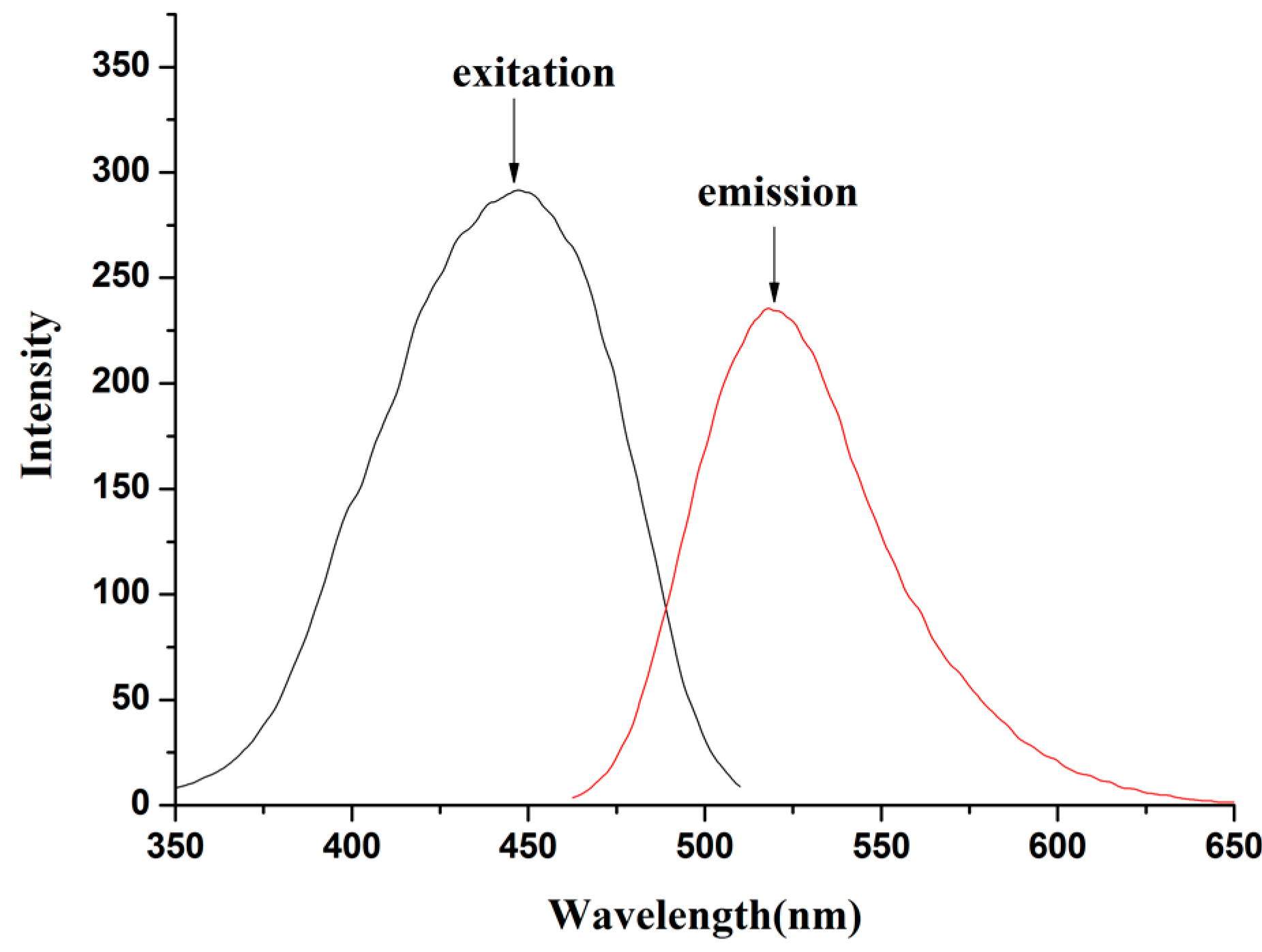
The photophysical characteristics of compound L in EtOH/H2O (4:1 v/v) were investigated. Figure 1 shows the excitation (λ = 447 nm) and emission (λ = 518 nm) spectra of compound L. The Stokes shift (υA–υF), an important parameter indicating the difference in properties and structure between the ground state (S0) and first excited state (S1), was calculated by Equation (1) to be 3062 cm−1.
The ability of the molecules to emit absorbed light energy is characterized quantitatively by the fluorescence quantum yield (ΦF). The quantum yields of fluorescence were calculated using coumarin 6G (Φsample = 0.94) in EtOH as the standard according to Equation (2), where Aref, Sref, and nref and Asample, Ssample, and nsample represent the absorbance at the exited wavelength, the integrated emission band area and the solvent refractive index of the standard and the sample, respectively [41,42]. Thus, the fluorescence quantum yield of probe L in EtOH/H2O (4:1 v/v) solution was 0.06.
The photophysical properties of compound L as a ligand in the presence of different metal cations were investigated in EtOH/H2O (4:1 v/v) to determine its potential application as a sensor for detecting these metal ions. The binding behavior of compound L toward different metal ions such as their corresponding chlorides, acetates, sulfides or nitrates was studied using fluorescence spectroscopy. The anions did not affect the spectroscopic results.
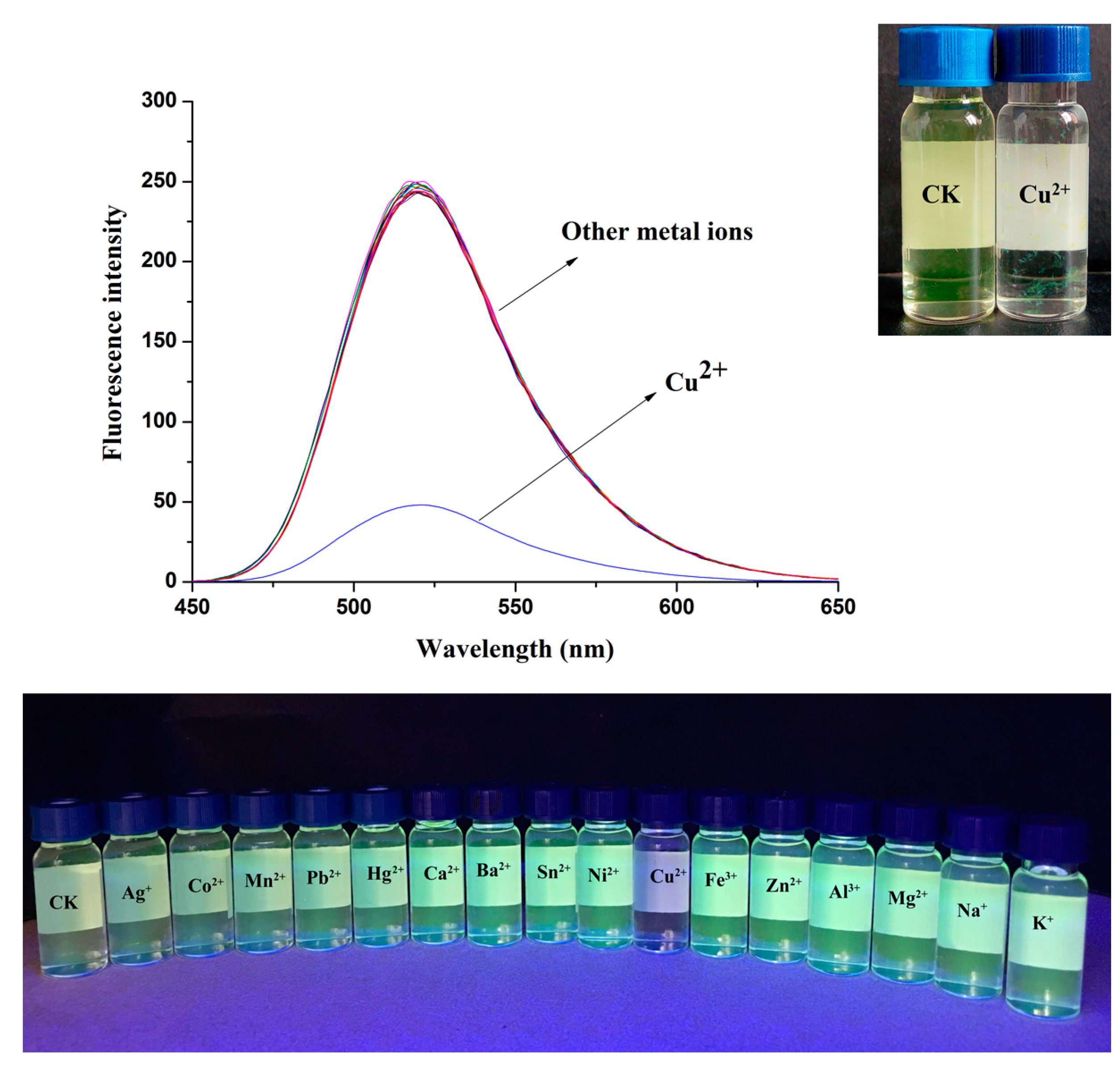
To evaluate the selectively of compound L for Cu2+, changes in the fluorescence intensity upon the addition of various metal ions under the same conditions were investigated. The fluorescence spectrum of compound L exhibited strong fluorescence emission when excited at 447 nm in EtOH/H2O (4:1 v/v; Figure 2). Under the same conditions as those used for adding the metal ions, significant fluorescence changes were observed in the presence of Cu2+. Fluorescence quenching was observed immediately after Cu2+ was added. When other metal ions were added, the fluorescence exhibit few changes. As shown in Figure 2, remarkable color changes ranging from yellow to colorless and yellow floccules floating in the solution could be distinguished by the naked eye. In addition, the introduction of Cu2+ to the mixture solution of compound L led to evident fluorescence quenching, while the rest of the metal cations exerted a negligible or very small influence under identical conditions, which was further evidenced by the illumination taken under a 365 nm portable UV lamp. On the basis of the literature, the fluorescence quench of probe L might have been due to the coordination to paramagnetic Cu2+ [43].
2.3. Calculation of Binding Constants and Different Concentrations of Cu2+
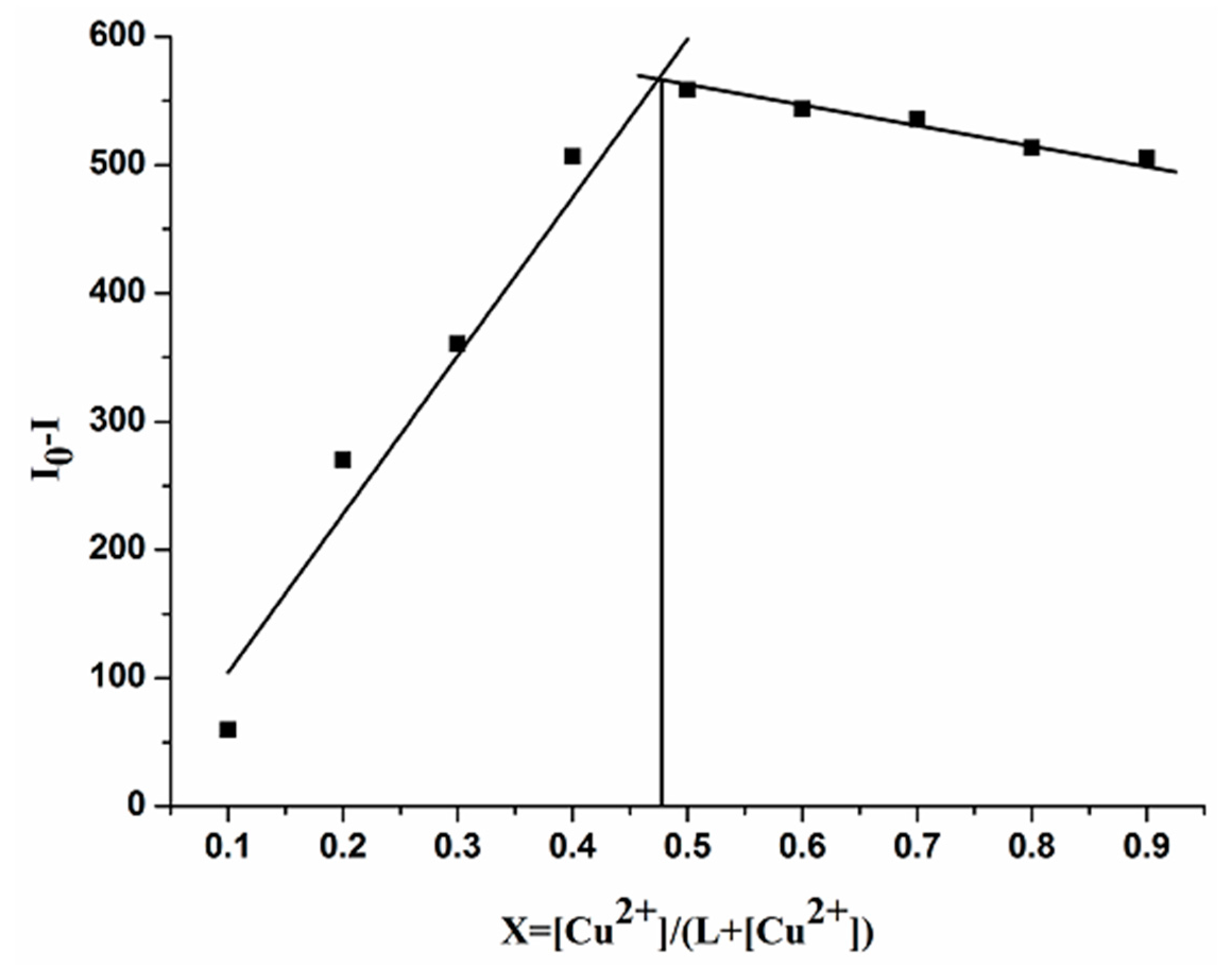
A Job plot was generated by continuously varying the mole fraction of Cu2+ from 0 to 1 in a solution of [L-Cu2+] with a total concentration of 50 μM to determine the stoichiometry of the [L-Cu2+] complex. The Job plot analysis revealed an approximate maximum at a 0.5 mole fraction, which indicated a 1:1 stoichiometry of the ligand L and Cu2+ complex (Figure 3).
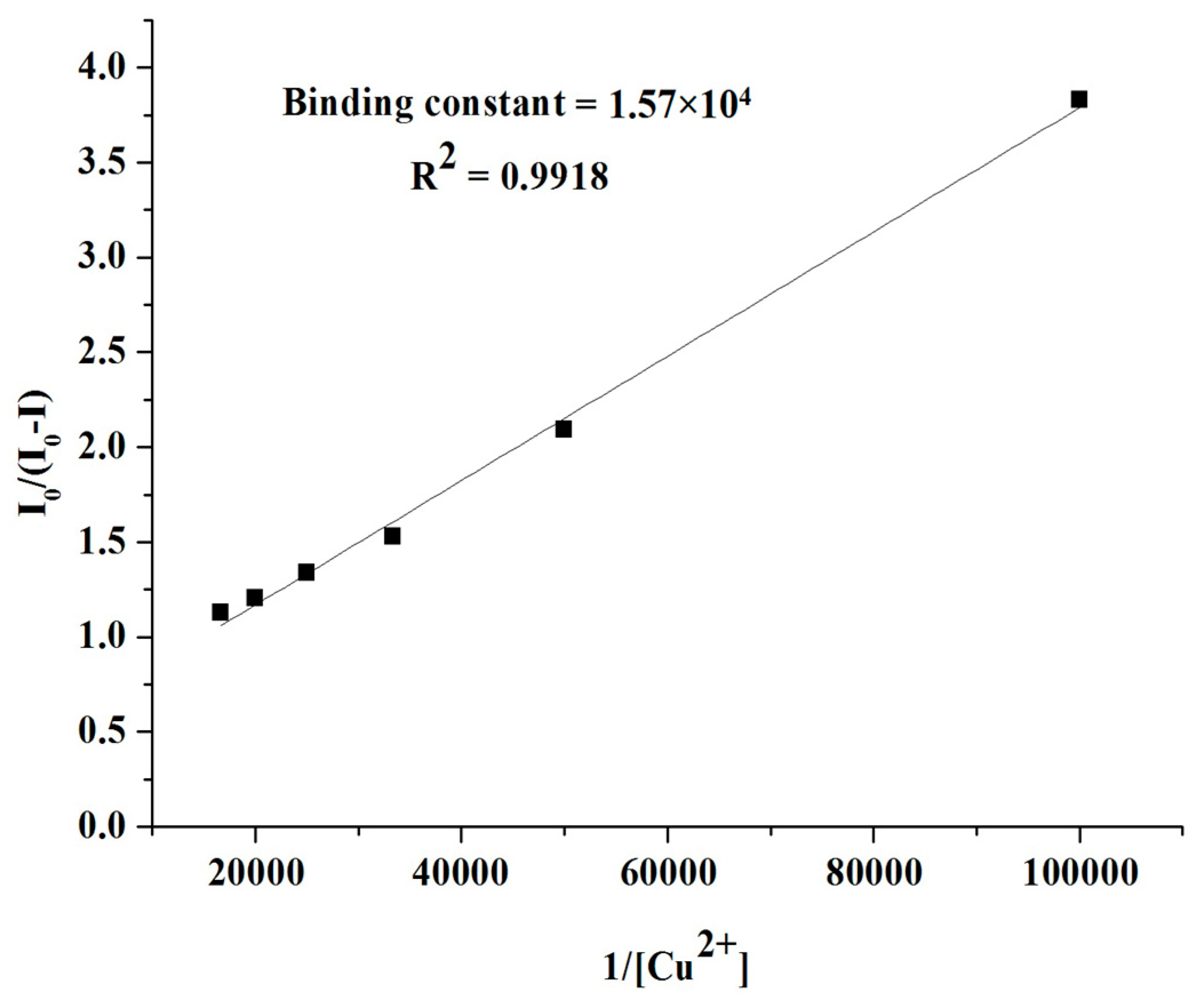
To better understand the sensing process, changes in the fluorescence spectra of compound L were measured along with the binding constant and different concentrations of Cu2+. The binding constant was calculated using the Stern-Volmer equation, I0/(I0 − I) = 1/A + 1/KA·1/[Q], on the basis of a 1:1 stoichiometry, where I0 is the fluorescence intensity of the free ligand L, I is the fluorescence intensity of the L-Cu2+ complex, Q is [Cu2+], A is a constant and K is a binding constant [44]. When the reciprocal of I0/(I0 − I) was plotted as a function of the 1/[Q] concentration, a linear relationship was obtained, (y = A + Bx), y = I0/(I0 − I), A = 1/A, B = 1/KA, x = 1/[Q], and K was calculated from A/B (Figure 4). Therefore, K, the binding association constant for Cu2+, was estimated to be 1.57 × 10⁴ M−1 in the EtOH/H2O (4:1 v/v) solution, as inferred from the fluorescence titration curves of compound L with Cu2+.
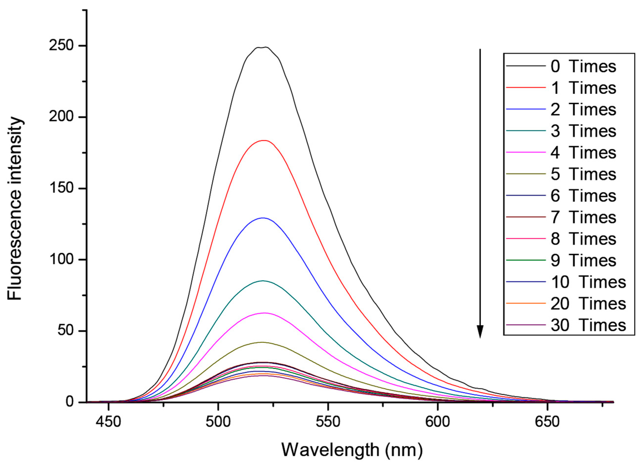
Upon the addition of Cu2+, the fluorescence was quenched. The changes in the fluorescence emission intensities of compound L (1 × 10⁻⁵ mol L−1) with different concentrations of Cu2+ (0–3 × 10⁻⁴ mol L−1) are presented in Figure 5. A progressive decrease in the fluorescence intensity was observed. The plot of the fluorescence intensity versus externally added Cu2+ (Figure 6) reaches saturation after a certain amount of externally added Cu2+. Curvilinearity was obtained for up to a value of 6 times (6 × 10−5 mol L−1) the amount of externally added Cu2+. The results indicated that compound L could detect Cu2+ at a micromole level.
2.4. Response Time
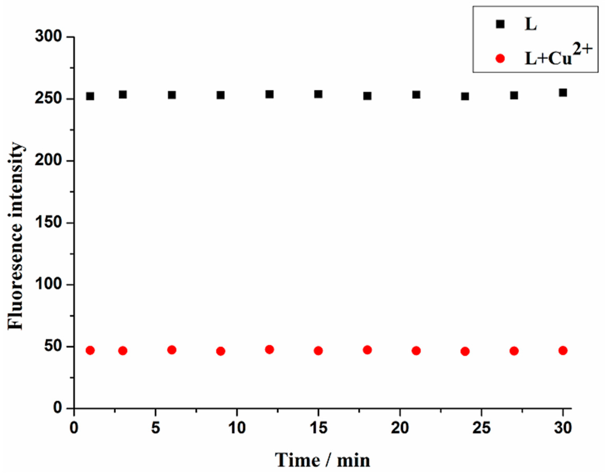
The response time is a significant practical parameter [45]. Time-dependence of probe L for sensing Cu2+ was examined and the results are illustrated in Figure 7. Upon the addition of Cu2+, the fluorescence intensity decreased to the minimum immediately and tended to be stable during the test time. No changes in the fluorescence of probe L in the absence of Cu2+ were detected with the increased time. This showed that probe L exhibited a reliable rapid response to Cu2+.
2.5. Effect of pH
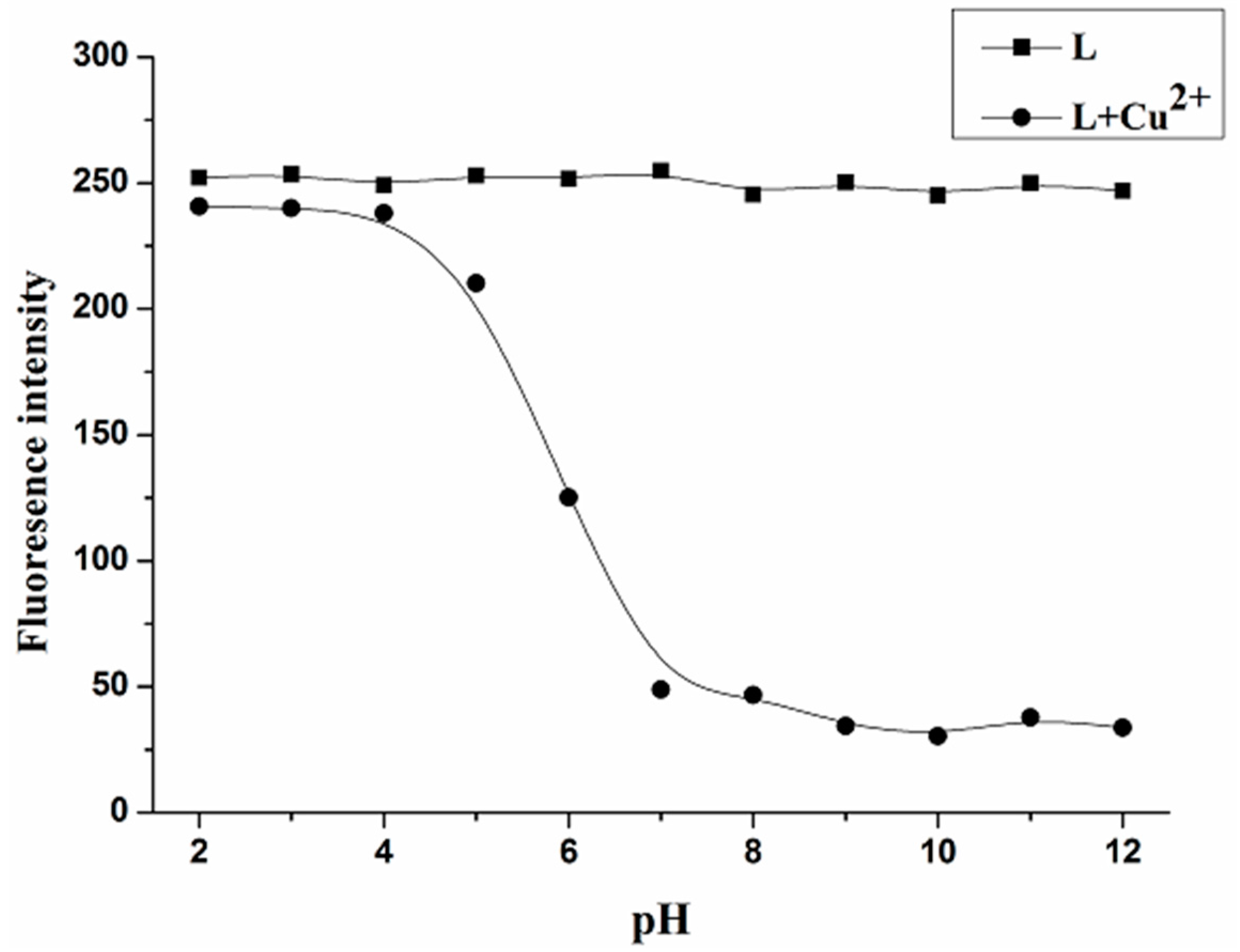
In order to investigate the application of probe L within complex media, the fluorescence spectra response of probe L was measured in the absence or presence of 5 × 10⁻⁵ mol L−1 Cu2+ under different pH conditions. As shown in Figure 8, the fluorescence intensity of probe L did not reveal any clear differences over a pH range of 2–12, which was consistent with the goal of our experimental design. The fluorescence of the [L-Cu2+] solution could not remarkably decrease when the pH values were lower than 5, possibly because the imino groups of probe L were protonated at acid conditions, resulting in weak coordination with Cu2+. In addition, the fluorescence intensity of the [L-Cu2+] solution showed a 5-fold change from pH 4 to pH 7, which suggested that it is sensitive to pH changes. These results indicated that probe L could be used to detect biological as well environmental Cu2+ in the range of pH from 7 to 12.
2.6. Selectivity
The selectivity is clearly one of the most important characteristics of a cation-selective chemosensor. Thus, the influence of other metal ions on the fluorescence intensity of the proposed Cu2+ chemosensor was investigated (Figure 9). In this study, Ag+, Na+, K+, Ba2+, Ca2+, Co2+, Mg2+, Mn2+, Ni2+, Pb2+, Sn2+, Zn2+, Al3+, Cu+, Hg2+ and Fe3+ (100 μM) were added to the compound L (10 μM)/Cu2+ (50 μM) solution, and the fluorescence was observed. The fluorescence emission intensity at 518 nm significantly decreased as a result of binding with Cu2+. The coexisting ions showed little influence on the detection of Cu2+. Common anions, including oxalate, sulfate, nitrate and chloride, did not interfere with the fluorescence intensity of the ligand or [L-Cu2+] complex. Compared to the other common cations, compound L exhibited good selectivity for Cu2+, indicating that L has a good anti-interference ability in detecting Cu2+.
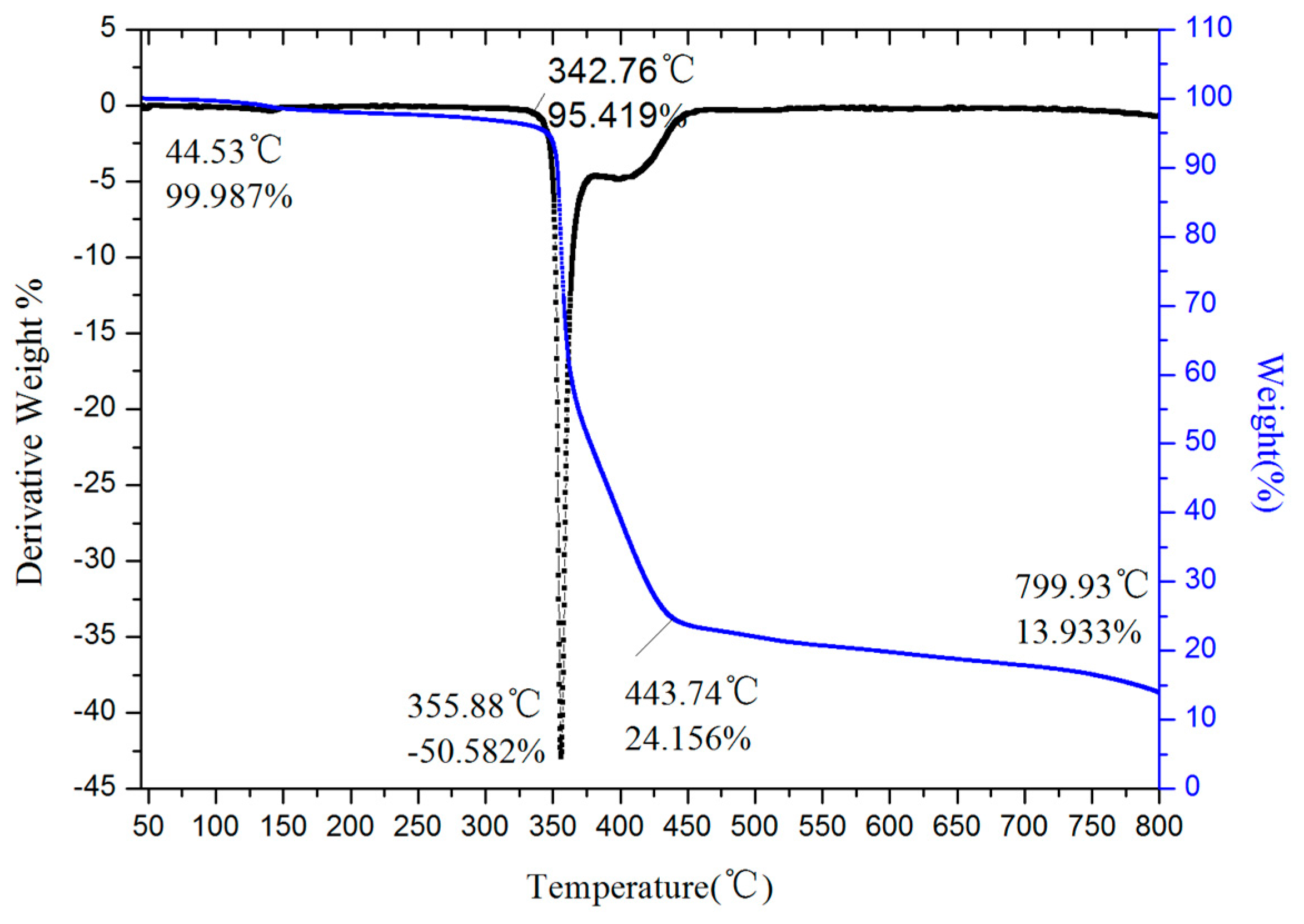
2.7. Thermal Studies
The stability of probe L and its Cu2+ complex was studied by thermogravimetry to prove the binding of ligand L with Cu2+ ions, and the results are presented in Figure 10 and Figure 11, respectively. The thermal stability of the Cu2+ complex (up to 324.76 °C) was better than that of the free ligand (up to 288.57 °C).
2.8. IR Spectra
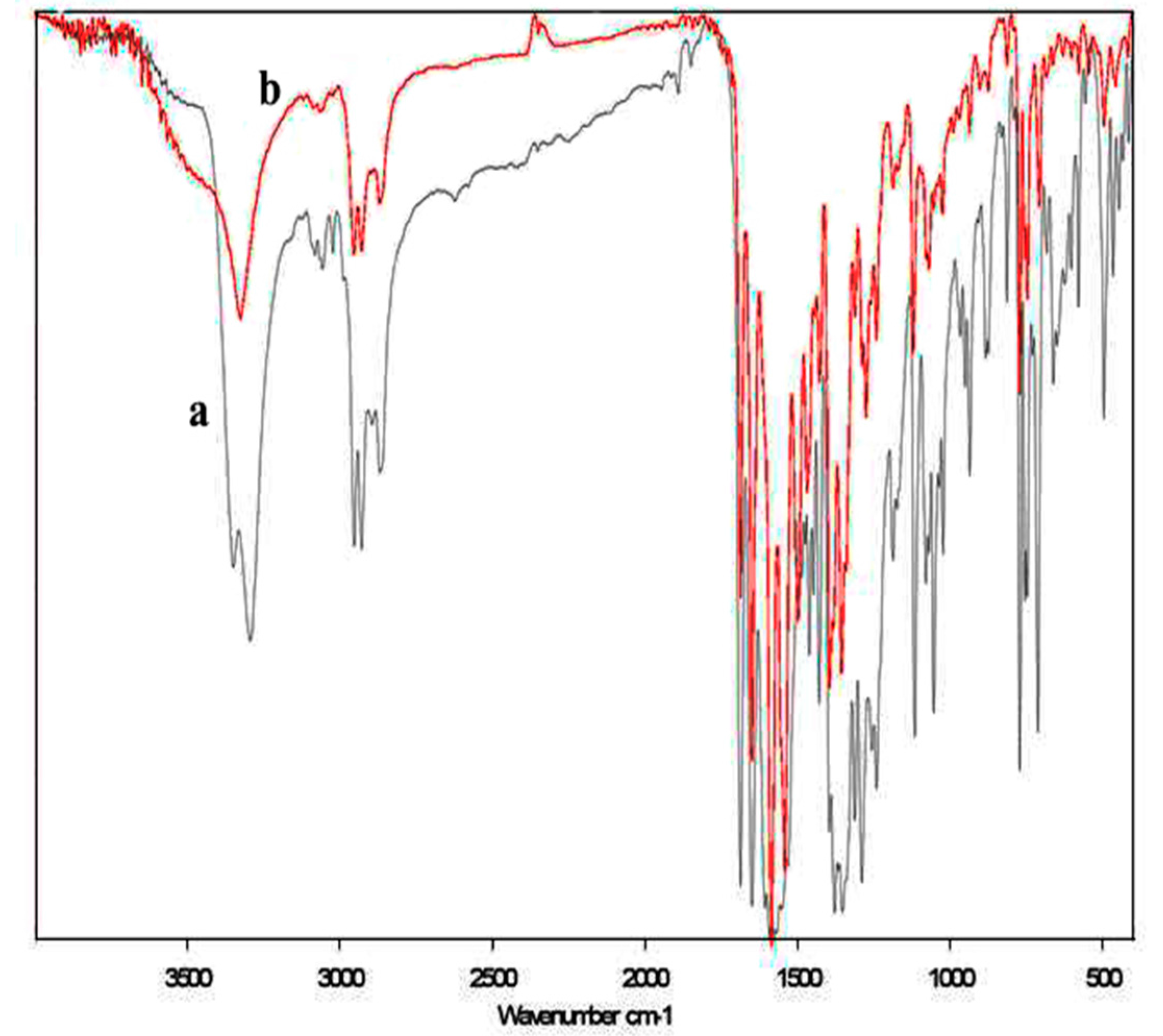
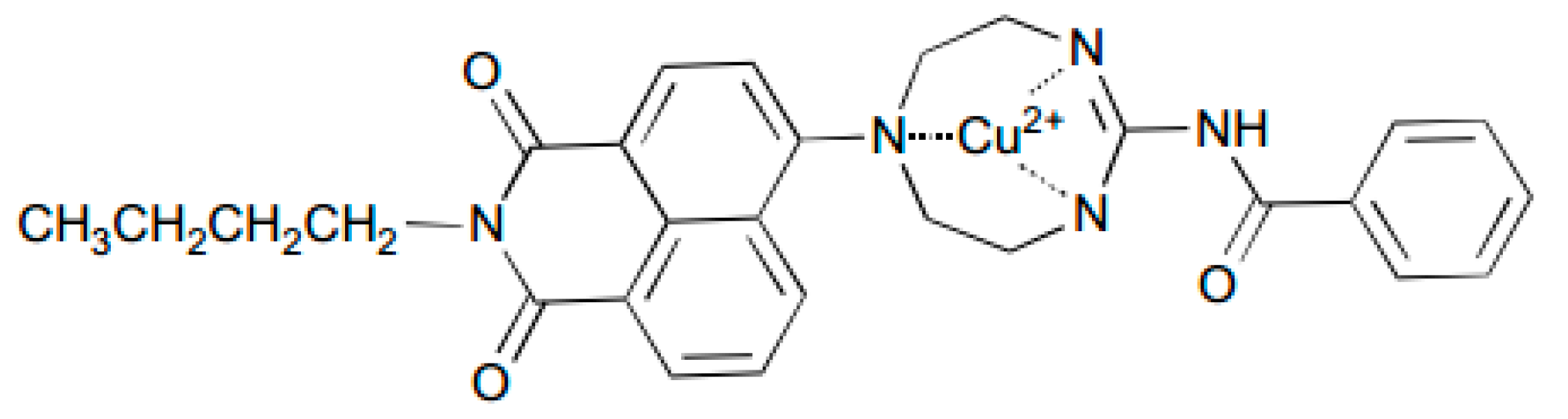
The structural matching played an important role in selective recognition. According to the examples in the literature, a similar mode of probes containing a tridentate structure binding with Cu2+ has been reported [46,47]. To further study the possible binding mode of compound L and Cu2+, IR spectroscopy was employed to elucidate the possible coordination mode. The IR spectra of compound L and the [L-Cu2+] complex are compared in Figure 12. The IR spectra of compound L (Figure 12a) has two slender peaks appearing between 3350 and 3290 cm−1. One of the extremely sharp peaks in this range vanished when the complex was formed (Figure 12b) and only appeared at 3325.72 cm−1. This result suggested that the aromatic amide was not involved in the coordination of [L-Cu2+]. However, the extremely sharp peak in the IR spectra corresponding to the imine disappeared. For fluorescence quenching, the N atom directly connected to naphthalene must be present in the coordination of the complex. On the basis of the aforementioned fact, a plausible binding mode of the complex was proposed, as shown in Scheme 3.
3. Experimental
3.1. Chemicals and Instruments
All of the solvents and reactants were commercially available and used without purification. The IR spectra were recorded on a Bruker ALPHA-T spectrometer (KBr, BRUKER Inc., Beijing, China). The melting points were determined using a Beijing Taike melting point apparatus (X-4) and were uncorrected, and 1H-NMR and 13C-NMR spectra were recorded on a Bruker AVANVE 300 or 400 MHz nuclear magnetic resonance spectrometer (Energy Chemical, Shanghai, China) using CDCl3 or DMSO-d6 as the solvent and TMS as the internal standard. The high-resolution mass spectrometry was recorded on a FT-ICR MS spectrometer (BRUKER Inc., Beijing, China). Fluorescence emission spectra were obtained using a PerkinElmer LS55 fluorescence spectrometer (PerkinElmer, Liantrisant, UK). The pH values were measured using a PHS-3C pH meter (Inesa, Shanghai, China). Thermal analysis was performed using a 449F3 thermal analyzer (Netzsch, Bavaria, Germany). The spectrogram data of the synthesized compounds could be found in the Supplementary Materials.
3.2. Testing Methods
Compound L was dissolved in EtOH to form a 10−3 M stock solution. Cu2+, Ag+, Na+, K+, Ba2+, Ca2+, Co2+, Mg2+, Mn2+, Ni2+, Pb2+, Sn2+, Zn2+, Al3+, Hg2+ and Fe3+ sources were dissolved in deionized water (H2O) to obtain 10−2 M stock solutions. For the fluorescence measurement of Cu2+, the mixed stock solutions were diluted to 10 mL with EtOH and H2O to form EtOH/H2O (4:1 v/v) solutions. For the titration experiments, compound L stock solution (100 μL) was mixed with a certain amount of Cu2+ stock solution and diluted to 10 mL with EtOH and H2O to form EtOH/H2O (4:1 v/v) solutions. The wide pH range solutions were prepared by adjustment of 0.05 mol L−1 Tris-HCl solution with HCl or NaOH solution. Thermal studies of compound L and its [L-Cu2+] complex were also performed to prove the binding of the ligand with Cu2+. All fluorescence spectra were recorded at 25 °C with the excitation wavelength set at 437 nm and the slit width set at 10 nm.
3.3. Synthesis of N-n-Butyl-4-bromo-1,8-naphthalimide (1)
N-n-Butyl-4-bromo-1,8-naphthalimide was prepared according to the reported method [48]. 4-Bromo-1,8-naphthalic anhydride (16.1 g, 58 mmol) and n-butyl amine (60 mmol, 4.4 g) were heated under reflux in EtOH (250 mL) with vigorous stirring for 12 h under N2. Then, the mixture was cooled, and the precipitate was filtered and recrystallized from ethanol to yield 13.5 g (84%) of a light-yellow product; m.p.: 105–106 °C; 1H-NMR (DMSO-d6, 300 MHz) δ (ppm): 8.46–8.52 (m, Ar-H, 2H), 8.27 (d, J = 7.9 Hz, Ar-H, 1H), 8.16 (d, J = 7.9 Hz, Ar-H, 1H), 7.92–7.97 (m, Ar-H, 1H), 4.01 (t, J = 7.3 Hz, N-CH2, 2H), 1.56–1.66 (m, CH2, 2H), 1.31–1.39 (m, CH2, 2H), and 0.93 (t, J = 7.3Hz, CH3, 3H).
3.4. Synthesis of N-n-Butyl-4-N′,N′-dihydroxyethyl-1,8-naphthalimide (2)
N-n-butyl-4-(N′,N′-dihydroxyethyl)amino-1,8-naphthalimide was obtained using a procedure similar to that reported by Guo et al. [49]. N-n-butyl-4-bromine-1,8-naphthalimide (15 g, 45.2 mmol) and diethanolamine (75 mL) were mixed in ethylene glycol monomethyl ether (100 mL). The mixture was refluxed for 6 h. The crude product was purified by column chromatography using silica gel, with EtOAc and light petroleum (1:4 v/v) as the eluent. Compound 2 was obtained as a yellow solid (3.28 g) in a 20% yield; m.p.: 129–130 °C; 1H-NMR (400 MHz, CDCl3) δ (ppm): 8.87 (d, J = 8.8 Hz, Ar-H, 1H), 8.52 (d, J = 7.2 Hz, Ar-H, 2H), 7.66 (t, J = 8.0 Hz, Ar-H, 1H), 7.38 (t, J = 8.0 Hz, Ar-H, 1H), 4.14 (t, J = 7.2 Hz, CH2, 2H), 3.87 (t, J = 5.2 Hz, CH2, 4H), 3.63 (t, J = 5.2 Hz, CH2, 4H), 2.81 (s, OH, 2H), 1.68–1.69 (m, CH2, 2H), 1.43–1.45 (m, CH2, 2H), 0.97 (t, J = 7.2 Hz, CH3, 3H); 13C-NMR (100 MHz, CDCl3) δ (ppm): 164.34, 163.91, 154.28, 131.18, 131.22, 130.85, 130.14, 127.24, 125.65, 122.92, 117.18, 117.05, 59.73, 59.73, 55.33, 55.33, 40.10, 30.20, 20.38, and 13.85.
3.5. Synthesis of N-n-Butyl-4-N′,N′-diaminoethyl-1,8-naphthalimide (4)
N-n-butyl-4-N′,N′-diaminoethyl-1,8-naphthalimide was prepared according to the reported procedure [50]. Compound 2 (2.0 g, 5.6 mmol), PPh3 (4.41 g, 16.8 mmol) and NaN3 (1.82 g, 28.0 mmol) were dissolved in anhydrous N,N-dimethylformamide (100 mL), the solution was cooled to 0 °C, and CBr4 (5.58 g, 16.8 mmol) was added portionwise over 10 min. Then, the reaction temperature was raised to room temperature, and the mixture was stirred for 12 h. The reaction mixture was diluted with water and extracted with ethyl acetate (4 × 50 mL). The organic layer was dried over anhydrous Na2SO4 and concentrated in vacuo. The crude compound was separated through silica gel column chromatography and afforded compound 3. The compound 3 (1.9 g, 4.67 mmol) and PPh3 (7.36 g, 28.1 mmol) were dissolved in 50 mL of pyridine and kept at room temperature for 1 h. Concentrated ammonium hydroxide (10 mL) was added, and the mixture was allowed to stand for an additional 2 h. Pyridine was removed through reduced pressure. Then, CH2Cl2 and 1 N HCl were added to the reaction mixture. The organic layer was washed with additional 1 N HCl, and the aqueous layers were combined, neutralized with KOH, and extracted with CH2Cl2. The combined organic layers were washed with water and brine and then dried over anhydrous Na2SO4 to afford compound 4 (1.11 g, 67%) as a yellow solid; m.p.: 99–101 °C; 1H-NMR (DMSO-d6, 300 MHz) δ (ppm): 8.74 (d, J = 0.3 Hz, Ar-H, 1H), 8.43 (d, J = 7.8 Hz, Ar-H, 1H), 8.27 (d, J = 8.4 Hz, Ar-H, 1H), 7.68 (t, J = 8.1 Hz, Ar-H, 1H), 6.81 (d, J = 8.4 Hz, Ar-H, 1H), 4.01 (t, J = 7.8 Hz, CH2, 2H), 3.44–3.48 (m, CH2, 2H), 2.87 (t, J = 5.7 Hz, CH2, 2H), 2.65–2.72 (m, CH2, 4H), 1.84 (s, NH2, 4H), 1.54–1.62 (m, CH2, 2H), 1.30–1.37 (m, CH2, 2H), and 0.92 (t, J = 7.5 Hz, CH3, 3H). HRMS-ESI m/z: found 355.2123 for [M + H]+ and calculated 354.2056 for C20H26N4O2.
3.6. Synthesis of N-n-Butyl-4-(1′-cyclooctene-1′,3′,6′-triazole)-1,8-naphthalimide (L)
N-n-butyl-4-(1′-cyclooctene-1′,3′,6′-triazole)-1,8-naphthalimide was obtained using a procedure similar to that reported in the literature [40]. Benzoyl isothiocyanate (0.92 g, 5.6 mmol) was added dropwise to a solution of N-n-butyl-4-N′,N′-diaminoethyl-1,8-naphthalimide (1.0 g, 2.8 mmol) in 30 mL of ketone and was stirred at 45 °C. The reaction mixture was stirred continuously for 45 min. After cooling, the solution was filtered, and the filter cake was washed with EtOH. The crude product was purified by chromatography to afford compound L (0.77 g, 57%) as a green solid; m.p.: 165–167 °C; 1H-NMR (CDCl3, 300 MHz) δ (ppm): 9.02 (s, NH, 1H), 8.29–8.42 (m, Ar-H, 4H), 7.54 (d, J = 6.6 Hz, Ar-H, 1H), 7.46 (t, J = 7.5 Hz, Ar-H, 3H), 6.57 (d, J = 8.7 Hz, Ar-H, 2H), 4.12 (t, J = 7.5 Hz, CH2, 4H), 3.60–3.86 (m, CH2, 4H), 3.55–3.61 (m, CH2, 2H), 1.63–1.70 (m, CH2, 2H), 1.38–1.45 (m, CH2, 2H); 1.26 (s, CNHC, 1H), 0.95 (t, J = 7.5 Hz, CH3, 3H). 13C-NMR (75 MHz, CDCl3) δ (ppm): 164.72, 164.26, 164.26, 150.97, 150.97, 134.24, 134.24, 131.02, 131.02, 129.54, 129.54, 128.35, 128.35, 128.35, 128.35, 124.25, 122.59, 110.26, 103.13, 103.13, 45.86, 41.31, 41.24, 41.05, 39.93, 30.32, 20.44 and 13.88. HRMS-ESI m/z: found 484.2332 for [M + H]+ and calculated 483.5616 for C28H29N5O3.
4. Conclusions
In summary, a new, rapidly responsive, colorimetric and “turn-off” fluorescent Cu2+ sensor, N-n-butyl-4-(1′-cyclooctene-1′,3′,6′-triazole)-1,8-naphthalimide, was successfully synthesized and characterized. Probe L exhibited a markedly quenched fluorescence in the presence of Cu2+ with a simultaneous color change (yellow to colorless) over a range of metal cations. The detection was highly sensitive and more selective than some reported probes and almost unaffected by the common coexisting metal ions [15,51]. In addition, Probe L showed a 1:1 stoichiometry (sensor/Cu2+) with a high binding constant. This sensor should be valuable for copper analysis in environmental samples and biological systems.
Supplementary Materials
Supplementary data associated with this article can be found in the online.
Acknowledgments
This work was supported by the Research Science Foundation in Technology Innovation of Harbin (2015RAYXJ010).
Author Contributions
Y. Fu and M.Q. Li developed the concept of the work. Z.Q. Wang and X.M. Liang carried out the synthetic work. Q. Chai conducted the fluorescence properties assay. Q. Chai and F. Ye wrote the paper.
Conflicts of Interest
The authors declare no conflict of interest.
References
- Thomas, S.W.; Joly, G.D.; Swager, T.M. Chemical sensors based on amplifying fluorescent conjugated polymers. Chem. Rev. 2007, 107, 1339–1386. [Google Scholar] [CrossRef] [PubMed]
- Martinez-Manez, R.; Sancenon, F. Fluorogenic and chromogenic chemosensors and reagents for anions. Chem. Rev. 2003, 103, 4419–4476. [Google Scholar] [CrossRef] [PubMed]
- Kim, H.N.; Ren, W.X.; Kim, J.S.; Yoon, J. Fluorescent and colorimetric sensors for detection of lead, cadmium, and mercury ions. Chem. Soc. Rev. 2012, 41, 3210–3244. [Google Scholar] [CrossRef] [PubMed]
- Duong, T.Q.; Kim, J.S. Fluoro- and chromogenic chemodosimeters for heavy metal ion detection in solution and biospecimens. Chem. Rev. 2010, 110, 6280–6301. [Google Scholar] [CrossRef] [PubMed]
- Ding, Y.B.; Tang, Y.Y.; Zhu, W.H.; Xie, Y.S. Fluorescent and colorimetric ion probes based on conjugated oligopyrroles. Chem. Soc. Rev. 2015, 44, 1101–1112. [Google Scholar] [CrossRef] [PubMed]
- Lippard, S.J.; Berg, J.M. Principles of Bioinorganic Chemistry; University Science Books: Mill Valley, CA, USA, 1994. [Google Scholar]
- Que, E.L.; Domaille, D.W.; Chang, C.J. Metals in neurobiology: Probing their chemistry and biology with molecular imaging. Chem. Rev. 2008, 108, 1517–1549. [Google Scholar] [CrossRef] [PubMed]
- Thiele, D.J.; Gitlin, A.D. Assembling the pieces. Chem. Biol. 2008, 4, 145–147. [Google Scholar] [CrossRef] [PubMed]
- Cho, J.; Pradhan, T.; Lee, Y.M.; Kim, J.S.; Kim, S. A calix[2] Triazole[2] Arene-based fluorescent chemosensor for probing the copper trafficking pathway in Wilson’s disease. Dalton Trans. 2014, 43, 16178–16182. [Google Scholar] [CrossRef] [PubMed]
- You, G.R.; Park, G.J.; Lee, J.J.; Kim, C. A colorimetric sensor for the sequential detection of Cu2+ and CN− in fully aqueous media: Practical performance of Cu2+. Dalton Trans. 2015, 44, 9120–9129. [Google Scholar] [CrossRef] [PubMed]
- Barnham, K.J.; Masters, C.L.; Bush, A.I. Neurodegenerative diseases and oxidative stress. Nat. Rev. Drug Discov. 2004, 3, 205–214. [Google Scholar] [CrossRef] [PubMed]
- Mare, S.; Penugonda, S.; Robinson, S.M.; Dohgu, S.; Banks, W.A.; Ercal, N. Copper complexing decreases the ability of amyloid beta peptide to cross the BBB and enter brain parenchyma. Peptides 2007, 28, 1424–1432. [Google Scholar] [CrossRef] [PubMed]
- Kim, B.E.; Nevitt, T.; Thiele, D.J. Mechanisms for copper acquisition, distribution and regulation. Nat. Chem. Biol. 2008, 4, 176–185. [Google Scholar] [CrossRef] [PubMed]
- Lee, J.C.; Gray, H.B.; Winkler, J.R. Copper(II) binding to alpha-synuclein, the Parkinson’s protein. J. Am. Chem. Soc. 2008, 130, 6898–6899. [Google Scholar] [CrossRef] [PubMed]
- Sun, S.H.; Hu, W.T.; Gao, H.F.; Qi, H.L.; Diang, L.P. Luminescence of ferrocene-modified pyrene derivatives for turn-on sensing of Cu2+ and anions. Spectochim. Acta A 2017, 184, 30–37. [Google Scholar] [CrossRef] [PubMed]
- Jung, H.S.; Kwon, P.S.; Lee, J.W.; Kim, J.I.; Hong, C.S.; Kim, J.W.; Yan, S.H.; Lee, J.Y.; Lee, J.H.; Joo, T.; et al. Coumarin-derived Cu2+ -selective fluorescence sensor: Synthesis, mechanisms, and applications in living cells. J. Am. Chem. Soc. 2009, 131, 2008–2012. [Google Scholar] [CrossRef] [PubMed]
- Liu, C.X.; Xu, J.; Yang, F.; Zhou, W.; Li, Z.X.; Wei, L.H.; Yu, M.M. Nanomolar Cu2+ and F− naked-eye detection with a 1,8-naphthalimide-based colorimetric probe. Sens. Actuators B 2015, 212, 364–370. [Google Scholar] [CrossRef]
- Khan, B.; Shah, M.R.; Ahmed, D.; Rabnawaz, M.; Anis, I.; Afridi, S.; Makhmoor, T.; Tahir, M.N. Synthesis, characterization and Cu2+ triggered selective fluorescence quenching of bis-calix[4]arene tetra-triazole macrocycle. J. Hazard. Mater. 2016, 309, 97–106. [Google Scholar] [CrossRef] [PubMed]
- Moussa, H.; Merlin, C.; Dezanet, C.; Balan, L.; Medjandi, G.; Ben-Attia, M.; Schneider, R. Trace amounts of Cu2+ ions influence ROS production and cytotoxicity of ZnO quantum dots. J. Hazard. Mater. 2016, 304, 532–542. [Google Scholar] [CrossRef] [PubMed]
- Faghihian, H.; Hajishabani, A.; Dadfarnia, S.; Zamani, H. Use of clinoptilolite loaded with 1-(2-pyridylazo)-2-naphthol as a sorbent for preconcentration of Pb(II), Ni(II), Cd(II) and Cu(II) prior to their determination by flame atomic absorption spectroscopy. Int. J. Environ. Anal. Chem. 2009, 89, 223–231. [Google Scholar] [CrossRef]
- Tomalova, I.; Foltynova, P.; Kanicky, V.; Preisler, J. MALDI MS and ICP MS detection of a single CE separation record: A tool for metalloproteomics. Anal. Chem. 2014, 86, 647–654. [Google Scholar] [CrossRef] [PubMed]
- Qing, Z.H.; Mao, Z.G.; Qing, T.P.; He, X.X.; Zou, Z.; He, D.G.; Shi, H.; Huang, J.; Liu, J.B.; Wang, K.M. Visual and portable strategy for copper(II) detection based on a striplike poly(thymine)-caged and microwell-printed hydrogel. Anal. Chem. 2014, 86, 11263–11268. [Google Scholar] [CrossRef] [PubMed]
- Liu, Z.M.; Jia, X.L.; Bian, P.P.; Ma, Z.F. A simple and novel system for colorimetric detection of cobalt ions. Analyst 2014, 139, 585–588. [Google Scholar] [CrossRef] [PubMed]
- Tang, B.; Niu, J.Y.; Yu, C.G.; Zhuo, L.H.; Ge, J.C. Highly luminescent water-soluble Cd Te nanowires as fluorescent probe to detect copper(II). Chem. Commun. 2005, 33, 4184–4186. [Google Scholar] [CrossRef] [PubMed]
- Zhu, Z.Q.; Su, Y.Y.; Li, J.; Li, D.; Zhang, J.; Song, S.P.; Zhao, Y.; Li, G.X.; Fan, C.H. Highly sensitive electrochemical sensor for mercury(II) ions by using a mercury-specific oligonucleotide probe and gold nanoparticle-based amplification. Anal. Chem. 2009, 81, 7660–7666. [Google Scholar] [CrossRef] [PubMed]
- Niamnont, N.; Khumsri, A.; Promchat, A.; Tumcharern, G.; Sukwattanasinitt, M. Novel salicylaldehyde derivatives as fluorescence turn-on sensors for cyanide ion. J. Hazard. Mater. 2014, 280, 458–463. [Google Scholar] [CrossRef] [PubMed]
- Zhang, J.R.; Zeng, A.L.; Luo, H.Q.; Li, N.B. Fluorescent silver nanoclusters for ultrasensitive determination of chromium(VI) in aqueous solution. J. Hazard. Mater. 2016, 304, 66–72. [Google Scholar] [CrossRef] [PubMed]
- Czamik, A.W. Fluorescent chemosensors for ion and molecule recognition. In ACS Symposium Series Sponsored by the Division of Organic Chemistry of the American Chemical Society at the 204th Meeting of the American Chemical Society; American Chemical Society: Washington, DC, USA, 1992; p. 538. [Google Scholar]
- Dujols, V.; Ford, F.; Czarnik, A.W. A long-wavelength fluorescent chemodosimeter selective for Cu(II) ion in water. J. Am. Chem. Soc. 1997, 119, 7386–7387. [Google Scholar] [CrossRef]
- Xiang, Y.; Tong, A.J.; Jin, P.Y.; Ju, Y. New fluorescent rhodamine hydrazone chemosensor for Cu(II) with high selectivity and sensitivity. Org. Lett. 2006, 8, 2863–2866. [Google Scholar] [CrossRef] [PubMed]
- Ding, Y.B.; Zhu, W.H.; Xie, Y.S. Development of Ion Chemosensors Based on Porphyrin Analogues. Chem. Rev. 2017, 117, 2203–2256. [Google Scholar] [CrossRef] [PubMed]
- Sirilaksanapong, S.; Sukwattanasinitt, M.; Rashatasakhon, P. 1,3,5-Triphenylbenzene fluorophore as a selective Cu2+ sensor in aqueous media. Chem. Commun. 2012, 48, 293–295. [Google Scholar] [CrossRef] [PubMed]
- Erdemir, S.; Tabakci, B.; Tabakci, M. A highly selective fluorescent sensor based on calix[4]arene appended benzothiazole units for Cu2+, S2− and HSO4− ions in aqueous solution. Sens. Actuators B Chem. 2016, 228, 109–116. [Google Scholar] [CrossRef]
- Hu, Y.; Ke, Q.; Yan, C.; Xu, C.H.; Huang, X.H.; Hu, S.L. A new fluorescence chemosensor for selective detection of copper ion in aqueous solution. Tetrahedron Lett. 2016, 57, 2239–2243. [Google Scholar] [CrossRef]
- Huang, L.; Cheng, J.; Xie, K.F.; Xi, P.X.; Hou, F.P.; Li, Z.P.; Xie, G.Q.; Shi, Y.J.; Liu, H.Y.; Bai, D.C.; et al. Cu2+-selective fluorescent chemosensor based on coumarin and its application in bioimaging. Dalton Trans. 2011, 40, 10815–10817. [Google Scholar] [CrossRef] [PubMed]
- Maher, N.J.; Diao, H.W.; O’Sullivan, J.; Fadda, E.; Heaney, F.; McGinley, J. Lower rim isoxazole-calix[4]arene derivatives as fluorescence sensors for copper(II) ions. Tetrahedron 2015, 71, 9223–9233. [Google Scholar] [CrossRef]
- Ganguly, A.; Ghosh, S.; Kar, S.; Guchhait, N. Selective fluorescence sensing of Cu(II) and Zn(II) using a simple Schiff base ligand: Naked eye detection and elucidation of photoinduced electron transfer (PET) mechanism. Spectrochim. Acta Part A 2015, 143, 72–80. [Google Scholar] [CrossRef] [PubMed]
- Quan, L.; Sun, T.T.; Lin, W.H.; Guan, X.G.; Zheng, M.; Xie, Z.G.; Jing, X.B. Bodipy fluorescent chemosensor for Cu2+ detection and its applications in living cells: Fast response and high sensitivity. J. Fluoresc. 2014, 24, 841–846. [Google Scholar] [CrossRef] [PubMed]
- Poss, M.A.; Iwanowicz, E.; Reid, J.A.; Lin, J.; Gu, Z.X. A mild and efficient method for the preparation of guanidines. Tetrahedron Lett. 1992, 33, 5933–5936. [Google Scholar] [CrossRef]
- Liu, B.; Tian, H. A selective fluorescent ratiometric chemodosimeter for mercury ion. Chem. Commun. 2005, 3156–3158. [Google Scholar] [CrossRef] [PubMed]
- Fischer, M.; Georges, J. Fluorescence quantum yield of rhodamine 6G in ethanol as a function of concentration using thermal lens spectrometry. Chem. Phys. Lett. 1996, 260, 115–118. [Google Scholar] [CrossRef]
- Ma, Q.J.; Zhang, X.B.; Han, Z.X.; Huang, B.; Jiang, Q.; Shen, G.L.; Yu, R.Q. A ratiometric fluorescent probe for zinc ions based on the quinoline fluorophore. Int. J. Environ. Anal. Chem. 2011, 91, 74–86. [Google Scholar] [CrossRef]
- Gao, C.J.; Liu, X.; Jin, X.J.; Wu, J.; Xie, Y.J.; Liu, W.S.; Yao, X.J.; Tang, Y. A retrievable and highly selective fluorescent sensor for detecting copper and sulfide. Sens. Actuators B Chem. 2013, 185, 125–131. [Google Scholar] [CrossRef]
- Karak, D.; Banerjee, A.; Sahana, A.; Guha, S.; Lohar, S.; Adhikari1, S.S.; Das, D. 9-Acridone-4-carboxylic acid as an efficient Cr(III) fluorescent sensor: Trace level detection, estimation and speciation studies. J. Hazard. Mater. 2011, 188, 274–280. [Google Scholar] [CrossRef] [PubMed]
- Xie, Y.S.; Ding, Y.B.; Li, X.; Wang, C.; Hill, J.P.; Ariga, K.; Zhang, W.B.; Zhu, W.H. Selective, sensitive and reversible “turn-on” fluorescent cyanide probes based on 2,2′-dipyridylaminoanthracene-Cu2+ ensembles. Chem. Commun. 2012, 48, 11513–11515. [Google Scholar] [CrossRef] [PubMed]
- Wang, H.X.; Yang, L.; Zhang, W.B.; Zhou, Y.; Zhao, B.; Li, X.Y. A colorimetric probe for copper(II) ion based on 4-amino-1,8-naphthalimide. Inorg. Chim. Acta 2012, 381, 111–116. [Google Scholar] [CrossRef]
- Gao, Y.G.; Tang, Q.; Shi, Y.D.; Zhang, Y.; Lu, Z.L. 1,8-naphthalimide modified [12]aneN compounds as selective and sensitive probes for Cu2+ ions and ATP in aqueous solution and living cells. Talanta 2016, 152, 438–446. [Google Scholar] [CrossRef] [PubMed]
- Zhang, W.J.; Huo, F.J.; Liu, T.; Wen, Y.; Yin, C.X. A rapid and highly sensitive fluorescent imaging materials for thiophenols. Dyes Pigm. 2016, 133, 248–254. [Google Scholar] [CrossRef]
- Guo, X.F.; Zhu, B.C.; Liu, Y.Y.; Zhang, Y.; Jia, L.H.; Qian, X.H. Synthesis and properties of N-butyl-4-(aza-15-crown-5)-1, 8-naphthalimide as a fluorescent probe. Chin. J. Org. Chem. 2006, 26, 504–507. [Google Scholar]
- Lee, S.; Lee, J.H.; Pradhan, T.; Lim, C.S.; Cho, B.R.; Bhuniya, S.; Kim, S.; Kim, J.S. Fluorescent turn-on Zn2+ sensing in aqueous and cellular media. Sens. Actuators B Chem 2011, 160, 1489–1493. [Google Scholar] [CrossRef]
- Zheng, X.; Lee, K.H.; Liu, H.G.; Park, S.Y.; Yoon, S.S.; Lee, J.Y.; Kim, Y.G. A bis(pyridine-2-ylmethyl)amine-based selective and sensitive colorimetric and fluorescent chemosensor for Cu2+. Sens. Actuators B Chem. 2016, 222, 28–34. [Google Scholar] [CrossRef]
Sample Availability: Samples of the compounds are available from the authors’ lab. |
Scheme 1.
Thiourea transformation into guanidine.

Scheme 2.
Synthetic route to compound L.

Figure 1.
Excitation and emission spectra of compound L (10 μM) in EtOH/H2O (4:1 v/v).

Figure 2.
Fluorescence changes of compound L (10 μM) in the absence (CK) and presence of metal cations (50 μM) in EtOH/H2O (4:1 v/v).
Figure 2.
Fluorescence changes of compound L (10 μM) in the absence (CK) and presence of metal cations (50 μM) in EtOH/H2O (4:1 v/v).

Figure 3.
Job plot to determine the stoichiometry of the [L-Cu2+] complex in solution.

Figure 4.
Stern-Volmer plot for calculating the binding constant of the chemosensor with Cu2+.

Figure 5.
Changes in the fluorescence spectra of compound L (10 μM) in EtOH/H2O (4:1 v/v) as a function of added [Cu2+].
Figure 5.
Changes in the fluorescence spectra of compound L (10 μM) in EtOH/H2O (4:1 v/v) as a function of added [Cu2+].

Figure 6.
Fluorescence intensity vs Cu2+; inset shows that up to 6-fold Cu2+ curvilinearity was sustained; λex: 437 nm; λem: 518 nm; slit width: 10 nm.
Figure 6.
Fluorescence intensity vs Cu2+; inset shows that up to 6-fold Cu2+ curvilinearity was sustained; λex: 437 nm; λem: 518 nm; slit width: 10 nm.

Figure 7.
Response time of 10 μM probe L in the absence or presence of 50 μM Cu2+ in EtOH/H2O (4:1 v/v); λex: 437 nm; λem: 518 nm; slit width: 10 nm.
Figure 7.
Response time of 10 μM probe L in the absence or presence of 50 μM Cu2+ in EtOH/H2O (4:1 v/v); λex: 437 nm; λem: 518 nm; slit width: 10 nm.

Figure 8.
Effect of pH on the fluorescence intensity of 10 μM probe L in the absence or presence of 50 μM Cu2+.
Figure 8.
Effect of pH on the fluorescence intensity of 10 μM probe L in the absence or presence of 50 μM Cu2+.

Figure 9.
Interference of foreign cations. Solvents: EtOH/H2O (4:1 v/v).

Figure 10.
Thermal studies of the fluorescence chemosensor.

Figure 11.
Thermal studies of the [L-Cu2+] complex with the fluorescent chemosensor.

Figure 12.
IR spectra of compound L before (a) and after (b) the addition of Cu2+.

Scheme 3.
Proposed coordination of compound L combined with Cu2+.


© 2017 by the authors. Licensee MDPI, Basel, Switzerland. This article is an open access article distributed under the terms and conditions of the Creative Commons Attribution (CC BY) license (http://creativecommons.org/licenses/by/4.0/).
Share and Cite
MDPI and ACS Style
Ye, F.; Chai, Q.; Liang, X.-M.; Li, M.-Q.; Wang, Z.-Q.; Fu, Y. A Highly Selective and Sensitive Fluorescent Turn-Off Probe for Cu2+ Based on a Guanidine Derivative. Molecules 2017, 22, 1741. https://doi.org/10.3390/molecules22101741
AMA Style
Ye F, Chai Q, Liang X-M, Li M-Q, Wang Z-Q, Fu Y. A Highly Selective and Sensitive Fluorescent Turn-Off Probe for Cu2+ Based on a Guanidine Derivative. Molecules. 2017; 22(10):1741. https://doi.org/10.3390/molecules22101741
Chicago/Turabian StyleYe, Fei, Qiong Chai, Xiao-Min Liang, Ming-Qiang Li, Zhi-Qiang Wang, and Ying Fu. 2017. "A Highly Selective and Sensitive Fluorescent Turn-Off Probe for Cu2+ Based on a Guanidine Derivative" Molecules 22, no. 10: 1741. https://doi.org/10.3390/molecules22101741