Article

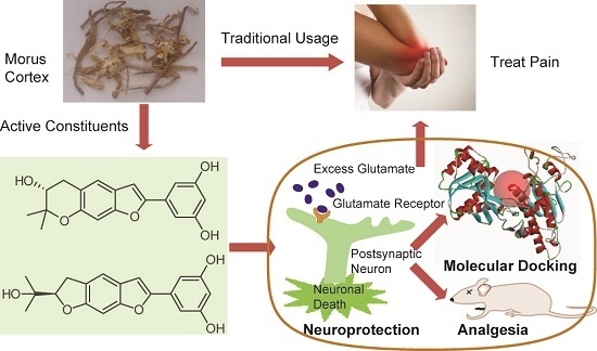
Bioactive Benzofuran Derivatives from Cortex Mori Radicis, and Their Neuroprotective and Analgesic Activities Mediated by mGluR1

1
State Key Laboratory of Bioactive Substance and Function of Natural Medicines, Institute of Materia Medica, Chinese Academy of Medical Sciences & Peking Union Medical College, Beijing 100050, China
2
GRU Cancer Center, Augusta University, Augusta, GA 30912, USA
3
Editorial Department, Shenyang Pharmaceutical University, Shenyang 110016, China
*
Author to whom correspondence should be addressed.
Molecules 2017, 22(2), 236; https://doi.org/10.3390/molecules22020236
Submission received: 30 December 2016 / Revised: 28 January 2017 / Accepted: 31 January 2017 / Published: 8 February 2017
(This article belongs to the Collection Bioactive Compounds)

Abstract

:
Four new benzofuran-type stilbene glycosides and 14 known compounds including 8 benzofuran-type stilbenes and 6 flavonoids were isolated from the traditional Chinese medicine, Cortex Mori Radicis. The new compounds were identified as (9R)-moracin P 3′-O-α-l-arabinopyranoside (1), (9R)-moracin P 9-O-β-d-glucopyranoside (2), (9R)-moracin P 3′-O-β-d-glucopyranoside (3), and (9R)-moracin O 10-O-β-d-glucopyranoside (4) based on the spectroscopic interpretation and chemical analysis. Three benzofuran-type stilbenes, moracin O (5), R (7), and P (8) showed significant neuroprotective activity against glutamate-induced cell death in SK-N-SH cells. In addition, moracin O (5) and P (8) also demonstrated a remarkable inhibition of the acetic acid-induced pain. The molecular docking with metabotropic glutamate receptor 1 (mGluR1) results indicated that these neuroprotective benzofuran-type stilbenes might be the active analgesic components of the genus Morus, and acted by mediating the mGluR1 pathway.

Graphical Abstract

1. Introduction

Cortex Mori Radicis, the root bark of some Morus species (e.g., M. alba, M. mongolica, M. cathayana, and M. australis) have been used in traditional Chinese medicine as an antidiabetic, diuretic, and expectorant agent. Many compounds were identified from Morus plants, such as Diels-Alder-type adducts, stilbenes, flavonoids, and alkaloids. Their antioxidant [1,2,3], anti-inflammatory [4,5], antimicrobial [6,7,8,9], anticarcinogenic [10,11,12], and antidiabetic [13] activities were reported frequently. Recently, the structures and bioactivities of polyphenols from Morus plants [14] and bioactive benzofurans [15] were reviewed. It should be noted that the extracts of some twigs [16], leaves [17], or root barks [18] of Morus species were used to relieve pain in folk medicine. To the best of our knowledge, only two compounds from genus Morus, cis-mulberroside A [19] and morusin [20], have been reported with analgesic activity to date, which is related to the traditional antirheumatic usage of Cortex Mori Radicis.
l-Glutamate is the major excitatory neurotransmitter in the central nervous system (CNS) and glutamate-mediated excitotoxicity plays a crucial role in neurodegenerative disorder, particularly in Parkinson’s disease, Alzheimer’s disease, epilepsy, spinal cord trauma, and ischemic stroke. As glutamate receptors are also found in peripheral tissue, they may be implicated in persistent or chronic pain, including inflammatory or joint-related pain (e.g., rheumatoid arthritis, osteoarthritis) and neuropathic pain resulting from injury and/or diseases of central (e.g., spinal cord injury) or peripheral nerves (e.g., diabetic neuropathy, radiculopathy) [21,22].
Neuroprotective effect of stilbenes, including piceid and resveratrol, has been widely reported [23], but the applications of benzofuran stilbene on CNS diseases [24] and analgesia were rarely studied. In this paper, we report four new benzofuran stilbene glycosides (14) and the neuroprotective and analgesic activities of their aglycones. Three benzofuran stilbenes showed significant protective activities against glutamate-induced neurotoxicity. Correspondingly, they also exhibited remarkable analgesic activities by inhibition of the acetic acid-induced pain. Structural determinations of compounds were carried out on the basis of spectroscopic data analyses. The primary action mechanism of neuroprotection and analgesia was also discussed with the help of the molecular docking with metabotropic glutamate receptor 1 (mGluR1).

2. Results and Discussion

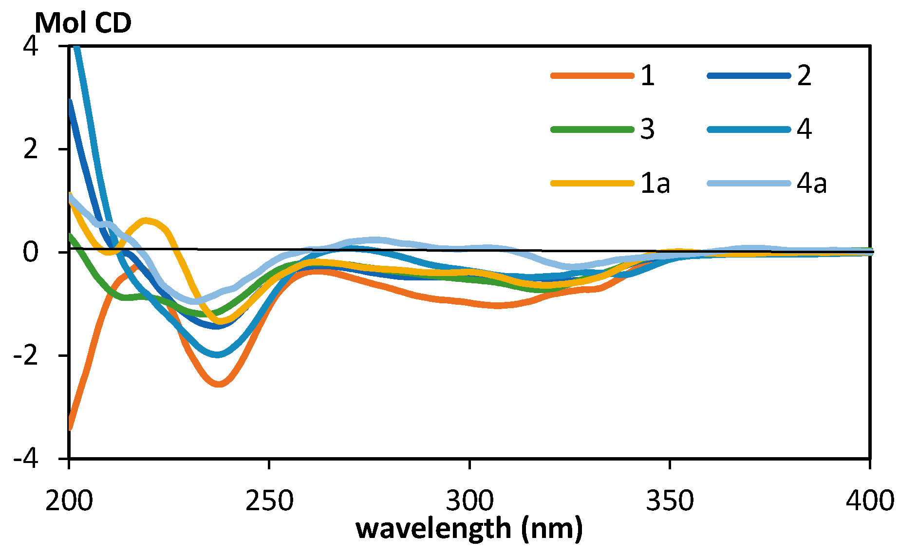
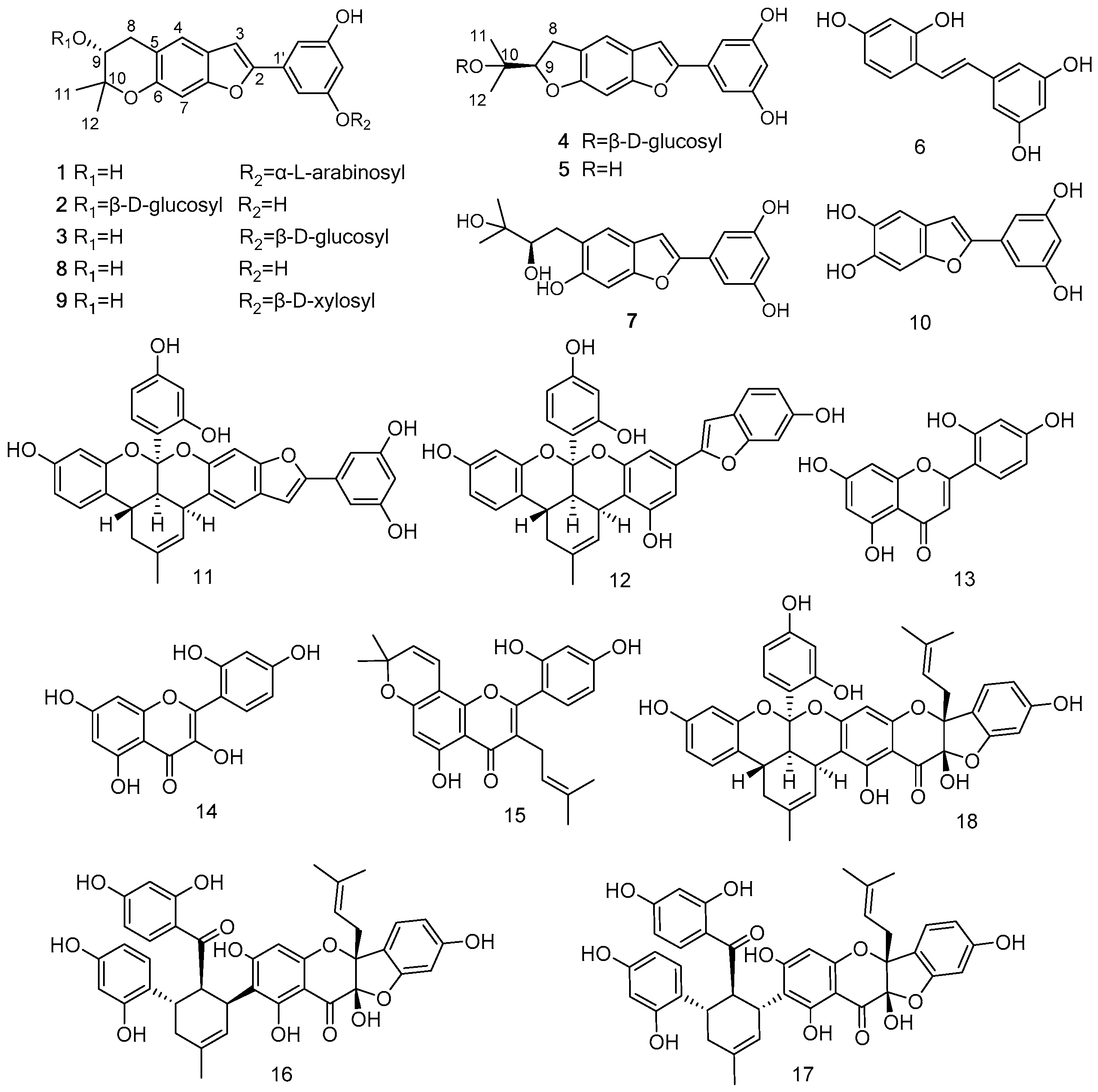
2.1. Structural Analysis

Eighteen compounds, including four new (14) and eight known (512) stilbenes and six known flavonoids (1318), were isolated from the EtOAc-soluble fraction of Cortex Mori Radicis (Figure 1). The known compounds were elucidated as moracin O (5) [25], oxyresveratrol (6) [26], moracin R (7) [27], moracin P (8) [25], mulberroside C (9) [28], wittifuran E (10) [28], isomulberrofuran G (11) [29], mulberrofuran G (12) [30], norartocarpetin (13) [31], morin (14) [32], morusin (15) [33], sanggenon C (16) [34], sanggenon D (17) [35], and cathayanin B (18) [36] by comparison with spectroscopic data in the literatures.
Compound 1, purified as a brown powder, had the molecular formula of C24H26O9 as established by HRESIMS m/z 459.1663 [M + H]+ (calcd for C24H27O9+, 459.1650). The 1H-NMR spectrum (Table 1) demonstrated a 2-arylbenzofuran skeleton with three meta-aromatic protons at δH 6.91 (1H, d, J = 2.1 Hz, H-2′), 6.89 (1H, d, J = 2.1 Hz, H-6′), and 6.43 (1H, t, J = 2.1 Hz, H-4′); two para-aromatic protons at δH 7.29 (1H, s, H-4), 6.93 (1H, s, H-7), and one singlet at δH 7.17 (1H, s, H-3). Two gem-dimethyl signals at δH 1.31 (3H, s, H-11), and 1.18 (3H, s, H-12), one methylene at δH 3.02 (1H, dd, J = 16.4, 6.0 Hz, H-8a), and 2.72 (1H, dd, J = 16.4, 7.0 Hz, H-8b) indicated the presence of an isoprenyl fragment. The remaining signals at δH 4.85 (1H, d, J = 6.7 Hz, H-1″) and δH 2.60–3.80 were assigned to a pentose moiety. Besides the five pentosyl signals, the 13C-NMR spectrum (Table 2) confirmed the presence of isoprenylated 2-arylbenzofuran skeleton including 14 olefinic carbons and 5 alkyl carbons. Further, HMBC correlations between H2-8 (δH 3.02, 2.72) and C-4 (δC 120.9) and C-6 (δC 151.2); H-9 (δH 3.67) and C-5 (δC 117.2) were observed, and these data elucidated the aglycone of 1 as moracin P [25]. The linkage of pentose was determined to be at the C-3′ position, deduced from the HMBC correlation between H-1″ (δH 4.85) and C-3′ (δC 158.8). In 1H-NMR spectra, the H-4″ of the pentose was a broad singlet and the coupling constants between H-4″ and H-5″, H-4″ and H-3″ were less than 4 Hz, indicated that H-4″ was equatorial. Subsequent acid hydrolysis identified the pentose as l-arabinose and the aglycone as (9R)-moracin P. The C-4″ (δC 68.0) indicated the arabinose was a pyranose [37]. Therefore, the chemical structure of 1 was established as (9R)-moracin P 3′-O-α-l-arabinopyranoside.
The molecular formulas of compounds 2 and 3 were both C25H28O10, established by HRESIMS (m/z 511.1588 [M + Na]+, calculated for C25H28O10Na+, 511.1575). Comparing 1H-NMR and 13C-NMR spectra (Table 1 and Table 2) of compounds 2 and 3 with those of compound 1, the structures of 2 and 3 were both identified as moracin P glycosides with a hexose in their structures, and further supported by HMBC analysis. The linkage of sugar in 2 was determined to be at C-9 position, deduced from the HMBC correlation between H-1″ (δH 4.33, d, J = 7.7 Hz) and C-9 (δC 73.4). The sugar in 3 was connected to C-3′ position, deduced from the HMBC correlation between H-1″ (δH 4.83 d, J = 7.6 Hz) and C-3′ (δC 159.1). Subsequent acid hydrolysis identified the aglycone as (9R)-moracin P and the sugar as d-glucose. Therefore, the structures of 2 and 3 were established as (9R)-moracin P 9-O-β-d-glucopyranoside (2) and (9R)-moracin P 3′-O-β-d-glucopyranoside (3), respectively.
The molecular formula of compound 4 was established to be C25H28O10 by HRESIMS, which showed the quasimolecular ion peak at m/z 511.1589 [M + Na]+ (calcd for C25H28O10Na+, 511.1575). The 1H-NMR and 13C-NMR spectroscopic data (Table 1 and Table 2) of 4 suggested that it was a stilbene glucoside similar to 2 and 3, except the dihydropyran ring. HMBC correlations between H2-8 (3.16, 3.35) and C-4 (δC 116.1) and C-6 (δC 158.1); H-9 (δH 4.75) and C-5 (δC 124.3) and C-6 (δC 158.1) were observed, and the aglycone of 4 was elucidated as moracin O [25]. The linkage of sugar was determined to be at the C-10 position, deduced from the HMBC correlation between H-1″ (δH 4.42, d, J = 7.8 Hz) and C-10 (δC 77.1). Acid hydrolysis identified the aglycone as (9R)-moracin O and the sugar as d-glucose. Therefore, the structure of 4 was established as (9R)-moracin O 10-O-β-d-glucopyranoside.
There was only one chiral carbon C-9 in aglycones of compounds 14. In order to determine their absolute configuration, compounds 14 were hydrolyzed by HCl, and the aglycones were partitioned by EtOAc. After drying the EtOAc-soluble fraction in vacuo, the residue was dissolved in MeOH to determine the optical rotation. All aglycones showed negative optical rotation values (see Supplementary Materials), which confirmed that aglycones 1a3a were (9R)-moracin P [38] and aglycone 4a was (9R)-moracin O [38,39].
Although the sugars were substituted at different position, the ECD (electronic circular dichroism) spectra of 14 and their aglycones 1a and 4a were quite similar (Figure 2). There were two Cotton effects in their ECD spectra. The broad negative Cotton effect over 280–340 nm was regarded as π→π* transition from MO86 to MO87 of 2-benzylbenzofuran. The negative Cotton effect at 240 nm was generated by π→π* transition from MO86 to MO90 of benzofuran moiety and π→π* transition from MO86 to MO89 of benzyl moiety, according to the MO analysis in the calculated ECD of 1a (Supplementary Materials). It seemed that the chiral carbon on the rigid skeleton attached to the benzofuran affected the Cotton effect at 240 nm more than the chiral center on the flexible benzyl moiety, deduced from the similar ECD spectra of 14, 1a, and 4a.

2.2. Neuroprotective Activities

Twelve compounds were evaluated by the 3-(4,5-dimethylthiazol-2-yl)-2,5-diphenyltetrazolium bromide (MTT) assay for the neuroprotective effects against glutamate-induced cellular damage on the human neuroblastoma SK-N-SH cells (Table 3). Moracin O (5), R (7), and P (8) exhibited significant protection with 40%–60% of cell viability at the concentration of 10 μM. Norartocarpetin (13) and morusin (15) exhibited modest protection effects with about 20% cell viability. Oxyresveratrol (6) and sanggenon C (16) at the concentration of 10 μM attenuated glutamate-induced toxicity weakly (about 12% cell viability), similar to resveratrol. It has been reported that moracin O (5) and moracin P (8) were promising drug candidates for cerebral ischemia and other neurodegenerative diseases due to their protective activities against oxygen-glucose deprivation (OGD)-induced cell death [40]. Therefore, we also tested the neuroprotective effects of compounds 15, 79 against OGD-induced damage and 3-nitropropionic acid (3-NP)-induced neuronal injury on human neuroblastoma SH-SY5Y cells using the MTT method. Our results demonstrated that only moracin O (5), P (8), and moracin P 3′-O-β-d-glucopyranoside (3) indicated weak neuroprotective activities against OGD-induced damage, and none of them protected cells from the 3-NP-induced damage (Table 3).

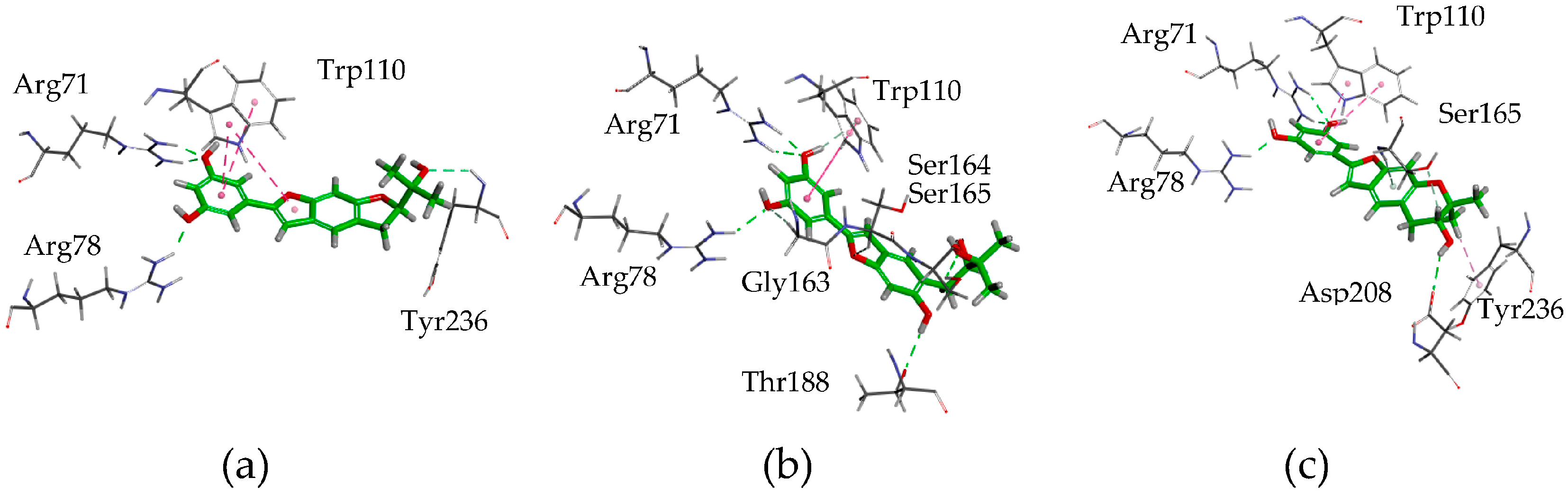
2.3. Postulating the Neuroprotective Pathway by Molecular Docking

In order to further understand the possible mechanisms of the benzofuran stilbenes against glutamate-induced neuronal death, 10 glutamate receptors, including various ionotropic and metabotropic types, were selected from 64 Homo sapiens GluRs in the Protein Data Bank (PDB) website to study the ligand binding to these receptors in a molecular-docking method.
Among the selected glutamate receptors, the mGluR1 (3KS9) was the only one that could be docked with all active compounds (58, 13, and 15) as well as glutamic acid. In contrast, all of the inactive compounds (14, 9) could not be docked into the binding site of the mGluR1 (3KS9). These results indicated that the neuroprotective activities of moracin O (5), R (7), and P (8), might be mediated by the mGluR1 pathway.
Molecular-docking results (Figure 3) showed each of the three ligands, moracin O (5), R (7), and P (8), had π–π interaction between the aromatic plane and Trp 110, and three to four H-bonds between hydroxyl groups of the ligands and the residues of Arg 71, Arg 78, Tyr 236 (or Asp 208, Ser165, Thr188) in mGluR1, which indicated that the aromatic group and the hydroxyl group play an important role in the bindings.

2.4. Analgesic Activities

Although some compounds from the genus Morus exhibited promising neuroprotective activity, Morus species have been rarely used to treat neurodegenerative diseases or other CNS diseases. As mGlu receptors had been considered to be valid targets for chronic pain control due to their ability of modulating rather than mediating excitatory synaptic activity [41], it was reasonable to hypothesize that these neuroprotective isoprenylated stilbenes might be the active analgesic components of Cortex Mori Radicis.
The three most effective neuroprotective stilbenes—moracin O (5), R (7), P (8)—and reported [20] flavonoid morusin (15) were assayed with the analgesic test and paracetamol was a positive control. As predicted, both moracin O (5) and P (8), with cyclic structural moieties, demonstrated more than 95% of inhibitions of acetic acid-induced pain at 80 mg/kg, which were as effective as morusin, much better than paracetamol. These results were consistent with the reported inhibition of morusin, which was about 73% at 60 mg/kg [20]. However, moracin R (7), which had an acyclic moiety, showed moderate inhibition (Table 4).
As compounds 14 and 9 were the glycosides of moracin O (5) and P (8), they might act as predrugs through oral administration, although they did not show neuroprotective effects in vitro.

3. Materials and Methods

3.1. Instruments

Melting points were determined on an XT5B melting point apparatus (Beijing Keyi Electric Light Instrument Factory, Beijing, China) and were uncorrected. Optical rotations were measured with a P-2000 polarimeter (Jasco, Tokyo, Japan). ECD spectra were recorded at room temperature with a J-815 spectropolarimeter (Jasco). UV spectra were collected in MeOH on a V-650 spectrophotometer (Jasco). IR spectra were recorded on a Nicolet 5700 spectrometer (Thermo, Madison, WI, USA) by the FT-IR transmission electron microscopy method. 1H- and 13C-NMR spectra were acquired using a DD2-500 spectrometer (Agilent, Santa Clara, CA, USA) or an AVIIIHD 600 spectrometer (Bruker, Billerica, MA, USA). HRESIMS were recorded on a 1200 series LC/6520 quadrupole time of flight (QTOF) spectrometer (Agilent). Column chromatography (CC) purification was performed using silica gel (160–200 mesh), Sephadex LH-20 (GE, Boston, MA, USA) and C18 (50 μm, YMC, Kyoto, Japan). CC fractions were analyzed by thin-layer chromatography (TLC) (silica gel GF254).

3.2. Plant Material

The Cortex Mori Radicis were bought from Anguo herb market, Hebei, China, which were collected from Hunan Province, China, in 2012. These samples were identified by Professor Lin Ma, Institute of Materia Medica, Chinese Academy of Medical Science and Peking Union Medical College, China. A voucher specimen (ID-S-2604) is deposited in the Institute of Materia Medica, Chinese Academy of Medical Science and Peking Union Medical College, China. The HPLC (high-performance liquid chromatography) profile of this plant material is in the Supplementary Materials.

3.3. Animals

Male ICR (Institute of Cancer Research) mice (18–22 g) from Vital River Laboratories (Beijing, China) were used in these experiments. The animals were housed in a soundproof room at an appropriate temperature (25 ± 1 °C) and humidity (50% ± 10%) for three days before the experiment. They were kept under a 12 h light/12 h dark cycle (light from 7:00 a.m. to 7:00 p.m.) with free access to food and water. Every effort was made to minimize the number of experimental animals and the discomfort that they might experience. All animals were treated humanely in compliance with the “Principles of Laboratory Animal Care” and the “Guide for the Care and Use of Laboratory Animals of Peking Union Medical College and Chinese Academy of Medical Sciences”. All experimental protocols were approved by the Animal Care and Use Committee of the College (Approval NO. 00000251).

3.4. Extraction and Isolation

Powdered Cortex Mori Radicis (50 kg) were soaked with EtOH/H2O (50:50, v/v) for 24 h and percolated with 300 L EtOH/H2O (50:50, v/v). Then, evaporation of the solvent under reduced pressure gave a liquid extract, which was suspended in H2O and partitioned with EtOAc. The EtOAc extract (ca. 1 kg) was applied to a silica gel column (160–200 mesh, 2 kg), with a gradient elution (CHCl3/MeOH = 1:0–1:1, v/v) to yield fractions D1–D22. Fraction D7 and D8 was subjected to a silica gel column and eluted with an eluent (CHCl3/acetone = 1:0–1:1, v/v) to give 15 subfractions. The fifth subfraction was purified by a Sephadex LH-20 column, eluted with MeOH, to yield compound 15 (32 mg). The 8–12th subfraction was separated by MCI gel using a mobile phase of MeOH–H2O and followed by preparative HPLC to afford compound 5 (25 mg), 7 (20 mg), 8 (53 mg), 13 (5 mg), 16 (15 mg), 17 (8 mg), and 18 (5 mg). Fraction D11 and D12 was applied to RP C18 column, this being eluted with an eluent (MeOH/H2O) to give 11 subfractions. Compound 6 (2.3 g) was precipitated from the third subfraction. The fourth subfraction was purified by preparative HPLC to yield compound 14 (320 mg). Fraction D13–D15 was applied to a Sephadex LH-20 column, using MeOH/H2O as eluent, to give eight subfractions. The third subfraction was purified by a Sephadex LH-20 column, eluted MeOH, and then purified by HPLC to yield compound 1 (7 mg), 2 (3 mg), 3 (2 mg), and 4 (4 mg). The fifth subfraction was purified by a Sephadex LH-20 column and preparative HPLC, to yield compound 10 (2 mg), 11 (3 mg), and 12 (3 mg). The seventh subfraction was subjected to a silica gel column and eluted by petroleum ether/EtOAc (1:0–1:1) to yield compound 9 (10 mg). The retention times of all isolated compounds were determined by the HPLC profile in Supplementary Materials.
Moracin P 3'-O-α-l-Arabinopyranoside (1). Brown powder, m.p. 135.6–137.4 °C; [ α ] D 20 −65.9 (c 0.1, MeOH); UV (MeOH) λmax (log ε) 219, 320 (3.54), 333 nm; CD (c 0.25, MeOH) [θ]237.5 −8440, [θ]307 −3409, [θ]329.5 −2380; IR νmax 3387, 2979, 2926, 1605, 1577, 1461, 1352, 1173, 1146, 1108, 1176, 947, 859, 799 cm1; 1H- and 13C-NMR data, Table 1 and Table 2; HRESIMS m/z 459.1663 [M + H]+ (calcd for C24H27O9+, 459.1650).
Moracin P 9-O-β-d-Glucopyranoside (2). Brown powder, m.p. 123.3–124.4 °C; [ α ] D 20 −183.4 (c 0.1, MeOH); UV (MeOH) λmax (log ε) 219, 319 (3.34), 333 nm; CD (c 0.25, MeOH) [θ]236.5 −4724, [θ]324 −1815; IR νmax 3358, 2977, 2930, 1606, 1579.7, 1461.9, 1354, 1150, 1107, 1080, 1036, 947, 845 cm1; 1H- and 13C-NMR, Table 1 and Table 2; HRESIMS m/z 511.1588 [M + Na]+ (calcd for C25H28O10Na+, 511.1575).
Moracin P 3'-O-β-d-Glucopyranoside (3). Brown powder, [ α ] D 20 −47.6 (c 0.1, MeOH); m.p. 136.3–137.2 °C; UV (MeOH) λmax (log ε) 219, 321 (3.28), 334 nm; CD (c 0.25, MeOH) [θ]234 −3966, [θ]317.5 −2390; IR νmax 3341, 2977, 2927, 1619.6, 1576.6, 1461.8, 1352, 1174, 1142, 1078, 1023, 996, 850 cm1; 1H- and 13C-NMR data, Table 1 and Table 2; HRESIMS m/z 511.1588 [M + Na]+ (calcd for C25H28O10Na+, 511.1575).
Moracin O 10-O-β-d-Glucopyranoside (4). Brown powder, m.p. 119.5–120.5 °C; [ α ] D 20 −148.3 (c 0.1, MeOH); UV (MeOH) λmax (log ε) 219, 322 (3.36), 336 nm; CD (c 0.25, MeOH) [θ]237 −6551, [θ]315 −1607; IR νmax 3349, 2979, 2922, 1619, 1578.8, 1456.8, 1359, 1163, 1084, 1042, 951, 876, 845 cm1; 1H- and 13C-NMR data, Table 1 and Table 2; HRESIMS m/z 511.1589 [M + Na]+ (calcd for C25H28O10Na+, 511.1575).

3.5. Acid Hydrolysis of Compounds 14

Compounds 14 (ca. 0.5 mg) were dissolved in 0.5 N HCl (0.1 mL) and heated at 90 °C for 2 h. The solution was neutralized with aqueous ammonia and partitioned with EtOAc (0.2 mL × 3). After drying the H2O-soluble fraction in vacuo, the residue was dissolved in pyridine (0.1 mL) containing l-cysteine methyl ester hydrochloride (0.5 mg) and heated at 60 °C for 1 h. A 0.1 mL solution of o-torylisothiocyanate (0.5 mg) in pyridine was added to the mixture, which was heated at 60 °C for 1 h. The reaction mixture was directly analyzed by analytical HPLC under the following conditions: column was Grace Prevail C18 (4.6 × 250 mm, 5 μm, Grace, Deerfield, IL, USA); the mobile phase was acetonitrile and 50 mM H3PO4 (25:75); flow rate was 0.8 mL/min; wavelength was 250 nm; column temperature was 35 °C. Under the above conditions, authentic d/l-glucose, d/l-arabinose gave HPLC peaks at tR 20.6/18.8 and 24.0/22.3 min, respectively. The tR of the sugars of the isolates obtained by acid hydrolysis gave similar results as those of standard sugars.

3.6. Neuroprotective Assay

Human neuroblastoma SH-SY5Y cells were grown in Dulbecco’s Modified Eagle’s Medium (DMEM) (Sigma-Aldrich, St. Louis, MO, USA) containing 10% fetal bovine serum (FBS), 100 U/mL penicillin/streptomycin. Cell cultures were incubated at 37 °C in a humid 5% CO2/95% air environment. SK-N-SH cells were cultured in 96-well microplates at a density of 1 × 10⁵ cells/well. Compounds were prepared in DMSO as 100 mM stock solution. Glutamate and 3-NP were freshly prepared prior to each experiment. Na2S2O4 was prepared in phosphate buffer solution (PBS) as 50 mM stock solution, stored in a refrigerator at −20 °C and kept away from light. The cells were preincubated with compounds for 4 h, and then the cells were separately incubated with 30 mM glutamate for 4 h, 5 mM Na2S2O4 with DMEM lacking d-glucose solution for 24 h, and 10 μM 3-NP solution for 24 h. After the scheduled time, MTT solution (5 mg/mL) was added for another 4 h at 37 °C. MTT formazan crystals were solubilized by DMSO and spectrophotometrically measured at 570 nm (the max. emission wavelength of detected samples was at 410 nm). All data presented in our study were obtained from at least three independent experiments and expressed as the mean ± SEM (standard error of the mean). Significant differences between groups were compared using the one-way ANOVA procedure followed by a least significant difference (LSD) post hoc test using SPSS ver. 10.0 software (SPSS Inc., Chicago, IL, USA). The differences were considered statistically significant at p < 0.05.

3.7. Acetic Acid-Induced Abdominal Constrictions

ICR mice (n = 10 each group) were pretreated intraperitoneally (i.p.) with the compounds (80 mg/kg) in a separate set or standard drug paracetamol (200 mg/kg) i.p. 30 min before acetic acid injection (0.7%). Control animals received a similar volume of the appropriate vehicle (10 mL/kg) used to dilute the compounds.
The mice were placed individually into glass beakers and 5 min were allowed to elapse. The mice were then observed for a period of 10 min and the number of writhes was recorded for each animal. For scoring purposes, a writhe was indicated by stretching of the abdomen with simultaneous stretching of at least one hind limb. The formula for computing percent inhibition was: average writhes in the control group minus writhes in the drug group divided by writhes in the control group times 100%.

3.8. Molecular Modeling

Computational molecular modeling studies were carried out using Discovery Studio 4.1.0 (Accelrys, San Diego, CA, USA). All structures of glutamate receptors (2NZS, 1S50, 3UA8, 4F39, 4NF4, 3KS9, 5CNI, 5CNK, 5C5C, and 3LMK) were downloaded from the PDB website. The binding site was defined from the PDB site records. The screening of glutamate receptors was carried out by positioning the ligand in the binding site using CDocker [42], and evaluated by –CDocker energy. The interaction of ligand and receptor was further calculated using Flexible Docking [43] and the conformation of ligand and protein-binding site was evaluated by –CDocker interaction energy.

4. Conclusions

In summary, 4 new benzofuran-type stilbene glycosides (14) along with 14 known compounds were isolated from the traditional Chinese medicines, Cortex Mori Radicis. The new compounds were identified as moracin P glycosides and moracin O glycosides. Three isoprenylated benzofuran-type stilbenes, moracin O (5), R (7), and P (8), showed significant neuroprotective activity against glutamate-induced cell death in SK-N-SH cells. The molecular-docking results indicated that the active stilbenes could be mediated by the mGluR1 pathway for their neuroprotective activities. As glutamate receptors may be implicated in persistent or chronic pain, we hypothesize that these neuroprotective isoprenylated stilbenes might be the active analgesic components of the genus Morus, and this opinion was confirmed by the inhibition of the acetic acid-induced pain by three stilbenes. Although the neuroprotective effect on glutamate-induced neuron death of benzofuran-type stilbenes such as moracin E was reported [44], this paper firstly postulated their mGluR1 pathway and correlated the neuroprotective activity with analgesic activity, which was related with the traditional antirheumatic usage of Morus species. As compounds 14 and 9 were the glycosides of moracin O (5) and P (8), they may act as predrugs through oral administration, although they did not show neuroprotective effect in vitro.
As only two analgesic compounds were reported in Morus plants, we believe that our findings gave a new alternative method to discover and evaluate the analgesic components in Morus species.

Supplementary Materials

The Supplementary materials (copies of MS, UV, ECD, IR, and NMR spectra of compounds 14, calculated ECD of 1a, and HPLC profile of plant material) are available online at: https://www.mdpi.com/1420-3049/22/2/236/s1.

Acknowledgments

We gratefully acknowledge financial support from the CAMS Innovation Fund for Medical Sciences (CIFMS) No. 2016-I2M-3-011 and the Fundamental Scientific Research Fund of Institute of Materia Medica No. 2016ZX350012.

Author Contributions

Y.-N.W. analyzed the NMR and ECD data; M.-F.L., A.-Q.L., M.-P.X. and L.L. performed the isolation. W.-Z.H., R.-M.X., J.G. and L.-L.M. perform the activity experiments; J.-J.Z., Y.P., and X.-L.W. designed the activity experiments; J.-G.S. and S.-J.W. calculated the molecular docking and wrote the paper.

Conflicts of Interest

The authors declare no conflict of interest.

References

  1. Abbas, G.M.; Abdel Bar, F.M.; Baraka, H.N.; Gohar, A.A.; Lahloub, M.F. A new antioxidant stilbene and other constituents from the stem bark of Morus nigra L. Nat. Prod. Res. 2014, 28, 952–959. [Google Scholar] [CrossRef] [PubMed]
  2. Ahmad, A.; Gupta, G.; Afzal, M.; Kazmi, I.; Anwar, F. Antiulcer and antioxidant activities of a new steroid from Morus alba. Life Sci. 2013, 92, 202–210. [Google Scholar] [CrossRef] [PubMed]
  3. Kapche, G.D.W.F.; Amadou, D.; Waffo Teguo, P.; Donfack, J.H.; Fozing, C.D.; Harakat, D.; Tchana, A.N.; Merillon, J.M.; Moundipa, P.F.; Ngadjui, B.T.; et al. Hepatoprotective and antioxidant arylbenzofurans and flavonoids from the twigs of Morus mesozygia. Planta Med. 2011, 77, 1044–1047. [Google Scholar] [CrossRef] [PubMed]
  4. Lim, H.J.; Jin, H.G.; Woo, E.R.; Lee, S.K.; Kim, H.P. The root barks of Morus alba and the flavonoid constituents inhibit airway inflammation. J. Ethnopharmacol. 2013, 149, 169–175. [Google Scholar] [CrossRef] [PubMed]
  5. Riviere, C.; Krisa, S.; Pechamat, L.; Nassra, M.; Delaunay, J.C.; Marchal, A.; Badoc, A.; Waffo-Teguo, P.; Merillon, J.M. Polyphenols from the stems of Morus alba and their inhibitory activity against nitric oxide production by lipopolysaccharide-activated microglia. Fitoterapia 2014, 97, 253–260. [Google Scholar] [CrossRef] [PubMed]
  6. Sohn, H.Y.; Son, K.H.; Kwon, C.S.; Kwon, G.S.; Kang, S.S. Antimicrobial and cytotoxic activity of 18 prenylated flavonoids isolated from medicinal plants: Morus alba L., Morus mongolica Schneider, Broussnetia papyrifera (L.) Vent, Sophora flavescens Ait and Echinosophora koreensis Nakai. Phytomedicine 2004, 11, 666–672. [Google Scholar] [CrossRef] [PubMed]
  7. Grienke, U.; Richter, M.; Walther, E.; Hoffmann, A.; Kirchmair, J.; Makarov, V.; Nietzsche, S.; Schmidtke, M.; Rollinger, J.M. Discovery of prenylated flavonoids with dual activity against influenza virus and Streptococcus pneumoniae. Sci. Rep. 2016, 6, 27156. [Google Scholar] [CrossRef] [PubMed]
  8. Pethakamsetty, L.; Ganapaty, S.; Bharathi, K.M. Phytochemical and antimicrobial examination of the root extracts of Morus Indica. Int. J. Pharm. Sci. Rev. Res. 2013, 21, 75–80. [Google Scholar]
  9. Fukai, T.; Oku, Y.; Hano, Y.; Terada, S. Antimicrobial activities of hydrophobic 2-arylbenzofurans and an isoflavone against vancomycin-resistant enterococci and methicillin-resistant Staphylococcus aureus. Planta Med. 2004, 70, 685–687. [Google Scholar] [CrossRef] [PubMed]
  10. Lim, S.L.; Park, S.Y.; Kang, S.; Park, D.; Kim, S.H.; Um, J.Y.; Jang, H.J.; Lee, J.H.; Jeong, C.H.; Jang, J.H.; et al. Morusin induces cell death through inactivating STAT3 signaling in prostate cancer cells. Am. J. Cancer Res. 2015, 5, 289–299. [Google Scholar] [PubMed]
  11. Zhu, J.J.; Yan, G.R.; Xu, Z.J.; Hu, X.; Wang, G.H.; Wang, T.; Zhu, W.L.; Hou, A.J.; Wang, H.Y. Inhibitory effects of (2′R)-2′,3′-dihydro-2′-(1-hydroxy-1-methylethyl)-2,6′-bibenzofuran-6,4′-diol on mushroom tyrosinase and melanogenesis in B16-F10 melanoma cells. Phytother. Res. 2015, 29, 1040–1045. [Google Scholar] [CrossRef] [PubMed]
  12. Tan, Y.X.; Liu, C.; Chen, R.Y. New 2-arylbenzofurans with selective cytotoxicity from Morus wittiorum. Phytochem. Lett. 2012, 5, 419–422. [Google Scholar] [CrossRef]
  13. Almeida, J.R.G.D.S.; Souza, G.R.; Araujo, E.C.D.C.; Silva, F.S.; Tolentino De Lima, J.; Ribeiro, L.A.D.A.; Nunes, X.P.; Barbosa Filho, J.M.; Quintans, L.J., Jr.; Viana Dos Santos, M.R. Medicinal plants and natural compounds from the genus Morus (Moraceae) with hypoglycemic activity: a review. In Glucose Tolerance; Chackrewarthy, S., Ed.; INTECH Open Access Publisher: Rijeka, Croatia, 2012; pp. 189–206. [Google Scholar]
  14. Yang, Y.; Tan, Y.X.; Chen, R.Y.; Kang, J. The latest review on the polyphenols and their bioactivities of Chinese Morus plants. J. Asian Nat. Prod. Res. 2014, 16, 690–702. [Google Scholar] [CrossRef] [PubMed]
  15. Naik, R.; Harmalkar, D.S.; Xu, X.; Jang, K.; Lee, K. Bioactive benzofuran derivatives: Moracins A-Z in medicinal chemistry. Eur. J. Med. Chem. 2015, 90, 379–393. [Google Scholar] [CrossRef] [PubMed]
  16. Guan, Q.; Zhang, H. Study on anti-inflammatory action and analgesic effect of single antirheumatic Chinese Medicine. Chin. J. Clin. Ration. Drug Use 2012, 5, 6–7. [Google Scholar]
  17. De, M.P.M.; Vilela, F.C.; Da, S.M.J.D.; Dos, S.M.H.; Alves da Silva, G.; Giusti Paiva, A. Antinociceptive effect of the extract of Morus nigra leaves in mice. J. Med. Food 2009, 12, 1381–1385. [Google Scholar]
  18. Feng, T.T.; Xie, T.B.; Lin, B.; Zhao, Z.; Zhou, Y. Analgesic and anti-inflammatory effects of total flavonoids in Cortex Mori. Shizhen Guoyi Guoyao 2013, 24, 2580–2582. [Google Scholar]
  19. Zhang, Z.; Shi, L. Anti-inflammatory and analgesic properties of cis-mulberroside A from Ramulus mori. Fitoterapia 2010, 81, 214–218. [Google Scholar] [CrossRef] [PubMed]
  20. De Souza, M.M.; Bittar, M.; Cechinel-Filho, V.; Yunes, R.A.; Messana, I.; Delle Monache, F.; Ferrari, F. Antinociceptive properties of morusin, a prenylflavonoid isolated from Morus nigra root bark. Z. Naturforsch. C J. Biosci. 2000, 55, 256–260. [Google Scholar] [CrossRef]
  21. Bardoni, R. Role of presynaptic glutamate receptors in pain transmission at the spinal cord level. Curr. Neuropharmacol. 2013, 11, 477–483. [Google Scholar] [CrossRef] [PubMed]
  22. Bleakman, D.; Alt, A.; Nisenbaum, E.S. Glutamate receptors and pain. Semin. Cell Dev. Biol. 2006, 17, 592–604. [Google Scholar] [CrossRef] [PubMed]
  23. Kim, Y.C. Neuroprotective phenolics in medicinal plants. Arch. Pharm. Res. 2010, 33, 1611–1632. [Google Scholar] [CrossRef] [PubMed]
  24. Cossio Mora, F.P.; Arias Echeverria, L.L.; Vara Salazar, Y.I.; Aldaba Arevalo, E.; San Sebastian Larzabal, E.; Zubia Olascoaga, A. Polysubstituted Benzofurans and Medicinal Applications Thereof. European Patent 2388255A1, 23 November 2011. [Google Scholar]
  25. Lee, H.Y.; Yum, J.H.; Rho, Y.K.; Oh, S.J.; Choi, H.S.; Chang, H.B.; Choi, D.H.; Leem, M.-J.; Choi, E.J.; Ryu, J.M.; et al. Inhibition of HCV replicon cell growth by 2-arylbenzofuran derivatives isolated from Mori Cortex Radicis. Planta Med. 2007, 73, 1481–1485. [Google Scholar] [CrossRef] [PubMed]
  26. Dai, S.G.; Yu, D.Q.; Lu, Z.M.; Chen, R.Y. Isolation and identification of phenolic compounds from Morus macroura Miq. Zhongguo Yaowu Huaxue Zazhi 2006, 16, 102–105. [Google Scholar]
  27. Kapche, G.D.W.F.; Fozing, C.D.; Donfack, J.H.; Fotso, G.W.; Amadou, D.; Tchana, A.N.; Bezabih, M.; Moundipa, P.F.; Ngadjui, B.T.; Abegaz, B.M. Prenylated arylbenzofuran derivatives from Morus mesozygia with antioxidant activity. Phytochemistry 2009, 70, 216–221. [Google Scholar] [CrossRef] [PubMed]
  28. Tan, Y.X.; Liu, C.; Chen, R.Y. 2-arylbenzofuran derivatives from Morus wittiorum. Yaoxue Xuebao 2008, 43, 1119–1122. [Google Scholar]
  29. Geng, C.A.; Ma, Y.B.; Zhang, X.M.; Yao, S.Y.; Xue, D.Q.; Zhang, R.P.; Chen, J.J. Mulberrofuran G and Isomulberrofuran G from Morus alba L.: Anti-hepatitis B Virus Activity and Mass Spectrometric Fragmentation. J. Agric. Food. Chem. 2012, 60, 8197–8202. [Google Scholar] [CrossRef] [PubMed]
  30. Fukai, T.; Hano, Y.; Hirakura, K.; Nomura, T.; Uzawa, J.; Fukushima, K. Constituents of the cultivated mulberry tree. XXV. Constituents of root bark of Morus lhou Koidz. V. Structures of two natural hypotensive Diels-Alder type adducts, mulberrofurans F and G, from the cultivated mulberry tree (Morus lhou Koidz.). Chem. Pharm. Bull. 1985, 33, 3195–3204. [Google Scholar] [CrossRef] [PubMed]
  31. Jeong, S.H.; Ryu, Y.B.; Curtis Long, M.J.; Ryu, H.W.; Baek, Y.S.; Kang, J.E.; Lee, W.S.; Park, K.H. Tyrosinase inhibitory polyphenols from roots of Morus lhou. J. Agric. Food. Chem. 2009, 57, 1195–1203. [Google Scholar] [CrossRef] [PubMed]
  32. Nasrollahzadeh, M.; Sajadi, S.M.; Maham, M.; Salaryan, P.; Enayati, A.; Sajjadi, S.A.; Naderi, K. Optimal extraction method of phenolics from the root of Euphorbia condylocarpa. Chem. Nat. Compd. 2011, 47, 434–435. [Google Scholar] [CrossRef]
  33. Luo, S.D.; Nemec, J.; Ning, B.M. Anti-HIV flavonoids from Morus alba. Yunnan Zhiwu Yanjiu 1995, 17, 89–95. [Google Scholar]
  34. Nomura, T.; Fukai, T.; Hano, Y.; Uzawa, J. Structure of sanggenon C, a natural hypotensive Diels-Alder adduct from Chinese crude drug “Sang-Bai-Pi” (Morus root barks). Heterocycles 1981, 16, 2141–2148. [Google Scholar] [CrossRef]
  35. Nomura, T.; Fukai, T.; Hano, Y.; Uzawa, J. Structure of sanggenon D, a natural hypotensive Diels-Alder adduct from Chinese crude drug “Sang-Bai-Pi” (Morus root barks). Heterocycles 1982, 17, 381–389. [Google Scholar] [CrossRef]
  36. Ni, G.; Zhang, Q.J.; Wang, Y.H.; Chen, R.Y.; Zheng, Z.F.; Yu, D.Q. Chemical constituents of the stem bark of Morus cathayana. J. Asian Nat. Prod. Res. 2010, 12, 505–515. [Google Scholar] [CrossRef] [PubMed]
  37. Agrawal, P.K. NMR Spectroscopy in the structural elucidation of oligosaccharides and glycosides. Phytochemistry 1992, 31, 3307–3330. [Google Scholar] [CrossRef]
  38. Dat, N.T.; Jin, X.; Lee, K.; Hong, Y.S.; Kim, Y.H.; Lee, J.J. Hypoxia-inducible factor-1 inhibitory benzofurans and chalcone-derived Diels-Alder adducts from Morus species. J. Nat. Prod. 2009, 72, 39–43. [Google Scholar] [CrossRef] [PubMed]
  39. Kaur, N.; Xia, Y.; Jin, Y.; Dat, N.T.; Gajulapati, K.; Choi, Y.; Hong, Y.-S.; Lee, J.J.; Lee, K. The first total synthesis of moracin O and moracin P, and establishment of the absolute configuration of moracin O. Chem. Commun. 2009, 1879–1881. [Google Scholar] [CrossRef] [PubMed]
  40. Lee, H.J.; Lyu, D.H.; Koo, U.; Lee, S.J.; Hong, S.S.; Kim, K.; Kim, K.H.; Lee, D.; Mar, W. Inhibitory effect of 2-arylbenzofurans from the Mori Cortex Radicis (Moraceae) on oxygen glucose deprivation (OGD)-induced cell death of SH-SY5Y cells. Arch. Pharm. Res. 2011, 34, 1373–1380. [Google Scholar] [CrossRef] [PubMed]
  41. Chiechio, S.; Nicoletti, F. Metabotropic glutamate receptors and the control of chronic pain. Curr. Opin. Pharmacol. 2012, 12, 28–34. [Google Scholar] [CrossRef] [PubMed]
  42. Wu, G.; Robertson, D.H.; Brooks, C.L., III; Vieth, M. Detailed analysis of grid-based molecular docking: A case study of CDOCKER-A CHARMm-based MD docking algorithm. J. Comput. Chem. 2003, 24, 1549–1562. [Google Scholar] [CrossRef] [PubMed]
  43. Koska, J.; Spassov, V.Z.; Maynard, A.J.; Yan, L.; Austin, N.; Flook, P.K.; Venkatachalam, C.M. Fully automated molecular mechanics based induced fit protein−ligand docking method. J. Chem. Inf. Model. 2008, 48, 1965–1973. [Google Scholar] [CrossRef] [PubMed]
  44. Jung, J.W.; Ko, W.M.; Park, J.H.; Seo, K.H.; Oh, E.J.; Lee, D.Y.; Lee, D.S.; Kim, Y.C.; Lim, D.W.; Han, D.; et al. Isoprenylated flavonoids from the root bark of Morus alba and their hepatoprotective and neuroprotective activities. Arch. Pharm. Res. 2015, 38, 2066–2075. [Google Scholar] [CrossRef] [PubMed]
  • Sample Availability: Samples of the compounds are available from the authors.
Figure 1. Structures of isolated compounds from Cortex Mori Radicis.
Molecules 22 00236 g001
Figure 2. Electronic circular dichroism (ECD) spectra of 1–4, 1a, and 4a.
Molecules 22 00236 g002
Figure 3. Binding models of moracin O (5), R (7), P (8) with the key residues of metabotropic glutamate receptor 1 (mGluR1) (3KS9). (a) Moracin O; (b) Moracin R; (c) Moracin P.
Molecules 22 00236 g003
Table 1. 1H-NMR (500 MHz, J in Hz) spectroscopic data of compounds 14 in DMSO-d6.
No.1234
37.17 s7.07 s7.15 s7.07 s
47.29 s7.30 s7.27 s7.34 s
76.93 s6.94 s6.92 s6.98 s
83.02 dd (16.4, 6.0),
2.72 dd (16.4, 7.0)
3.14 m b,
2.83 dd (16.6, 6.7)
3.01 dd (16.5, 5.2),
2.70 dd (16.5, 8.1)
3.16 m c,
3.35 m *
93.67 dd (7.0, 6.0)3.95 dd (6.7, 5.9)3.66 dd (8.1, 5.2)4.75 t (8.8)
111.31 s1.34 s1.31 s1.28 s
121.18 s1.24 s1.18 s1.21 s
2′6.91 d (2.1)6.68 d (1.5)6.96 br s6.67 d (2.0)
4′6.43 t (2.1)6.21 t (1.5)6.44 br s6.21 t (2.0)
6′6.89 d (2.1)6.68 d (1.5)6.88 br s6.67 d (2.0)
1″4.85 d (6.7)4.33 d (7.7)4.83 d (7.6)4.42 d (7.8)
2″3.60 m a2.94 dd (8.0, 7.7)3.22 t (7.6)2.88 t (8.2)
3″3.49 dd (8.5, 3.2)3.14 m b3.27 t (8.5)3.16 m c
4″3.70 brs3.03 t (9.1)3.16 t (9.0)3.04 t (8.0) d
5″3.60 dd (11.2, 1.5) a,
3.75 dd (11.2, 3.2)
3.14 m b3.35 m *3.04 d (8.0) d
6″----3.68 br d (11.3),
3.43 dd (11.3, 6.2)
3.71 br.d (11.4),
3.48 m
3.35 m *,
3.45 br.d (11.3)
a–d Signals were overlapped with each other. * Signals were overlapped by water or solvent peak.
Table 2. 13C-NMR spectroscopic data of compounds 1–4 in DMSO-d6.
Position1 a2 b34Position1 a2 b34
2154.0153.8154.1154.21′131.5131.5131.6131.6
3101.8101.1101.7101.72′103.4102.4103.5102.2
3a121.9122.2122.0122.03′158.8158.8159.1158.8
4120.9121.0120.9116.14′103.9102.8103.8102.6
5117.2116.5117.2124.35′158.8158.8158.8158.8
6151.2150.8151.2158.16′104.8102.4104.8102.2
798.498.598.492.31″100.8100.2100.897.4
7a153.8154.6153.9154.22″72.473.873.373.5
831.227.631.329.83″70.377.076.677.0
967.473.468.188.94″68.070.369.770.1
1077.376.377.377.15″65.577.077.176.6
1125.825.825.823.46″ 61.460.760.9
1220.321.320.421.8
Measured at a 125 MHz or b 150 MHz.
Table 3. Neuroprotective effects of compounds (10 μM) from Cortex Mori Radicis against the injured neuron cells.
CompoundsCell Viability (%) a
Injured reagentl-Glu bNa2S2O4 c3-NP c
Control100100100
Injured control0.0 ###,d0.0 ###,e0.0 ###,f
Moracin P 3’-O-α-l-arabinopyranoside (1)−5.0 ± 2.6−0.4 ± 1.3----
Moracin P 9-O-β-d-glucopyranoside (2)−2.0 ± 1.54.4 ± 1.34----
Moracin P 3’-O-β-d-glucopyranoside (3)1.9 ± 3.37.1 ± 2.52----
Moracin O 10-O-β-d-glucopyranoside (4)−4.4 ± 0.4−1.4 ± 0.5----
Moracin O (5)56.0 ± 5.1 **12.4 ± 1.5----
Oxyresveratrol (6)12.7 ± 5.4--------
Moracin R (7)50.0 ± 4.5 **0.9 ± 1.4−3.3 ± 2.6
Moracin P (8)40.1 ± 4.4 *9.0 ± 2.10.3 ± 3.8
Mulberroside C (9)−6.0 ± 2.00.0 ± 2.1----
Norartocarpetin (13)21.7 ± 3.9--------
Morusin (15)20.2 ± 4.8--------
Sanggenon C (16)13.5 ± 4.4--------
Resveratrol g12.0 ± 2.4--------
a The cell viability was calculated as 100 × (tested group − damaged group)/(control group − damaged group). The cell line was b SK-N-SH or c SH-SY5Y. The cell survival percentage was d 58.4 ± 3.1%, e 67.8% ± 1.5%, f 60.1% ± 1.8% compared with the control group. g Acted as positive control only in the glutamate-induced cell death assay. ### p < 0.001 versus control group; * p < 0.05, ** p < 0.01 versus injured control group.
Table 4. Analgesic effects of neuroprotective stilbenes on acetic acid-induced pain.
SamplesDosage (mg/kg, i.p.)Mean Numbers of Writhes ± SDInhibition Ratio (%)
Control017 ± 6----
Moracin O (5)800 ± 1 *98%
Moracin R (7)805 ± 3 *76%
Moracin P (8)801 ± 1 *95%
Morusin (15)801 ± 1 *95%
Paracetamol2001 ± 1 *95%
* p < 0.01 versus control group.

