Article

On-Line Screening, Isolation and Identification of Antioxidant Compounds of Helianthemum ruficomum

1
Unité de Recherche Valorisation des Ressources Naturelles, Molécules Bioactives et Analyses Physicochimiques et Biologiques, Université des Frères Mentouri, Constantine 1, Route d’Aïn El Bey, 25000 Constantine, Algeria
2
Chimie Analytique des Molécules Bioactives, Institut Pluridisciplinaire Hubert Curien (UMR 7178 CNRS/UDS), 74 route du Rhin, 67400 Illkirch, France
3
Institut des Sciences Chimiques de Rennes, CNRS UMR 6226, Université de Rennes 1, 263 Avenue du Général Leclerc, CS 74205, 35042 Rennes CEDEX, France
*
Author to whom correspondence should be addressed.
Molecules 2017, 22(2), 239; https://doi.org/10.3390/molecules22020239
Submission received: 15 December 2016 / Revised: 28 January 2017 / Accepted: 2 February 2017 / Published: 8 February 2017

Abstract

:
Many Helianthemum species (Cistaceae) are recognized for their various medicinal virtues. Helianthemum ruficomum is an endemic species to the septentrional Sahara on which no report is available so far. The purpose of this work was to investigate the chemical composition and the radical scavenging capacity of this species and its isolated components. Collected from Mougheul (south-west of Algeria), the aerial parts were macerated with 80% EtOH/H2O, after evaporation, the remaining extract was diluted with H2O and extracted with petroleum ether, chloroform, ethyl acetate and n-butanol. EtOAc and n-BuOH extracts were evaluated for their free radical scavenging capacity by on-line HPLC-ABTS•+ assay. The obtained data which were confirmed by TEAC and ORAC assays, allowed guiding the fractionation of these extracts by CC, TLC and reverse phase HPLC. Among the components, 14 were isolated and identified by spectroscopic analyses: protocatechuic acid (1), trans-tiliroside (2), cis-tiliroside (3), astragalin (4), picein (7), vanillic acid 4-O-β-d-glucopyranoside (8), lavandoside (9), 4-hydroxybenzoic acid 4-O-β-d-glucopyranoside (10), nicotiflorin (11), rutin (12), vicenin-2 (13), narcissin (14) and stigmasterol (5) and β-sitosterol (6) as a mixture (71% and 29%, respectively). Compounds 5, 7, 8, 9, 10 and 14 were new for the genus Helianthemum. The antioxidant power of all the isolated compounds was also evaluated by HPLC-ABTS•+, TEAC and ORAC assays. The results clearly indicated high antioxidant potential of the extracts and tested compounds of this species especially, compounds 1, 4, 8, 9, 10 and 12.

1. Introduction

Currently, there is an increasing interest in the research and prospection of new sources of natural antioxidants as safe additives in food industry or ingredients of functional foods, neutraceuticals and pharmaceuticals. Free radicals are major contributors in aging and play a key role in degenerative diseases [1,2,3]. Sahara species can develop metabolite responses against drought stress and ROS produced by extensive UV exposition [4,5,6]. According to this finding and the results of our previous studies on Saharan species which showed the presence of high content of bioactive compounds and positive antioxidant and antiproliferative properties [7,8,9,10], we investigate in this study, extracts of an endemic species of the septentrional Sahara, Helianthemum ruficomum from the Cistaceae family by on-line HPLC-ABTS•+ method in order to evaluate their antioxidant capacities.
Cistaceae consists of 8 genera and about 180 species [11]. Helianthemum genus (Cistaceae) contains approximately 110 species [12], some of them are important medicinal plants used in several countries for different purposes [13,14,15,16,17]. This genus is reported to possess anti-inflammatory, antimicrobial, antiprotozoal and antioxidant properties [18,19,20,21,22]. Even if this genus was not studied much from the phytochemical viewpoint, some species have been previously examined for bioactive components like flavonoids, phenolic acids, lignans and essential oils [23,24,25,26,27,28].
To the best of our knowledge, the species Helianthemum ruficomum (Viv.) Spreng (synonyms: Cistus ruficomus Viv., Helianthemum arnaizii Sennen, H. desiderii Sennen, H. hirtum subsp. ruficomum (Viv.) Maire, H. eremophilum Pomel, H. hirtum var. deserti Coss., H. desertorum Willk. [29,30,31] has not been previously studied. In this work, ethyl acetate and n-butanol soluble parts of the aqueous-EtOH extract of the aerial parts were investigated using liquid chromatography with post-column reaction allowing a direct on-line detection of radical scavenging power of molecular species. A special focus was done on the evaluation of free radical scavenging capacities (ABTS•+, TEAC, ORAC assays and on-line HPLC-ABTS•+) of extracts and isolated compounds. The structures of the isolated compounds were elucidated using ESI-HRMS, molecular absorption spectroscopy, extensive application of one- and two-dimensional NMR spectroscopy and comparison with literature data.

2. Results and Discussion

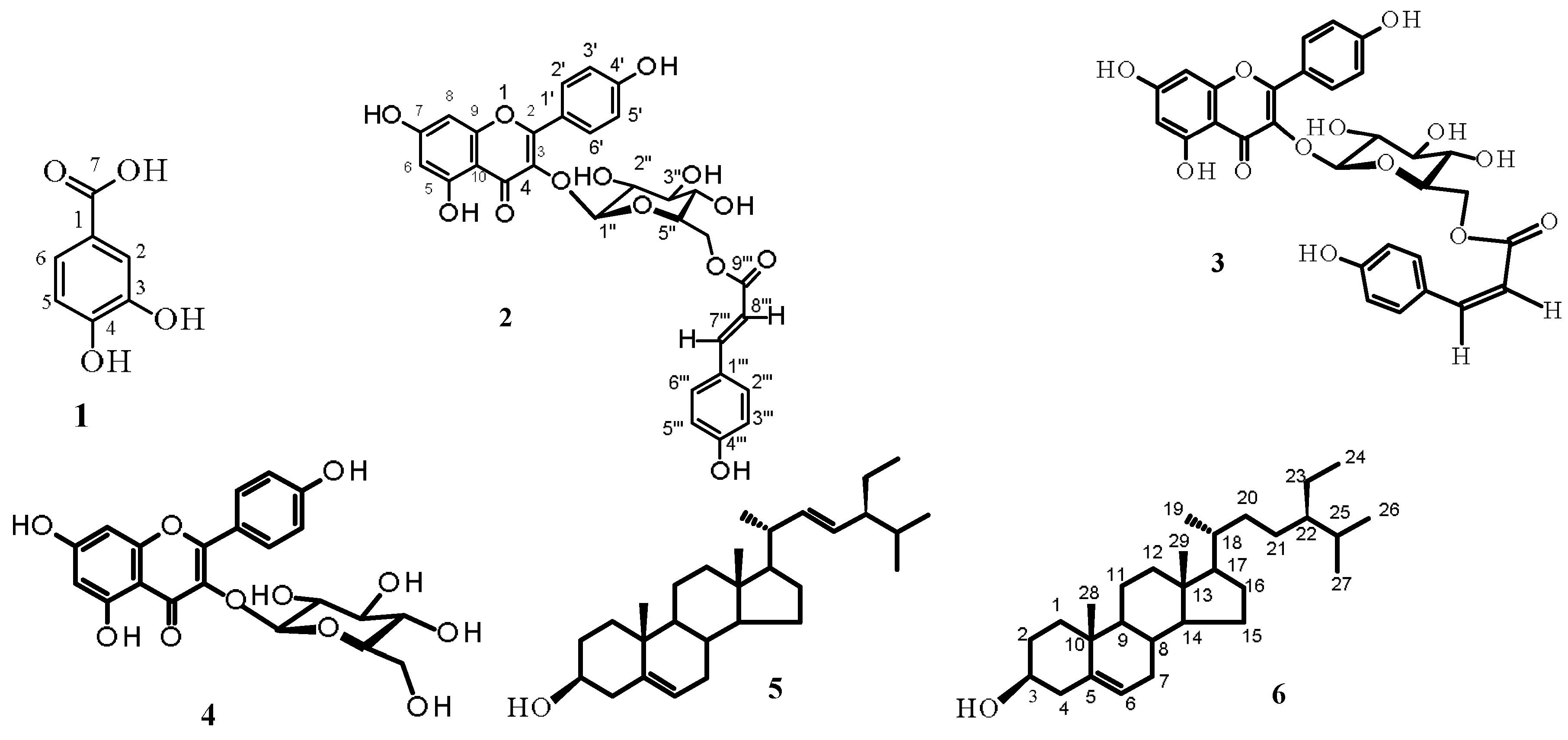
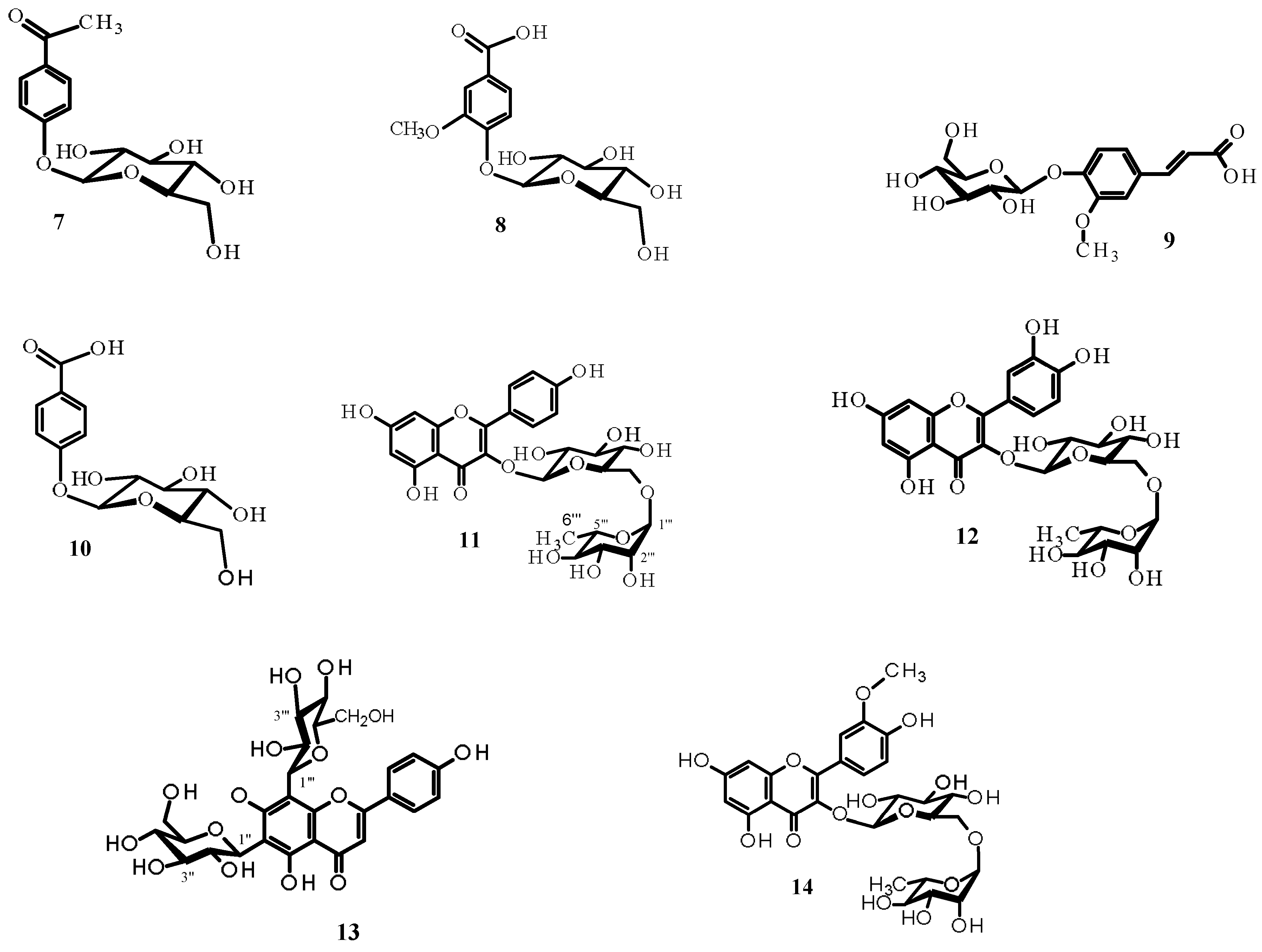
2.1. Isolation and Structure Elucidation of Compounds

Chromatographic procedures (CC and TLC of silica gel 60 and semi-preparative reverse phase HPLC) led to the isolation from the aerial parts of Helianthemum ruficomum, of five phenolic acids: protocatechuic acid (1) [32], picein (7) [33], vanillic acid 4-O-β-d-glucopyranoside (8) [34], lavandoside (trans-Ferulic acid 4-O-β-d-glucopyranoside) 9 [35,36], 4-hydroxybenzoic acid 4-O-β-d-glucopyranoside (10) [37]; seven flavonoid glycosides: trans-tiliroside (2) [38,39], slightly contamined by its stereoisomer cis-tiliroside, cis-tiliroside (3) [38,39] contamined by trans-tiliroside, astragalin (4) [40], nicotiflorin (11) [41], rutin (12) [41], vicenin-2 (13) [42], narcissin (14) [41], and a mixture (71%–29%) of stigmasterol (5) and β-sitosterol (6), respectively [43], (Figure 1). The compounds were identified by spectral analysis, mainly ESI-HRMS, UV and NMR experiments (1H, 13C, DEPT, DOSY, COSY, NOESY, HSQC and HMBC) and comparison of their spectroscopic data with those reported in the literature. To the best of our knowledge, compounds 5, 7, 8, 9, 10 and 14 were new for the genus Helianthemum. In addition, all the isolated compounds were described for the first time from this species. The spectra of all the compounds are available in the Supplementary Materials.
Protocatechuic acid (1): ESI-HRMS(+): m/z 155.03358 [M + H]+, accurate mass 154.0263; formula: C7H6O4; 1H-NMR (400 MHz, CD3OD) δH (ppm) 7.35 (1H, d, J = 1.8 Hz, H-2), 7.30 (1H, dd, J = 8.0, 1.8 Hz, H-6), 6.72 (1H, d, J = 8.0 Hz, H-5); 13C-NMR (100 MHz, CD3OD) δC (ppm) 168.87 (C-7), 149.76 (C-4), 144.72 (C-3), 123.09 (C-1), 122.56 (C-6), 116.91 (C-2), 114.95 (C-5).
trans-Tiliroside (2): Yellow powder, ESI-HRMS(+): m/z 617.12741 [M + Na]+, 595.14507 [M + H]+, formula: C30H26O13; 1H-NMR (400 MHz, DMSO-d6) δH (ppm) aglycone: 7.98 (2H, d, J = 8.8 Hz, H-2′, H-6′), 6.84 (2H, d, J = 8.8 Hz, H-3′, H-5′), 6.26 (1H, d, J = 1.6 Hz, H-8), 6.05 (1H, d, J = 1.6 Hz, H-6), sugar moiety: 5.41 (1H, d, J = 6.8 Hz, H-1″), 4.28 (1H, dd, J = 12.0, 2.8 Hz, H-6″a), 4.03 (1H, dd, J =12.0, 5.2 Hz, H-6″b), 3.40 (1H, m, H-5″), 3.30 (1H, m, H-3″), 3.25 (1H, m, H-2″), 3.19 (1H, m, H-4″), p-coumaroyl moiety: 7.38 (2H, d, J = 8.8 Hz, H-2′′′, H-6′′′), 7.36 (1H, d, J = 16.0 Hz, H-7′′′), 6.79 (2H, d, J = 8.8 Hz, H-3′′′, H-5′′′), 6.13 (1H, d, J = 16.0 Hz, H-8′′′); 13C-NMR (100 MHz, DMSO-d6) δC (ppm) aglycone: 177.31 (C-4), 166.69 (C-7), 161.54 (C-5), 160.38 (C-4′), 157.07 (C-9), 157,06 (C-2), 133.40 (C-3), 131.18 (C-2′, C-6′), 121.28 (C-1′), 115,53 (C-3′, C-5′), 103.30 (C-10), 99.99 (C-6), 94.55 (C-8), sugar moiety: 101.79 (C-1″), 76.71 (C-3″), 74.66 (C-5″), 74.60 (C-2″), 70.39 (C-4″), 63.46 (C-6″), p-coumaroyl moiety: 166.69 (C-9′′′), 156.36 (C-4′′′), 145.13 (C-7′′′), 130.64 (C-2′′′, C-6′′′), 125.30 (C-1′′′), 116.31 (C-3′′′, C-5′′′), 114.04 (C-8′′′). This compound was slightly contamined by its stereoisomer cis-tiliroside.
cis-Tiliroside (3): This compound was obtained as a mixture with trans-tiliroside (2) (42% trans-tiliroside- 58% cis-tiliroside). Yellow powder, ESI-HRMS(+): m/z 595.14494 [M + H]+, formula: C30H26O13; 1H-NMR (500 MHz, CD3OD) δH (ppm) aglycone : 7.99 (2H, d, J = 8.9 Hz, H-2′, H-6′), 6.80 (2H, d, J = 8.9 Hz, H-3′, H-5′), 6.31(1H, d, J = 2.0 Hz, H-8), 6.18 (1H, d, J = 2.0 Hz, H-6), sugar moiety: 5.18 (1H, d, J = 7.5 Hz, H-1″), 4.28 (1H, dd, J = 12.0, 2.8 Hz, H-6″a), 4.18 (1H, dd, J = 12.0, 5.2 Hz, H-6″b), 3.45 (1H, m, H-5″), 3.43 (1H, m, H-3″), 3.40 (1H, m, H-2″), 3.28 (1H, m, H-4″), p-coumaroyl moiety: 7.51(2H, d, J = 8.8 Hz, H-2′′′, H-6′′′), 7.31(2H, d, J = 8.0 Hz, H-3′′′, H-5′′′), 6.80 (1H, d, J = 12.3 Hz, H-7′′′), 5.51(1H, d, J = 12.3 Hz, H-8′′′); 13C-NMR (125 MHz, CD3OD) δC (ppm) aglycone: 177.98 (C-4), 163.61 (C-7), 161.54 (C-5), 159.84 (C-4′), 157.24 (C-2),157.18 (C-9), 133.78 (C-3), 130.87 (C-2′, C-6′), 121.34 (C-1′), 115.42 (C-3′, C-5′), 102.71 (C-10), 97.40 (C-6), 94.91 (C-8), sugar moiety: 101.94 (C-1″), 76.61 (C-3″), 74.27 (C-5″), 74.15 (C-2″), 70.30 (C-4″), 62.65 (C-6″), p-coumaroyl moiety: 166.35 (C-9′′′), 159.68 (C-4′′′), 132.37 (C-2′′′, C-6′′′), 129.80 (C-3′′′, C-5′′′), 126.15 (C-1′′′), 114.65 (C-7′′′), 114.34 (C-8′′′).
Astragalin (4): ESI-HRMS(+): m/z 449.10709 [M + H]+, formula: C21H20O11; 1H-NMR (400 MHz, CD3OD) δH (ppm) aglycone: 7.94 (2H, d, J = 8.8 Hz, H-2′, H-6′), 6.79 (2H, d, J = 8.8 Hz, H-3′, H-5′), 6.28 (1H, d, J = 2.0 Hz, H-8), 6.09 (1H, d, J = 2.0 Hz, H-6), sugar moiety: 5.13 (1H, d, J = 7.3 Hz, H-1″), 4.61 (1H, dd, J = 12.0, 2.4 Hz, H-6″a), 4.46 (1H, dd, J = 12.0, 5.2 Hz, H-6″b), 3.35 (1H, m, H-2″), 3.34 (1H, m, H-3″), 3.24 (1H, t, J = 8.0 Hz, H-4″), 3.12 (1H, m, H-5″); 13C-NMR (100 MHz, CD3OD) δC (ppm) aglycone: 178.06 (C-4), 164.65 (C-7), 161.52 (C-5), 160.27 (C-4′), 157.73 (C-2), 157.04 (C-9), 134.15 (C-3), 131.05 (C-2′, C-6′), 121.33 (C-1′), 114.91 (C-3′, C-5′), 104.36 (C-10), 98.74 (C-6), 93.64 (C-8), sugar moiety: 102.79 (C-1″), 77.06 (C-5″), 76.64 (C-3″), 74.34 (C-2″), 69.98 (C-4″), 61.21 (C-6″).
Stigmasterol (5): This compound was obtained as a mixture with the compound 6; HRMS: m/z 412.3713 [M]+, formula: C29H48O; 1H-NMR (500 MHz, CDCl3) δ (ppm): 5.28 (1H, brd, J = 5.1 Hz, H-6), 5.11 (1H, dd, J =14.9, 8.5 Hz, H-21), 4.97 (1H, dd, J = 14.9, 8.2 Hz, H-20), 3.45 (1H, m, H-3), 1.03 (3H, s, H3-29), 0.94 (3H, d, J = 6.2 Hz H3-19), 0.88 (3H, t, J = 7.1 Hz, H3-24), 0.86 (3H, d, J = 6.6 Hz, H3-26), 0.82 (3H, d, J = 6.6 Hz, H3-27), 0.73 (3H, s, H-28); 13C-NMR (125 MHz, CDCl3) δC (ppm) 141.12 (C, C-5), 137.95 (CH, C-20), 129.93 (CH, C-21), 121.73 (CH, C-6), 72.22 (CH, C-3), 56.73 (CH, C-14), 56.18 (CH, C-17), 50.25 (CH, C-9), 46.13 (CH, C-22), 42.40 (CH2, C-4), 42.38 (C, C-13), 40.60 (CH, C-18), 39.92 (CH2, C-12), 37.63 (CH2, C-1), 36.60 (C, C-10), 32.11 (CH2, C-2), 31.83 (CH2 and CH, C-7 and C-8), 29.64 (CH, C-25), 29.31 (CH2, C-16), 25.45 (CH2, C-23), 24.44 (CH2, C-15), 21.72 (CH3, C-19), 21.50 (CH2, C-11), 20.23 (CH3, C-26), 19.82 (CH3, C-27), 18.93 (CH3, C-28), 12.25 (CH3, C-29), 12.14 (CH3, C-24).
β-Sitosterol (6): White crystals; HRMS(+): m/z 414.3864 [M]+, formula: C29H50O; 1H-NMR (500 MHz, CDCl3) δH (ppm) 5.34 (1H, brd, J = 6.4 Hz, H-6), 3.54 (1H, m, H-3), 1.02 (3H, s, H3-29), 0.94 (3H, d, J = 6.5 Hz, H3-19), 0.85 (3H, d, J = 7.2 Hz, H3-24), 0.84 (3H, t, J = 6.4 Hz, H3-26), 0.81 (3H, d, J = 6.4 Hz, H3-27), 0.68 (3H, s, H3-28); 13C-NMR (125 MHz, CDCl3) δC (ppm) 140.75 (C, C-5), 121.63 (CH, C-6), 71.80 (CH, C-3), 56.78 (CH, C-14), 56.12 (CH, C-17), 50.19 (CH, C-9), 45.89 (CH, C-22), 42.34 (C, C-13), 42.34 (CH2, C-4), 39.83 (CH2, C-12), 37.28 (CH2, C-1), 36.52 (C, C-10), 36.14 (CH, C-18), 33.99 (CH2, C-20), 31.92 (CH2 and CH, C-7 and C-8), 31.65 (CH2, C-2), 29.24 (CH, C-25), 28.24 (CH2, C-16), 26.20 (CH2, C-21), 24.30 (CH2, C-15), 23.13 (CH2, C-23), 21.12 (CH2, C-11), 19.79 (CH3, C-28), 19.38 (CH3, C-27), 19.07 (CH3, C-26), 18.78 (CH3, C-19), 11.98 (CH3, C-24), 11.86 (CH3, C-29). This compound was obtained as a mixture with stigmasterol (5) (29 and 71%, respectively).
Picein (7): ESI-HRMS(+): m/z 321.09386 [M + Na]+, 619.19864 [2M + Na]+, accurate mass 298.10461; formula: C14H18O7, m/z 137.05910 [M + H − 162]+ indicating the presence of a hexose moiety linked to the rest of the molecule by an O-glycosidic bond; 1H-NMR (400 MHz, DMSO-d6) δH (ppm) aglycone: 7.91 (2H, d, J = 8.6 Hz, H-2, H-6), 7.11 (2H, d, J = 8.6 Hz, H-3, H-5), 2.51 (3H, s, H3-7), sugar moiety: 4.99 (1H, d, J = 7.2 Hz, H-1′), 3.69 (1H, brd, J = 12.1 Hz, H-6′a), 3.48 (1H, dd, J = 12.1, 5.9 Hz, H-6′b), 3.42 (1H, m, H-5′), 3.35 (1H, m, H-3′), 3.30 (1H, dd, J = 9.2, 7.2 Hz, H-2′), 3.19 (1H, t, J = 9.2 Hz, H-4′); 13C-NMR (100 MHz, DMSO-d6) δC (ppm) aglycone: 198.99 (C-7), 162.25 (C-4), 132.07 (C-1), 131.73 (C-2, C-6), 117.18 (C-3, C-5), 27.64 (C-8), sugar moiety: 100.79 (C-1′), 77.93 (C-5′), 77.26 (C-3′), 74.16 (C-2′), 70.72 (C-4′), 61.74 (C-6′).
Vanillic acid 4-O-β-d-glucopyranoside (8): ESI-HRMS(+): m/z 353.08341 [M + Na]+, formula: C14H18O9, m/z 169.04893 [M + H − 162]+ indicating the presence of a hexose moiety linked to the rest of the molecule by an O-glycosidic bond; 1H-NMR (400 MHz, CD3OD) δH (ppm) aglycone: 7.56 (1H, dd, J = 8.4, 2.0 Hz, H-6), 7.51 (1H, d, J = 2.0 Hz, H-2), 7.12 (1H, d, J = 8.4 Hz, H-5), 3.83 (3H, s, OCH3), sugar moiety: 5.04 (1H, d, J = 7.2 Hz, H-1′), 3.85 (1H, brd, J = 12.0 Hz, H-6′a), 3.67 (1H, dd, J = 12.0, 5.6 Hz, H-6′b), 3.55 (1H, t, J = 8 Hz, H-2′), 3.53 (1H, m, H-3′), 3.48 (1H, m, H-5′), 3.42 (1H, m, H-4′); 13C-NMR (100 MHz, CD3OD) δC (ppm) aglycone: 167.65 (C-7), 148.67 (C-4), 146.96 (C-3), 123.03 (C-1), 122.35(C-6′), 113.32 (C-5), 111.59 (C-2), 54.21 (OCH3), sugar moiety: 98.72 (C-1′), 75.01 (C-5′), 74.49 (C-3′), 71.64 (C-2′), 68.17 (C-4′), 59.34 (C-6′).
Lavandoside (trans-ferulic acid 4-O-β-d-glucopyranoside, 9): ESI-HRMS(+): m/z 379.09896 [M + Na]+, 735.20725 [2M + Na]+ according to the molecular formula C16H20O9, m/z 195.06445 [M + H − 162]+ indicating the presence of a hexose moiety linked to the rest of the molecule by an O-glycosidic bond; 1H-NMR (400 MHz, CD3OD) δH (ppm) aglycone: 7.62 (1H, d, J = 16.4 Hz, H-7), 7.25 (1H, brs, H-2), 7.19 (1H, d, J = 8.0 Hz, H-5), 7.18 (1H, brd, H-6), 6.41 (1H, d, J = 16.4 Hz, H-8), 3.91 (3H, s, OCH3), sugar moiety: 4.99 (1H, d, J = 7.3 Hz, H-1′), 3.89 (1H, dd, J = 10.2, 3.9 Hz, H-6′a), 3.71 (1H, dd, J = 10.2, 5.8 Hz, H-6′b), 3.54 (1H, m, H-2′), 3.52 (1H, t, J = 8.0 Hz, H-3′), 3.45 (1H, m, H-5′), 3.43 (1H, m, H-4′); 13C-NMR (100 MHz, CD3OD) δC(ppm) aglycone: 169.39 (C-9), 149.58 (C-3), 148.51 (C-4), 144.45 (C-7), 129.31 (C-1), 121.99 (C-6), 116.84 (C-8), 116.01 (C-5), 111.03 (C-2), 55.40 (OCH3), sugar moiety: 100.82 (C-1′), 76.87 (C-5′), 76.44 (C-3′), 73.42 (C-2′), 69.89 (C-4″), 61.07 (C-6′).
4-Hydroxybenzoic acid 4-O-β-d-glucopyranoside (10): ESI-HRMS(−): m/z 299.0772 [M − H], formula: C13H16O8; 1H-NMR (500 MHz, CD3OD) δH (ppm) aglycone: 7.98 (2H, d, J = 9.0 Hz, H-2, H-6), 7.14 (2H, d, J = 9.0 Hz, H-3, H-5), sugar moiety: 5.03 (1H, d, J = 7.3 Hz, H-1′), 3.92 (1H, dd, J = 12.5, 2.5 Hz, H-6′a), 3.73 (1H, dd, J = 12.5, 5.5 Hz, H-6′b), 3.52 (1H, m, H-5′), 3.51 (1H, m, H-3′), 3.50 (1H, m, H-2′), 3.44 (1H, m, H-4′); 13C-NMR (125 MHz, CD3OD) δC (ppm) aglycone: 169.89 (C-7), 160.86 (C-4), 131.10 (C-2, C-6), 126.89 (C-1), 115.57 (C-3, C-5), sugar moiety: 100.34 (C-1′), 76.84 (C-5″), 76.55 (C-3″), 73.46 (C-2″), 69.91 (C-4′), 61.06 (C-6′).
Nicotiflorin (11): ESI-HRMS(+): m/z 595.16524 [M + H]+, 617.14788 [M + Na]+, formula: C27H30O15; 1H-NMR (400 MHz, DMSO-d6) δH (ppm): aglycone: 7.94 (2H, d, J = 8.8 Hz, H-2′, H-6′), 6.88 (d, 2H, J = 8.8 Hz, H-3′, H-5′), 6.38 (1H, d, J = 2.0 Hz, H-8), 6.16 (1H, d, J = 2.0 Hz, H-6), O-glucopyranosyl moiety: 5.34 (1H, d, J = 7.6 Hz, H-1″), 3.67 (1H, dd, J = 10.2, 3.6 Hz, H-6″a), 3.42 (1H, m, H-3″), 3.31(1H, dd, J = 10.2, 3.6 Hz, H-6″b), 3.25 (1H, m, H-5″), 3.04 (1H, m, H-4″), 3.23 (1H, m, H-2″), O-rhamnopyranosyl moiety: 4.40 (1H, d, J = 1.2 Hz, H-1′′′), 3.42 (1H, m, H-2′′′), 3.24 (1H, m, H-3′′′), 3.21 (1H, m, H-5′′′), 3.06 (1H, m, H-4′′′), 0.92 (3H, d, J = 6.4 Hz, H3-6′′′); 13C-NMR (100 MHz, DMSO-d6) δC (ppm) aglycone: 177.67 (C-4), 164.22 (C-7), 161.14 (C-5), 159.92 (C-4′), 157.11 (C-2), 156.90 (C-9), 133.55 (C-3), 131.37 (C-2′, C-6′), 121.58 (C-1′), 115,54 (C-3′, C-5′), 104.45 (C-10), 99.08 (C-6), 94.31 (C-8), O-glucopyranosyl moiety: 101.70 (C-1″), 76.03 (C-5″), 72.05 (C-4″), 70.53 (C-2″), 70.34 (C-3″), 67.43 (C-6″), O-rhamnopyranosyl moiety: 101.19 (C-1′′′), 70.81 (C-3′′′), 70.53 (C-4′′′), 70.34 (C-2′′′), 68.64 (C-5′′′), 17.83 (C-6′′′).
Rutin (12): ESI-HRMS(+): m/z 611.15894 [M + H]+, 633.1404 [M + Na]+, formula: C27H30O16; 1H-NMR (400 MHz, CD3OD) δH (ppm) aglycone: 7.68 (1H, d, J = 2.4 Hz, H-2′), 7.62 (1H, dd, J = 8.4, 2.4 Hz, H-6′), 6.88 (1H, d, J = 8.4 Hz, H-5′), 6.36 (1H, d, J = 2.0 Hz, H-8), 6.18 (1H, d, J = 2.0 Hz, H-6), O-glucopyranosyl moiety: 5.11 (1H, d, J = 7.2 Hz, H-1″), 3.82 (1H, dd, J = 9.6, 3.6 Hz, H-6″a), 3.50 (1H, t, J = 8.0 Hz, H-2″), 3.43 (1H, m, H-5″), 3.39 (1H, m, H-6″b), 3.35 (1H, m, H-3″), 3.29 (1H, m, H-4″), O-rhamnopyranosyl moiety: 4.55 (1H, d, J = 1.2 Hz, H-1′′′), 3.68 (1H, m, H-2′′′), 3.57 (1H, dd, J = 6.1, 3.5 Hz, H-3′′′), 3.45 (1H, dd, J = 11.0, 2.1 Hz, H-5′′′), 3.33 (1H, m, H-4′′′), 1.15 (3H, d, J = 6.4 Hz, H3-6′′′); 13C-NMR (100 MHz, CD3OD) δC (ppm) aglycone: 177.93 (C-4), 164.50 (C-7), 161.42 (C-5), 157.92 (C-9), 157.01 (C-2), 148.36 (C-4′), 144,36 (C-3′), 134.32 (C-3), 122.29 (C-1′), 121.77 (C-6′), 116.44 (C-2′), 114.74 (C-5′), 104.25 (C-10), 98.64 (C-6), 93.60 (C-8), O-glucopyranosyl moiety: 103.48 (C-1″), 76.81 (C-5″), 75.77 (C-3″), 74.38 (C-2″), 70.03 (C-4″), 67.23 (C-6″), O-rhamnopyranosyl moiety: 101.03 (C-1′′′), 70.92 (C-3′′′), 70.71 (C-2′′′), 70.03 (C-4′′′), 68.34 (C-5′′′), 16.55 (C-6′′′).
Vicenin-2 (13): HRESI-MS(+): m/z 595.16515 [M + H]+, formula: C27H30O15; 1H-NMR (400 MHz, DMSO-d6) δH (ppm) aglycone: 7.99 (2H, d, J = 8.4 Hz, H-2′, H6′), 6.91 (2H, d, J = 8.4 Hz, H-3′, H-5′), 6.76 (1H, s, H-3), 6-C-glucopyranosyl moiety: 4.80 (1H, d, J = 9.7 Hz, H-1″), 3.75 (1H, dd, J = 10.9, 3.4 Hz, H-6″a), 3.50 (1H, dd, J = 10.9, 6.2 Hz, H-6″b), 3.48 (1H, m, H-2″), 3.37 (1H, t, J = 8.6 Hz, H-4″), 3.30 (1H, t, J = 8.6 Hz, H-3″), 3.23 (1H, m, H-5″), 8-C-glucopyranosyl moiety: 4.75 (1H, d, J = 9.7 Hz, H-1′′′), 3.87 (1H, m, H-2′′′), 3.63 (2H, m, H-6′′′), 3.36 (1H, t, J = 8.6 Hz, H-4′′′), 3.33 (1H, m, H-5′′′), 3.28 (1H, m, H-3′′′); 13C-NMR (100 MHz, DMSO-d6) δC (ppm) aglycone: 182.78 (C-4), 161.60 (C-2), 160.67 (C-4′), 159.53 (C-7), 159.07 (C-5), 155.49 (C-9), 129.51 (C-2′, C-6′), 125.99 (C-1′), 116.25 (C-3′,C-5′), 103.61 (C-10), 103.16 (C-6), 103.08 (C-3), 101.27 (C-8), 6-C-glucopyranosyl moiety: 82.39 (C-5″), 79.32 (C-3″), 74.56 (C-1″), 72.43 (C-2″), 69.53 (C-4″), 61.92 (C-6″), 8-C-glucopyranosyl moiety: 81.36 (C-5′′′), 79.32 (C-3′′′), 73.82 (C-1′′′), 70.98 (C-2′′′), 69.53 (C-4′′′), 61.78 (C-6′′′).
Narcissin (14): This compound was slightly contamined by compound 9. ESI-HRMS(+): m/z 625.17636 [M + H]+, formula: C28H32O16; 1H-NMR (400 MHz, CD3OD) δH (ppm) aglycone: 7.95 (1H, d, J = 1.6 Hz, H-2′), 7.63 (1H, dd, J = 8.0, 1.6 Hz, H-6′), 6.92 (1H, d, J = 8.0 Hz, H-5′), 6.40 (1H, d, J = 1.5 Hz, H-8), 6.21 (1H, d, J = 1.5 Hz, H-6), 3.96 (3H, s, OCH3-3′), O-glucopyranosyl moiety: 5.24 (1H, d, J = 7.2 Hz, H-1″), 3.81 (1H, dd, J = 10.4, 3.4 Hz, H-6″a), 3.47 (1H, m, H-2″), 3.45 (1H, m, H-5″), 3.41 (1H, dd, J = 10.4, 6.5, H-6″b), 3.39 (1H, m, H-3″), 3.28 (1H, t, J = 9.3 Hz, H-4″), O-rhamnopyranosyl: 4.55 (1H, d, J = 1.2 Hz, H-1′′′), 3.63 (m, 1H, H-2′′′), 3.48 (1H, m, H-3′′′), 3.27 (1H, t, J = 9.3 Hz, H-4′′′), 3.0 (1H, m, H-5′′′), 1.12 (3H, d, J = 6.2 Hz, H3-6′′′); 13C-NMR (100 MHz, CD3OD) δH (ppm) aglycone: 177.91 (C-4), 164.57 (C-7), 161.55 (C-5), 157.46 (C-2), 157.05 (C-9), 149.43 (C-4′), 146.60 (C-3′), 134.12 (C-3), 122.62 (C-6′), 121.60 (C-1′), 114.72 (C-5′), 113.22 (C-2′), 104.32 (C-10), 98.59 (C-6), 93.56 (C-8), 55.42 (OCH3-3′), O-glucopyranosyl moiety: 103.11 (C-1″), 76.80 (C-5″), 75.97 (C-3″), 74.54 (C-4″), 74.38 (C-2″), 67.16 (C-6″), O-rhamnopyranosyl: 101.11 (C-1′′′), 72.38 (C-4′′′), 70.69 (C-3′′′), 70.24 (C-2′′′), 68.39 (C-5′′′), 16.50 (C-6′′′).

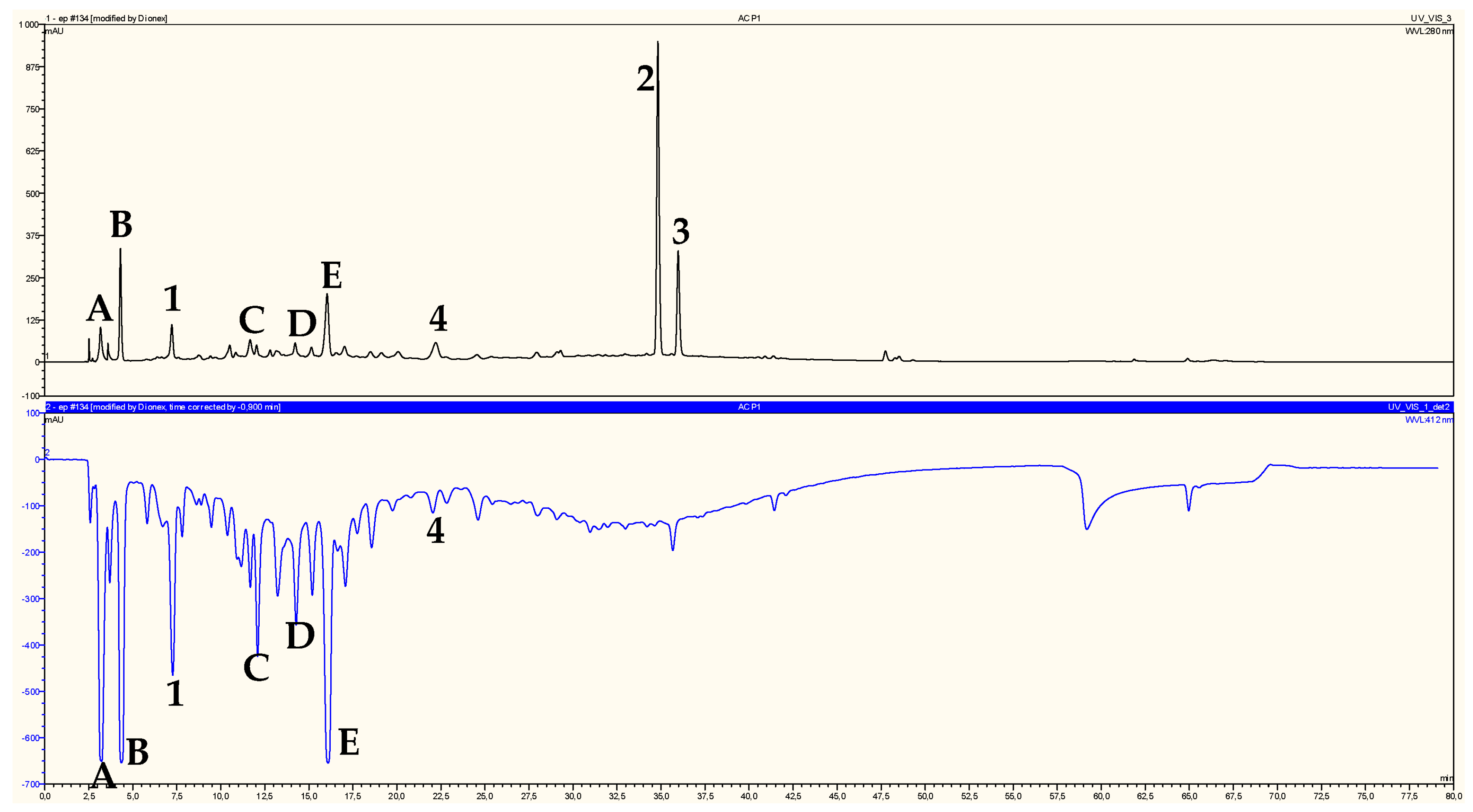
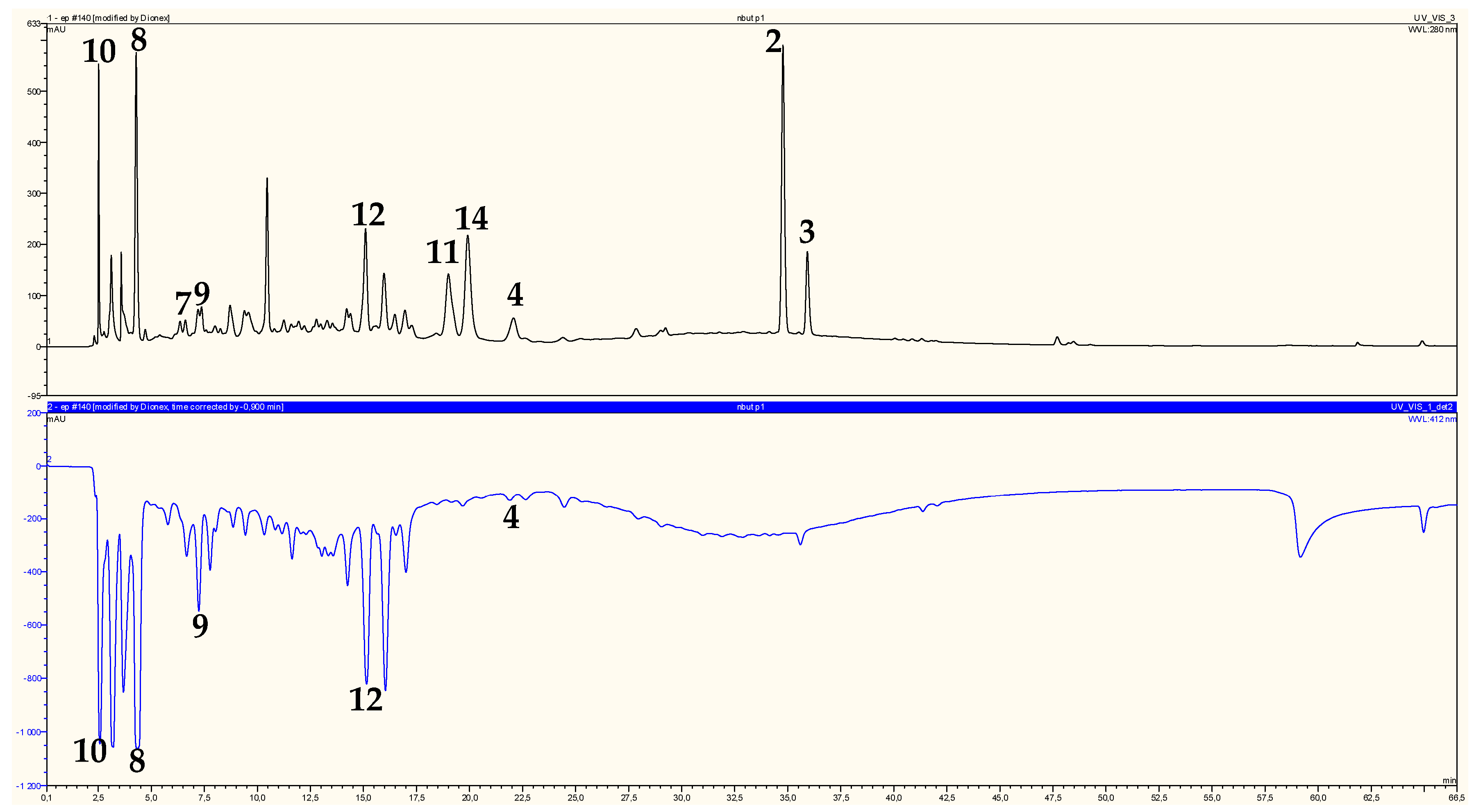
2.2. Identification of Chromatographic Peaks and Antioxidant Activity of Plant Extracts and Pure Compounds

On-line HPLC-ABTS•+ profiles of ethyl acetate and n-butanol extracts of H. ruficomum are reported in Figure 2 and Figure 3, respectively. These profiles showed a wealth of the two extracts in phenolic compounds detected by their absorbancy at 280 nm. The identification of chromatographic peaks was carried out after separation and purification by chromatographic techniques and re-injection of the pure isolated compounds (HPLC-ABTS•+) under the same conditions as the extracts. Regarding antioxidant activity, compounds protocatechuic acid (1), astragalin (4), vanillic acid 4-O-β-d-glucopyranoside (8), lavandoside (9), 4-hydroxybenzoic acid 4-O-β-d-glucopyranoside (10) and rutin (12) showed relatively high radical scavenging capacity (Table 1 and Table 2), in particular vanillic acid 4-O-β-d-glucopyranoside (8) and 4-hydroxybenzoic acid 4-O-β-d-glucopyranoside (10) were the most active (833 and 823 mAU respectively) (Table 2). The antioxidant activity of the n-BuOH extract is largely due to the presence of the three phenolic acids 8, 9, 10 and the flavonoid glycoside rutin (12) which represent 88.15% of the total activity (Table 2). The phenolic compound 1 and the flavonoid glucoside 4 represent only 10.34% of the total antioxidant activity of EtOAC extract (Table 1) as the compounds A, B, C, D and E which also showed high antioxidant activity (Figure 2) could not be isolated in pure state and then were not identified. These five components represented 62.21% of the ethyl acetate extract antioxidant activity (Table 1). trans-Tiliroside (2), cis-tiliroside (3), nicotiflorin (11) and narcissin (14) which showed a relatively high molecular absorbance on the upper chromatogram (Figure 3), exhibited little or no radical scavenging capacities. These results were confirmed off-line by ABTS, ORAC and TEAC assays. The measure of the free radical scavenger capacity of the two studied extracts of Helianthemum ruficomum by off-line TEAC test, confirmed these results and showed that EtOAc and n-BuOH extracts exhibited comparable activities (TEAC 432 and 431 µMolTE/mg, respectively, Table 3). This may be in relation with the strongest amount of phenolic and flavonoid compounds in these extracts. The off-line ABTS assay of the isolated compounds showed also that protocatechuic acid (1), vanillic acid 4-O-β-d-glucopyranoside (8), 4-hydroxybenzoic acid 4-O-β-d-glucopyranoside (10) and rutin (12) were the most potent antioxidant with on-line ABTS assay values: 341, 833, 823 and 594 mAU respectively (Table 1 and Table 2), off-line ABTS values: 202, 75, 254 and 644 mAU respectively (Table 3), ORAC essay values: 690, 651, 435 and 613 µMolTE/mg respectively (Table 3) and TEAC assay values: 469, 106, 408 and 556 µMolTE/mg (Table 3). The higher activity of rutin (12) in comparison to the other flavonoid glycosides present in these extracts may be due to the presence of the ortho di-OH system on the ring B of this molecule [44]. In addition, the higher activity of protocatechuic acid (1) in comparison with vanillic acid 4-O-β-d-glucopyranoside (8) and 4-hydroxybenzoic acid 4-O-β-d-glucopyranoside (10) may be due the lack of glycosylation, which has been found to diminish the radical scavenging activity [45]. This was also observed for ferulic acid glucoside (9), which showed relatively low antioxidant activity in comparison with ferulic acid [46,47].
The presence in this species, of phenolic acids and flavonoids was in good agreement with major studies reported on Helianthemum species [14,24,26,27]. The strong accumulation by this species, of trans-tiliroside which has been demonstrated to exert multiple biological effects [48,49] and the high antioxidant potential of its extracts and tested compounds, emphasized the possible relevance of this plant for Algerian traditional medicine. Moreover, it is important to note that no report has been published so far on eventual ethnomedical uses of this species. This may be due to a low distribution of this species.

3. Materials and Methods

3.1. General Procedure

Ultraviolet spectra were recorded using a Shimadzu model UV-1700 spectrophotometer. NMR spectra were obtained by Bruker model Avance 400 and AMX-500 spectrometers (Bruker BioSpin, Rheinstetten, Germany) with standard pulse sequences, operating at 400 and 500 MHz for 1H and 100 and 125 MHz for 13C, respectively. MeOH-d4, DMSO-d6, or CDCl3 were used as solvents with TMS as internal standard. High resolution mass spectra (ESI-HRMS) were performed on a Agilent 6520 Accurate Mass Q-TOF (Agilent Corporation, Santa Clara, CA, USA) and a µ-QToF spectrometer (Bruker Daltonics, Wissembourg, France). Column chromatography (CC) was carried out with Si gel Fluka (cat. 60737, 40–63 μm), and column fractions were monitored by TLC Si gel 60 F254, 0.2 mm, Macherey Nagel (cat. 818–333) by detection with a spraying reagent (CH3CO2H/H2O/H2SO4; 80:16:4) followed by heating at 100 °C. Preparative TLC was carried out on Si gel 60 PF254+366 (20 cm × 20 cm, 1 mm thickness, Analtech cat. 02014).

3.2. Plant Material

The plant material was collected from the area of Mougheul in the south-west of Algeria, latitude: N 32°1′23.6928″ (+32°1′23.6828″), longitude W 2°13′3.0648″ (−2°13′3.0648″) and authenticated by M. Mohamed Benabdelhakem, director of the nature preservation agency, Bechar on the basis of Quezel and Santa [23]. A voucher specimen (HCC0512-MOG-ALG-60) has been deposited at the Herbarium of the VARENBIOMOL research unit, Université des Frères Mentouri Constantine 1.

3.3. Extraction and Isolation

Air-dried aerial parts (1448 g) of Helianthemum ruficomum (Cistaceae) were macerated at room temperature with EtOH/H2O (80:20, v/v) for 72 h, three times. After filtration, the filtrates were concentrated in vacuum (up to 35 °C) and dissolved in distilled H2O (900 mL) under magnetic stirring and then put at the refrigerator for one night. After filtration, the resulting solution was extracted successively with petroleum ether, CHCl3, EtOAc and n-BuOH. The organic phases were filtered using common filter paper and concentrated in vacuum up to 35 °C to obtain the following dry extracts: petroleum ether (0.135 g), CHCl3 (0.9 g), EtOAc (5.142 g), n-BuOH (22.938 g). A part of the EtOAc extract (4 g) was dissolved in 5 mL of MeOH and subjected to column chromatography on silica gel (60–200 mesh, 160 g) eluted with CHCl3/MeOH step gradients to yield 41 fractions (F1–F41) obtained by combining the eluates on the basis of TLC analysis. Fraction F12 (57.8 mg) (CHCl3/MeOH, 96:4), was submitted to preparative plates of silica gel (CHCl3/MeOH, 12:1) to give Protocatechuic acid (1) (1 mg). Fraction F16 (113.7 mg) (CHCl3/MeOH, 94:6), was submitted to preparative plates of silica gel (CHCl3/MeOH, 7:1) to give cis-tiliroside (1.7 mg). Fraction F17 (930.6 mg) (CHCl3/MeOH, 93:7), was subjected to preparative plates of silica gel (CHCl3/MeOH, 2:0.5) and purified on analytical plates of silica gel GF254 (AcOEt/MeOH/H2O, 6:1:1) to give trans-tiliroside (80 mg) and astragalin (3 mg).
A part of the n-buthanol extract (16 g) was dissolved in 15 mL of MeOH and subjected to a column chromatography of silica gel (high-purity grade pore size 60 Å, 70-230 mesh, 63–200 µm, 478 g) eluted with CH2Cl2/MeOH step gradients to yield 33 fractions (F1–F33) obtained by combining the eluates on the basic of TLC and analytical HPLC analysis. Fraction F4 showed the formation of a white precipitate which was filtered (29.2 mg) and washed with methanol to give stigmasterol and β-sitosterol (5.3 mg) as a mixture (71 and 29%, respectively). From the fractions: F9 (288.2 mg), F11 (1281.7 mg), F13 (2837.3 mg), F14 (2054 mg), F24 (211 mg) and F32 (77 mg), aliquots were dissolved in methanol and submitted to a semi preparative HPLC separation using thermo column hypersil gold C18 (5 µm, 250 mm × 10 mm), with a mobile phase delivered at 5 mL/min consisting of mixture of Milli-Q water containing 0.1% formic acid (solvent A) and acetonitrile containing 0.1% formic acid (solvent B). The gradient was as follow: 0 min, 0% B; 30 min, 25% B; maintained during 7 min, to obtain picein (7) (2.1 mg) from F9, vanillic acid 4-O-β-d-glucopyranoside (8) (8 mg) and lavandoside (9) (1.2 mg) from F11, 4-hydroxybenzoic acid 4-O-β-d-glucopyranoside (10) (1.6 mg) and nicotiflorin (11) (2 mg) from F13, Rutin (12) (70 mg) from F14, vicenin-2 (13) (1 mg) from F24 and narcissin (14) (1.2 mg) from F32.

3.4. Solvents and Chemicals

Solvents: chloroform, ethyl acetate, formic acid and methanol reagent grade were purchased from VWR (Fontenay-sous-bois, France); acetonitrile HPLC grade was purchased from fisher scientific; Milli-Q water (18.2 MΩ) was generated by Millipore synergy system (Molsheim, France).
Chemicals: 2,2′-azino-bis(3-ethylbenzthiazoline-6-sulfonic acid) diammonium salt (ABTS•+), (C18H24N6O6S4) was purchased from Biochemica Applichem (Darmstadt, Germany); 2,2′-azobis(2-methylpropionamidine) dihydrochloride (AAPH), (C8H18N6 2HCl); (+)-6-hydroxy-2,5,7,8-tetramethylchromane-2-carboxylic acid (trolox), (C14H18O4); fluorescein (C20H12O5), were purchased from Sigma-Aldrich (Steinheim, Germany); sodium chloride was purchased from VWR; potassium chloride and potassium persulfate were purchased from Prolabo (Paris, France); potassium dihydrogen phosphate, disodium hydrogen phosphate and disodium dihydrogen phosphate were purchased from Merck (Darmstadt, Germany).

3.5. On-Line HPLC-ABTS•+ Assay

The ABTS•+ assay was based on the procedure described by Re (1999) [50] and Siddhuraju (2006) [51]. ABTS (7 mM) was dissolved in 20 mL of Milli-Q water, to which potassium persulfate (2.5 mM) was added, generating the radical cation ABTS•+ overnight. The solution was left overnight at 4 °C protected from light exposure. This solution was used within 4 days, dilutes in the Phosphate Buffer Saline (PBS) solution (pH 7.4) in order to reach an absorbance of 1.2 at 412 nm. Phosphate Buffer Saline (PBS) was prepared by dissolving in Milli-Q water, 80 g NaCl, 14.4 g Na2HPO4, 2.4 g KH2PO4 and 2 g KCl; the volume was completed with Milli-Q water to 1L. The pH was adjusted to 7.4 using NaOH 0.1 mol/L. The radical cation ABTS•+ solution (10 mL) was diluted in PBS (50 mL) and completed to 500 mL with Milli-Q water before use. The diode array detector (DAD) was connected to a mixing tee followed by a reaction coil (Peek, 20 m × 0.25 mm) loaded in a temperature controlled oven. Post-column reaction is operated by delivering (0.5 mL/min) the ABTS•+ reagent with an Ultimate 3000 variable wavelength detector through the mixing tee. After the reaction coil, the flow pass through a second molecular absorption photometric detector set at 412 nm to detect the reduced form of ABTS•+ radical and thus a reduced absorbancy. The result is presented as a double chromatogram, the upper part representing the phenolic compounds, detected by their absorbancy at 280 nm, while the lower part representing the free radical scavenging activity of these phenolic compounds. A negative peak indicates that a compound having radical scavenging activity elutes out of the chromatographic column and react with the ABTS•+ radical cation. The area of the chromatographic negative peak gives an indication on the radical-scavenging activity of the considered compound. The column used in the separation of EtOAc and n-BuOH extracts, was a Kromasil C18 with a 5 µm particle size, 4.60 mm × 250 mm (column temperature: 25 °C). The mobile phase delivered at 1 mL/min, was composed of 0.1% formic acid in H2O milli-Q (solvent A) and acetonitrile containing 1% formic acid (solvent B). Gradient was as follow: 0 min, 10% B; 10 min, 20% B; 20 min, 20% B; 50 min, 50% B; 55 min, 50% B; 56 min, 80% B; 66 min, 80% B; 67 min, 10% B, maintained during 13 min. Each phenolic compound was injected into the LC-ABTS•+ and quantified by reference to its appropriate authentic standard by absorption at 280 nm, whereas the antioxidant potential was calculated as the concentration of trolox required to produce an equivalent negative peak area by absorption at 412 nm and expressed as trolox equivalent antioxidant capacity (TEAC) or µMolTE/mg.

3.6. Oxygen Radical Absorbance Capacity (ORAC)

The ORAC assay, developed and validated by Ou et al. (2001) [52], was performed as described by Davalos (2004) [53] with minor modification Volden (2008) [54]. The assay measures the oxidative degradation of fluorescein by peroxyl radicals initiated by 2′,2-azobis(2-methylpropionamidine) dihydrochloride (AAPH) at 37 °C. Free radical scavenging molecules protect fluorescein from the oxidative degradation and until exhaustion, slow reduction of the fluorescence signal by inducing latency. The area under the curve of the kinetics of fluorescence is directly proportional to the amount and effectiveness of the free radical scavengers present in a sample. The results are therefore expressed as trolox equivalent (µMolTE/mg) of dry extract. The products are dissolved in a mixture of water/methanol (70/30) at 1 mg/mL (1000 ppm), and then have to be diluted with water (between 25 and 500 ppm) before being placed in triplicate in 96 wells micro-plate up to 10 µL/well. A trolox standard range between 25 and 500 µMol/L was also filed in triplicate. An aqueous solution of 150 µL fluorescein (8.5 × 10⁻⁶ mol/L) was added per well. An automatic dispenser then permits the initiation of the reaction by the addition of AAPH (30 µL, 153 µMol/L) to each well from the initiation of the generation of radicals by the addition of AAPH, the intensity of fluorescence emitted is measured every 5 min for 2 h with a wavelength of excitation between 400 and 600 nm.

4. Conclusions

The present study allowed the isolation, structural elucidation and antioxidant evaluation of phenolics and flavonoids from Helianthemum ruficomum, an endemic Saharan species on which no report is available so far. In this work, 14 compounds were isolated and identified from the ethyl acetate and n-butanol soluble parts of the aqueous EtOH extract, five phenolics: protocatechuic acid (1), picein (7), vanillic acid 4-O-β-d-glucopyranoside (8), lavandoside (9), 4-hydroxybenzoic acid 4-O-β-d-glucopyranoside (10); seven flavonoid glycosides: trans-tiliroside (2), slightly contamined by its stereoisomer cis-tiliroside, cis-tiliroside (3) contamined by trans-tiliroside, astragalin (4), nicotiflorin (11), rutin (12), vicenin-2 (13), narcissin (14); and a mixture (71–29%) of stigmasterol (5) and β-sitosterol (6) respectively. All the compounds were identified by spectral analysis, mainly ESI-HRMS, UV and NMR experiments (1H, 13C, DEPT, DOSY, COSY, NOESY, HSQC and HMBC) and comparison of their spectroscopic data with those reported in the literature. Compounds 5, 7, 8, 9, 10 and 14 were new for the genus Helianthemum. The investigated extracts and isolated compounds were evaluated for their free radical scavenging capacity by on-line HPLC-ABTS•+ screening. The antioxidant properties were confirmed by ORAC and TEAC assays. The results clearly indicated high antioxidant potential of the extracts and tested compounds of this species and agreed with literature data that free radical scavenging activity depends on the molecular structure, the number and position of the hydroxyl groups of tested compounds. Moreover, given the large amounts isolated and purified in this work, of trans-tiliroside and rutin which besides its numerous recognized biological activities, is used as oral complement; it becomes important to note that this Helianthemum species might be developed industrially for its rich content of these bioactive components. For this reason, this plant could be a good candidate for culture as a crop.

Supplementary Materials

1H-NMR, 13C-NMR, DEPT, COSY, HSQC, HMBC, NOESY, and MS spectra are available as supplementary materials. Supplementary materials can be accessed at: https://www.mdpi.com/1420-3049/22/2/239/s1.

Acknowledgments

This work was carried out with the support of “Programme algéro-français de bourses en doctorat PROFAS B+” and by the General Directorate for Scientific Research and Technological Development (DGRSDT), Ministry of Higher Education and Scientific Research (MESRS), Algeria.

Author Contributions

S.B., E.M. and F.B. conceived and designed the experiments; S.B. chose and collected the plant material; Y.C. performed the experiments; F.B., E.M., S.B., P.M., M.Z. and Y.C. analyzed the data; E.M. contributed with reagents, materials, and analysis tools; F.B. wrote the paper. All authors read, approved and revised the final manuscript.

Conflicts of Interest

The authors declare no conflict of interest.

References

  1. Singh, R.; Singh, B.; Singh, S.; Kumar, N.; Kumar, S.; Arora, S. Umbelliferone—An antioxidant isolated from Acacia nilotica (L.) Willd. Ex. Del. Food. Chem. 2010, 120, 825–830. [Google Scholar] [CrossRef]
  2. Deshpande, S. Role of antioxidants in prevention of age-related macular degeneration. J. Med. Nutr. Nutraceut. 2012, 1, 83–86. [Google Scholar] [CrossRef]
  3. Neera, J.P.; Singh, S.; Singh, J. Role of free radicals and antioxidants in human health and disease. Int. J. Curr. Res. Rev. 2013, 5, 14–22. [Google Scholar]
  4. Agrawal, S.B.; Singh, S.; Agrawal, M. Ultraviolet-B induced changes in gene expression and antioxidants in plants. Adv. Bot. Res. 2009, 52, 47–86. [Google Scholar]
  5. Simova-Stoilova, L.; Vaseva, I.; Grigorova, B.; Demirevska, K.; Feller, U. Proteolytic activity and cysteine protease expression in wheat leaves under severe soil drought and recovery. Plant Physiol. Biochem. 2010, 48, 200–206. [Google Scholar] [CrossRef] [PubMed]
  6. Zlatev, Z.S.; Lidon, F.C.; Ramalho, J.C.; Yordanov, I.T. Comparison of resistance to drought of three bean cultivars. Biol. Plant. 2006, 50, 389–394. [Google Scholar] [CrossRef]
  7. Boussaha, S.; Bekhouche, K.; Boudjerda, A.; Leόn, F.; Koldaş, S.; Yaglioglu, A.S.; Demirtas, I.; Brouard, I.; Marchioni, E.; Zama, D.; et al. Chemical constituents, in vitro antioxidant and antiproliferative activities of Perralderia coronopifolia Coss. subsp. eu-coronopifolia M. var. typica M. extract. Rec. Nat. Prod. 2015, 9, 312–322. [Google Scholar]
  8. Mohamadi, S.; Zhao, M.; Amrani, A.; Marchioni, E.; Zama, D.; Benayache, F.; Benayache, S. On-line screening and identification of antioxidant phenolic compounds of Saccocalyx satureioides Coss. et Dur. Ind. Crop. Prod. 2015, 76, 910–919. [Google Scholar] [CrossRef]
  9. Bougandoura, A.; D’Abrosca, B.; Ameddah, S.; Scognamiglio, M.; Mekkiou, R.; Fiorentino, A.; Benayache, S.; Benayache, F. Chemical constituents and in vitro anti-inflammatory activity of Cistanche violacea Desf. (Orobanchaceae) extract. Fitoterapia 2016, 109, 248–253. [Google Scholar] [CrossRef] [PubMed]
  10. Djebbari, R.; Chemam, Y.; Amrani, A.; Lassed, S.; Boubekri, N.; Zama, D.; Benayache, F.; Benayache, S. In vitro antioxidant activities of n-butanol extract of Helianthemum confertum. Int. J. Phytomed. 2015, 7, 119–122. [Google Scholar]
  11. Guzmán, B.; Vargas, P. Historical biogeography and character evolution of Cistaceae (Malvales) based on analysis of plastid rbcL and trnL-trnF sequences. Org. Divers. Evol. 2009, 9, 83–99. [Google Scholar] [CrossRef]
  12. Mabberly, D.J. The Plant Book; Cambridge University Pres: Cambridge, UK, 1997. [Google Scholar]
  13. Meckes, M.; Torres, J.; Calzada, F.; Rivera, J.; Camorlinga, M.; Lemus, H.; Rodríguez, G. Antibacterial properties of Helianthemum glomeratum, a plant used in Maya traditional medicine to treat diarrhea. Phytother. Res. 1997, 11, 128–131. [Google Scholar] [CrossRef]
  14. Rubio-Moraga, Á.; Argandoña, J.; Mota, B.; Pérez, J.; Verde, A.; Fajardo, J.; Gómez-Navarro, J.; Castillo-López, R.; Ahrazem, O.; Gómez-Gómez, L. Screening for polyphenols, antioxidant and antimicrobial activities of extracts from eleven Helianthemum taxa (Cistaceae) used in folk medicine in south-eastern Spain. J. Ethnopharmacol. 2013, 148, 287–296. [Google Scholar] [CrossRef] [PubMed]
  15. Meckes, M.; Villarreal, M.L.; Tortoriello, J.; Berlin, B.; Berlin, E.A. A microbiological evaluation of medicinal plants used by the Maya people of Southern Mexico. Phytother. Res. 1995, 9, 244–250. [Google Scholar] [CrossRef]
  16. Calzada, F.; Alanís, A.D.; Meckes, M.; Tapia-Contreras, A.; Cedillo-Rivera, R. In vitro susceptibility of Entamoeba histolytica and Giardia lamblia to some medicinal plants used by the people of Southern Mexico. Phytother. Res. 1998, 12, 70–72. [Google Scholar] [CrossRef]
  17. Rigat, M.; Bonet, M.A.; Garcia, S.; Garnatje, T.; Vallès, J. Studies on pharmaceutical ethnobotany in the high river Tervalley (Pyrenees, Catalonia, Iberian Peninsula). J. Ethnopharmacol. 2007, 113, 267–277. [Google Scholar] [CrossRef] [PubMed]
  18. Meckes, M.; Calzada, F.; Tapia-Contreras, A.; Cedillo-Rivera, R. Antiprotozoal propertiesof Helianthemum glomeratum. Phytother. Res. 1999, 13, 102–105. [Google Scholar] [CrossRef]
  19. Barbosa, E.; Calzada, F.; Campos, R. Antigiardial activity of methanolic extracts from Helianthemum glomeratum Lag. and Rubus coriifolius Focke in suckling mice CD-1. J. Ethnopharmacol. 2006, 108, 395–397. [Google Scholar] [CrossRef] [PubMed]
  20. Tawaha, K.; Alali, F.Q.; Gharaibeh, M.; Mohammad, M.; El-Elimat, T. Antioxidant activity and total phenolic content of selected Jordanian plant species. Food Chem. 2007, 104, 1372–1378. [Google Scholar] [CrossRef]
  21. Bouzergoune, F.; Bitam, F.; Aberkane, M.C.; Mosset, P. Preliminary phytochemical and antimicrobial activity investigations on the aerial parts of Helianthemum kahiricum. Chem. Nat. Compd. 2013, 49, 751–752. [Google Scholar] [CrossRef]
  22. Alsabri, S.G.; Rmeli, N.B.; Zetrini, A.A.; Mohamed, S.B.; Meshri, M.I.; Aburas, K.M.; Bensaber, S.M.; Mrema, I.A.; Mosbah, A.A.; Allahresh, K.A.; et al. Phytochemical, anti-oxidant, anti-microbial, anti-inflammatory and anti-ulcer properties of Helianthemum lippii. J. Pharmacogn. Phytochem. 2013, 2, 86–96. [Google Scholar]
  23. Benabdelaziz, I.; Marcourt, L.; Benkhaled, M.; Wolfender, J.L.; Haba, H. Antioxidant and antibacterial activities and polyphenolic constituents of Helianthemum sessiliflorum Pers. Nat. Prod. Res. 2016, 15, 686–690. [Google Scholar] [CrossRef] [PubMed]
  24. Calzada, F.; Alanis, A.D. Additional antiprotozoal flavonol glycosides of the aerial parts of Helianthemum glomeratum. Phytother. Res. 2007, 21, 78–80. [Google Scholar] [CrossRef] [PubMed]
  25. Barbosa, E.; Calzada, F.; Campos, R. In vivo antigiardial activity of three flavonoids isolated of some medicinal plants used in Mexican traditional medicine for the treatment of diarrhea. J. Ethnopharmacol. 2007, 109, 552–554. [Google Scholar] [CrossRef] [PubMed]
  26. Benabdelaziz, I.; Haba, H.; Lavaud, C.; Harakat, D.; Benkhaled, M. Lignans and other constituents from Helianthemum sessiliflorum Pers. Rec. Nat. Prod. 2015, 9, 342–348. [Google Scholar]
  27. Calzada, F.; Lopez, R.; Meckes, M.; Cedillo-Rivera, R. Flavonoids of the aerial parts of Helianthemum glomeratum. Int. J. Pharmacogn. 1995, 33, 351–352. [Google Scholar] [CrossRef]
  28. Javidnia, K.; Nasiri, A.; Miri, R.; Jamalian, A. Composition of the essential oil of Helianthemum kahiricum Del. from Iran. J. Essent. Oil Res. 2007, 19, 52–53. [Google Scholar] [CrossRef]
  29. Quezel, P.; Santa, S. Nouvelle Flore de l’Algérie et des Régions Désertiques Méridionales; Editions du C.N.R.S: Paris, France, 1963; p. 710. [Google Scholar]
  30. Ozenda, P. Flore du Sahara Septentrional et Central; CNRS: Paris, France, 1958; p. 350. [Google Scholar]
  31. The Plant List (2010). Version 1. Available online: http://www.theplantlist.org/ (accessed on 1 January 2017).
  32. An, L.J.; Guan, S.; Shi, G.F.; Bao, Y.M.; Duan, Y.L.; Jiang, B. Protocatechuic acid from Alpinia oxyphylla against MPP+-induced neurotoxicity in PC12 cells. Food Chem. Toxicol. 2006, 44, 436–443. [Google Scholar] [CrossRef] [PubMed]
  33. Ushiyama, M.; Furuya, T. Glycosylation of phenolic compounds by root culture of Panax ginseng. Phytochemistry 1989, 28, 3009–3013. [Google Scholar] [CrossRef]
  34. Cui, C.B.; Tezuka, Y.; Kikuchi, T.; Nakano, H.; Tamaoki, T.; Park, J.H. Constituents of a fern, Davallia mariesii Moore. IV.1) Isolation and structures of a novel norcarotane sesquiterpene glycoside, a chromone glucuronide and two epicatechin glycosides. Chem. Pharm. Bull. 1992, 40, 2035–2040. [Google Scholar] [CrossRef]
  35. Kurkin, V.A.; Lamrini, M.; Klochkov, S.G. Lavandoside from Lavandula spica flowers. Chem. Nat. Compd. 2008, 44, 169–170. [Google Scholar] [CrossRef]
  36. Yang, F.; Chen, R.; Feng, L.; Li, H.D.; Zhang, H.; Liang, J.Y. Chemical constituents from the aerial part of Peganum nigellatrum. Chin. J. Nat. Med. 2010, 8, 199–201. [Google Scholar] [CrossRef]
  37. Strack, D.; Heilemann, J.; Wray, V.; Dirks, H. Structures and accumulation patterns of soluble and insoluble phenolics from Norway spruce needles. Phytochemistry 1989, 28, 2071–2078. [Google Scholar] [CrossRef]
  38. Mekhelfi, T.; Kerbab, K.; Guella, G.; Zaiter, L.; Benayache, S.; Benayache, F. Phytochemical constituents of Thymelaea microphylla Coss. et Dur. from Algeria. Pharm. Lett. 2014, 6, 152–156. [Google Scholar]
  39. Kumarasamy, Y.; Cox, P.J.; Jaspars, M.; Rashid, M.A.; Sarker, S.D. Bioactive flavonoid glycosides from the seeds of Rosa canina. Pharm. Biol. 2003, 41, 237–242. [Google Scholar] [CrossRef]
  40. Wei, Y.; Xie, Q.; Fisher, D.; Sutherland, I.A. Separation of patuletin-3-O-glucoside, astragalin, quercetin, kaempferol and isorhamnetin from Flaveria bidentis (L.) Kuntze by elution-pump-out high-performance counter-current chromatography. J. Chromatogr. A 2011, 1218, 6206–6211. [Google Scholar] [CrossRef] [PubMed]
  41. Olennikov, D.N.; Tankhaeva, L.M.; Partilkhaev, V.V.; Rokhin, A.V. Chemical constituents of Caragana bungei shoots. Rev. Bras. Farmacogn. Braz. J. Pharmacogn. 2012, 22, 490–496. [Google Scholar] [CrossRef]
  42. Xie, C.; Veitch, N.C.; Houghton, P.J.; Simmonds, M.S.J. Flavone C-glycosides from Viola yedoensis Makino. Chem. Pharm. Bull. 2003, 51, 1204–1207. [Google Scholar] [CrossRef] [PubMed]
  43. Chaturvedula, V.S.P.; Prakash, I. Isolation of stigmasterol and β-sitosterol from the dichloromethane extract of Rubus suavissimus. Int. Curr. Pharm. J. 2012, 1, 239–242. [Google Scholar] [CrossRef]
  44. Rice-Evans, C.A.; Miller, N.J.; Paganga, G. Structure-antioxidant activity relationships of flavonoids and phenolic acids. Free Radic. Biol. Med. 1995, 20, 933–956. [Google Scholar] [CrossRef]
  45. Pratt, D.E. Natural antioxidants of soybean oil seeds. In Autoxidation in Food and Biological Systems; Simic, M.G., Karel, M., Eds.; Plenum Press: New York, NY, USA, 1980; pp. 283–293. [Google Scholar]
  46. Rice-Evans, C.A.; Miller, N.J.; Paganga, G. Antioxidant properties of phenolic compounds. Trends Plant Sci. 1997, 2, 152–159. [Google Scholar] [CrossRef]
  47. Wojdylo, A.; Oszmiański, J.; Czemerys, R. Antioxidant activity and phenolic compounds in 32 selected herbs. Food Chem. 2007, 105, 940–949. [Google Scholar] [CrossRef]
  48. Goto, T.; Teraminami, A.; Lee, J.Y.; Ohyama, K.; Funakoshi, K.; Kim, Y.I.; Hirai, S.; Uemura, T.; Yu, R.; Takahashi, N.; et al. Tiliroside, a glycosidic flavonoid, ameliorates obesity-induced metabolic disorders via activation of adiponectin signaling followed by enhancement of fatty acid oxidation in liver and skeletal muscle in obese-diabetic mice. J. Nutr. Biochem. 2012, 23, 768–776. [Google Scholar] [CrossRef] [PubMed]
  49. Schinella, G.R.; Tournier, H.A.; Máñez, S.; De Buschiazzo, P.M.; Del Carmen Recio, M.; Ríos, J.L. Tiliroside and gnaphaliin inhibit human low density lipoprotein oxidation. Fitoterapia 2007, 78, 1–6. [Google Scholar] [CrossRef] [PubMed]
  50. Re, R.; Pellegrini, N.; Protegente, A.; Pannala, A.; Yang, M.; Rice-Evans, C. Antioxidant activity applying an improved ABTS radical cation decolorization assay. Free Radic. Biol. Med. 1999, 26, 1231–1237. [Google Scholar] [CrossRef]
  51. Siddhuraju, P. The antioxidant activity and free radical-scavenging capacity of phenolics of raw and dry heated moth bean (Vigna aconitifolia) (Jacq.) Marechal seed extracts. Food Chem. 2006, 99, 149–157. [Google Scholar] [CrossRef]
  52. Ou, B.; Hampsch-Woodill, M.; Prior, R. Development and validation of an improved oxygen radical absorbance capacity assay using fluorescein as the flurescent probe. J. Agric. Food Chem. 2001, 49, 4619–4626. [Google Scholar] [CrossRef] [PubMed]
  53. Dávalos, A.; Gómez-Cordovés, C.; Bartolomé, B. Extending applicability of the oxygen radical absorbance capacity (ORAC-fluorescein) assay. J. Agric. Food Chem. 2004, 52, 48–54. [Google Scholar] [CrossRef] [PubMed]
  54. Volden, J.; Borge, G.I.A.; Bengtsson, G.B.; Hansen, M.; Thygesen, I.E.; Wicklund, T. Effect of thermal treatment on glucosinolates and antioxidant-related parameters in red cabbage (Brassica oleracea L. ssp. capitata f. rubra). Food Chem. 2008, 109, 595–605. [Google Scholar] [CrossRef]
  • Sample Availability: Samples of some compounds are available from the authors.
Figure 1. Structures of compounds (114) isolated from Helianthemum ruficomum.
Molecules 22 00239 g001aMolecules 22 00239 g001b
Figure 2. Chromatographic determination of antioxidant compounds (upper chromatogram) and their corresponding antioxidant activity (lower chromatogram) in ethyl acetate extract of H. ruficomum. Protocatechuic acid (1), trans-tiliroside (2), cis-tiliroside (3), astragalin (4), unidentified compounds (A, B, C, D and E).
Molecules 22 00239 g002
Figure 3. Chromatographic determination of antioxidant compounds (upper chromatogram) and their corresponding antioxidant activity (lower chromatogram) in n-butanol extract of H. ruficomum. Picein (7), vanillic acid 4-O-β-d-glucopyranoside (8), lavandoside (9), 4-hydroxybenzoic acid 4-O-β-d-glucopyranoside (10), nicotiflorin (11), rutin (12), narcissin (14).
Molecules 22 00239 g003
Table 1. Free radical scavenging activities of the phenolic compounds in the EtOAc extract of H. ruficomum based on ABTS•+ and their participation (%) in total antioxidant capacities with ABTS assay.
PeaksCompoundsABTS (µgTE/mL)Radical-Scavenging Activity (mAU)Scavenging Activity Percent (%)
ANot identified131.8354617.40
BNot identified137.4756916.86
1Protocatechuic acid81.563418.37
CNot identified63.962695.48
DNot identified42.571813.97
ENot identified118.0549018.50
4Astragalin11.48541.97
Table 2. Free radical scavenging activities of the phenolic compounds in n-butanol extract of H. ruficomum, based on ABTS•+ and their participation (%) in total antioxidant capacities with ABTS assay.
PeaksCompoundsABTS (µgTE/mL)Radical-Scavenging Activity (mAU)Scavenging Activity Percent (%)
8Vanillic acid 4-O-β-d-glucopyranoside201.9183335.27
9Lavandoside81.663419.37
104-hydroxybenzoic acid 4-O-β-d-glucopyranoside199.5082320.31
12Rutin143.5959423.20
Table 3. Free radical scavenging activities of the pure compounds and extracts of H. ruficomum based on ABTS, Oxygen Radical Absorbance Capacity (ORAC) and trolox equivalent antioxidant capacity (TEAC) assays.
PeaksPure Compounds or ExtractsABTS (µgTE/mL)Radical-Scavenging Activity (mAU)ORAC (µMolTE/mg)TEAC (µMolTE/mg)
EtOAc extract--18.43 ± 12.58432.06 ± 15.84
n-BuOH extract--5.64 ± 0.21430.66 ± 80.25
1Protocatechuic acid47.59202690.40 ± 45.67469.48 ± 15.71
4Astragalin1.4813300.006 ± 82.71144.69 ± 15.36
8Vanillic acid 4-O-β-d-glucopyranoside16.4975650.91 ± 185.18105.56 ± 8.95
9Lavandoside91.4938137.44 ± 24.7412.14 ± 7.34
104-Hydroxybenzoic acid 4-O-β-d-glucopyranoside60.45254434.86 ± 36.43407.94 ± 32.73
12Rutin155.66644612.77 ± 165.45555.66 ± 20.79
(Results expressed as mean ± SD, n = 3).

