Article

Phytochemical Study of Tapirira guianensis Leaves Guided by Vasodilatory and Antioxidant Activities

1
Laboratório de Biologia do Reconhecer, Centro de Biociências e Biotecnologia, Universidade Estadual do Norte Fluminense Darcy Ribeiro, Av. Alberto Lamego, 2000, Parque Califórnia, Campos dos Goytacazes, 28013-602 Rio de Janeiro, Brazil
2
Laboratório Integrado de Pesquisa, Universidade Federal do Rio de Janeiro, Campus Macaé, Av. Aluízio da Silva Gomes, 50, Novo Cavaleiros, Macaé, 27930-560 Rio de Janeiro, Brazil
3
Laboratório de Produtos Bioativos, Universidade Federal do Rio de Janeiro, Campus Macaé, Polo Novo Cavaleiro—IMCT, R. Alcides da Conceição, 159, Novo Cavaleiros, Macaé, 27933-378 Rio de Janeiro, Brazil
4
Núcleo de Estudos em Ecologia e Desenvolvimento Sócio-Ambiental de Macaé, Universidade Federal do Rio de Janeiro, Av. São José Barreto, 764—São José do Barreto. Macaé, 27965-045 Rio de Janeiro, Brazil
5
Instituto de Pesquisa de Produtos Naturais Walter Mors, Centro de Ciências da Saúde, Universidade Federal do Rio de Janeiro, Brazil
6
Núcleo de Pesquisa em Produtos Naturais e Sintéticos, Departamento de Física e Química, Faculdade de Ciências Farmacêuticas de Ribeirão Preto, Universidade de São Paulo, Av. do Café s/n. 14040-020 Ribeirão Preto, Brazil
7
Laboratório de Química, Universidade Federal do Rio de Janeiro—Campus Macaé, Av. Aluízio da Silva Gomes, 50, Novo Cavaleiros. Macaé, 27930-560 Rio de Janeiro, Brazil
8
Laboratório de Produtos Naturais e Ensaios Biológicos, Departamento De Produtos Naturais e Alimentos, Faculdade de Farmácia, Universidade Federal do Rio de Janeiro, 21941-902 Rio de Janeiro, Brazil
*
Authors to whom correspondence should be addressed.
Molecules 2017, 22(2), 304; https://doi.org/10.3390/molecules22020304
Submission received: 22 December 2016 / Revised: 31 January 2017 / Accepted: 1 February 2017 / Published: 18 February 2017

Abstract

:
The aim of this research was to perform a phytochemical study of the methanol leaves extract of T. guianensis (MET) guided by vasodilatory and antioxidant activities. The chemical profile of MET and the ethyl acetate fraction (EA fraction) was determined by HPLC-UV-MS and EA fraction guided fractionation by reverse-phase chromatography. The vasorelaxant effects of MET, fractions, sub-fractions and constituents were assessed on rat aorta pre-contracted with phenylephrine. Antioxidant activity was evaluated by using a DPPH assay. The results show that MET-induced vasodilation was dependent on NO/cGMP; and that the PI3K/Akt pathway seems to be the main route involved in eNOS activation. The EA fraction showed greater vasodilatory and antioxidant potency and was submitted to further fractionation. This allowed the isolation and characterization of quercetin, quercetin 3-O-(6″-O-galloyl)-β-d-galactopyranoside and 1,4,6-tri-O-galloyl-β-d-glucose. Also, galloyl-HHDP-hexoside and myricetin deoxyhexoside were identified by HPLC-UV-MS. These compounds are being described for the first time for T. guianensis. 1,4,6-tri-O-galloyl-β-d-glucose and quercetin 3-O-(6″-O-galloyl)-β-d-galactopyranoside showed no vasodilatory activity. Quercetin and myricetin glycoside seems to contribute to the MET activity, since they have been reported as vasodilatory flavonoids. MET-induced vasodilation could contribute to the hypotensive effect of T. guianensis previously reported.

1. Introduction

Hypertension is one of the key risk factors for cardiovascular diseases, which are the main cause of death worldwide [1]. Hypertension is characterized by a chronic elevation of arterial blood pressure, in which abnormally increased vascular tone plays a major role in the maintenance of high blood pressure [1].
The endothelium regulates the contractility of vascular smooth muscle by releasing relaxing and contracting factors and the loss of its normal function results in increased vascular tone [2]. Endothelial dysfunction is a common feature of hypertensive patients, a condition that includes reduced endothelium-dependent vasodilation, morphological changes of vascular smooth muscle and a hypercoagulatory state [3,4,5]. Elevated levels of reactive oxygen species are a key player in the pathogenesis of endothelium dysfunction leading to decreased nitric oxide (NO) bioavailability, the main endothelium relaxant factor, and endothelial NO synthase (eNOS) uncoupling [5,6].
Phenolic compounds are plant secondary metabolites widely distributed in Nature that are known for their beneficial effects in many processes involved in the pathogenesis of cardiovascular diseases. Epidemiological assessments show reduction in the incidence of these diseases, besides beneficial effects observed in preclinical and clinical studies. Phenolic compounds have been related to vasodilatory activity [7], hypotensive effect [8,9], improvement of endothelial dysfunction, reduction of oxidative stress [10,11,12] and attenuation of vascular aging [13]. Moreover, the cardiovascular action of herbal and non-herbal products is largely attributed to the presence of phenolic compounds [12].
Tapirira genus (Anacardiaceae) is composed of approximately 40 species mainly distributed in South America [14]. Among them, Tapirira guianensis Aubl. species is popularly known in Brazil as “pau-pombo” or “tapiririca”. It is used in traditional medicine against leprosy, diarrhea and syphilis [15]. Moreover, in Brazilian Amazonian coastal areas, the natural wood and an inner bark mixture of T. guianensis are used for the treatment of infant oral thrush and throat and mouth sore, respectively [16].
Phytochemical studies have reported the isolation of different secondary metabolites from T. guianensis, some with already described biological activities. David et al. [15] reported the presence of β-sitosterol, in addition with two new compounds identified as 2-[10(Z)-heptadecenyl]-1,4-hydroquinone and (4R,6R)-dihydroxy-4-[10(Z)-heptadecenyl]-2-cyclohexenone in a methanol extract of the seeds, which showed cytotoxic activity against different human cancer cell lines. Flavonoids, norisoprenoids and terpenes have been isolated from T. guianensis leaves, including the flavonoids kaempferol 3-α-rhamnoside, kaempferol-3-α-arabinofuranoside, quercetin-3-α-rhamnoside and kaempferol [17]. Roumy et al. [18] identified new cyclic alkyl polyol derivatives from T. guianensis bark with anti-protozoal and anti-bacterial activities.
The ethanol extract of leaves and twigs of T. guianensis induced a 38.5% reduction of arterial blood pressure in anaesthetized rats, indicating its potential as a hypotensive agent. This effect could be related to the presence of triterpenoids and flavonoids in the extract [19]. However, there are no studies about the mechanisms involved in the cardiovascular action of T. guianensis, as well as about the compounds responsible for that effect. Thus, the aim of the present study was to investigate the vasodilatory and antioxidant activities of the methanol extract of T. guianensis (MET) and to identify the compounds involved in these effects.

2. Results

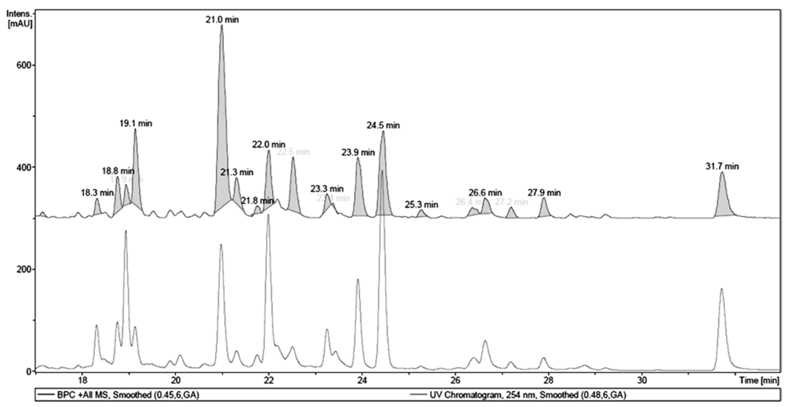
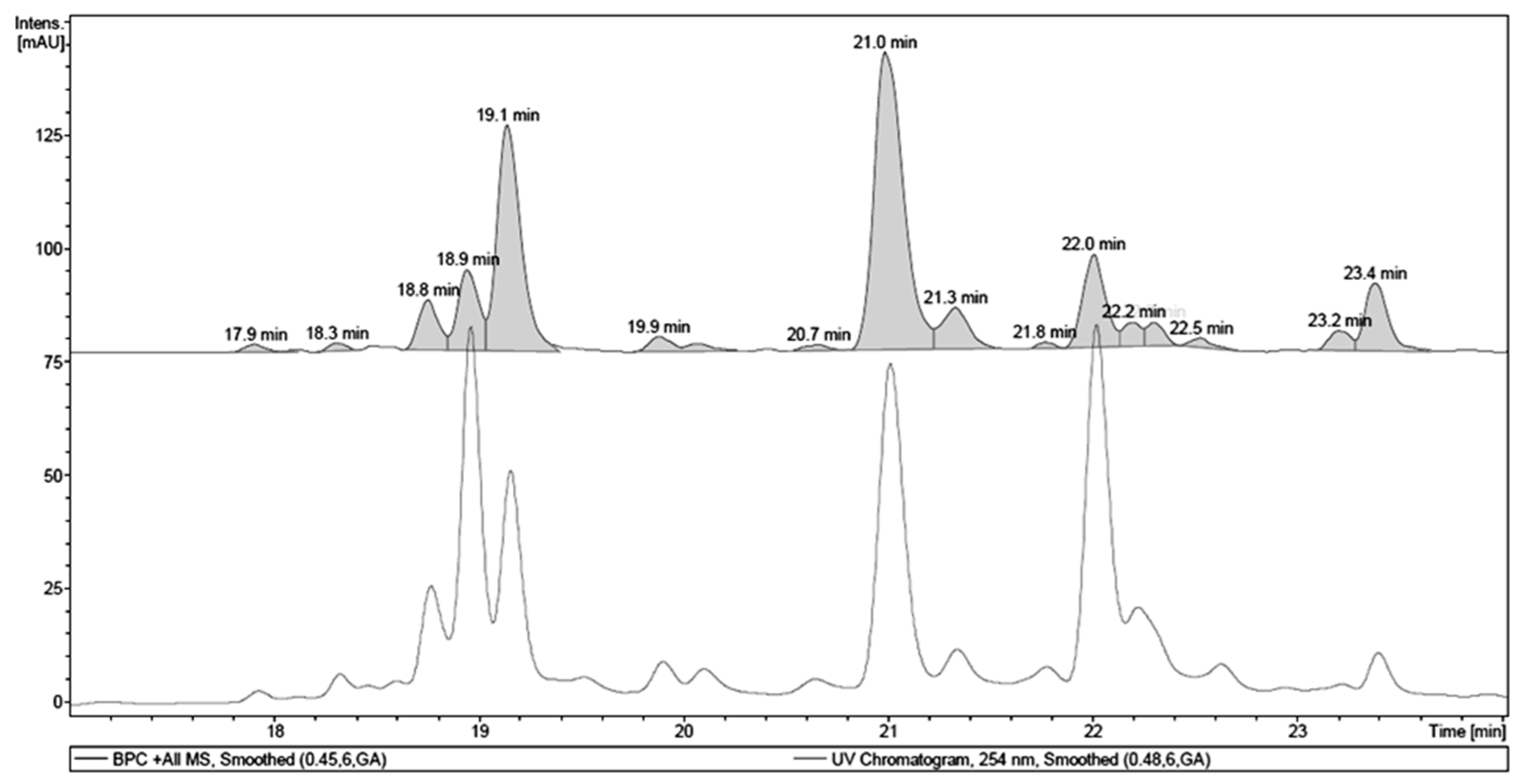
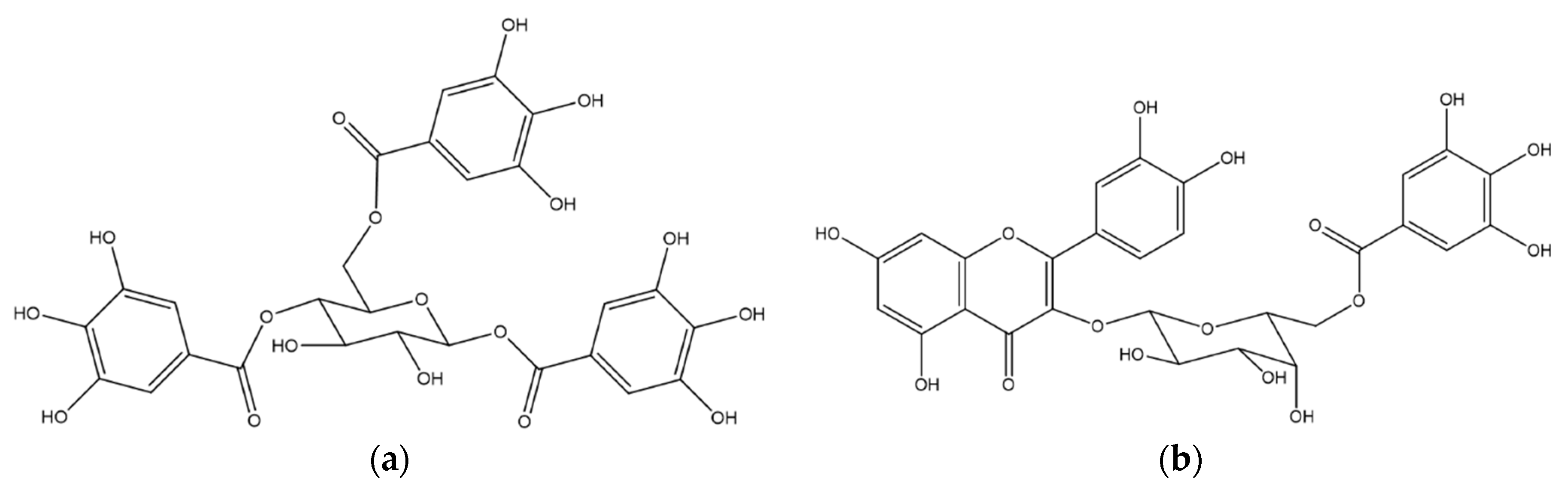
2.1. Chemical Analysis of MET

Initially, MET was fractionated affording a main active fraction obtained by partition with EA. The EA fraction was analyzed by LC-UV-MS (Figure 1) and showed a very similar phenolic profile when compared to MET. Figure 2 shows UV and MS chromatograms of EA, where the main UV chromatogram peaks, tR 21.0, 22.0, 23.9, 24.5 and 31.7 min, showed a characteristic flavonol UV spectrum, with λMAX at 250–280 and 350–380 nm [20], with the exception of the peak at 18.9 min. MS analyses of each main peak allowed the identification of hydrolysable tannins and flavonols, specifically quercetin and myricetin glycosides, as shown in Figure 1 and Table 1.
Chromatographic fractionation of the EA fraction afforded four phenolic sub-fractions: 3, 4, 5 and 6. Two of them, 3 and 6, corresponded to pure compounds, the hydrolysable tannin 1,4,6-tri-O-galloyl-β-d-glucopyranose (EA1) and the flavonoid quercetin (EA3), respectively. From sub-fraction 4, the flavonoid 3-O-(6″-O-galloyl)-β-d-galactopyranoside (EA2) was isolated and characterized In addition, sub-fraction 4 was also analyzed by LC-UV-MS. Thus, it was possible to identify the isolated compounds EA1 and EA2 and other major components: one hydrolysable tannin, galloyl-HHDP-hexoside, and one flavonoid, myricetin deoxyhexoside (Figure 2 and Table 2).

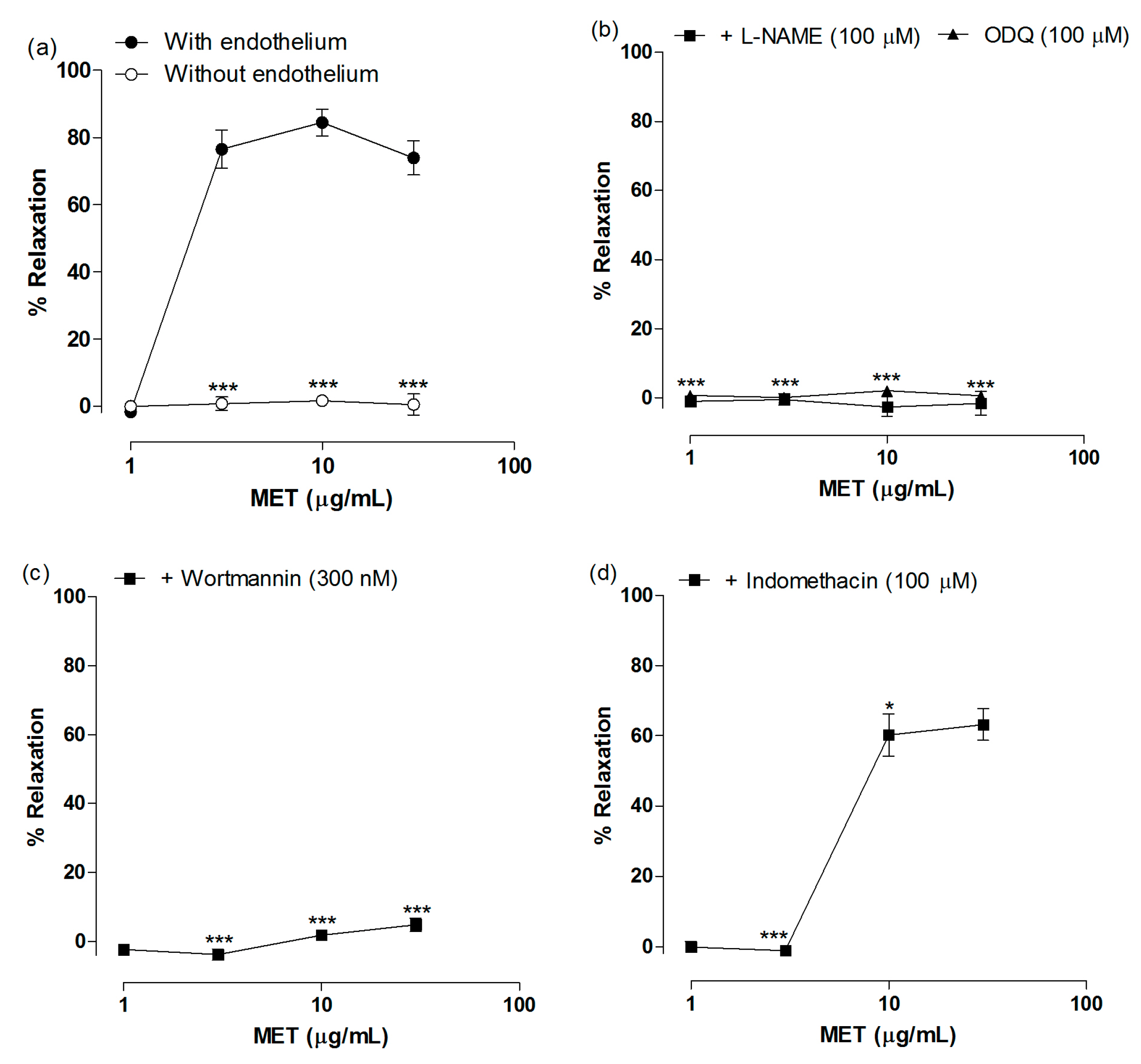
2.2. Vasodilatory Effect of MET

MET induced intense relaxation of aortic rings with endothelium pre-contracted with Phe. At 10 µg/mL, MET produced a relaxation of 84.46% ± 4.0% (Figure 3a). The concentration of MET necessary to reduce by 50% Phe-induced contraction of aorta (IC50) was 4.46 ± 0.97 µg/mL. To test whether the vasorelaxation induced by MET was endothelium-dependent, it was tested in endothelium-denuded aortic rings.
Removal of endothelium completely inhibited MET-induced vasodilation (Figure 3a). Thus, we evaluated the involvement of the endothelium-derived factors NO and PGI2. Pretreatment of aortic rings with L-NAME and ODQ abolished MET-induced vasodilation, indicating that the NO/cGMP pathway is crucial for the effect (Figure 3b). Moreover, similar results were obtained with the pretreatment with wortmannin (Figure 3c), indicating that MET induces eNOS activation mainly via activation of PI3K/Akt pathway and eNOS phosphorylation. Indomethacin induced a rightward shift of MET concentration-response curve (Figure 3d), demonstrating that the extract also acts through the activation of PGI2 production.

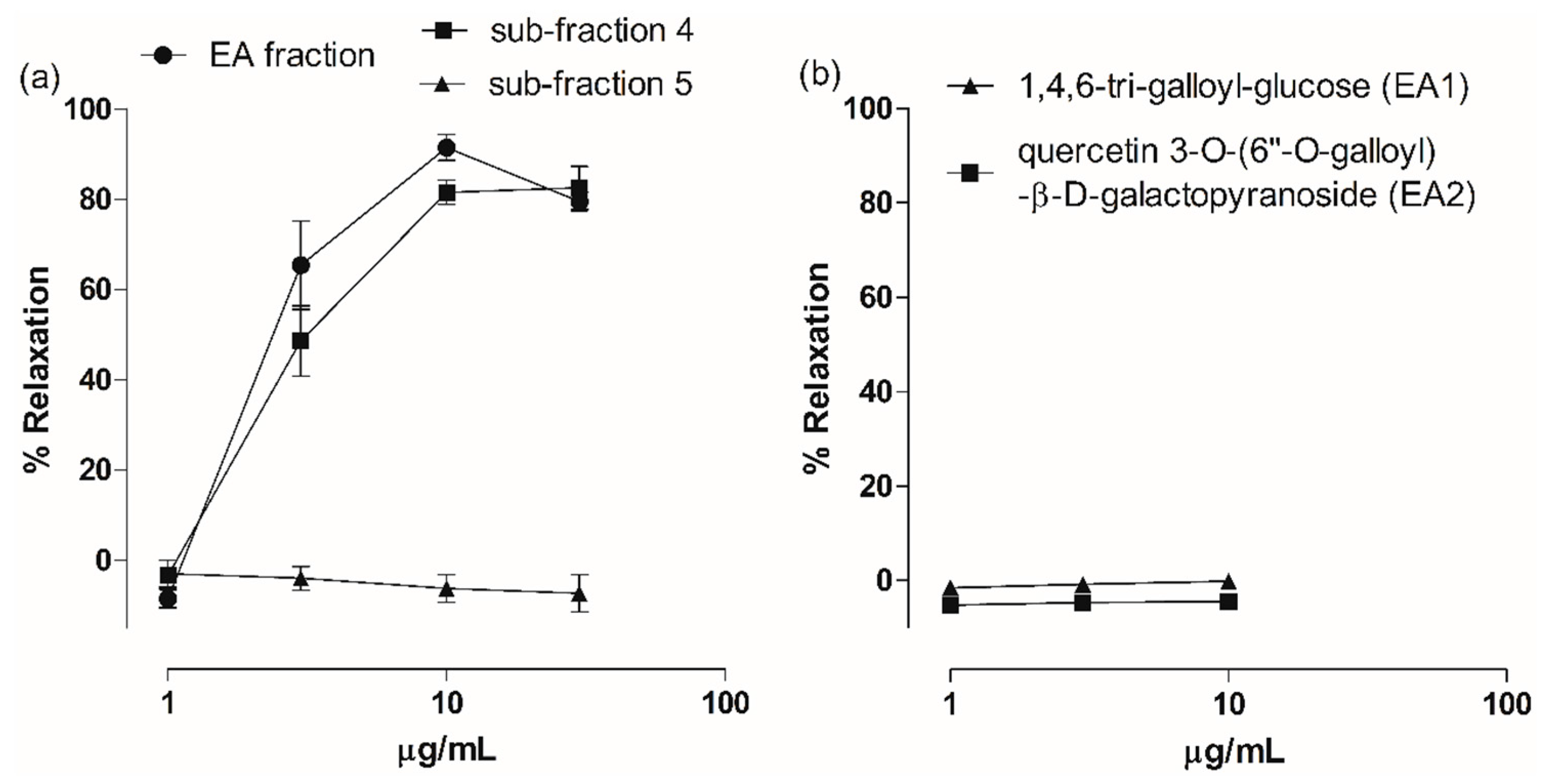
2.3. Vasodilatory Effect of Fractions, Sub-Fractions and Isolated Compounds

All fractions obtained from liquid-liquid partitioning of MET were evaluated in aortas with endothelium and EA fraction was the most effective in producing vasodilation. At 30 µg/mL, the DCM and EA fractions induced 53.56% ± 5.87% and 79.52% ± 2.86% relaxation (Figure 4a). The EA fraction (IC50 3.09 ± 0.37 µg/mL) exhibited higher potency than the DCM fraction (IC50 44.81 ± 6.77 µg/mL). Vascular relaxation induced by HN, BT and Aq fractions was inferior to 50%.
Sub-fraction 4 was evaluated and produced significant relaxation of rat aorta, similar to EA fraction-induced vasodilation. At 10 µg/mL, sub-fraction 4 induced 81.53% ± 2.70% of relaxation (IC50 3.99 ± 0.70 µg/mL) (Figure 4a). On the other hand, sub-fraction 5 had no vasodilator effect. In addition, the isolated hydrolysable tannin EA1 and the flavonoid EA2 were evaluated because their vasodilatory activity is not described in literature yet. As shown in Figure 4b, they had no effect on aorta contractility.

2.4. Antioxidant Effect of MET and Fractions

In vitro antioxidant activity of MET and its fractions were evaluated by using the DPPH assay. Ginkgo biloba extract Egb 761®, with an EC50 of 22.91 ± 0.66 µg/mL (Table 3) was used as positive control. MET and its fractions showed antioxidant activity with different potency (Table 3). MET (EC50 3.12 ± 0.20 µg/mL) exhibited the highest potency, while EA fraction (EC50 5.33 ± 0.16 µg/mL) and BT fraction (EC50 6.05 ± 0.19 µg/mL) were the most potent fractions. MET and all fractions exhibited greater potency than Egb 761®.

3. Discussion

Here we have presented a phytochemical study guided by vasodilatory and antioxidant activities of a methanol leaves extract of T. guianensis. Phytochemical analyses revealed that MET is a phenolic-rich extract, containing tannins and flavonoids. The compounds isolated and characterized by 1H and 13C-NMR were 1,4,6-tri-O-galloyl-β-d-glucose (EA1), quercetin 3-O-(6″-O-galloy)-β-d-galactopyranoside (EA2) and quercetin (EA3). Also, galloyl-HHDP-hexoside and myricetin deoxyhexoside were identified by LC-UV-MS. As far as we know, this is the first report of the occurrence of these compounds in T. guianensis.
Although no ethnopharmacological studies describing the medicinal use of T. guianensis for cardiovascular diseases have been found, one animal study showed promising results [19]. The ethanol extract of leaves from T. guianensis was able to reduce blood pressure in Sprague-Dawley rats, probably due the action of triterpenoids and flavonoids presents in the extract [19]. Our data showed that MET induces intense vasodilation in Wistar rat aortic rings, which could be, at least in part, responsible for the hypotensive effect described by Jiménez et al. [19].
MET-induced vasodilation was endothelium-dependent, since it was abolished by removal of the endothelium. Vascular endothelial cells produce and release relaxant factors, NO, PGI2 and endothelium-derived hyperpolarizing factor (EDHF), which play an important role in the regulation of vascular smooth muscle tone [21,22]. The contribution of NO, PGI2 and EDHF for vascular relaxation is heterogeneous and varies according to the size of the blood vessel [22,23]. NO has a more pronounced role in large arteries, such as aorta and epicardial coronary arteries, while the contribution of PGI2 does not change according to the type of vessel and EDHF is more important in the control of vessel diameter in smaller arteries and arterioles [22,23].
Considering endothelium-dependent mechanisms, most of vasodilator compounds isolated from plants produce vasodilation by activating the NO/cGMP pathway, whereas PGI2 plays a minor role in the mechanism of action of these compounds [7]. MET-induced vasodilation was completely inhibited in the presence of L-NAME and OQD, while indomethacin partially inhibited MET effect, indicating that vascular relaxation was mediated predominantly by NO.
Classically, eNOS activation is dependent on intracellular calcium concentration increase, as occurs when acetylcholine and histamine activates endothelial receptors. However, eNOS can also be activated by phosphorylation on specific amino acids residues. Some agonists such as estrogen and insulin induce vasodilation by activating the PI3K/Akt pathway, with subsequent phosphorylation of eNOS on Ser1177 [24]. MET seems to induce vasodilation by activating the PI3K/Akt pathway, since its effect was significantly blocked in aortic rings pretreated with wortmannin.
Among MET fractions, EA fraction presented greater vasodilatory and antioxidant potency and was submitted to further fractioning. Sub-fraction 4 showed a vasodilator profile similar to EA fraction, while sub-fractions 3 (EA1) and 5 were not active. Sub-fraction 6 was identified as the pure compound quercetin (EA3), extensively studied for its pharmacological properties. Quercetin was shown to produce both endothelium-dependent, with the involvement of NO and PGI2, and endothelium-independent vasodilation [25,26,27]. Thus, this flavonol is, at least partially, responsible for the vasodilatory activity of MET.
Fractionation of sub-fraction 4 resulted in the isolation of quercetin 3-O-(6″-O-galloyl)-β-d-galactopyranoside (EA2). LC-MS analysis showed that the isolated hydrolysable tannin 1,4,6-tri-O-galloyl-β-d-glucose (EA1, sub-fraction 3) is also present in sub-fraction 4. However, both of them did not present vasodilatory activity in rat aorta. Galloyl tannins and flavonoid galloyl glycosides have been shown to induce vasodilation and to inhibit the activity of angiotensin converting enzyme [28,29,30]. Penta-O-galloyl-β-glucoside induced NO-mediated vascular relaxation of rat aorta [8] and quercetin 3-O-β-d-galactopyranoside (hyperoside) showed vasodilatory activity in rat basilar artery [30], suggesting that the vasodilation effect is influenced by the number and the position of galloyl groups both in tannins and flavonoids [31,32].
In addition, galloyl-HHDP-hexoside and myricetin deoxyhexoside were identified in sub-fraction 4. Myricetin deoxyhexoside is a glycoside of myricetin, usually found in plants as myricetin-3-O-rhamnoside, also known as myricitrin. It was shown to attenuate endothelial cells apoptosis through PI3K/Akt signaling [33,34], but no vasodilatory activity has already been described. Also, part of the antihypertensive action of Tetraclinis articulates (Cupressaceae) was assigned to myricitrin [35].
Besides vascular effects, phenolic compounds are well known antioxidant agents, which is of particular interest since oxidative stress is a common feature of cardiovascular diseases [36]. MET and EAF showed a potent in vitro ability to scavenge the free radical DPPH, that could be attributed to the phenolic compounds identified. 1,4,6-tri-O-galloyl-β-d-glucose (EA1), quercetin 3-O-(6″-O-galloy)-β-d-galactopyranoside (EA2), quercetin (EA3) and myricitrin have already been shown to have antioxidant activity [37,38,39,40]. In addition, antioxidant effect of quercetin and its metabolites was described in vascular smooth muscle cells from normotensive and spontaneously hypertensive rats, by inhibiting the membrane NADPH oxidase activity [41].

4. Materials and Methods

4.1. Plant Material and Preparation of Crude Extract and Fractions

Leaves of Tapirira guianensis (Anacardiaceae) were collected on January 2012 at Parque Nacional da Restinga de Jurubatiba, Quissamã, Rio de Janeiro, Brazil, under legal authorization (SISBIO 39673-2). Botanical identification was performed by Dr. Tatiana Ungaretti Paleo Konno and a voucher specimen was deposited at the Universidade Federal do Rio de Janeiro Herbarium under the number RFA38757. T. guianensis leaves (3 Kg) were dried, triturated and extracted with methanol by maceration to yield the total dry crude extract MET (320.20 g), i.e., dry extract yield of 10.67% (w/w). A sample (50 g) of MET was solubilized in MeOH/distilled water (9:1) in agitation. The solution obtained was submitted to successive partitions with organic solvents in the following order: n-hexane (HN; eight partitions; 6.5287 g), dichloromethane (DCM; four partitions; 1.7312 g), ethyl acetate (EA; 11 partitions; 26.1552 g) and n-butanol (BT; four partitions; 7.2052 g). After partition with n-butanol the remaining aqueous fraction (Aqr; 6.0103 g) was obtained and it was submitted to lyophilization.

4.2. Chromatographic Separation of EA Fraction

An aliquot of EA fraction (2 g) was re-suspended in distilled water and chromatographed (H2O/MeOH gradient) on silanized silica column (0.063–0.200 mm, Merck, Darmstadt, Germany) affording eight sub-fractions, four of them, 3, 4, 5 and 6, presented UV chromatographic profiles characteristic of phenolic compounds. Sub-fraction 3, containing one pure compound codified as EA1 (10.1 mg), eluted with H2O/MeOH 20%, and was identified as the hydrolysable tannin 1,4,6-tri-O-galloyl-β-d-glucose (Figure 5a) by 1H- and 13C-NMR in comparison with the literature [42]. The semi-pure fraction sub-fraction 4 (100.7 mg) was re-suspended in distilled water and was purified on an RP-18 silanized silica (40–63 µM, Merck®; H2O/MeOH gradient) affording one isolated flavonoid, codified as EA2 (9.6 mg), eluted with H2O/MeOH 40%, and identified as quercetin 3-O-(6″-O-galloy)-β-d-galactopyranoside (Figure 5b) by 1H- and 13C-NMR in comparison with the literature [43]. Sub-fraction 6, containing one pure compound, codified as EA3 (17.5 mg), eluted with H2O/MeOH 40%, and identified as quercetin by 1H-NMR in comparison with the literature [44]. EA1, EA2 and EA3 NMR data are provided in the online Supplementary Material.

4.3. LC-MS Analyses of EA Fraction

The LC system, as previously described [45], was coupled to a mass spectrometer ESI-IT (Bruker Daltonics, Billerica, MA, USA), fitted with an electrospray ionization source operating in the positive mode, and an ion trap analyzer. The chromatographic conditions used were as follows: Luna C18 column (250 × 4.6 mm, 5 μm, Phenomenex, Torrance, CA, USA), sample injection volume of 10 μL at 1 mg/mL, flow rate 1.0 mL/min at 25 °C, and H2O containing 0.1% (v/v) formic acid (solvent A) and acetonitrile (solvent B) as the mobile phase. The elution gradient was 5% B in 0–3 min; 5%–10% B in 3–10 min; 10% B in 10–12 min; 10%–20% B in 12–15 min; 20% B in 15–17 min; 5%–10% B in 3–10 min; 10% B in 10–12 min; 10%–20% B in 12–15 min; 20% B in 15–17 min 35%–45% B in 32–40 min; 45% B in 40–42 min; 45%–60% B in 42–50 min; 60% B in 50–52 min; 60%–80% B in 52–60 min; 80% B in 60–62 min; 80%–100% B in 62–70 min; 100% B in 70–72 min; 100%–5% B in 72–75 min; 5% B in 75–76 min. The mass spectrometer parameters used were: capillary voltage, 3.5 kV; desolvation temperature, 330 °C; gas flow, 10 L/min; pressure, 70 psi, collision energy of 0.7 eV. Nitrogen was used as both the drying and nebulizing gas.

4.4. Preparation of Rat Aortic Rings for iSometric Tension Recording

All experimental protocols were approved by the Animal Care and Use Committee at Universidade Federal do Rio de Janeiro on 14 March 2012, under the license MACAÉ01. Thoracic aorta was dissected from male Wistar rats (200–250 g) and adipose and connective tissues were carefully removed. Aorta was cut into 3–4 mm rings, which were suspended in organ baths filled with Krebs-Henseleit solution (mM: 118.0 NaCl; 4.7 KCl; 1.2 KH2PO4; 1.2 MgSO4; 2.5 CaCl2; 25 NaHCO3 and 11.0 glucose; pH 7.4; 37 °C) continuously oxygenated with carborgen gas (95% O2, 5% CO2). Each aorta ring was mounted between two hooks in which one was attached to a force transducer (MLT0201; AD Instruments, Sydney, Australia), which signal was digitalized (Power Lab 4/30; AD Instruments) and stored on a computer for analysis using the software LabChart Pro (AD Instruments). After an equilibrium period of 1.5 h under 1 g resting tension, aortic rings were contracted with phenylephrine (Phe; 10 µM) and the presence of functional endothelium was confirmed by a relaxation response to acetylcholine (10 µM) greater than 80%. In some rings, the endothelium was mechanically removed, which was confirmed by the lack of relaxation in response to acetylcholine [46]. Concentration-response curves to MET, fractions and constituents were obtained in Phe-contracted rings.
In order to determine the involvement of NO pathway in vasodilatory activity of MET, aorta with endothelium were pretreated for 15 min with L-NAME (100 µM), an inhibitor of NO synthase; ODQ (100 µM), an inhibitor of soluble guanylyl cyclase (sGC); or wortmannin (300 nM), and inhibitor of phosphatidylinositol 3 kinase (PI3K). To verify the involvement of PGI2 pathway, aorta with endothelium was pretreated for 15 min with indomethacin (100 µM).

4.5. 1,1-Diphenyl-2-picrylhydrazyl (DPPH) Assay

The DPPH scavenging activity of MET and fractions was measured according to Nascimento et al. [47], with modifications. Methanolic solutions of plant extract and fractions at different concentrations (1–200 µg/mL) were mixed with a methanolic 300 µM DPPH solution in 96-wells microtiter plates and kept for 30 min at room temperature in the dark. Absorbances were measured at 517 nm (UVM 340 spectrophotometer, Biochrom ASYS, Cambridge, UK) using methanol as blank. Ginkgo biloba standardized extract of leaves (EGb 761®) was used as positive control. Absorbance values were converted into the percentage of antioxidant activity (AA%) by using the following formula: AA % =   100 { [ ( ABSsample     ABSblank )   ×   100 ]   /   ABScontrol } . To determine the concentration necessary to induce 50% of maximal response (EC50), results obtained from three separate experiments in triplicate were fitted by non-linear regression.

4.6. Statistical Analysis

Data are expressed as means ± S.E.M. Relaxation response is expressed as percentage of maximal tension observed in the presence of phenylephrine. Analyzes were performed using Prism 5.0 software (GraphPad Software, La Jolla, CA, USA). All data were analyzed using Kolmogorov-Smirnov normality test and all data have shown a Gaussian distribution. One-way analysis of variance followed by Newman-Keuls post-hoc test was used for comparison between concentration response curves. Differences between groups were considered statistically significant when p < 0.05.

5. Conclusions

Our findings suggest that T. guianensis leaves could be a source of phenolic compounds with pharmacological potential, since MET could reduce vascular tone through NO-dependent vasodilation and reduce oxidative stress by its antioxidant activity. Tannins and flavonoids were identified in MET, and quercetin and myricetin glycoside seems to contribute for MET activity as they have been reported as vasodilatory flavonoids.

Supplementary Materials

Supplementary materials are available online. 1H- and 13C-NMR spectroscopic data of the isolated compounds.

Acknowledgments

This work was supported by FAPERJ (E-26/102.238/2013) and CNPq research grants. This work was also supported by fellowship from FAPERJ to Amélia M. G. Rodrigues.

Author Contributions

A.M.G.R. performed chemical and pharmacological experiments and drafted the manuscript; D.O.G. and I.C.R.L. participated in the analysis of the data and supervising experiments; T.B., F.A.A. and N.P.L. were responsible for LC-MS analyses and L.W.T. for NMR data; T.U.P.K. was responsible for botanical identification; J.M.R. and M.F.M. participated in the project design and manuscript preparation.

Conflicts of Interest

The authors declare no conflict of interest.

References

  1. World Health Organization. A Global Brief on Hypertension. Document Number: WHO/DCO/WHD/2013.2. Available online: http://ish-world.com/downloads/pdf/globalbrief hypertension.pdf (accessed on 23 October 2016).
  2. Vanhoutte, P.M.; Shimokawa, H.; Tang, E.H.; Feletou, M. Endothelial dysfunction and vascular disease. Acta Physiol. 2009, 196, 193–222. [Google Scholar] [CrossRef] [PubMed] [Green Version]
  3. Giles, T.D.; Sander, G.E.; Nossaman, B.D.; Kadowitz, P.J. Impaired vasodilatation in the pathogenesis of hypertension: Focus on nitric oxide, endothelial-derived hyperpolarizing factors, and prostaglandins. J. Clin. Hypertens. 2012, 14, 198–205. [Google Scholar] [CrossRef] [PubMed]
  4. Triggle, C.R.; Samuel, S.M.; Ravishankar, S.; Marei, I.; Arunachalam, G.; Ding, H. The endothelium: Influencing vascular smooth muscle in many ways. Can. J. Biochem. Physiol. 2012, 90, 713–738. [Google Scholar] [CrossRef] [PubMed]
  5. Cohen, R.A.; Tong, X.Y. Vascular oxidative stress: The common link in hypertensive and diabetic vascular disease. J. Cardiovasc. Pharmacol. 2010, 55, 308–316. [Google Scholar] [CrossRef] [PubMed]
  6. Baradaran, A.; Nasri, H.; Rafieian-Kopaei, M. Oxidative stress and hypertension: Possibility of hypertension therapy with antioxidants. J. Res. Med. Sci. 2014, 19, 358–367. [Google Scholar] [PubMed]
  7. Luna-Vázquez, F.J.; Ibarra-Alvarado, C.; Rojas-Molina, A.; Rojas-Molina, I.; Zavala-Sánchez, M.A. Vasodilator compounds derived from plants and their mechanisms of action. Molecules 2013, 18, 5814–5857. [Google Scholar] [CrossRef] [PubMed]
  8. Xie, Y.W.; Xu, H.X.; Dong, H.; Fiscus, R.R.; But, P.P. Role of nitric oxide in the vasorelaxant and hypotensive effects of extracts and purified tannins from Geum japonicum. J. Ethnopharmacol. 2007, 109, 128–133. [Google Scholar] [CrossRef] [PubMed]
  9. Tapas, A.R.; Sakarkar, D.M.; Kakde, R.B. Flavonoids as nutraceuticals. Trop. J. Pharm. Res. 2008, 7, 1089–1099. [Google Scholar] [CrossRef]
  10. Annapurna, A.; Reddy, C.S.; Akondi, R.B.; Rao, S.R. Cardioprotective actions of two bioflavonoids, quercetin and rutin, in experimental myocardial infarction in both normal and streptozotocin-induced type I diabetic rats. J. Pharm. Pharmacol. 2009, 61, 1365–1374. [Google Scholar] [CrossRef] [PubMed]
  11. Grassi, D.; Desideri, G.; Ferri, C. Flavonoids: Antioxidants against atherosclerosis. Nutrients 2010, 2, 889–902. [Google Scholar] [CrossRef] [PubMed]
  12. Salvamani, S.; Gunasekaran, B.; Shaharuddin, N.A.; Ahmad, S.A.; Shukor, M.Y. Antiartherosclerotic effects of plant flavonoids. BioMed Res. Int. 2014, 2014, 480258. [Google Scholar] [CrossRef] [PubMed]
  13. Roos, C.M.; Zhang, B.; Palmer, A.K.; Ogrodnik, M.B.; Pirtskhalava, T.; Thalji, N.M.; Hagler, M.; Jurk, D.; Smith, L.A.; Casaclang-Verzosa, G.; et al. Chronic senolytic treatment alleviates established vasomotor dysfunction in aged or atherosclerotic mice. Aging Cell 2016, 15, 973–977. [Google Scholar] [CrossRef] [PubMed]
  14. Missouri Botanical Garden. Available online: http://www.tropicos.org (accessed on 5 May 2015).
  15. David, J.M.; Chávez, J.P.; Chai, H.B.; Pezzuto, J.M.; Cordell, G.A. Two new cytotoxic compounds from Tapirira guianensis. J. Nat. Prod. 1998, 61, 287–289. [Google Scholar] [CrossRef] [PubMed]
  16. Coelho-ferreira, M. Medicinal knowledge and plant utilization in an Amazonian coastal community of Marudá, Pará State (Brazil). J. Ethnopharmacol. 2009, 126, 159–175. [Google Scholar] [CrossRef] [PubMed]
  17. Correia, S.J.; David, J.M.; Da Silva, L.P.; David, J.P.; Lopes, L.M.X.; Guedes, M.L.S. Flavonoides, norisoprenóides e outros terpenos das folhas de Tapirira guianensis. Quim. Nova 2008, 31, 2056–2059. [Google Scholar] [CrossRef]
  18. Roumy, V.; Fabre, N.; Portet, B.; Bourdy, G.; Acebey, L.; Vigor, C.; Valentin, A.; Moulis, C. Four anti-protozoal and anti-bacterial compounds from Tapirira guianensis. Phytochemistry 2009, 70, 305–311. [Google Scholar] [CrossRef] [PubMed]
  19. Jiménez, G.; Hasegawa, M.; Rodríguez, M.; Estrada, O.; Méndez, J.; Castillo, A.; Gonzalez-Mujica, F.; Motta, N.; Vásquez, J.; Romero-Vecchione, E. Biological screening of plants of the Venezuelan Amazons. J. Ethnopharmacol. 2001, 77, 77–83. [Google Scholar] [CrossRef]
  20. Greenham, J.; Harborne, J.B.; Williams, C.A. Identification of lipophilic flavones and flavonols by comparative HPLC, TLC and UV spectral analysis. Phytochem. Anal. 2003, 14, 100–118. [Google Scholar] [CrossRef] [PubMed]
  21. Cines, D.B.; Pollak, E.S.; Buck, C.A.; Loscalzo, J.; Zimmerman, G.A.; McEver, R.P.; Pober, J.S.; Wick, T.M.; Konkle, B.A.; Schwartz, B.S.; et al. Endothelial cells in physiology and in the pathophysiology of vascular disorders. Blood 1998, 91, 3527–3561. [Google Scholar] [PubMed]
  22. Shimokawa, H. 2014 Willians Harvey lecture: Importance of coronary vasomotion abnormalities—From bench to bedside. Eur. Heart J. 2014, 35, 3180–3193. [Google Scholar] [CrossRef] [PubMed]
  23. Urakami-Harasawa, L.; Shimokawa, H.; Nakashima, M.; Egashira, K.; Takeshita, A. Importance of endothelium-derived hyperpolarizing factor in human arteries. J. Clin. Investig. 1997, 100, 2793–2799. [Google Scholar] [CrossRef] [PubMed]
  24. Fleming, I.; Busse, R. Molecular mechanisms involved in the regulation of the endothelial nitric oxide synthase. Am. J. Physiol. Regul. Integr. Comp. Physiol. 2003, 284, R1–R12. [Google Scholar] [CrossRef] [PubMed]
  25. Duarte, J.; Pérez-Vizcaíno, F.; Zarzuelo, A.; Jiménez, J.; Tamargo, J. Vasodilator effects of quercetin isolated rat vascular smooth muscle. Eur. J. Pharmacol. 1993, 239, 1–7. [Google Scholar] [CrossRef]
  26. Ajay, M.; Gilani, A.U.; Mustafa, M.R. Effects of flavonoids on vascular smooth muscle of the isolated rat thoracic aorta. Life Sci. 2003, 74, 603–612. [Google Scholar] [CrossRef] [PubMed]
  27. Nishida, S.; Satoh, H. Possible involvement of Ca2+ activated K+ channels, SK channel, in the quercetin-induced vasodilation. Korean J. Physiol. Pharmacol. 2009, 13, 361–365. [Google Scholar] [CrossRef] [PubMed]
  28. Goto, H.; Shimada, Y.; Akechi, Y.; Kohta, K.; Hattori, M.; Terasawa, K. Endothelium-dependent vasodilator effect of extract prepared from of roots of Paeonia lactiflora on isolated rat aorta. Planta Med. 1996, 62, 436–439. [Google Scholar] [CrossRef] [PubMed]
  29. Kang, D.G.; Moon, M.K.; Choi, D.H.; Lee, J.K.; Kwon, T.O.; Lee, H.S. Vasodilatory and anti-inflammatory effects of the 1,2,3,4,6-penta-O-galloyl-β-d-glucose (PGG) via a nitric oxide-cGMP pathway. Eur. J. Pharmacol. 2005, 524, 111–119. [Google Scholar] [CrossRef] [PubMed]
  30. Fan, Y.; Chen, Z.W.; Guo, Y.; Wang, Q.H.; Song, B. Cellular mechanisms underlying hyperin-induced relaxation of rat basilar artery. Fitoterapia 2011, 82, 626–631. [Google Scholar] [CrossRef] [PubMed]
  31. Zhu, M.; Phillipson, D.; Greengrass, P.M.; Bowery, N.E.; Cai, Y. Plants polyphenols: biologically active compounds or non-selective binders to proteins? Phytochemistry 1997, 44, 441–447. [Google Scholar] [CrossRef]
  32. Lee, T.H.; Liu, D.Z.; Hsu, F.L.; Wu, W.C.; Hou, W.C. Structure-activity relationships of five myricetin galloylglycosides from leaves of Acacia confusa. Bot. Stud. 2006, 47, 37–43. [Google Scholar]
  33. Zhang, B.; Chen, Y.; Shen, Q.; Liu, G.; Ye, J.; Sun, G.; Sun, X. Myricitrin Attenuates High Glucose-Induced Apoptosis through Activating Akt-Nrf2 Signaling in H9c2 Cardiomyocytes. Molecules 2016, 21. [Google Scholar] [CrossRef] [PubMed]
  34. Qin, M.; Luo, Y.; Meng, X.B.; Wang, M.; Wang, H.W.; Song, S.Y.; Ye, J.X.; Pan, R.L.; Yao, F.; Wu, P.; et al. Myricitrin attenuates endothelial cell apoptosis to prevent atherosclerosis: An insight into PI3K/Akt activation and STAT3 signaling pathway. Vasc. Pharmacol. 2015, 70, 23–34. [Google Scholar] [CrossRef] [PubMed]
  35. Zidane, A.; Tits, M.; Angenot, L.; Wauters, J.N.; Frederich, M.; Dib, I.; Mekhfi, H.; Aziz, M.; Bnouham, M.; Legssyer, A.; et al. Phytochemical analysis of Tetraclinis articula in relation to its vasorelaxant property. J. Mater. 2014, 5, 1368–1375. [Google Scholar]
  36. Lum, H.; Roebuck, K.A. Oxidant stress and endothelial cell dysfunction. Am. J. Physiol. Cell Physiol. 2001, 280, C719–C741. [Google Scholar] [PubMed]
  37. Cai, Y.Z.; Mei, S.; Jie, X.; Luo, Q.; Corke, H. Structure-radical scavenging activity relationships of phenolic compounds from traditional Chinese medicinal plants. Life Sci. 2006, 78, 2872–2888. [Google Scholar] [CrossRef] [PubMed]
  38. Gao, D.F.; Xu, M.; Yang, C.R.; Xu, M.; Zhang, Y.J. Phenolic antioxidants from the leaves of Camellia pachyandra Hu. J. Agric. Food Chem. 2010, 58, 8820–8824. [Google Scholar] [CrossRef] [PubMed]
  39. Tan, H.P.; Wong, D.Z.; Ling, S.K.; Chuah, C.H.; Kadir, H.A. Neuroprotective activity of galloylated cyanogenic glucosides and hydrolysable tannins isolated from leaves of Phyllagathis rotundifolia. Fitoterapira 2012, 83, 223–229. [Google Scholar] [CrossRef] [PubMed]
  40. Domitrović, R.; Rashed, K.; Cvijanović, O.; Vladimir-Knežević, S.; Škoda, M.; Višnić, A. Myricitrin exhibits antioxidant, anti-inflammatory and antifibrotic activity in carbon tetrachloride-intoxicated mice. Chem. Biol. Interact. 2015, 230, 21–29. [Google Scholar] [CrossRef] [PubMed]
  41. Jimenez, R.; Lopez-Sepulveda, R.; Romero, M.; Toral, M.; Cogolludo, A.; Perez-Vizcaino, F.; Duarte, J. Quercetin and its metabolites inhibit the membrane NADPH oxidase activity in vascular smooth muscle cells from normotensive and spontaneously hypertensive rats. Food Funct. 2015, 6, 409–413. [Google Scholar] [CrossRef] [PubMed]
  42. Nawwar, M.A.M.; Hussein, S.A.M.; Merfort, I. NMR spectral analysis of polyphenols from Punica granatum. Phytochemistry 1994, 36, 793–798. [Google Scholar] [CrossRef]
  43. Braca, A.; Politi, M.; Sanogo, R.; Sanou, H.; Morelli, I.; Pizza, C.; De Tommasi, N. Chemical composition and antioxidant activity of phenolic compounds from wild and cultivated Sclerocarya birrea (Anacardiaceae) leaves. J. Agric. Food Chem. 2003, 51, 6689–6695. [Google Scholar] [CrossRef] [PubMed]
  44. Huang, W.; Wan, C.; Zhou, S. Quercetin—A flavonoid compound from Sarcopyramis bodinieri var delicate with potential apoptotic activity in HepG2 liver cancer cells. Trop. J. Pharm. Res. 2013, 12, 529–533. [Google Scholar] [CrossRef]
  45. Ferreira, L.L.D.M.; Gomes, M.V.; Paes, B.M.; Do Carmo, P.L.; Konno, T.U.; Esteves, F.A.; Lopes, N.P.; Tomaz, J.C.; Leal, I.C.; Guimarães, D.O.; et al. The hydroalcoholic extract of leaves of Mandevilla moricandiana induces NO-mediated vascular relaxation. Planta Med. 2016. [Google Scholar] [CrossRef] [PubMed]
  46. Raimundo, J.M.; Trindade, A.P.; Velozo, L.S.; Kaplan, M.A.; Sudo, R.T.; Zapata-Sudo, G. The lignan eudesmin extracted from Piper truncatum induced vascular relaxation via activation of endothelial histamine H1 receptors. Eur. J. Pharmacol. 2009, 606, 150–154. [Google Scholar] [CrossRef] [PubMed]
  47. Nascimento, J.C.; Lage, L.F.O.; Camargos, C.R.D.; Amaral, J.C.; Costa, L.M.; De Souza, A.N.; Oliveira, F.Q. Antioxidant determination activity by DPPH method and assay for total flavonoids in leaves extracts of Bauhinia variegata L. Rev. Bras. Farm. 2011, 92, 327–332. [Google Scholar]
  • Sample Availability: Not available.
Figure 1. LC-UV-MS chromatogram of the ethyl acetate fraction of the methanol extract of T. guianensis leaves.
Molecules 22 00304 g001
Figure 2. LC-UV-MS chromatogram of sub-fraction 4 from the ethyl acetate fraction of the methanol extract of T. guianensis leaves.
Molecules 22 00304 g002
Figure 3. Mechanism of action of vasodilatory activity of the methanol extract of T. guianensis leaves (MET). (a) Concentration-response curves for MET in aortas with and without endothelium; (b) Effect of MET in endothelium-intact rings pretreated with L-NAME (100 μM) or ODQ (100 μM); (c) Effect of MET in endothelium-intact rings pretreated with wortmannin (300 nM); (d) Effect of MET in endothelium-intact rings pretreated with indomethacin (10 μM). Data are mean ± standard error of mean (S.E.M.) (n = 5–7). * p < 0.05 and *** p < 0.0001 compared to with endothelium.
Molecules 22 00304 g003
Figure 4. Effects of ethyl acetate fraction (EA fraction), sub-fractions and isolated compounds on aortic rings with endothelium. (a) Concentration-response curves for EA fraction, sub-fraction 4 and sub-fraction 5; (b) Concentration-response curves for 1,4,6-tri-O-galloyl-glucose (EA1) and quercetin 3-O-(6″-O-galloyl)-β-d-galactopyranoside (EA2). Data are mean ± S.E.M. (n = 5–6).
Molecules 22 00304 g004
Figure 5. Chemical structure of compounds isolated from T. guianensis leaves. (a) 1,4,6-tri-O-galloyl-β-d-glucose (EA1); (b) quercetin 3-O-(6″-O-galloyl)-β-d-galactopyranoside (EA2).
Molecules 22 00304 g005
Table 1. MS analyses of the T. guianensis EA fraction with emphasis on the major (254 nm chromatogram) and isolated compounds.
Retention Time (tR, min)IdentityPseudomolecular Ion m/zFragment m/z (Loss)λMAX (nm)
18.91,4,6-tri-O-galloyl-β-d-glucose a-467.12 (170)276
21.0quercetin 3-O-(6″-O-galloyl)-β-d-galactopyranoside a617.17 [M + H]+-263, 352
22.0myricetin deoxyhexoside465.13 [M + H]+319.06 (146)257, 350
23.9quercetin pentoside435.12 [M + H]+303.05 (132)256, 352
24.5quercetin dideoxyhesoside595.20 [M + H]+449.13 (146) 303.06 (146)255, 348
31.7Quercetin a303.05 [M + H]+-255, 369
a Isolated compound.
Table 2. MS analyses of T. guianensis EA active sub-fraction 4 with emphasis on major (254 nm chromatogram) and isolated compounds.
Retention Time (tR, min)IdentityPseudomolecular Ion m/zFragment m/z (Loss)λMAX (nm)
18.91,4,6-tri-O-galloyl-β-d-glucose a-467.08 (170)277
19.1galloyl-HHDP-hexoside633.10 [M + H]+-266
21.0quercetin 3-O-(6″-O-galloyl)-β-d-galactopyranoside a617.17 [M + H]+-263, 352
22.0myricetin deoxyhexoside465.13 [M + H]+319.06 (146)261, 350
a Isolated compound.
Table 3. Antioxidant effect of MET and its fractions assessed by the DPPH assay.
SamplesEC50 (µg/mL)
MET3.12 ± 0.20 a
HN fraction40.30 ± 0.39 c
DCM fraction19.83 ± 0.90 d
EA fraction5.33 ± 0.16 b
BT fraction6.05 ± 0.19 b
Aq fraction14.33 ± 0.15 e
Egb 761®22.91 ± 0.66 f
EC50, Concentration required to induce 50% maximal response; MET, methanol extract of T. guianensis leaves; HN fraction, hexane fraction; DCM fraction, dichloromethane fraction; EA fraction, ethyl acetate fraction; BT fraction, butanol fraction; Aq fraction, aqueous fraction; Egb 761®, Ginkgo biloba extract). The results are means ± S.E.M. Statistical analyses were calculated and values with different superscript letters (a–f) are significantly different (p < 0.0001); determined by a Tukey test.

