Article

Synthesis, X-ray Single Crystal Structure, Molecular Docking and DFT Computations on N-[(1E)-1-(2H-1,3-Benzodioxol-5-yl)-3-(1H-imidazol-1-yl)propylidene]-hydroxylamine: A New Potential Antifungal Agent Precursor

by
Reem I. Al-Wabli
1,
Alwah R. Al-Ghamdi
1,
Hazem A. Ghabbour
1,2,
Mohamed H. Al-Agamy
3,4,
James Clemy Monicka
5,
Issac Hubert Joe
6 and
Mohamed I. Attia
1,7,*
1
Department of Pharmaceutical Chemistry, College of Pharmacy, King Saud University, P.O. Box 2457, Riyadh 11451, Saudi Arabia
2
Department of Medicinal Chemistry, Faculty of Pharmacy, Mansoura University, Mansoura 35516, Egypt
3
Department of Pharmaceutics, College of Pharmacy, King Saud University, P.O. Box 2457, Riyadh 11451, Saudi Arabia
4
Microbiology and Immunology Department, Faculty of Pharmacy, Al-Azhar University, Cairo 11884, Egypt
5
Muslim Arts College, Thiruvithancode 629174, Tamil Nadu, India
6
Centre for Molecular and Biophysics Research, Mar Ivanios College, Thiruvananthapuram 695015, Kerala, India
7
Medicinal and Pharmaceutical Chemistry Department, Pharmaceutical and Drug Industries Research Division, National Research Centre (ID: 60014618), El Bohooth Street, Dokki, Giza 12622, Egypt
*
Author to whom correspondence should be addressed.
Molecules 2017, 22(3), 373; https://doi.org/10.3390/molecules22030373
Submission received: 4 January 2017 / Revised: 12 February 2017 / Accepted: 21 February 2017 / Published: 28 February 2017
(This article belongs to the Section Medicinal Chemistry)

Abstract

:
Mycoses are serious health problem, especially in immunocompromised individuals. A new imidazole-bearing compound containing an oxime functionality was synthesized and characterized with different spectroscopic techniques to be used for the preparation of new antifungal agents. The stereochemistry of the oxime double bond was unequivocally determined via the single crystal X-ray technique. The title compound 4, C13H13N3O3·C3H8O, crystallizes in the monoclinic space group P21with a = 9.0963(3) Å, b = 14.7244(6) Å, c = 10.7035(4) Å, β = 94.298 (3)°, V = 1429.57(9) Å3, Z = 2. The molecules were packed in the crystal structure by eight intermolecular hydrogen bond interactions. A comprehensive spectral analysis of the title molecule 4 has been performed based on the scaled quantum mechanical (SQM) force field obtained by density-functional theory (DFT) calculations. A molecular docking study illustrated the binding mode of the title compound 4 into its target protein. The preliminary antifungal activity of the title compound 4 was determined using a broth microdilution assay.

1. Introduction

The incidence of systemic fungal infections (mycoses) has increased drastically in recent years, mainly in immunosuppressed or immunocompromised individuals with AIDS, cancer or undergoing organ transplantation [1,2]. Failure of the available antifungal agents to treat fungal infections is primarily due to dramatic increase in resistance to the conventional antifungal drugs leading to morbidity and mortality in patients facing life-threatening fungal infections. In order to overcome this serious problem, the development of new alternative antifungal drug therapies with improved efficacy, broader activity and favorable safety profile has attracted a great deal of interest [3,4].
Azole-based compounds having either an imidazole or triazole pharmacophore moiety in their structure, constitute the mainstay of the antifungal chemotherapeutic agents used in the clinic [5,6]. Azoles competitively inhibit cytochrome P450-dependent lanosterol 14α-demethylase (CYP51) resulting in depletion of ergosterol in fungi making them unable to grow in a normal way [7,8]. A screening the literature revealed that most of the available imidazole-bearing antifungal agents have two carbon spacers between the imidazole moiety and an aromatic residue, while few antifungals have a three carbon spacer between the pharmacophore and the aromatic part [9,10,11].
On the other hand, the benzodioxole moiety is found in a sizable number of biologically active compounds with a wide range of activities [12,13,14,15,16]. The title molecule features both the 1,3-benzodioxole moiety and the imidazole nucleus connecting to each other through a three carbon bridge. Therefore, the current investigation deals with the synthesis, molecular characterization and single crystal X-ray structure of a new oxime derivative, namely N-[(1E)-1-(2H-1,3-benzodioxol-5-yl)-3-(1H-imidazol-1-yl)propylidene]hydroxylamine (4) to be utilized as a potential precursor for imidazole-bearing antifungal agents. The stereochemistry of the imine functionality in the title molecule 4 was certainly determined via the single crystal X-ray crystallography technique. In addition, density-functional theory (DFT) computations were also performed as a useful tool to investigate the electronic structure and molecular geometry of the title molecule 4. Molecular docking studies were conducted in order to predict the biological activity of compound 4.

2. Results and Discussion
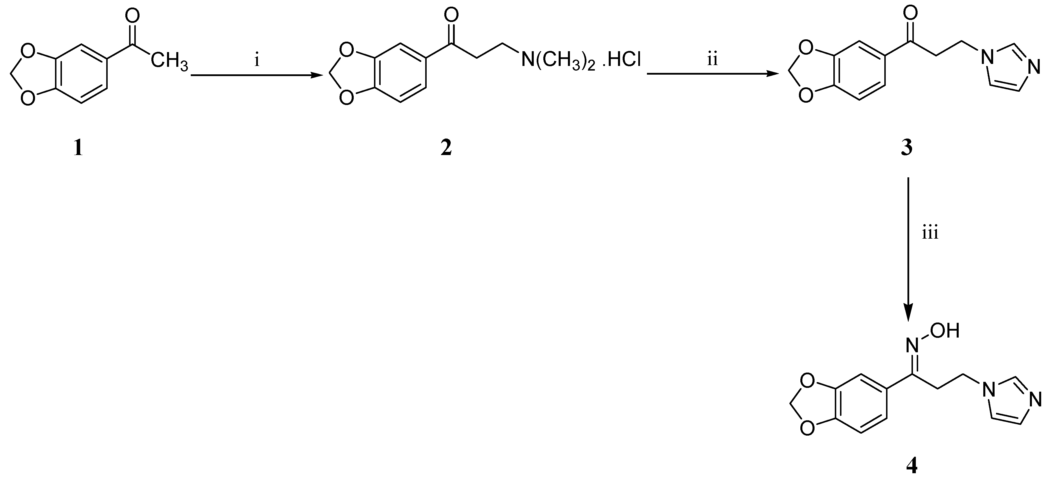
2.1. Chemistry

Scheme 1 illustrates the synthetic pathway which was adopted to synthesize the target compound 4. The synthesis commenced with a Mannich reaction under acidic conditions using the commercially available 1-(2H-1,3-benzodioxol-5-yl)ethanone (1). Subsequently, the formed Mannich base hydrochloride 2 was smoothly transformed in aqueous solution into the ketone 3 via a nucleophilic substitution reaction using imidazole. The target oxime 4 was ultimately obtained using the standard procedure for oxime formation with hydroxylamine hydrochloride in ethanol in the presence of potassium hydroxide [17].

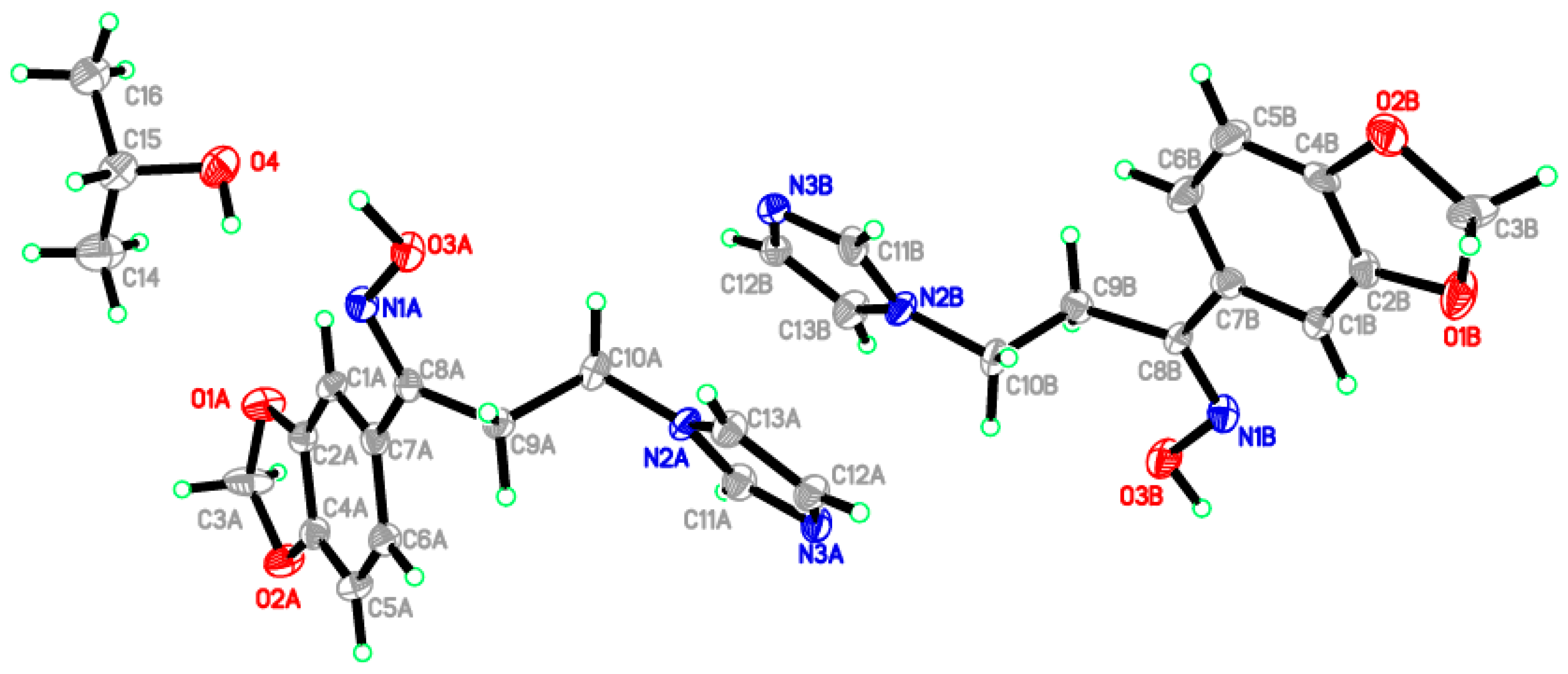
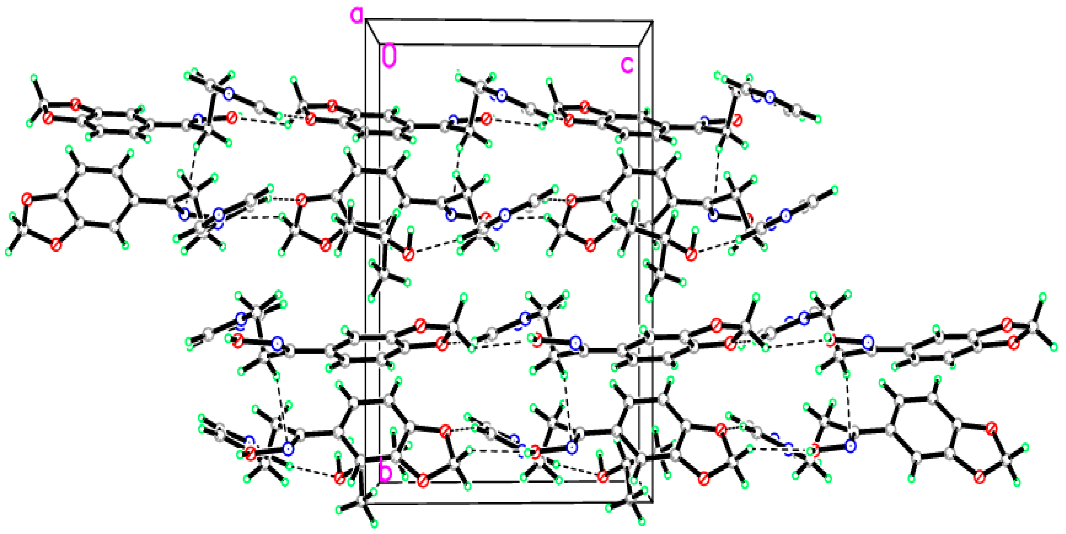
2.2. Crystal Structure of the Title Compound 4

The configuration of the target compound 4 was confirmed via X-ray crystallography. A suitable single colorless crystal of dimensions, 0.40 × 0.23 × 0.11 mm, was selected for X-ray diffraction analysis. The labeled displacement ellipsoid plot of this molecule is shown in Figure 1. Figure 2 depicts the packing of the molecules in the crystal structure.
The single crystal X-ray molecular structure therefore conformed the assigned (E)-configuration of the imine group in the target compound. The crystal structure contains two independent molecules with one isopropanol molecule as a solvent in the asymmetric unit. The benzodioxole ring (C1/C2/O1/C3/O2/C4–C7) forms dihedral angles of 44.04(3)° and 20.04(2)° with the imidazole ring (N2/C11/N3/C12/C13) for molecules A and B, respectively. The crystal structure is stabilized by eight hydrogen bonds along the b and c-axis (Table 1).

2.3. Structural Geometry Analysis

Equilibrium structural geometry of the title molecule 4 has been evaluated by a potential energy surface (PES) scan study. The flexible dihedral angles of C11–C12–N13–C14, C11–C10–C5–C4 and C11–C10–N18–O19 were scanned from 0° to 360° and their optimum energy was studied to identify stable conformations of the title compound 4. The global minimum energy is –893.45 and –893.49 Hartree for the conformers of compound 4 in the gas and solution phases, respectively (various conformers of compound 4 are shown in Figure S1). From this PES analysis, we have identified the minimum energy conformer of this molecule which was chosen for the subsequent studies. The optimized structure of the studied compound 4 with atoms numbering is depicted in Figure 3. Optimized bond lengths, bond angles and dihedral angles have been presented in Table 2. The formation of intramolecular hydrogen bonding is exposed by the intramolecular contacts to H22···N18 occur with H···O distance of 2.455 Å, which is shorter than the van der Waals separation between the O and H atoms (2.75 Å) [18]. Shortening of the C–N bond lengths N13–C14, N13–C17, N16–C17 and N16–C15 is typical for double bonds and it is due to resonance interactions. The linear fitting graphs (Figure S2) are drawn to study the correlation between the average experimental values and computed results. Statistical analysis revealed that the results of the computed solvation model are in a good agreement with the average experimental XRD values. Therefore, this method was considered to compute spectral vibrations, natural bond orbital and Frontier orbital energy analyses for the title molecule 4.

2.4. Natural Bond Orbital Analysis

Natural bond orbital (NBO) analysis describes the (hyper)conjugative interactions between donor–acceptor orbitals in order to understand intramolecular charge–transfer phenomenon of the molecular system [19]. Selective donor–acceptor interactions of the oxime 4 are listed along with their occupancy and stabilization energy values in Table 3. The hyperconjugative interactions of π(C1–C6)→π*(C2–C3), π(C1–C6)→π*(C4–C5), π(C2–C3)→π*(C1–C6), π(C2–C3)→π*(C4–C5), π(C4–C5)→π*(C1–C6), and π(C4–C5)→π*(C2–C3) were 20.27, 17.86, 18.94, 19.07, 17.00 and 17.47 kcal/mol, respectively. This could be attributed to the charge delocalization leading to ring resonance effect. The lone pair conjugative interactions of LP(2)O7→π*(C1–C6), LP(2)O9→π*(C2–C3), LP1(N13)→π*(C14–C15), and LP1(N13)→π*(N16–C17) have stabilization energy of 25.80, 27.07, 30.67, and 46.28 kcal/mol, respectively. The large E(2) values revealed the occurrence of strong electron delocalization over the ring moieties. These interactions were observed as an increase in the electron density (ED) of the C–C antibonding orbital, which weakens the respective bonds. The C10=N18 bond length (1.2861 Å) is significantly shorter than the other –CN bonds and the electron density (ED) of this antibonding orbital was decreased to 0.18707, which is an evidence for the rehybridization [20]. More conjugative and hyperconjugative interactions were formed in the lone-pair, C–C and C–C bond orbital overlap which confirms the intramolecular charge-transfer (ICT) causing the stabilization of the molecular structure of the title oxime 4.

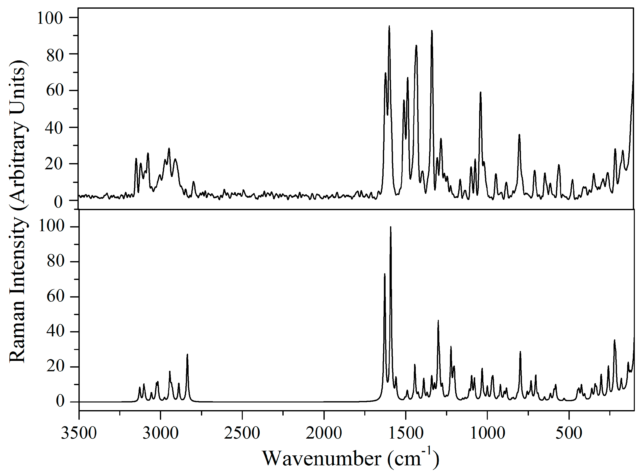
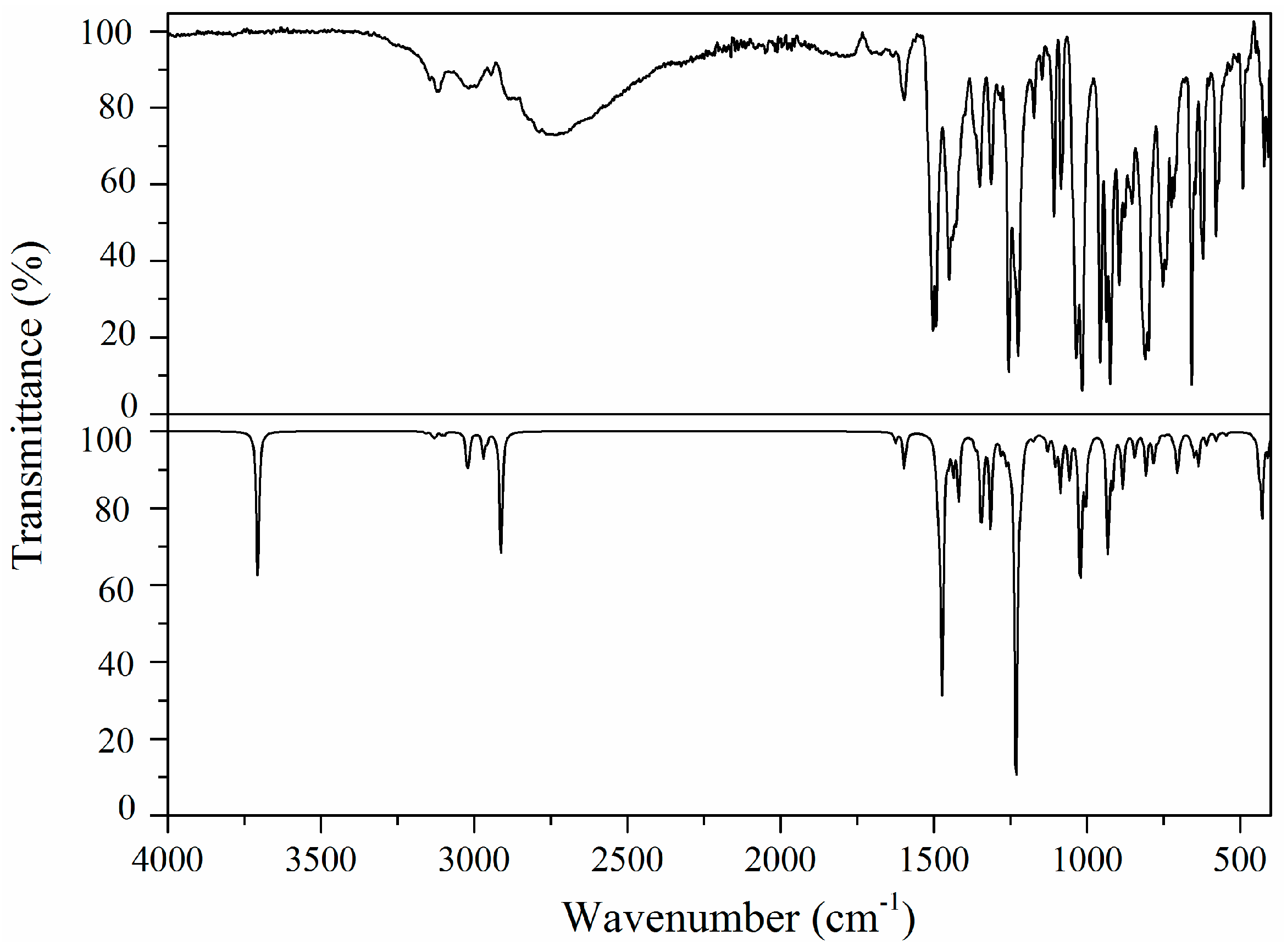
2.5. Vibrational Analysis

The title molecule 4 consists of 32 atoms and their characteristic vibrations are described by 90 normal modes. FT-Raman and FT-IR spectra of the title compound 4 are shown in Figure 4 and Figure 5, respectively. Theoretical and experimental spectral data of the target oxime 4 are presented in Table 4, including calculated and fundamental wavenumbers, FT-IR- and FT-Raman intensities along with the tentative vibrational assignment. The calculated vibrational wavenumbers were corrected by scaled quantum mechanical force-field (SQMFF) method [21] by selective scaling factor approach [22]. Using this force-field method, the calculated wavenumbers are reproducing the experimental values with a mean deviation of 15 cm–1. Internal valence coordinates and scaling factors information of the title compound 4 modes are given in Tables S1 and S2, respectively.

2.5.1. Imidazole Ring Vibrations

The calculated IR and Raman imidazole C–H stretching vibrations at 3118 and 3145 cm–1 are in close agreement with the experimental wavenumbers observed at 3144, 3125, 3120 cm–1 [23,24]. The FT-IR bands observed at 1503, 1350, 1280 and 1108 cm–1 and the FT-Raman ones at 1516, 1346, 1291, 1107 and 1050 cm–1 are definitely attributed to imidazole CH in-plane bend modes which are coupled with several other modes. The FT-IR bands at 752 and 725 cm–1 and the weak FT-Raman bands at 721 cm–1, can be assigned to the out-of-plane C–H wagging modes, as supported by theliterature data [25]. The C14=C15 stretching mode is the most characteristic vibration of the heterocyclic imidazole ring which was observed as medium intensity bands at 1493 (FT-IR) and at 1494 (FT-Raman) cm–1. The FT-IR and FT-Raman vibrations of the imidazole ring out-of-plane torsion modes appeared at 658, 578 (FT-IR) and 573 (FT-Raman) cm–1.

2.5.2. Methylene Group Vibrations

The neighboring rings π-system and nitrogen lone pair affects the spectral behavior of sp3 hybridized methylene moiety. Methylene asymmetric and symmetric C–H stretching vibrations usually appear in the region 2960–2840 cm–1 [26]. The asymmetric (2970 and 2946 cm–1) and symmetric (2908 and 2845 cm–1) stretching contributing bands appeared as a medium intensity band in the FT-Raman spectrum of the title compound 4. Lowering of symmetric stretching wavenumbers could be attributed to the hyperconjugative interaction between the nitrogen lone pair and σ*(C–H) bond. The rocking, wagging and twisting vibrational modes of compound 4 appeared in the region of 1400–900 cm–1 [27]. The CH2 scissoring mode appeared as a characteristic band near 1450 and 1439 cm–1 in the FT-IR spectrum and at 1440 cm–1 in the FT-Raman spectrum of the title oxime 4. The normal coordinate analysis (NCA) supports the FT-Raman bands at 1254 and 1271 cm–1 and the FT-IR bands at 1255 cm–1 for the unambiguous assignment of CH2 twisting and wagging modes.

2.5.3. Benzodioxole Ring Vibrations

Aromatic C–H stretching mode appears in the region of 3000–3100 cm–1. Weak FT-Raman bands identified at 3073 and 3001 cm–1 have been assigned to the C–H stretching mode. Usually the bands due to ring C–H in-plane and out-of-plane bending vibrations are observed in the region of 1000–1300 and 750–1000 cm–1, respectively. In the title compound 4, the C–H in-plane bending vibration has been observed as a medium intensity FT-IR band at 1313 cm–1 and as a weak band at 1314 cm–1 in the FT-Raman spectrum. The medium to weak intensity bands observed at 854 (FT-IR), 850 (FT-Raman), 814 (FT-Raman) and 808 (FT-IR) cm–1 have been assigned to C–H out-of-plane bending mode. Ring C–C stretching modes have been identified in the FT-IR spectrum of the target compound 4 at 1595, 1428 and 1173 cm–1 while they appeared at 1606 and 1174 cm–1 in the FT-Raman spectrum. The ring asymmetric deformation and torsion modes have been identified at 716, 556, 492 and 417 cm–1 in the FT-IR and at 490 cm–1 in the FT-Raman spectra of the title molecule 4.

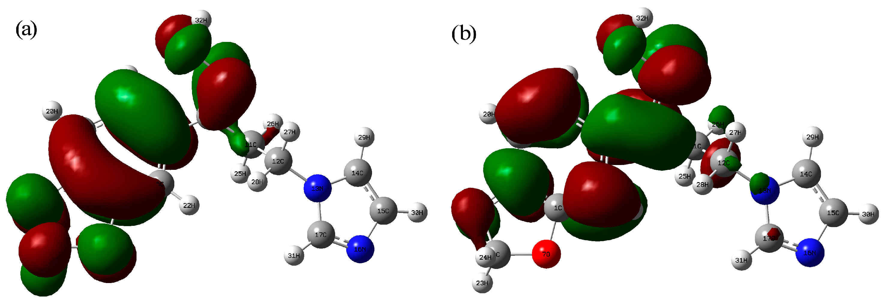
2.6. Frontier Molecular Orbital Analysis

A detailed knowledge of molecular electron density distribution and electron motion are necessary to understand molecular recognition and chemical reactivity of the molecule. The LUMO and HOMO orbital energy analysis of the title compound 4 has been computed using DFT method in the solution phase (isopropanol). The electron charge cloud is located at the piperonal ring in highest occupied molecular orbital (HOMO) while it is located mainly at the phenyl ring in the lowest unoccupied molecular orbital (LUMO). The energy of the HOMO is −6.23 eV and LUMO is −1.60 eV giving rise to a HOMO-LUMO energy gap of 4.63 eV. The HOMO-LUMO energy gap value supports the intramolecular charge-transfer interactions within the title molecule. The HOMO and LUMO orbital diagram is shown in Figure 6.

2.7. NMR Chemical Shift Analysis

The carbon and hydrogen chemical shift values of the title oxime 4 were calculated based on Gauge-independent atomic orbital (GIAO) method at B3LYP/6-311++G(d,p) level of theory [28]. The computed values are presented in Table 5. The correlation graphs were plotted over the observed and predicted chemical shift values of the title molecule 4 and the linear fitting plots are shown in Figure S3.
The phenyl carbons signals usually appear in the region of 120–140 ppm. The C1 and C2 in the title molecule 4 were observed at 148.0 and 148.3 ppm, respectively. This downfield chemical shift values revealed that, these carbon atoms bounded with the electronegative oxygen atoms. In general, imidazole ring protons signals occur in the region of 6–8 ppm. The 1H-NMR spectrum of the target oxime 4 showed signals at 6.89, 7.19 and 7.66 ppm which correspond to the protons of the imidazole ring. The aromatic piperonal ring protons were observed at 6.84 (as doublet), 7.06 (as doublet) and 7.13 (as singlet) ppm. There is a good agreement between the calculated and observed chemical shift values of the title molecule 4 with a correlation coefficient (R2) values = 0.994 and 0.953 for 13C and 1H, respectively.

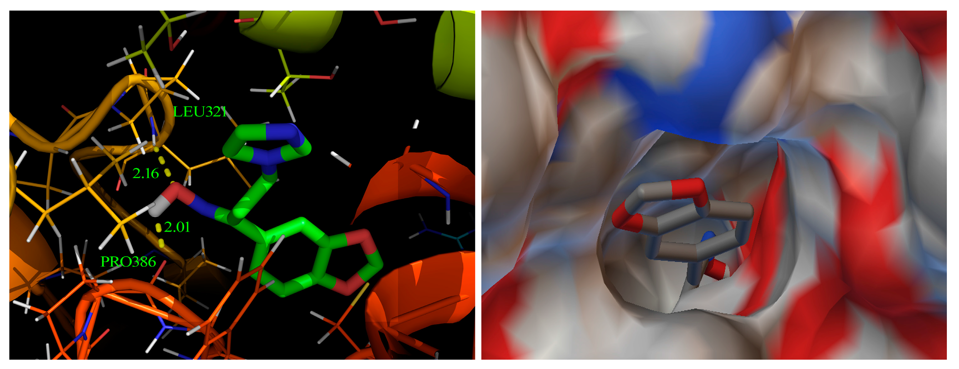
2.8. Molecular Docking Study

Molecular docking is an important technique to predict the biological activity of chemical compounds [29]. The target compound 4 was energy minimized using DFT method with the help of Gaussian program [30]. The target protein (cytochrome P450-dependent (CYP51) lanosterol 14α-demethylase enzyme) for antifungal azoles has been identified based on a multilevel neighborhoods of atoms’ (MNAs) algorithm model by the PASS online server [31]. This target protein (PDB code: 1EA1) was downloaded from the research collaboratory structural bioinformatics (RCSB) protein data bank [32]. The target compound 4 was docked using AutoDock Tools 1.5.4 (The Scripps Research Institute, La Jolla, CA, USA) interfaced with the AutoDock 4.2 program in the rigid docking methodology [33,34]. The binding free energy and inhibition constant of the proper conformation of the title compound 4 was predicted to be −5.06 kcal/mol and 195.75 μM, respectively. The hydrogen bond interaction of protein-ligand complex is shown in Figure 7. The amino acid residues LEU321 and PRO386 of the target protein are bounded with the title compound 4 by an intermolecular hydrogen bonding interaction. The docking study illustrated the affinity of compound 4 toward its target protein with a good binding energy value (–5.06 kcal/mol) and hence its suitability as a potential precursor to prepare new antifungal agents.

2.9. Antifungal Activity of the Title Compound 4

Table 6 presents the preliminary antifungal activity of the tested oxime 4 as well as the reference standard drug, ketoconazole. Compound 4 exhibited a MIC value of 987.43 μmol/L against C. albicans, C. tropicalis, C. parapsilosis and Aspergillus niger in the broth microdilution assay. While inactive itself, taken together the body of evidence suggests that compound 4 could be used as a starting material to prepare new antifungal agents with better antifungal profile.

3. Experimental

3.1. General

Melting points were measured using a Gallenkamp melting point device, and are uncorrected. Infrared (IR) spectra (as KBr disks) were recorded on FT-IR Spectrum BX device (Perkin Elmer, Ayer Rajah Crescent, Singapore). The NMR samples were dissolved in either CDCl3 or DMSO-d6 and the NMR spectra were recorded using a Bruker NMR spectrometer (Bruker, Reinstetten, Germany), at 500 MHz for 1H and 125.76 MHz for 13C at the Research Center, College of Pharmacy, King Saud University, Saudi Arabia. Chemical shifts are expressed in δ-values (ppm) relative to TMS as an internal standard. Mass spectra were recorded using a Quadrupole 6120 LC/MS equipped with an electrospray ionization (ESI) source (Agilent Technologies, Palo Alto, CA, USA). Silica gel TLC (thin layer chromatography) plates (silica gel precoated aluminium cards with 254 nm fluorescent indicator) from Merck (Darmstadt, Germany) were used for thin layer chromatography. Visualization was performed by illumination with a UV light source (254 nm).

3.2. Synthesis

3.2.1. Synthesis of 1-(2H-1,3-Benzodioxol-5-yl)-3-(1H-imidazol-1-yl)propan-1-one (3)

A catalytic amount of concentrated hydrochloric acid (0.5 mL) was added to a mixture containing paraformaldehyde (0.81 g, 9.0 mmol), dimethylamine hydrochloride (2.20 g, 27 mmol) and1-(2H-1,3-benzodioxol-5-yl)ethanone (1, 3.28 g, 20 mmol) in absolute ethanol (15 mL). The reaction mixture was refluxed for two hours, cooled and acetone (30 mL) was added to precipitate the Mannich base hydrochloride (2) which was collected by filtration and dried. Imidazole (1.36 g, 20 mmol) was added to a solution of compound 2 (2.58 g, 10 mmol) in water (10 mL) and the solution was refluxed for five hours, cooled and the precipitated solid was filtered off to afford compound 3. Compound 3 was re-crystallized from ethanol to give 1.15 g (47%) of the pure pivotal ketone 3 as a white solid m.p. 150–152 °C. IR (KBr): ν (cm–1) 3115, 2968, 1757 (C=O), 1637 (C=N), 1600, 1494, 1255, 750; 1H-NMR (CDCl3): δ (ppm) 3.37 (t, J = 6.5 Hz, 2H, –CH2–CH2–N), 4.42 (t, J = 6.5 Hz, 2H, –CH2–CH2–N), 6.05 (s, 2H, –O–CH2–O–), 6.84 (d, J = 8.0 Hz, 1H, Ar–H), 6.99 (s, 1H, –N–CH=CH–N=), 7.04 (s, 1H, –N–CH=CH–N=), 7.39 (d, J = 1.5 Hz, 1H, Ar–H), 7.50 (dd, J = 1.5, 8.0 Hz, 1H, Ar–H), 7.64 (s, 1H, –N–CH=N–); 13C-NMR (CDCl3): δ (ppm) 39.6 (–CH2–CH2–N), 41.7 (–CH2CH2–N), 102.0 (–O–CH2–O–), 107.7 (Ar–CH), 108.0 (Ar–CH), 119.2 (–N–CH=CH–N=), 124.4, 129.1 (Ar–CH, –N–CH=CH–N=), 131.0, (Ar–C), 137.4 (–N–CH=N–), 148.4, 152.3 (Ar–C), 194.6 (C=O); MS m/z (ESI): 245.0 [M + H]+.

3.2.2. Synthesis of (1E)-1-(2H-1,3-Benzodioxol-5-yl)-N-hydroxy-3-(1H-imidazol-1-yl)propan-1-imine (4)

Potassium hydroxide (1.12 g, 20 mmol) was added to a mixture containing hydroxylamine hydrochloride (1.39 g, 20 mmol), ketone 3 (2.44 g, 10 mmol) in ethanol (10 mL). The reaction mixture was refluxed under stirring for 18 hours, cooled to room temperature and the insoluble matter was collected by filtration. The filtrate was concentrated under reduced pressure and the residue was poured onto ice-cold water (15 mL). The obtained solid was filtered off and dried to yield 1.7 g (64%) of the target oxime 4 as a white solid m.p. 142–144 °C. Re-crystallization of the oxime 4 from isopropanol gave colorless single crystals which were suitable for X-ray analysis. 1H-NMR (DMSO-d6): δ(ppm) 3.14 (t, J = 7.0 Hz, 2H, –CH2–CH2–N), 4.17 (t, J = 7.0 Hz, 2H, –CH2–CH2–N), 6.04 (s, 2H, –O–CH2–O–), 6.84 (d, J = 7.5 Hz, 1H, Ar–H), 6.89 (s, 1H, –N–CH=CH–N=), 7.06 (dd, J = 1.5, 8.0 Hz, 1H, Ar–H), 7.13 (d, J = 1.0 Hz, 1H, Ar–H), 7.19 (s, 1H, –N–CH=CH–N=), 7.66 (s, 1H, –N–CH=N–), 11.41 (s, 1H, OH); 13C-NMR (DMSO–d6): δ(ppm) 28.2 (–CH2–CH2–N), 43.3 (–CH2CH2–N), 101.7 (–O–CH2–O–), 106.1, 108.5 (Ar–CH), 119.9 (–N–CH=CH–N=), 120.5, 128.3 (Ar–CH, –N–CH=CH–N=), 130.3 (Ar–C), 137.5 (–N–CH=N–), 148.0, 148.3 (Ar–C), 153.7 (C=N–OH); MS m/z (ESI): 260.1 [M + H]+.

3.3. Crystal Structure Determination

Slow evaporation of the alcoholic (isopropanol) solution of the title compound 4 furnished its colourless block single crystals. The X-ray diffraction measurement of the target oxime 4 was conducted on a SMART APEXII CCD diffractometer (Bruker, Karlsruhe, Germany) equipped with graphite monochromatic CuK\a radiation (λ = 1.54178 Å) at 296 (2) K. Cell refinement and data reduction were done by Bruker SAINT [35]. SHELXS-97 [36] was used to solve and refine the title structure. The final refinement of the crystal structure of the title oxime 4 was performed by full- matrix least-squares techniques with anisotropic thermal data for non hydrogen atoms on F2. All the hydrogen atoms were placed in the calculated positions and constrained to ride on their parent atoms. Multi-scan absorption correction was applied by the use of SADABS software [35]. The crystallographic data and refinement information are summarized in Table 7. Crystallographic data of compound 4 have been deposited with the Cambridge Crystallographic Data Center (supplementary publication number CCDC-1508986). Copies of the data may be obtained free of charge from the Director, CCDC, 12 Union Road, Cambridge, CB2 1EZ, UK ([email protected]).

3.4. FT-IR and FT-Raman Measurements

The FT-Raman spectrum of the oxime 4 was recorded in the spectral range of 3500–50 cm–1 using a Bruker RFS-27 FT-Raman spectrophotometer (Bruker, Billerica, MA, USA). The 1064 nm line of Nd:YAG laser operating at 100 mW power was used for excitation. The FT-IR spectrum of the oxime 4 was recorded with a spectral resolution of 2 cm–1 in the 4000–400 cm–1 range. Solid sample in KBr pellets was used in the FT-IR measurements.

3.5. Quantum Chemical Calculations

Optimized structural geometry and harmonic vibrational wavenumbers have been calculated at DFT/B3LYP/6-311++G(d,p) level of basis set in the gas phase. The polarizable continuum model (PCM) using the integral equation formalism (IEF) variant is the self-consistent reaction field (SCRF) to predict the structural parameters and vibrational wavenumbers of the title molecule 4 and isopropanol has defined as the solvent in implicit solvation model. This calculation has been performed in the presence of isopropanol by placing the title molecule 4 in a cavity within the solvent reaction field. Normal coordinate analysis (NCA) has been performed to obtain detailed explanation of the molecular motion relating to the normal modes using the MOLVIB program version 7.0 written by Sundius [37,38]. According to the scaled quantum mechanical force-field (SQMFF) procedure [21], selective scaling has been performed in the natural internal coordinate representation [22]. The simulated IR and Raman spectra of the title compound 4 have been plotted using pure Lorentizian band shapes with a bandwidth of full width height maximum of 10 cm–1. Second order interactions between the filled orbital of one subsystem and vacant orbitals of another subsystem have been understood with the aid of natural bonding orbitals (NBO) analysis [19] using NBO 3.1 program [39] as implemented in the Gaussian ‘09 package [30] at the DFT/B3LYP level. Molecular docking analysis using AutoDock 4.2 program [33] predicted the antifungal activity of the title compound 4.

3.6. Antifungal Activity

3.6.1. Materials

The reference standard antifungal drug, ketoconazole, was purchased from Sigma-Aldrich Co. (St. Louis, MO, USA). Liquid RPMI 1640 medium supplemented with l-glutamine was obtained from Gibco-BRL, Life Technologies (Paisley, Scotland). Sabouraud Dextrose Agar (SDA) was obtained from Merck Co. (Darmstadt, Germany). Dimethyl sulfoxide (100%) was used to dissolve ketoconazole, and/or the tested compound 4 to give an initial concentration of 2048 mg/L.

3.6.2. Organisms

The used fungal strains are Candida albicans (ATCC 90028), Candida tropicalis (ATCC 66029), Candida parapsilosis (ATCC 22019) and Aspergillus niger (ATCC 16404).

3.6.3. Preparation of Fungal Inocula

The inocula of the standard mold Aspergillus niger strain have been prepared by removing the sporulated A. niger from the Sabouraud Dextrose agar slant with a microbiological loop and the spores have been suspended in 10 mL of sterile water. The suspension has been filtered through sterile gauze to remove hyphae. The resulting suspension of conidia has been vigorously mixed using a vortex. The suspension has been adjusted to 1 × 10⁵ CFU/mL using spectrophotometer. This fungal suspension has been diluted 1:5 with RPMI medium to obtain suspensions having 2× of the required final concentration. This conidial suspension had a final concentration of 1 × 10⁴ CFU/mL when mixed with the tested solution of compound 4. On the other hand, the inocula of the standard yeast strains of C. albicans, C. tropicalis and C. parapsilosis have been prepared by suspending five representative colonies, obtained from 24 to 48 h culture on Sabauraud Dextrose agar medium, in sterile distilled water. The final inoculum concentration must be between 0.5 × 10⁵ and 2.5 × 10⁵ CFU/mL.

3.6.4. Preparation of the Tested Compound Solution

Briefly, a twofold dilution series of the tested compound 4 has been prepared in a double strength RPMI 1640 culture medium. Ten serial dilutions were prepared to give concentrations ranged from 1024 mg/L to 2 mg/L.

3.6.5. Antifungal Susceptibility Studies

Minimum Inhibitory Concentrations (MICs) have been determined by broth microdilution testing as described previously by EUCAST [40]. The experiment was carried out in duplicate. Briefly, one mL of RPMI 1640 medium from each of the bottle containing the corresponding concentration of the tested compound 4 has been transferred into sterile 7 mL Sterilin tubes (Thermo Fisher Scientific, Waltham, MA, USA). The RPMI 1640 medium containing 1024 mg/L of the tested compound 4 has been dispensed to tube 1, the medium containing 512 mg/L has been dispensed to tube 2, the medium containing 256 mg/L has been dispensed to tube 3 and so on to tube 10 for the medium containing 2 mg/L of the tested compound 4. One mL of the medium has been dispensed in tubes 11 (positive control) and 12 (negative control). One mL of the diluted inoculum suspension has transferred to each tube except tube 12 to bring the tested compound 4 dilutions to the required final test concentrations. The tubes were incubated at 35 °C for 72 h. The MIC of the tested compound 4 was determined visually by recording the degree of growth inhibition in each tube.

4. Conclusions

The synthesis and spectroscopic characterization of N-[(1E)-1-(2H-1,3-benzodioxol-5-yl)-3-(1H-imidazol-1-yl)propylidene]hydroxylamine (4) as new antifungal precursor has been reported. Computational studies on the target oxime 4 revealed that the theoretical wavenumbers are in a fair agreement with the observed wavenumbers except those associated with H-bonding. CH2 symmetric stretching wavenumber is red-shifted due to the hyperconjugative interaction between the nitrogen lone pair and σ*(C–H) bond. Single crystal X-ray analysis of the target molecule 4 confirmed the (E)-configuration of its imine double bond. A molecular docking study predicted the binding mode of compound 4 into its target protein and hence its usefulness as a potential precursor for new imidazole-bearing antifungal agents featuring both benzodioxole and imidazole pharmacophore moieties.

Supplementary Materials

Supplementary materials are available online.

Acknowledgments

The authors would like to extend their sincere appreciation to the Deanship of Scientific Research at King Saud University for its funding of this research through the Research Group Project No. RGP-196.

Author Contributions

R.I.A.-W. and A.R.A.-G. synthesized and characterized the title molecule. H.A.G. carried out the X-ray analysis of the title molecule. M.H.A.-A. performed the in vitro antifungal screening for the title compound. J.C.M. and I.H.J. conducted the computational work. M.I.A. proposed the work, prepared the single crystals of the title compound and prepared the manuscript for publication. All authors discussed the contents of the manuscript.

Conflicts of Interest

The authors have declared no conflict of interest.

References

  1. Caston-Osorio, J.J.; Rivero, A.; Torre-Cisneros, J. Epidemiology of invasive fungal infection. Int. J. Antimicrob. Agents 2008, 32 (Suppl. 2), S103–S109. [Google Scholar] [CrossRef]
  2. Cuenca-Estrella, M.; Bernal-Martinez, L.; Buitrago, M.J.; Castelli, M.V.; Gomez-Lopez, A.; Zaragoza, O.; Rodriguez-Tudela, J.L. Update on the epidemiology and diagnosis of invasive fungal infection. Int. J. Antimicrob. Agents 2008, 32 (Suppl. 2), S143–S147. [Google Scholar] [CrossRef]
  3. Denning, D.W.; Kibbler, C.C.; Barnes, R.A. British Society for Medical Mycology proposed standards of care for patients with invasive fungal infections. Lancet Infect. Dis. 2003, 3, 230–240. [Google Scholar] [CrossRef]
  4. Kathiravan, M.K.; Salake, A.B.; Chothe, A.S.; Dudhe, P.B.; Watode, R.P.; Mukta, M.S.; Gadhwe, S. The biology and chemistry of antifungal agents: A review. Bioorg. Med. Chem. 2012, 20, 5678–5698. [Google Scholar] [CrossRef] [PubMed]
  5. Pfaller, M.A.; Messer, S.A.; Hollis, R.J.; Jones, R.N. Antifungal activities of posaconazole, ravuconazole, and voriconazole compared to those of itraconazole and amphotericin B against 239 clinical isolates of Aspergillus spp. and other filamentous fungi: Report from SENTRY Antimicrobial Surveillance Program, 2000. Antimicrob. Agents Chemother. 2002, 46, 1032–1037. [Google Scholar] [PubMed]
  6. Spreghini, E.; Maida, C.M.; Tomassetti, S.; Orlando, F.; Giannini, D.; Milici, M.E.; Scalise, G.; Barchiesi, F. Posaconazole against Candida glabrata isolates with various susceptibilities to fluconazole. Antimicrob. Agents Chemother. 2008, 52, 1929–1933. [Google Scholar] [CrossRef] [PubMed]
  7. Aoyama, Y.; Yoshida, Y.; Sato, R. Yeast cytochrome P-450 catalyzing lanosterol 14 α-demethylation. II. Lanosterol metabolism by purified P-450(14)DM and by intact microsomes. J. Biol. Chem. 1984, 259, 1661–1666. [Google Scholar] [PubMed]
  8. Vanden Bossche, H.; Koymans, L. Cytochromes P450 in fungi. Mycoses 1998, 41 (Suppl. 1), 32–38. [Google Scholar] [CrossRef] [PubMed]
  9. Aboul-Enein, M.N.; El-Azzouny, A.A.; Attia, M.I.; Saleh, O.A.; Kansoh, A.L. Synthesis and anti-Candida potential of certain novel 1-[(3-substituted-3-phenyl)propyl]-1H-imidazoles. Arch. Pharm. 2011, 344, 794–801. [Google Scholar] [CrossRef] [PubMed]
  10. Roman, G.; Mares, M.; Nastasa, V. A novel antifungal agent with broad spectrum: 1-(4-biphenylyl)-3-(1H-imidazol-1-yl)-1-propanone. Arch. Pharm. 2013, 346, 110–118. [Google Scholar] [CrossRef] [PubMed]
  11. Attia, M.I.; Radwan, A.A.; Zakaria, A.S.; Almutairi, M.S.; Ghoneim, S.W. 1-Aryl-3-(1H-imidazol-1-yl)propan-1-ol esters: Synthesis, anti-Candida potential and molecular modeling studies. Chem. Cent. J. 2013, 7, 1–12. [Google Scholar] [CrossRef] [PubMed]
  12. Cotinguiba, F.; Regasini, L.O.; da Silva Bolzani, V.S.; Debonsi, H.; Passerini, G.D.; Cicarelli, R.M.B.; Kato, M.J.; Furlan, M. Piperamides and their derivatives as potential antitrypanosomal agents. Med. Chem. Res. 2009, 18, 703–711. [Google Scholar] [CrossRef]
  13. Himaja, M.; Vandana, K.; Ranjitha, A.; Ramana, M.; Karigar, A. Synthesis, docking studies and antioxidant activity of 1, 3-benzodioxole-5-carboxyl amino acids and dipeptides. Int. Res. J. Pharm. 2011, 2, 57–61. [Google Scholar]
  14. Leite, A.C.; da Silva, K.P.; de Souza, I.A.; de Araujo, J.M.; Brondani, D.J. Synthesis, antitumour and antimicrobial activities of new peptidyl derivatives containing the 1,3-benzodioxole system. Eur. J. Med. Chem. 2004, 39, 1059–1065. [Google Scholar] [CrossRef] [PubMed]
  15. Aboul-Enein, M.N.; El-Azzouny, A.A.; Attia, M.I.; Maklad, Y.A.; Amin, K.M.; Abdel-Rehim, M.; El-Behairy, M.F. Design and synthesis of novel stiripentol analogues as potential anticonvulsants. Eur. J. Med. Chem. 2012, 47, 360–369. [Google Scholar] [CrossRef] [PubMed]
  16. Attia, M.I.; El-Brollosy, N.R.; Kansoh, A.L.; Ghabbour, H.A.; Al-Wabli, R.I.; Fun, H.-K. Synthesis, single crystal X-ray structure, and antimicrobial activity of 6-(1,3-benzodioxol-5-ylmethyl)-5-ethyl-2-{[2-(morpholin-4-yl)ethyl]sulfanyl}pyrimidin-4(3H)-one. J. Chem. 2014, 2014, 1–8. [Google Scholar] [CrossRef]
  17. Fun, H.-K.; Quah, C.K.; Attia, M.I.; Almutairi, M.S.; Ghoneim, S.W. (E)-N-[3-(imidazol-1-yl)-1-phenyl-propylidene]hydroxylamine. Acta Cryst. E 2012, 68, o627. [Google Scholar] [CrossRef] [PubMed]
  18. Sato, H.; Murakami, R.; Noda, I.; Ozaki, Y. Infrared and Raman spectroscopy and quantum chemistry calculation studies of C–H⋯O hydrogen bondings and thermal behavior of biodegradable polyhydroxyalkanoate. J. Mol. Struct. 2005, 744, 35–46. [Google Scholar] [CrossRef]
  19. Reed, A.E.; Curtiss, L.A.; Weinhold, F. Intermolecular interactions from a natural bond orbital, donor-acceptor viewpoint. Chem. Rev. 1988, 88, 899–926. [Google Scholar] [CrossRef]
  20. Alabugin, I.V.; Manoharan, M.; Peabody, S.; Weinhold, F. Electronic basis of improper hydrogen bonding: A subtle balance of hyperconjugation and rehybridization. J. Am. Chem. Soc. 2003, 125, 5973–5987. [Google Scholar] [CrossRef] [PubMed]
  21. Rauhut, G.; Pulay, P. Transferable scaling factors for density functional derived vibrational force fields. J. Phys. Chem. 1995, 99, 3093–3100. [Google Scholar] [CrossRef]
  22. Fogarasi, G.; Zhou, X.; Taylor, P.W.; Pulay, P. The calculation of ab initio molecular geometries: Efficient optimization by natural internal coordinates and empirical correction by offset forces. J. Am. Chem. Soc. 1992, 114, 8191–8201. [Google Scholar] [CrossRef]
  23. Billes, F.; Endrédi, H.; Jalsovszky, G. Vibrational spectroscopy of diazoles. J. Mol. Struct. THEOCHEM 1999, 465, 157–172. [Google Scholar] [CrossRef]
  24. Loeffen, P.W.; Pettifer, R.F.; Fillaux, F.; Kearley, G.J. Vibrational force field of solid imidazole from inelastic neutron scattering. J. Chem. Phys. 1995, 103, 8444–8455. [Google Scholar] [CrossRef]
  25. Naumov, P.; Ristova, M.; Šoptrajanov, B.; Zugik, M. Vibrational spectra of bis(acetato)tetrakis(imidazole)copper(II). J. Mol. Struct. 2001, 598, 235–243. [Google Scholar] [CrossRef]
  26. Smith, B.C. Infrared Spectral Interpretation: A Systematic Approach; CRC Press: New York, NY, USA, 1999. [Google Scholar]
  27. Mesu, J.G.; Visser, T.; Soulimani, F.; Weckhuysen, B.M. Infrared and Raman spectroscopic study of pH-induced structural changes of l-histidine in aqueous environment. Vib. Spectrosc. 2005, 39, 114–125. [Google Scholar] [CrossRef]
  28. Ditchfield, R. Molecular orbital theory of magnetic shielding and magnetic susceptibility. J. Chem. Phys. 1972, 56, 5688–5691. [Google Scholar] [CrossRef]
  29. Baxter, C.A.; Murray, C.W.; Waszkowycz, B.; Li, J.; Sykes, R.A.; Bone, R.G.; Perkins, T.D.; Wylie, W. New approach to molecular docking and its application to virtual screening of chemical databases. J. Chem. Inf. Comput. Sci. 2000, 40, 254–262. [Google Scholar] [CrossRef] [PubMed]
  30. Frisch, M.J.; Trucks, G.W.; Schlegel, H.B.; Scuseria, G.E.; Robb, M.A.; Cheeseman, J.R.; Scalmani, G.; Barone, V.; Mennucci, B.; Petersson, G.A.; et al. Gaussian-09; Revision A.02; Gaussian, Inc.: Wallingford, CT, USA, 2009. [Google Scholar]
  31. Lagunin, A.; Stepanchikova, A.; Filimonov, D.; Poroikov, V. PASS: Prediction of activity spectra for biologically active substances. Bioinformatics 2000, 16, 747–748. [Google Scholar] [CrossRef] [PubMed]
  32. Podust, L.M.; Poulos, T.L.; Waterman, M.R. Crystal structure of cytochrome P450 14α-sterol demethylase (CYP51) from Mycobacterium tuberculosis in complex with azole inhibitors. Proc. Nat. Acad. Sci. USA 2001, 98, 3068–3073. [Google Scholar] [CrossRef] [PubMed]
  33. Morris, G.M.; Huey, R.; Lindstrom, W.; Sanner, M.F.; Belew, R.K.; Goodsell, D.S.; Olson, A.J. AutoDock4 and AutoDockTools4: Automated docking with selective receptor flexibility. J. Comput. Chem. 2009, 30, 2785–2791. [Google Scholar] [CrossRef] [PubMed]
  34. Sanner, M.F. Python: A programming language for software integration and development. J. Mol. Graph. Model. 1999, 17, 57–61. [Google Scholar] [PubMed]
  35. APEX2, SAINT and SADABS; Brucker AXS: Madison, WI, USA, 2009.
  36. Sheldrick, G.M. A short history of SHELX. Acta Cryst. A 2008, 64, 112–122. [Google Scholar] [CrossRef] [PubMed]
  37. Sundius, T. Molvib-A flexible program for force field calculations. J. Mol. Struct. 1990, 218, 321–326. [Google Scholar] [CrossRef]
  38. Sundius, T. Scaling of ab initio force fields by MOLVIB. Vib. Spectrosc. 2002, 29, 89–95. [Google Scholar] [CrossRef]
  39. Glendening, E.D.; Reed, A.E.; Carpenter, J.E.; Weinhold, F. NBO, version 3.1; Theoretical Chemistry Institute and Department of Chemistry, University of Wisconsin: Madison, WI, USA, 1998. [Google Scholar]
  40. Rodriguez-Tudela, J.L.; Arendrup, M.C.; Barchiesi, F.; Bille, J.; Chryssanthou, E.; Cuenca-Estrella, M.; Dannaoui, E.; Denning, D.W.; Donnelly, J.P.; Dromer, F.; et al. EUCAST Definitive Document EDef 7.1: method for the determination of broth dilution MICs of antifungal agents for fermentative yeasts: Subcommittee on Antifungal Susceptibility Testing (AFST) of the ESCMID European Committee for Antimicrobial Susceptibility Testing (EUCAST). Clin. Microbiol. Infect. 2008, 14, 398–405. [Google Scholar]
  • Sample Availability: Samples of the synthesized compounds are available from the corresponding author.
Scheme 1. Synthesis of the target oxime 4. Reagents and conditions: (i) HN(CH3)2.HCl, (CH2O)n, conc. HCl, ethanol, reflux, 2 h; (ii) Imidazole, water, reflux, 5 h; (iii) H2NOH.HCl, KOH, ethanol, reflux, 18h.
Molecules 22 00373 sch001
Figure 1. ORTEP diagram of the title compound drawn at 40% ellipsoids for non-hydrogen atoms showing two molecules and one isopropanol molecule as a solvent.
Molecules 22 00373 g001
Figure 2. Crystal packing showing intermolecular hydrogen bonds as dashed lines along the b and c axes.
Molecules 22 00373 g002
Figure 3. Optimized molecular structure of compound 4.
Molecules 22 00373 g003
Figure 4. Experimental (upper) and simulated (lower) FT-Raman spectra of compound 4.
Molecules 22 00373 g004
Figure 5. Experimental (upper) and simulated (lower) FT-IR spectra of compound 4.
Molecules 22 00373 g005
Figure 6. (a) HOMO (b) LUMO orbital of compound 4.
Molecules 22 00373 g006
Figure 7. Binding pose of compound 4 with its target protein.
Molecules 22 00373 g007
Table 1. Hydrogen-bond geometry (Å, °) of the title molecule 4.
D–H···AD–HH···AD···AD–H···A
O3A–H3OA···N3A i1.00(5)1.73(5)2.722(5)176(6)
O3B–H3OB···N3B ii0.98(6)1.70(6)2.672(5)175(8)
O4–H1O4···N1A0.91(7)2.38(7)3.127(6)139(6)
C3A–H3AB···O3A iii0.99002.49003.128(6)122.00
C3B–H3BB···O3B iv0.99002.36003.080(6)129.00
C11A–H11A···O4 ii0.95002.27003.179(7)159.00
C13A–H13A···O2A iv0.95002.56003.459(6)158.00
C13B–H13B···O2B iii0.95002.52003.424(6)160.00
C9B–H9BB···N1A v0.99002.58003.502(6)154.00
Symmetry codes: (i) x − 1, y, z; (ii) x + 1, y, z; (iii) x, y, z − 1; (iv) x, y, z + 1; (v) −x + 1, y + 1/2, −z + 1.
Table 2. Optimized structural geometry parameters for compound 4.
Bond Length (Å)Bond Angle (°)Dihedral Angle (°)
ParametersCalculatedExp.ParametersCalculatedExp.ParametersCalculatedExp.
Gas PhaseSolution PhaseGas PhaseSolution PhaseGas PhaseSolution Phase
C1–C21.39471.39451.379C1–C2–C3121.40121.44121.60C1–C2–C3–C4–0.32–0.291.20
C2–C31.37531.37641.362C1–C2–O9109.69109.67110.05C1–C2–C3–H20179.6179.72–179.12
C3–C41.40491.4051.395C1–C6–C5117.55117.55117.05C1–C2–O9–C810.3710.84–4.10
C4–C51.40011.40051.394C1–C6–H22121.74121.53121.62C1–O7–C8–O916.9117.41–4.55
C5–C61.41811.41831.411C1–O7–C8105.45105.50105.41C1–O7–C8–H23135.54135.89115.78
C6–C11.37071.37191.370C2–C1–C6122.44122.37122.65C2–C1–O7–C8–10.64–10.951.95
C1–O71.37431.3741.376C2–C1–O7109.26109.35109.85C2–C3–C4–C50.390.51–1.00
C2–O91.36951.36951.382C2–C3–C4116.93116.98117.25C2–C3–C4–H21–179.8–179.96178.51
C3–H201.08211.08220.950C2–O9–C8105.31105.44105.57C2–C1–C6–C50.210.091.80
C4–H211.08141.08150.950C3–C2–O9128.89128.87128.35C2–C1–C6–H22–179.59179.94–179.81
C5–C101.48461.48471.482C2–C3–H20121.55121.72121.41C3–C4–C5–C6–0.17–0.441.05
C6–H221.08051.08080.950C4–C3–H20121.52121.30121.44C3–C2–C1–O7–178.62–178.62179.60
C8–O71.43171.43721.424C3–C4–C5122.28122.18121.95C3–C4–C5–C10178.73178.73177.75
C8–O91.43601.44011.418C3–C4–H21117.57117.55119.03C3–C2–O9–C8–170.62–170.62177.8
C8–H231.08921.08770.990C5–C4–H21120.15120.27119.03C4–C5–C6–C10.130.132.30
C8–H241.09661.09430.990C4–C5–C6119.38119.48119.62C4–C5–C6–H22–179.72–179.72179.78
C10–C111.51271.51331.519C4–C5–C10121.01120.74121.35C4–C5–C10–C1116.3716.3719.05
C10–N181.28611.28681.288C5–C10–C11121.63121.26119.86C4–C5–C10–N18–163.31–163.31164.15
C11–C121.54351.54271.523C5–C10–N18116.24116.28117.56C6–C1–C2–C30.0020.004–1.60
C11–H251.08971.08940.990C5–C6–H22120.71120.91121.63C6–C1–C2–O9178.67178.67178.55
C11–H261.09121.09150.990C6–C5–C10119.60119.78118.95C6–C1–O7–C8170.55170.55–178.00
C12–N131.45741.46201.459C6–C1–O7128.27128.27127.61C6–C5–C4–H21–179.7–179.96–179.51
C12–H271.09021.08940.990O7–C8–O9107.29106.84109.45C6–C5–C10–C11–164.46–164.46159.60
C12–H281.09241.09110.990O7–C8–H23109.53109.56109.83C6–C5–C10–N1815.8515.8517.25
N13–C141.38151.38021.370O7–C8–H24109.50109.44109.81O7–C1–C2–O90.060.131.40
N13–C171.36781.36291.349O9–C8–H23109.38109.46109.80O7–C1–C6–C5178.42178.54179.65
C14–C151.37161.37131.360O9–C8–H24109.17109.18109.81O7–C1–C6–H22–1.73–1.640.59
C14–H291.07781.07760.950C10–C11–C12111.71111.99111.31O9–C2–C3–C4–178.68–178.61178.55
C15–N161.37511.37951.377C10–C11–H25110.75110.32109.42O9–C2–C3–H201.321.52–1.53
C15–H301.07901.07920.950C10–C11–H26108.46108.41109.42C10–C5–C6–C1–179.05–179.29–178.45
N16–C171.31421.31971.323C11–C10–N18122.12122.46122.83C10–C5–C4–H21–0.79–1.78–1.11
C17–H311.08021.07980.950C11–C12–N13112.49112.05111.10C10–C5–C6–H221.090.901.52
N18–O191.40671.40611.409C12–C11–H25109.89109.89109.42C5–C4–C3–H20–179.53–179.49178.63
O19–H320.96310.96470.985C12–C11–H26108.47108.56109.43C12–N13–C14–C15176.72177.67176.55
The used numbering of atoms is as shown in Figure 3.
Table 3. Second-order perturbation theory analysis of Fock matrix in natural bond orbital basis for compound 4.
Donor (i)Occupancy(e)Acceptor (j)Occupancy(e)E(2) a kcal/molE(j) – E(i) b (a.u)F(i,j) c (a.u)
π(C1–C6)1.70639π*(C2–C3)0.3642420.270.290.070
π(C1–C6)1.70639π*(C4–C5)0.3784717.860.300.067
π(C2–C3)1.69340π*(C1–C6)0.3303618.940.300.068
π(C2–C3)1.69340π*(C4–C5)0.3784719.070.300.069
π(C4–C5)1.69274π*(C1–C6)0.3303617.000.290.063
π(C4–C5)1.69274π*(C2–C3)0.3642417.470.280.063
π(C4–C5)1.69274π*(C10–N18)0.1870717.620.280.064
π(C14–C15)1.85899π*(N16–C17)0.3792614.880.280.061
LP2(O7)1.85977π*(C1–C6)0.3303625.800.360.090
LP2(O9)1.85270π*(C2–C3)0.3642427.070.350.093
LP1(N13)1.55644π*(C14–C15)0.3060630.670.290.087
LP1(N13)1.55644π*(N16–C17)0.3792646.280.280.103
LP1(N18)1.95533σ*(C10–C11)0.032618.700.830.076
LP1(O19)1.99160σ*(C11–H25)0.012850.671.110.024
LP2(O19)1.90883π*(C10–N18)0.1870715.970.350.068
The used numbering of atoms is as shown in Figure 3. a: E(2) means energy of stabilization interactions; b: Energy difference between donor-to-acceptor, i and j NBO orbitals; c: F(i,j) is the Fock matrix element between i and j NBO orbitals.
Table 4. Calculated vibrational wavenumbers, observed FT-IR and FT-Raman frequencies, FT-IR and FT-Raman intensities and their assignments with PED % for compound 4.
WavenumberIntensityAssignment with PED % (≥10%)
Expt.Calc.IR a (km·mol–1)Raman b (m2·sr−1)
νIR (cm1)νRaman (cm1)νScal (cm1)
3830131.1187.45ν (O19–H32) (100)
3144314531281.4978.37νring II (C–H) (99)
3121312031182.0547.851νring II (C–H) (99)
3114311831034.32410.20νring II (C–H) (99)
-307330963.0356.57νring II (C–H) (99)
3018-30561.2865.39νring I (C–H) (99)
3007300130262.14910.90νring I (C–H) (99)
2993-30173.21511.70νring I (C–H) (99)
-297029779.2302.62νas (CH2) Me III(93)
29452946294429.93417.60νas (CH2) Me I (75) + νs (CH2) Me I (25)
--293423.67111.10νs (CH2) Me III (98)
--29274.5317.86νas (CH2) Me II (93)
-290828896.87310.60νs (CH2) Me II (98)
-28452836118.55727.20νs (CH2) Me I (74) + νas (CH2) Me I (26)
-1629162914.07873.10ν (C10–N18) (71)
15951606159124.766100.00νring I (CC) (53) + β (CH) Ring I (16)
--15813.98520.60νring I (CC) (58) + β (CH) Ring I (14)
--15600.33614.40Sci(HC) Me I (89)
15031516150444.6732.85β (CH) Ring II (41) + ν (C17–N16) (28) +ν (NC) (12)
--1492179.1976.28β (CH) Ring I (50) + νring I (CC) (32)
14931494149017.7366.84ν (C14–C15) (35) + β (CH) Ring II (29) +ν (NC) (11)
1450-145714.7404.35Sci (CH2) Me II (37) + τ (CN) (31) + Sci (CH2) Me III (16)
--14443.32621.40ω (CH2) Me I (88)
14391440144225.30319.50Sci (CH2) Me II + τ (CN) (25) + Sci (CH2) Me III (14)
1428-142455.3765.77νring I (CC) (44) + β (CH) Ring I (34)
14011403139017.02113.50τ (CN) (47) + ω (CH2) Me III (30)
1364-13692.3765.68ν (NC) (28) + τ (CN) (17) + ω (CH2) Me III (12) + ρ (CH2) Me III (12) + twi (CH2) Me III (12)
1350134613412.47715.10β (CH) Ring II (39) + ν (C17–N16) (31) + ν (NC) (15)
13131314132438.62610.90β (CH) Ring I (40) + ν (CC) (37) + δa (Ring I) (10)
--130190.15346.50νring I (CC) (32) + β (CH) Ring I (15)
1280129112939.74728.50β (CH) Ring II (55) + ν (C17–N16) (16)
-1271127618.73310.70ω (CH2) Me II (62)
1255125412514.9213.83Twi (CH2) Me II (34) + τ (CN) (24) + ρ (CH2) Me III (10) + Twi (CH2) Me III (10)
--122526.08928.90ω (CH2) Me I (37)
122512321224199.12531.10β (NOH) (23) + CO (13) + CC (12)
--122278.68830.70ν (NC) (16) + CN (13) + β (CH) Ring II (13) + Twi (CH2) Me I (11)
--1208103.08319.30β (NOH) (23) + νring I (CC) (16) + ν (CO) (10) + β (CH) Ring I (10)
--1202120.69919.90νring I (CC) (30) + ν (C5–C10) (10) + β (NOH) (10)
1173117411514.0282.22νring I (CC) (25) + β (CH) Ring I (25)
1147114111366.7922.61ν (NC) (28) + Twi (CH2) Me I (14) + νring I (CC) (14) + β (CH) Ring I (12)
--11245.6822.44ρ (CH2) Me I (56) + τ (CO) (10) + νring I (CC) (10)
11081107111112.7147.30ν (NC) (32) + β (CH) Ring II (25) + ν (C14–C15) (11)
10851083109754.77915.00δ (Ring I) (20) + ν (CO) (16) + ν (NC) (14)
-1050107951.30113.80β (CH) Ring II (43) + ν (NC) (32) + ν (C14–C15)(15)
10351030103212.85719.20τ (CN) (25) + ρ (CH2) Me III (21) + Twi (CH2) Me III (21)
1016-1019121.7784.74ν (OC) (36) + τ (CN) (11)
957956100050.3869.32ν (CC) (26) + νring I (CC) (14)
--9726.19314.00ν (CC)(66)
936-96616.46014.90δ (Ring I) (34) + ν (NC) (27) + ν (CN) (13)
--9353.5793.29ω (CH) Ring I (86)
924922921100.2579.39ν (N18–O19) (34) + τ (CN) (14) +ρ (CH2) Me III (10) + Twi (CH2) Me III (10)
89589389654.2086.21ν (CO) (70) + δ (Ring I) (14)
878-88361.0518.07ν (CO) (24) + ν (N18–O19) (16) + δ (Ring I) (13)
85485085124.4342.27ω (CH) Ring I (77)
--8412.1122.68ω (CH) Ring II(87)
--8187.1044.86γ (Ring II) (89)
80881480921.3607.71ω (CH) Ring I (84)
799-79813.38728.60νring I (CC) (33) + ν (CO) (18) + ν (CO) (13)
752-78532.3194.73ω (CH) Ring II (77) + ORO (11)
743-7562.9566.19τ (CN) (40) + ρ (CH2) Me III (17) + twi (CH2) Me III (17)
--7432.7425.72τ (CN) (26) + ρ (CH2) Me III (11) + twi (CH2) Me III (11)
--7339.47812.10τ (CN) (37) + ρ (CH2) Me III (15) + twi (CH2) Me III (15)
72572170633.33614.00ω (CH) Ring II (82) + τ (CN) (14)
716-7043.01915.40γ (Ring I) (18) + δ (Ring I) (11) + γ (Ring I) (11)
--6905.7225.12puc (Ring I) (61) + ω (CC) (13) + τa (Ring I) (10)
-6926505.4782.91ω (CC) (15) + τa (Ring I) (12) + τ (Ring I) (10)
65865862917.0501.48τa (Ring II) (69) + τ (CN) (17)
62162661418.6724.91δ (Ring II) (17) + ν (C12–N13) (15) + ν (CC) (11)
57857359211.3477.42τa (Ring II) (62) + ω (CC) (11)
556-58214.2999.94δ (Ring I) (30)
4924905305.9472.11τ (CN) (20) + ω (CC) (14) + puc (14)
--4475.3857.04γ (Ring I) (20) + OC (19) + δ (Ring I) (15)
43344944019.0547.96β (CNO) (21) + δ (Ring I) (15) + ν (CC) (10)
425-42486.6099.89τ (NO) (72)
417-40513.2614.70τa (Ring I) (36) + butt (35) + τ (Ring I) (14)
-3873611.4867.82τ (CN) (26) + β (CN) (20) + τ (CN) (10)
--3411.03410.40τ (CN) (30) + Sci (13) + OC (12)
-3303330.7539.09τ (CN) (67) + β (CN) (11)
-3033034.67615.80τ (CN) (38) + τ (CN) (15) + butt (11)
-2772601.20120.60τ (CN) (69)
-2302220.66134.50τ (CN) (51) + ρ (OC) (10)
--2150.93029.90τa (Ring II) + (24) + τ (Ring I) (24)
--1800.38413.60τ (CN) (73) + β (CC) (10)
--1380.18322.60τ (CN) (37) + Sci (20)
--1267.96118.10τ (Ring I) (40) + τ (CN)(18) + τ (CN) (18) + τ (CN) (14)
--915.187119.00τ (CN) (27) + τ (Ring I) (17) + ω (CC) (12) + τ (CN)(10)
--721.075156.00τ (CN) (99)
--640.692162.00τ (CN) (78)
--291.601104.00τ (CN) (98)
--201.176312.00τ (CN) (99)
--160.169328.00τ (CN) (86)
a: IR intensity; b: Raman intensity; Ring I: benzodioxole ring; Ring II: imidazole ring; ν: stretching; Me: methylene; νs: symmetric stretching; νas: asymmetric stretching; β: bending; τ: torsion; puc: puckering; ω: wagging; twi: twisting; ρ: rocking; Sci: scissoring; γ: out-of-plane bending; δ: in-plane bending; butt: butterfly mode; τa: out-of-plane torsion.
Table 5. Calculated and experimental chemical shift values for compound 4.
Carbon Atoms (13C)Hydrogen Atoms (1H)
AtomsValue (ppm)AtomsValue (ppm)
Calc.Exp.Calc.Exp.
C1127.25148.0H205.816.84
C2127.81148.3H217.017.06
C389.62108.5H225.897.13
C4107.84120.5H235.406.04
C5107.85130.3H245.206.04
C689.80106.1H251.923.14
C887.78101.7H262.233.14
C10132.24153.7H273.404.17
C1129.6628.2H283.644.17
C1238.5443.3H296.056.89
C14100.78119.9H305.947.19
C15109.30128.3H316.387.66
C17115.68137.5
The used numbering of atoms is as shown in Figure 3.
Table 6. Antifungal activity of compound 4 and ketoconazole against different Candida species and Aspergillus niger.
Compound No.MIC (μmol/L)
C. albicansC. tropicalisC.parapsilosisAspergillus niger
4987.43987.43987.43987.43
Ketoconazole7.5315.0515.0515.05
Table 7. The crystallographic data and refinement information.
Chemical Formula2(C13H13N3O3)·C3H8O
Molecular weight578.62
Crystal system, space groupMonoclinic, P21
Temperature (K)296
a, b, c (Å)9.0963(3), 14.7244(6), 10.7035(4)
β(°)94.298(3)
V3)1429.57(9)
Z2
Radiation typeCu Kα
µ (mm–1)0.81
Crystal size (mm)0.40 × 0.23 × 0.11
Data collection
DiffractometerBruker APEX-II CCD diffractometer
Absorption correctionMulti-scan, SADABS Bruker 2014
Tmin, Tmax0.740, 0.916
No. of measured, independent and observed [I > 2σ(I)] reflections6654, 3851, 2788
Rint0.039
Refinement
R[F2> 2σ(F2)] a, wR(F2) b, S0.059, 0.144, 1.03
No. of reflections3851
No. of parameters393
No. of restraints1
H-atom treatmentH atoms treated by a mixture of independent and constrained refinement
Δρmax, Δρmin (e·Å–3)0.26, −0.23
a: R is the residual factor for the reflections; b: wR is the weighted residual factor for all the reflections.

