Article

Chemoresistance to Cancer Treatment: Benzo-α-Pyrene as Friend or Foe?

1
International Centre for Genetic Engineering and Biotechnology (ICGEB), Cape Town Component, Wernher and Beit Building (South), University of Cape Town Medical Campus, Anzio Road, Observatory 7925, Cape Town, South Africa
2
Division of Medical Biochemistry and Institute of Infectious Disease and Molecular Medicine, Faculty of Health Sciences, University of Cape Town, Anzio Road, Observatory 7925, Cape Town, South Africa
3
Pharmacogenomics and Drug Metabolism Group, Division of Human Genetics, Department of Pathology, Faculty of Health Sciences, University of Cape Town, Anzio Road, Observatory 7925, Cape Town, South Africa
4
Division of Haematology, Department of Pathology, Faculty of Health Sciences, University of Cape Town, Anzio Road, Observatory 7925, Cape Town, South Africa
*
Author to whom correspondence should be addressed.
These authors contributed equally to this work.
Molecules 2018, 23(4), 930; https://doi.org/10.3390/molecules23040930
Submission received: 3 April 2018 / Revised: 13 April 2018 / Accepted: 15 April 2018 / Published: 17 April 2018

Abstract

:
Background: Environmental pollution such as exposure to pro-carcinogens including benzo-α-pyrene is becoming a major problem globally. Moreover, the effects of benzo-α-pyrene (BaP) on drug pharmacokinetics, pharmacodynamics, and drug resistance warrant further investigation, especially in cancer outpatient chemotherapy where exposure to environmental pollutants might occur. Method: We report here on the effects of benzo-α-pyrene on esophageal cancer cells in vitro, alone, or in combination with chemotherapeutic drugs cisplatin, 5-flurouracil, or paclitaxel. As the study endpoints, we employed expression of proteins involved in cell proliferation, drug metabolism, apoptosis, cell cycle analysis, colony formation, migration, and signaling cascades in the WHCO1 esophageal cancer cell line after 24 h of treatment. Results: Benzo-α-pyrene had no significant effect on WHCO1 cancer cell proliferation but reversed the effect of chemotherapeutic drugs by reducing drug-induced cell death and apoptosis by 30–40% compared to drug-treated cells. The three drugs significantly reduced WHCO1 cell migration by 40–50% compared to control and BaP-treated cells. Combined exposure to drugs was associated with significantly increased apoptosis and reduced colony formation. Evaluation of survival signaling cascades showed that although the MEK-ERK and Akt pathways were activated in the presence of drugs, BaP was a stronger activator of the MEK-ERK and Akt pathways than the drugs. Conclusion: The present study suggest that BaP can reverse the effects of drugs on cancer cells via the activation of survival signaling pathways and upregulation of anti-apoptotic proteins such as Bcl-2 and Bcl-xL. Our data show that BaP contribute to the development of chemoresistant cancer cells.

1. Introduction

Globally, environmental pollution is increasing, resulting in increased atmospheric levels of pollutants such as members of the polycyclic aromatic hydrocarbon family. The polycyclic aromatic hydrocarbon family contains hundreds of chemicals, of which benzo-α-pyrene (BaP) is the most studied due to its high levels in the environment and toxicity [1,2]. Sources of BaP include natural events such as the eruption of volcanoes, forest fires, and other sources such as exhaust fumes from cars and buses, cigarette smoke, and fried or roasted foods [3,4,5,6,7,8,9]. Areas that experience high volumes of vehicles such as cities and towns have higher levels of BaP in the environment than rural areas [10,11,12,13]. On average, people are exposed to about 20–800 ng of BaP per day [14,15,16,17]. Other estimates are much higher for example in the United States the estimated daily exposure amount is 2.2 µg per day [18]. BaP is a well-characterized procarcinogen [19,20,21,22,23] that is converted to reactive metabolites that can bind to nucleic acids and proteins and can result in uncontrolled proliferation of the cells [24,25,26]. Since the oesophagus is exposed to BaP ingested with food and water [27,28,29,30], we considered it relevant to evaluate the influence of BaP on esophageal cancer cell response to chemotherapeutic drugs.
One of the least studied yet very aggressive cancers is esophageal cancer [31,32,33,34]. Although it is the 8th most common type of cancer, it is the 6th leading cancer in terms of cancer deaths [32,33,34]. Esophageal squamous cell carcinoma (ESCC) is the predominant histological cancer type in Africa, Asia, and Latin America. ESCC is usually detected in its advanced stages. With a five-year survival rate of roughly 10–20%, ESCC is one of the deadliest cancers known [35,36]. Chemotherapy with cytotoxic drugs remains the bedrock of cancer treatment including ESCC. Several chemotherapeutic drugs have been approved for the treatment of ESCC and these include cisplatin, 5-flurouracil, and paclitaxel. Chemotherapy is given in cycles, usually a 2–4-week period, followed by a resting period to give the body enough time to recover. Patients normally undergo several cycles during treatment. Unless the case is serious, most patients are treated as outpatients. During this treatment period, patients are exposed to further environmental insults including chemicals in air, ingested food, and the water they drink. Few, if any, studies have ever investigated the influence of environmental pollutants on the response of cancer patients to chemotherapy. The mechanism(s) by which cancer cells develop chemoresistance is debatable with many reports implicating cancer stem cells and the tumor microenvironment [37,38,39,40,41].
This study focused on three chemotherapeutic drugs, namely cisplatin, 5-flurouracil, and paclitaxel, commonly used in the treatment of a wide range of malignancies, including breast, gastrointestinal, esophageal, cervical, skin, and lung cancer [42,43,44]. Combinations of these drugs, together with radiotherapy is the gold standard in the treatment of esophageal cancer [45,46]. The influence of environmental pollutants such as BaP on the response of esophageal cancer cells to combinations of these drugs has not been investigated before. Thus, the aim of the study was to investigate the influence of BaP on the response of WHCO1 esophageal cancer cells to chemotherapeutic drugs, cisplatin, 5-fluorouracil, or paclitaxel in vitro.

2. Results

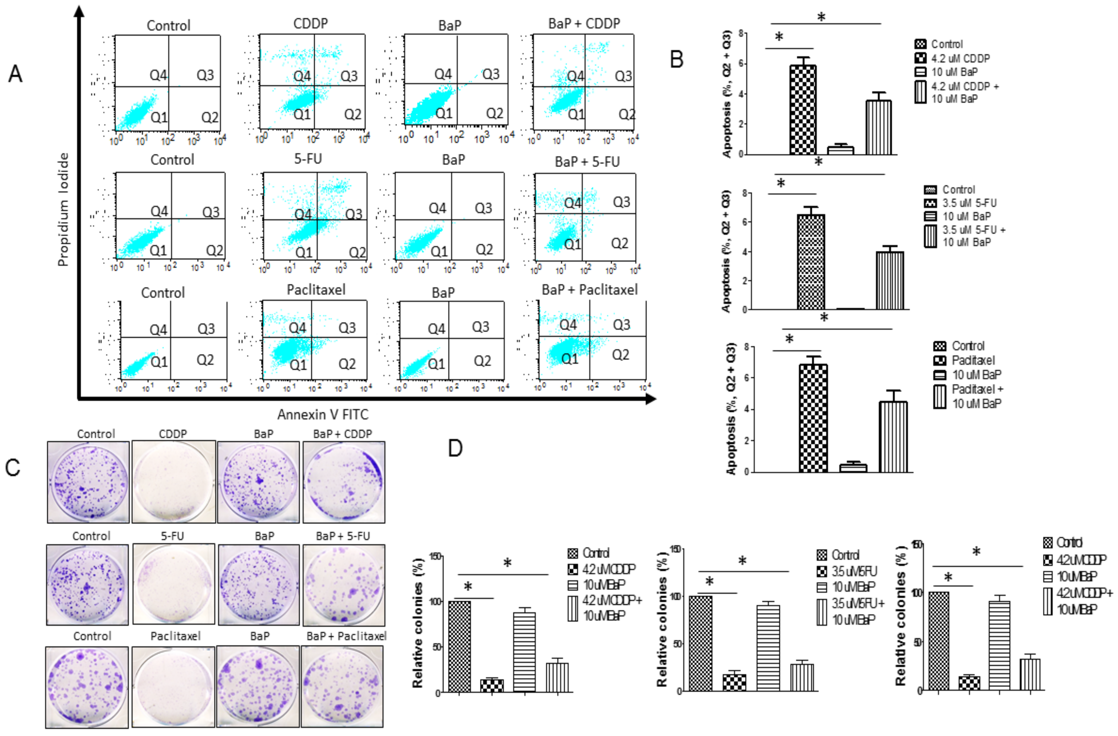
2.1. Benzo-α-Pyrene Reverse Individual Drug-Induced Cell Death and Aapoptosis in WHCO1 Cells

The first step was to evaluate the effect of BaP on the response of WHCO1 cancer cells to individual drugs followed by the evaluation of BaP effect on the response of WHCO1 cancer cells to combinations of drugs (Figure 1A,B). The IC50 of cisplatin, measured via the MTT assay, was 18.5 ± 6.4 µM whilst that of 5-fluorouracil was 14.1 ± 3.8 µM. Both IC50 values were measured in WHCO1 cells over 24 h. The IC50 was determined as the concentration of drug needed to kill 50% of cells over 24 h. Similar IC50 values have been obtained before [47,48,49,50,51,52,53,54]. Our preliminary experiments showed that the use of concentrations above 10 µM would result in considerable cell death, and we used concentrations less than half the IC50 value (Supplementary Figure S1A–C). Paclitaxel IC50 is reported to be much lower than that of cisplatin and 5-fluorouracil [55,56,57]. We used 3.5 µM 5-fluorouracil, 4.2 µM cisplatin, and 2 µM for paclitaxel. The concentration for benzo-α-pyrene (10 µM) was based on our cell viability assays and from the literature value [58,59]. No morphological changes were observed in WHCO1 cells treated with 0.1% DMSO and 10 µM BaP alone (data not shown). Cell viability assays using different doses of BaP, from 0 µM to as high as 120 µM showed that the viability of WHCO1 cells decreased when the concentration of BaP was above 80 µM after 24 h of incubation (Supplementary Figure S1D). Two different methods were used to determine the effects of cisplatin, 5-fluorouracil, paclitaxel, and BaP on WHCO1 cell proliferation. Immunoblot analysis of proliferation markers, Ki67 and PCNA, showed that at early time points in the experiment there is a differential response to the presence of cisplatin, 5-fluorouracil, and paclitaxel (Supplementary Figure S2A,C,E). At 24 h after the start of the experiment, cisplatin, 5-fluorouracil, and paclitaxel caused downregulation of both Ki67 and PCNA. This downregulation was reversed in the presence of BaP, showing that BaP can increase WHCO1 cancer cell proliferation (Supplementary Figure S2A,C,E). Cell counting assays substantiated our immunoblot findings (Supplementary Figure S2B,D,F). Similar cell counting results were obtained using another esophageal cancer cell line, WHCO5 (Supplementary Figure S3A–C).
Annexin V and propidium iodide double staining confirmed the induction of apoptosis in the presence of cisplatin, 5-fluorouracil, and paclitaxel (Figure 2A,B, early apoptosis and late apoptosis shown in Q2 and Q3). Indeed, apoptotic cell death was also characterized by cytoplasmic blebbing (data not shown). BaP on its own did not cause any apoptosis. BaP, when combined with either cisplatin, 5-fluorouracil, or paclitaxel, seems to confer protection against apoptosis, as the percentage of cells undergoing apoptosis decreased. Cisplatin, 5-fluorouracil, and paclitaxel reduced colony formation significantly and BaP reverses the effect of each individual drug (Figure 2C,D). Drug-treated cells displayed significantly smaller and fewer colonies than control cells and those treated with BaP (Figure 2C,D). BaP in combination with drugs reduced drug-induced apoptosis and ameliorated the reduction in colony formation (Figure 2C,D). Again, using a different esophageal cancer cell line, WHCO5, we obtained similar apoptosis and colony formation results (Supplementary Figure S4A–D). Since results for the two esophageal cancer cell lines, WHCO1 and WHCO5, were similar, only WHCO1 cells were used in subsequent studies.

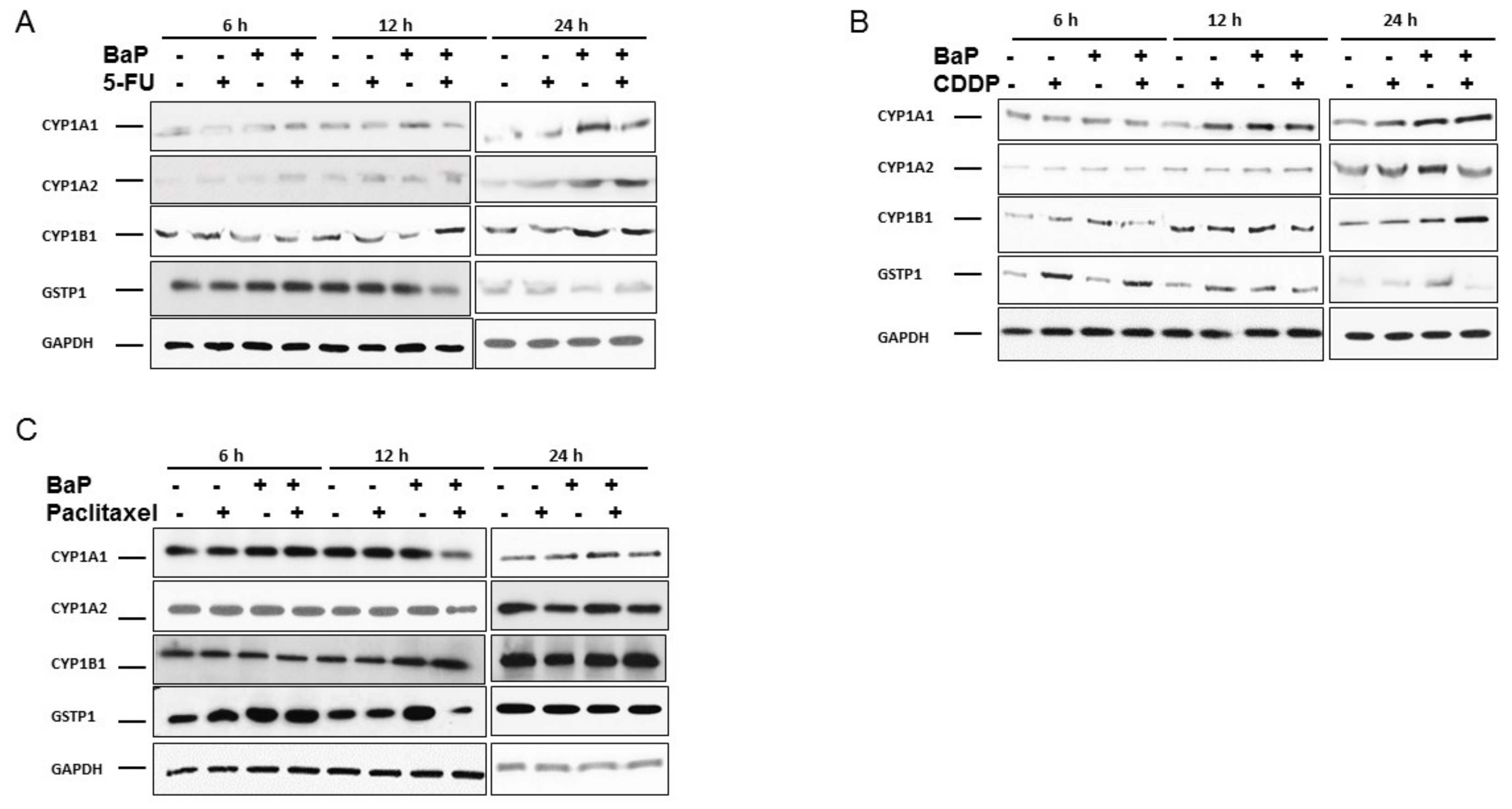
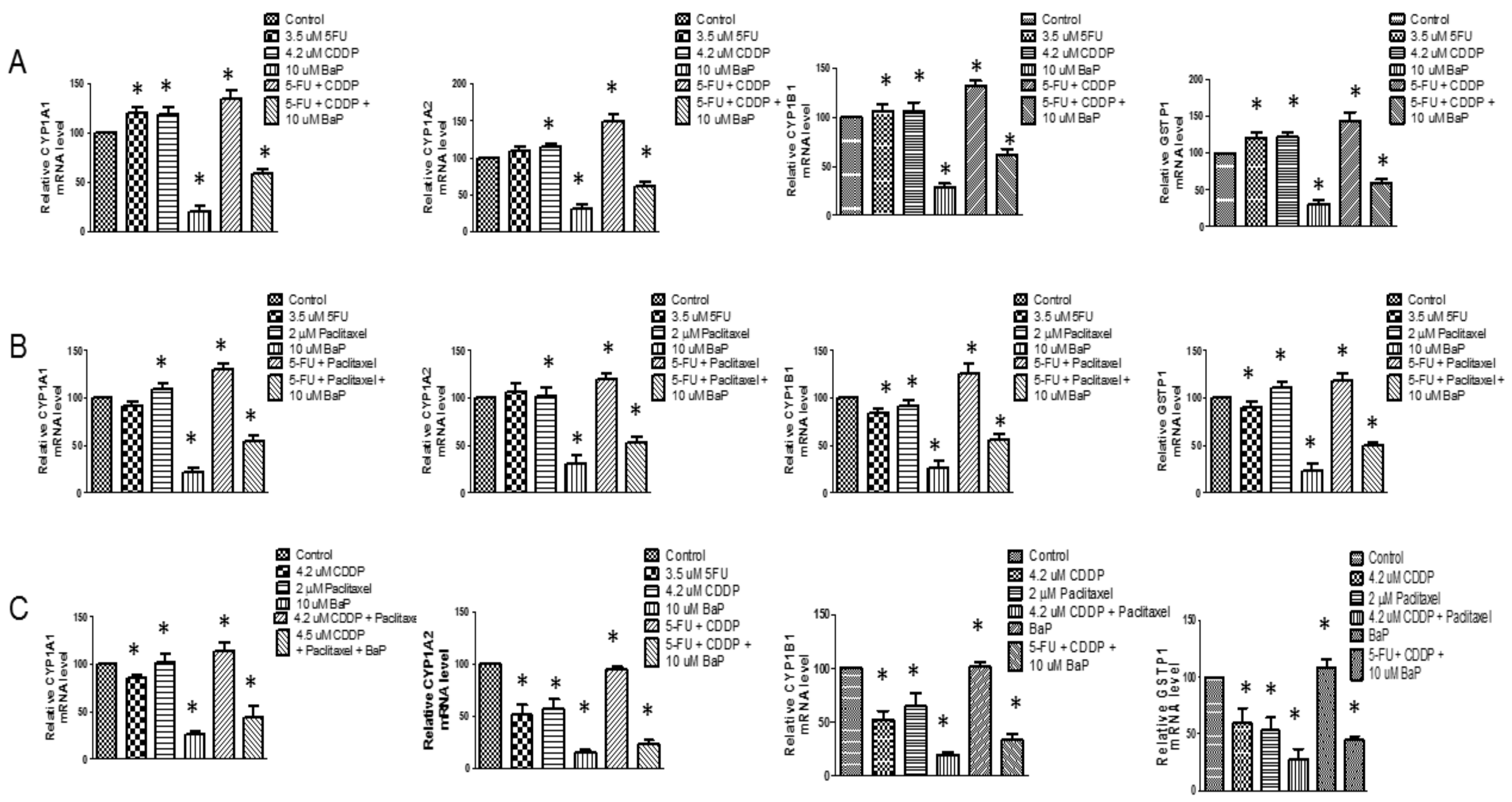
2.2. Cisplatin, 5-Fluorouracil, and Paclitaxel Differentially Affected the Expression of CYP1A1, CYP1A2, CYP1B1, and GSTP1 in WHCO1 Ccells

CYPs are members of the xenobiotic metabolizing enzymes involved in drug metabolism. We evaluated how the presence of cisplatin, 5-fluorouracil, paclitaxel, and BaP would affect the expression of four of these enzymes. At 6 h of incubation, BaP did not affect CYP1A1 protein levels. At 12 h and 24 h, however, the presence of BaP caused significant increases in CYP1A1 protein levels (Figure 3A). The treatment of WHCO1 cells with 5-fluorouracil and BaP resulted in a significant increase in CYP1A2 protein levels especially after 24 h (Figure 3A). 5FU caused differential CYP1B1 gene expression in the presence of BaP at 6 and 12 h of incubation. After 24 h, BaP induced a significant increase in CYP1B1 protein levels (Figure 3A).
Cisplatin-treated cells showed significant increase in CYP1A1 protein levels only after 12 h of incubation (Figure 3B). The use of both cisplatin and BaP resulted in a significant increase in CYP1A1 and CYP1B1, higher than when each is used separately, thus having a synergistic effect on CYP1A1 and CYP1B1 gene expression (Figure 3B). Cisplatin and BaP induced a significant upregulation of CYP1A2 protein levels only after 12 h of incubation (Figure 3B). The presence of cisplatin caused significant increases in GSTP1 protein levels at all time points during the experiment (Figure 3B). Paclitaxel-treated cells showed no change in CYP1A1 protein levels (Figure 3C). After 12 h of incubation with both paclitaxel and BaP, CYP1A1 protein levels decreased significantly. The same trend was observed in the expression of CYP1A2. There was a differential expression of GSTP1 in the presence of paclitaxel and BaP (Figure 3C). In summary, BaP is associated with increased CYP1A1 and CYP1A2 gene expression. These genes are involved in drug metabolism and their increased expression might result in reduced drug efficacy.

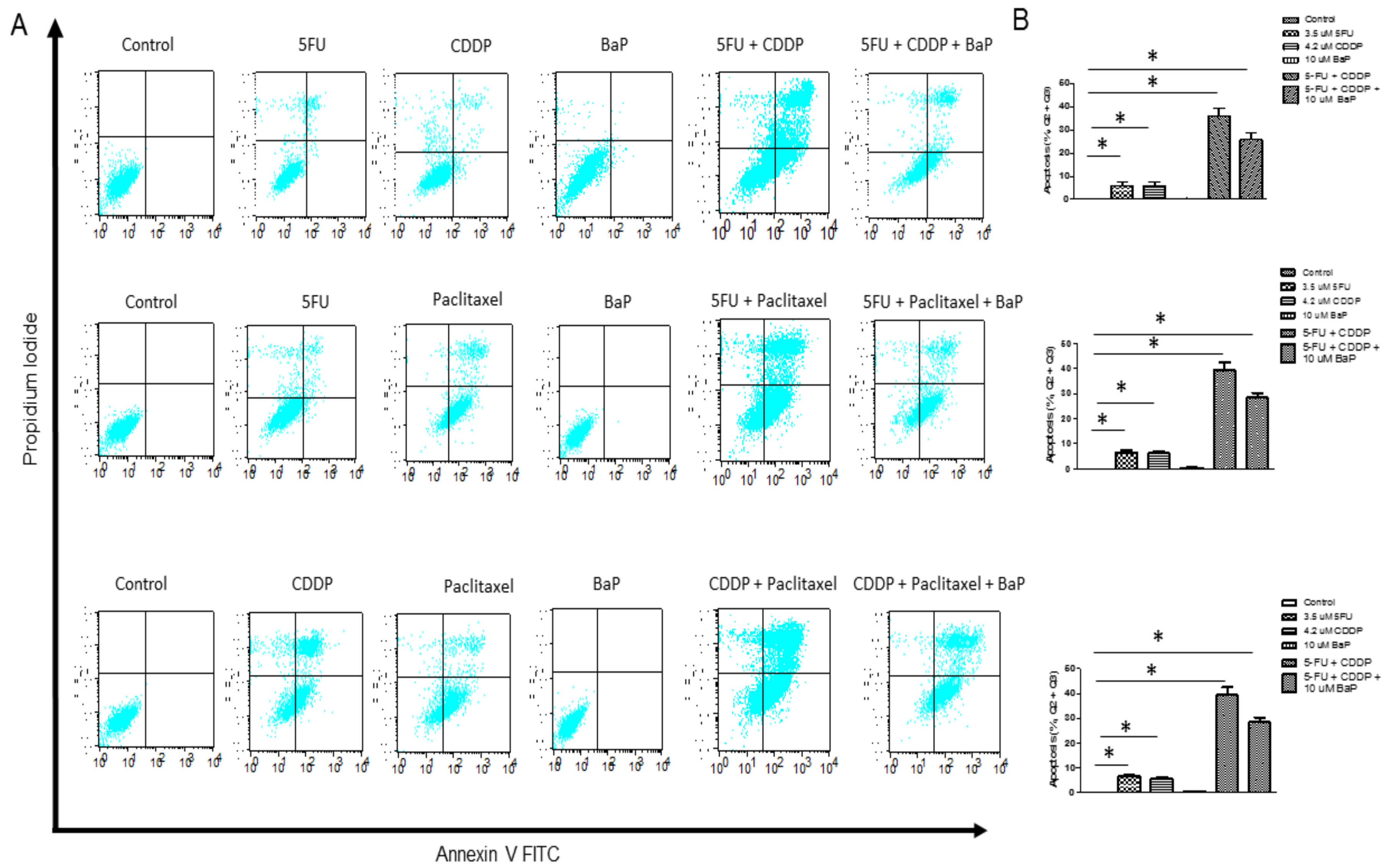
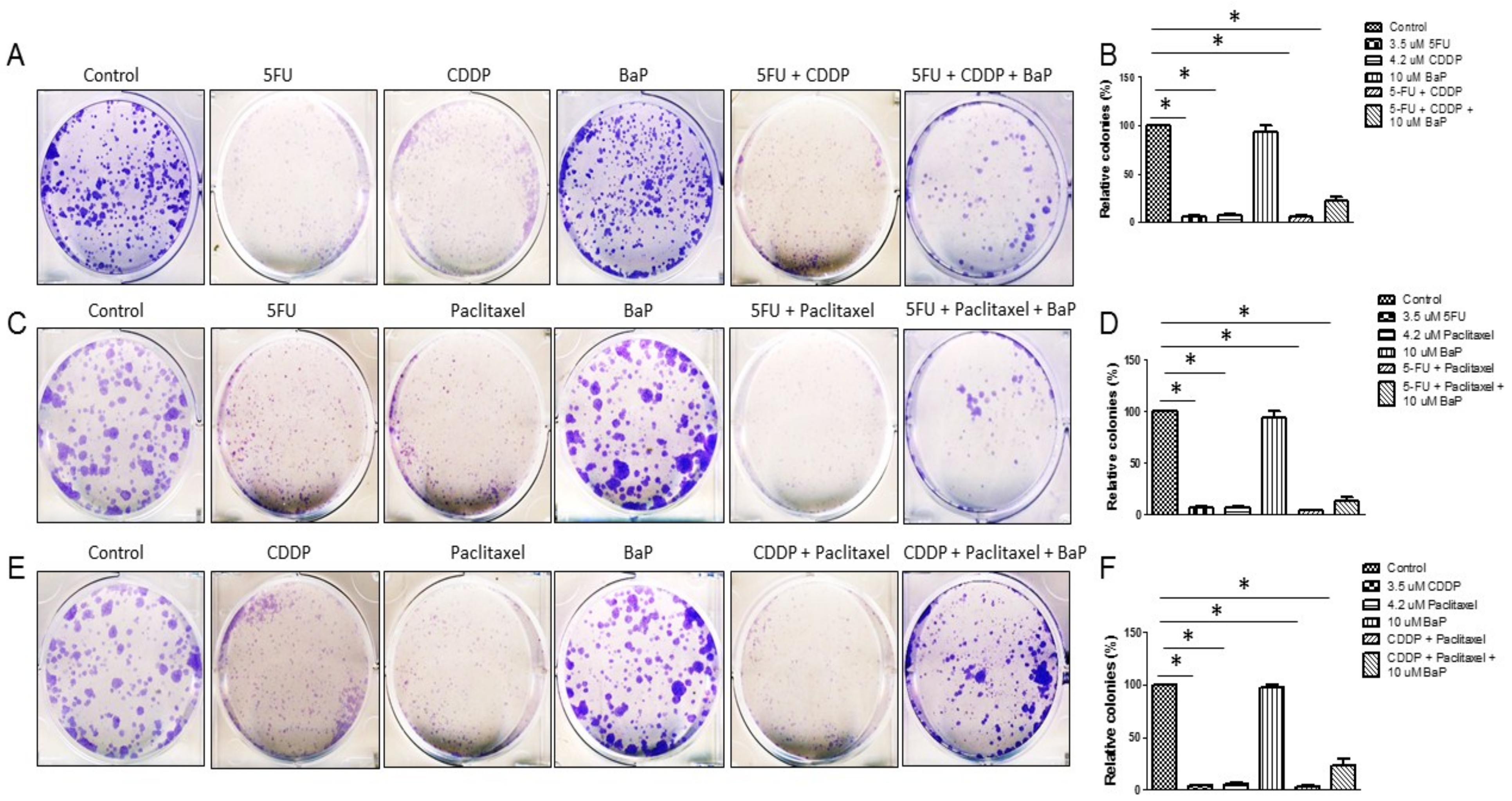
2.3. BaP Protects WHCO1 Cancer Cells from the Effects of Cisplatin, 5-fluorouracil, and Paclitaxel Combination Therapy

Chemotherapy is given as combinations of drugs and, to increase the relevance of our study, we evaluated the influence of BaP exposure on the response of WHCO1 esophageal cancer cells to combinations of chemotherapeutic drugs. As expected, drug-treated cells showed reduced proliferation compared to controls (Supplementary Figure S5A,B). A combination of cisplatin and 5-fluorouracil further reduced proliferation of WHCO1 cells compared to individual drugs (Supplementary Figure S5A,B). Similar results were obtained when WHCO1 cells were treated with 5-fluorouracil and paclitaxel (Supplementary Figure S5C,D) and a combination of cisplatin and paclitaxel reduced WHCO1 cell proliferation further compared to the effect of the individual drugs (Supplementary Figure S5E,F). Treatment of WHCO1 cells with a combination of 5-fluorouracil and cisplatin induced increased apoptosis compared to the individual drugs (Figure 4A,B, top panel). BaP had a protective effect on WHCO1 cancer cells treated with cisplatin and 5-fluorouracil as exposure of cancer cells to drugs in addition to BaP reduced the percentage of cells undergoing apoptosis (Figure 4A,B, top panel). Indeed, similar results were obtained when combinations of 5-fluorouracil and paclitaxel (Figure 4A,B, middle panel), and cisplatin and paclitaxel were used (Figure 4A,B, lower panel). The presence of BaP reversed the increased apoptosis observed when combinations of cisplatin, 5-fluorouracil, and paclitaxel were used.
Commitment of cells to the apoptotic pathway depends on the balance between protein mediators of cell death (Bax included) and anti-apoptotic proteins such as Bcl-2 and Bcl-xL. As shown in Supplementary Figure S6, there is a drug-induced decrease in Bcl-2 and Bcl-xL mRNA levels. BaP alone caused an upregulation of Bcl-2 and Bcl-xL mRNA levels and reversed by 30–40% the effects of drugs combinations on the expression of the same genes (Supplementary Figure S6A–F). Drug-treated cells showed significantly reduced colony formation (Figure 5A–F). Combinations of cisplatin, 5-fluorouracil, and paclitaxel further reduced colony formation compared to individual drugs. BaP had no effect on WHCO1 cell colony forming abilities (Figure 5A–F). The presence of BaP reduced the effect of drugs combination on WHCO1 cell colony formation as larger and more colonies were formed compared to both individual and combinations of drugs (Figure 5A–F).

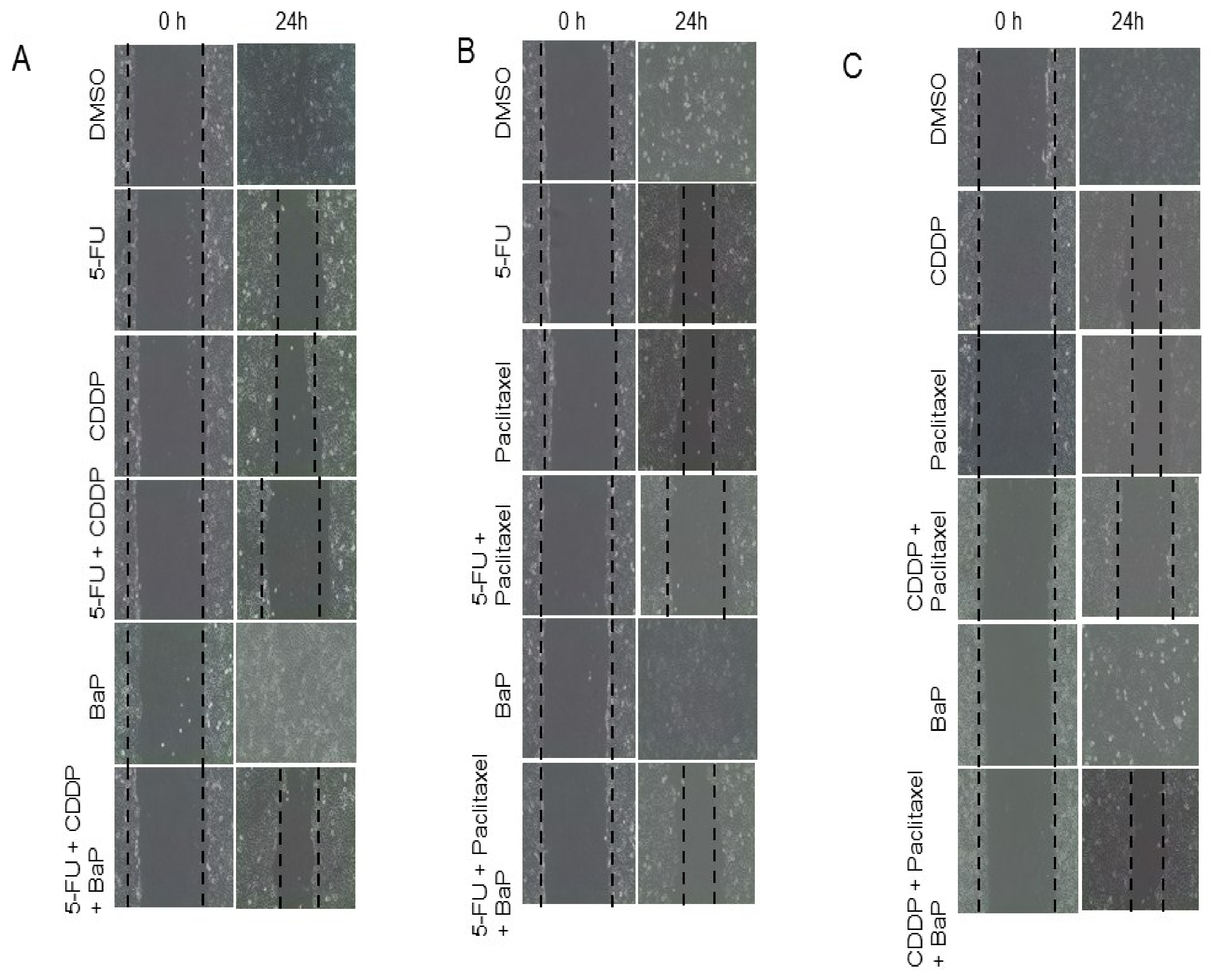
2.4. Effect of Combinations of Cisplatin, 5-Fluorouracil, Paclitaxel, and BaP on WHCO1 Cell Migration and Invasion

To determine whether BaP would influence the effect of cisplatin, 5-fluorouracil, and paclitaxel on WHCO1 cell migration, we performed the wound-healing assay. The scratch wound assays showed that BaP-treated cells migrated at the same rate as control WHCO1 cells (Figure 6A–C; Supplementary Figure S7A–C). In contrast, drug-treated cells did not migrate much and the wound gap remained (Figure 6A–C; Supplementary Figure S7A–C). WHCO1 cells exposed to combinations of cisplatin, 5-fluorouracil, and paclitaxel migrated very little compared to controls. Combinations of cisplatin, 5-fluorouracil, and paclitaxel together with BaP resulted in WHCO1 cells migrating much further than combinations of drugs only (Figure 6A–C; Supplementary Figure S7A–C). The influence of BaP on WHCO1 cancer cell invasion in the presence of drugs was investigated using the trans-well assay. Matrigel invasion assays showed that same number of control cells and BaP-treated cells invaded through the Matrigel whilst drug combination-treated cells showed decreased invasion abilities compared to control cells (Supplementary Figure S7D–F). BaP reversed the effects of drugs on the invasive abilities of WHCO1 cells (Supplementary Figure S7D–F).

2.5. Chemotherapeutic Drugs Differentially Affect CYPs Expression and Survival Signaling Pathways

The treatment of WHCO1 cells with combinations of cisplatin and 5-fluorouracil resulted in increased expression of CYP1A1 mRNA after 24 h (Figure 7A). Each individual drug also increased CYP1A1 mRNA expression (Figure 7A). BaP alone and in combinations with cisplatin and 5-fluorouracil caused significant increases in CYP1A1 mRNA levels (Figure 7A). The treatment of WHCO1 cells with cisplatin and 5-fluorouracil caused significant increases in CYP1B1 mRNA levels, while there were no major changes to the levels of CYP1A2 and GSTP1 mRNA levels (Figure 7A). A combination of 5-fluorouracil and paclitaxel resulted in increased CYP1A1 and CYP1A2 mRNA levels (Figure 7B), with an even higher increase when 5-fluorouracil, paclitaxel, and BaP were combined. There were changes to CYP1B1 and GSTP1 mRNA levels (Figure 7B). Combinations of cisplatin and paclitaxel caused significant increases in CYP1A1, CYP1A2, and CYP1B1 mRNA levels, but adding BaP to this mixture did not result in any further increase in the expression of these genes. There was however an increase in GSTP1 when BaP was added to the combination of cisplatin and paclitaxel (Figure 7C).
Signal transduction pathways including the Akt and MEK-ERK, involved in cancer cell survival, can be activated by chemotherapeutic drugs and can affect cell proliferation, gene expression, and invasion. Immunoblot analysis showed that, individually, cisplatin, 5-fluorouracil, and paclitaxel activated the MEK-ERK pathway while combination-treated cells showed a downregulation of the MEK-ERK signaling pathway, probably due to the toxic effects of the two drugs (Figure 8A–C). The 5-fluorouracil and paclitaxel-treated cells showed upregulation of the p-Akt (Figure 8C). BaP on the other hand significantly activated the MEK-ERK pathway rather than the Akt pathway (Figure 8A–C). No major changes were detected in the activation levels of the PI3K/Akt pathway in the presence of cisplatin (Figure 8A,C). Importantly, BaP reversed the downregulation of p-ERK 1, 2 induced by drug combinations (Figure 8A–C). In summary, BaP protect cancer cells from the effect of drugs through activating survival pathways such as the MEK-ERK and PI3K/Akt pathways.

3. Discussion

Globally, environmental pollution is increasing, resulting in increased atmospheric levels of chemicals such as BaP. Investigations into the role of BaP in carcinogenesis is well studied [20,21,60,61,62,63,64]. BaP contamination poses a serious health hazard to humans and those affected by diseases [25,65,66,67,68]. It is well documented that exposure to BaP and other chemicals found in soot, cigarette smoke, and exhaust fumes is associated with cancer in humans [5,6,7,8,9,58,69,70]. Due to its lipophilic nature, BaP can easily cross the cell membrane and accumulate in cells where it complexes with the aryl hydrocarbon receptor (AhR), stimulating its metabolism through induction of cytochrome P450 isoforms 1A1 (CYP1A1) and 1B1 (CYP1B1) [8,9,10].
People are exposed to different amounts of BaP depending on their daily activities. Ambient air contains BaP concentrations ranging from 20 mg/m3 to 100 mg/m3 [71]. Daily food consumption especially of foods such as fried chicken, smoked meat, and even potato chips contain higher levels of up to 125 ng/person/day [72]. The long half-life and the lipophilicity of BaP result in its accumulation in the human body to higher levels. Occupational exposure can result in very high levels of BaP accumulating in the human body. Limited studies have provided data on pollutants in rural areas [6]. In the rural areas the source of BaP would be burning wood for example. Concentrations of BaP depended on the season and ranged from 0.057 to 1.526 ng m3 in the cold season whilst it ranged from 0.009 to 0.111 ng m3 during the warm season in a study done in Croatia [6]. A major determinant of urban air genotoxicity is the fumes from traffic [73,74,75,76,77]. On average individual polycyclic aromatic hydrocarbons concentrations in ambient air in urban areas range from 1 to 30 ng/m3, with concentrations shooting up to 10–100 ng/cm3 being reported in road tunnels and large cities [78,79,80,81,82,83,84].
Cancer patients undergoing treatment such as chemotherapy are normally treated as outpatients. This exposes them to environmental insults such as BaP, especially smokers and passive smokers. Given that exposure to BaP, a well-characterized procarcinogen, is unavoidable, basic reasoning demands that the influence of such compounds on the efficacy of chemotherapeutic agents be investigated. The effect of BaP on patients undergoing chemotherapy has not been studied before. In addition, the in vitro effect of BaP on the response of WHCO1 esophageal cancer cells to the presence of two drugs studied at the same time hasn’t been investigated. To understand the toxicity of polycyclic aromatic hydrocarbons, an investigation of downstream genes including the CYP450 enzymes is important [85,86]. Being one of the most environmentally relevant polycyclic aromatic hydrocarbon, we used BaP as a reference to investigate the changes in WHCO1 cancer cell gene expression in response to the presence of chemotherapeutic agents in the presence of a pollutant.
Esophageal squamous cell carcinoma has a very low five-year survival rate [35,36] and the disease relapses and becomes resistant to most available chemotherapeutic drugs [35,36]. This poor prognosis emanates from the lack of efficacy of chemotherapeutic drugs and also the late detection of the disease. The anticancer drugs used in the study are the most common ones used in esophageal cancer and have different modes of actions. Firstly, cisplatin, also known as Platinol (CDDP), is an alkylating agent used for the treatment of many cancers such as esophageal, breast, cervical, and bladder cancer [87,88,89]. Secondly, 5-fluorouracil, an analogue of pyrimidine, is considered one of the most effective anticancer drug available [90,91]. Lastly, paclitaxel, also known as Taxol, is a member of the taxane family of several chemotherapeutic agents used to treat several cancers [92,93,94].
Different concentrations of these drugs have been used in several experimental setups [53,95,96,97,98,99,100]. Several studies have shown that the use of cisplatin and 5-fluorouracil combination is effective and safe in patients with advanced squamous cell oral cancer and head and neck carcinomas [101,102,103]. In addition, cisplatin and paclitaxel have been used to induce apoptosis in several cell lines including ovarian carcinoma cells [104,105,106]. A recent study evaluated the use of paclitaxel and 5-fluorouracil regimen to treat patients with advanced oesophageal carcinoma [107]. Whilst most of these studies show that it is safe and effective to use these combinations of drugs to treat the above mentioned cancers, none of these studies evaluated how such treatment regimens will be affected by the presence of polycyclic aromatic hydrocarbons such as BaP, making our study very relevant. A study to determine the toxicity of cisplatin as a treatment of malignant pleural effusions showed that a great variability of intrapleural cisplatin concentration depends on plasma diffusion [108]. Previous studies have shown that low concentrations of Paclitaxel such as 3 µM inhibited cell proliferation as effective as high concentrations [109,110]. Similar to our results, paclitaxel has been shown to cause G2 cell cycle arrest [109,110].
Our initial investigations, using individual drugs, showed that BaP reverses the effects of cisplatin, 5-fluorouracil, and paclitaxel on WHCO1 esophageal cancer cell proliferation by 20–30% compared to that of controls. Several studies have shown that BaP has proliferation-inducing properties. Dutta and colleagues found that BaP concentrations up to 80 µM did not cause any changes to glial cell viability after 48 h of BaP administration, compared to control cells [111]. Beside inducing proliferation and reversing the effects of cisplatin, 5-fluorouracil, and paclitaxel on WHCO1 cancer cell proliferation, BaP also reversed the effect of the same drugs on WHCO1 cancer cell apoptosis and colony formation. The observed decrease in cellular proliferation in the presence of drugs is expected as one of the modes of action of the drugs is inhibition of cellular growth and the killing of fast growing cells [112,113]. Contrary to our results, BaP has been found to induce apoptosis in several cells including neurons and hepatocytes [114,115,116,117].
In summary, our results show a differential expression of CYPs in WHCO1 cells in response to the presence of drugs with more upregulation than downregulation of genes. The precise mechanism responsible for the up-regulation caused by the drugs remains to be elucidated. Earlier studies have shown that 5-fluorouracil induces the activity of cyclin A-cyclin-dependent kinase 2 (Cdk2), an important cell cycle regulatory protein [118,119]. Interestingly, inhibition of the AhR has been shown to down-regulate cyclin and Cdk-2 expression [120,121,122]. Thus, it is possible that an up-regulation in Cdk2 activity, in this case caused by cisplatin and 5-fluorouracil, could be linked to a corresponding up-regulation in AhR activity ultimately resulting in increased CYP1 expression [121,122]. Alternatively, 5-fluorouracil has also been shown to activate the NF-E2 p45-regulated factor (Nrf2) signaling pathway. The regulatory region of the Nrf2 gene encompasses several AhR-binding response elements [123,124,125].
Several reports have shown that BaP has an effect on genes involved in cell cycle, migration, DNA repair, and apoptosis [126,127,128,129,130,131,132,133]. Proteins such as Bcl-2, Bax, and Bcl-xL are downstream of DNA damage and repair processes and the levels of these proteins determine whether cells undergo apoptosis or not [134]. Overall, our results show decreased Bcl-2 and Bcl-xL when cells were treated with drugs and even more decrease when drugs combinations were used. It is possible that this BaP-induced increase in Bcl-21 and Bcl-xL reverses drug-induced apoptosis. Several reports have shown that Bcl-2 and Bcl-xL are involved in the development of resistance to paclitaxel in cancer cells [135,136,137,138,139,140,141,142].
Several signaling pathways have been shown to be activated by the presence of reactive oxygen species, a by-product of cellular activation by BaP [111,143,144,145,146,147,148,149]. The PI3K-Akt signaling pathway is one of the most important pathways involved in the proliferation and survival of cells [150,151,152]. Our data show that indeed the MEK-ERK and PI3K-Akt signaling pathways are activated by both drugs and BaP, with BaP the stronger activator. The present study suggest that BaP can reverse the effects of drugs on cancer cells, including apoptosis, and this may be mediated by the activation of survival pathways such as MEK-ERK and Akt pathways in addition to upregulation of proteins such as Bcl-2 and Bcl-xL. This can result in resistant cancer cells, adding more weight to the growing concerns to the problem of environmental pollution.

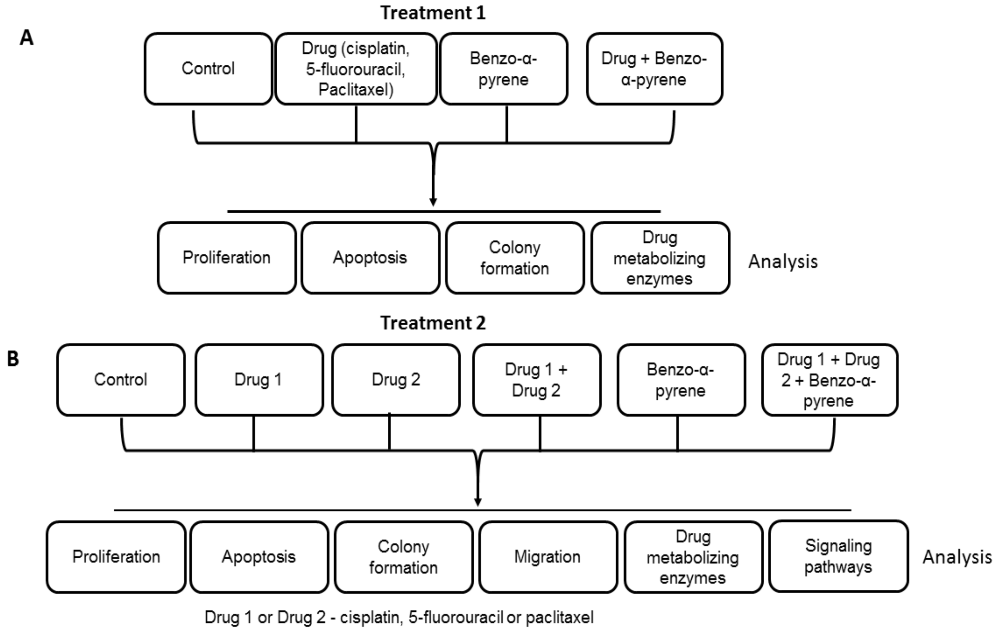
4. Materials and Methods

4.1. Cell Culture and Treatments

WHCO1 and WHCO5 cells, derived from primary oesophageal squamous cell carcinoma in South Africa [153], were maintained in Dulbecco’s Modified Eagle Medium (DMEM) (Sigma-Aldrich, St. Louis, MO, USA) supplemented with 10% foetal bovine serum (FBS) (GIBCO, Invitrogen, Carlsbad, CA, USA), penicillin (100 U/mL), and streptomycin (100 µg/mL) (Sigma-Aldrich, St. Louis, MO, USA) at 37 °C in a humidified atmosphere of 5% CO2. These cells have been extensively used to study carcinogenesis and drug discovery [95,154]. WHCO1 and WHCO5 cells (5 × 10⁵ cells/well) were seeded in 6 cm dishes and allowed to attach. The cell culture media was replaced with fresh media prior to the start of treatment. Cell were treated by the addition of cisplatin (Mw 300.05; CAS 15663-27-1; Sigma, Marlborough, MA, USA), 5-fluorouracil (Mw 130.08 g/mol; CAS 51-21-8; Sigma), paclitaxel (Mw 853.91 g/mol; CAS 33069-62-4; Sigma), or BaP (Mw 252.31; CAS 50-32-8; Sigma) at the indicated concentrations for different time periods [53]. Drug concentrations at less than half of the IC50 were used in evaluating gene expression changes. The concentrations used were: 3.5 µM 5-fluorouracil; 4.2 µM cisplatin, 2 µM paclitaxel, 10 µM BaP, and combinations of 3.5 µM 5-fluorouracil, 4.2 µM cisplatin, 2 µM paclitaxel, and 10 µM BaP; and 0.1% DMSO (control). Incubation with drugs was done for 6, 12, and 24 h. For proliferation assays WHCO1 and WHCO5 cells were incubated for periods of up to 72 h.

4.2. Cell Viability Assay

Cell viability of WHCO1 and WHCO5 cancer cells after exposure to 0.1% DMSO (control), cisplatin, 5-flurouracil, paclitaxel, and BaP was determined by the MMT assay. The MTT reagent (3-[4,5-dimethylthiazol-2yl]-2,5-diphenyl-tetrazolium bromide) was first dissolved in phosphate-buffered saline (PBS) and stored at 20 °C in the freezer. WHCO1 and WHCO5 cancer cells (5 × 10³/well) were plated in 96-well plates and incubated overnight. Different concentrations of cisplatin, 5-flouorouracil, paclitaxel, or combinations, and 10 µM BaP were added to the media and cells were cultured for 24 h. The MTT reagent (20 µL) was added to each well and incubation continued for 4 h. After clearing the supernatant, DMSO was then added to each well and cells were shaken for 15 min. The resulting colour changes were read as optical density on a microplate reader. Cell viability was also determined by counting viable cells using the Countess Cell Counter. Assays were performed at the same time and done in triplicates. Results are reported as mean ± S.D. Experiments were repeated at least twice.

4.3. Microscopy

Cell morphology was observed and photographed using a light microscope (Olympus CKX41 with SC30 camera, Olympus Optical Co., Tokyo, Japan). Images were taken at 10X and 20X magnification. WHCO1 and WHCO5 cancer cells were treated as described above and observed after 24 h of incubation.

4.4. Annexin V Assay for Apoptosis

Apoptosis was evaluated through double staining with Annexin V and Propidium Iodide according to manufacturer’s instructions followed by flow cytometric analysis. WHCO1 and WHCO5 cells were incubated with cisplatin, 5-fluorouracil, paclitaxel, and BaP for 24 h and washed with PBS. Cells were harvested by trypsinisation using 0.05% trypsin-EDTA and resuspended in Annexin V binding buffer and stained with Annexin V conjugated to FITC and Propidium Iodide as described by the manufacturer’s instructions. Incubation in the dark was done for 15 min at 25 °C. The Beckman Coulter Flow Cytometer (Beckman Coulter, Life Sciences, Indianapolis, IN, USA) was used for flow cytometric analysis. Data acquisition was done on 2 × 10⁴ events per treatment condition. Analysis was performed using the Cellquest software (Version 5.1, Becton Dickinson, Franklin Lakes, NJ, USA).

4.5. Scratch Wound Assay

The effect of cisplatin, 5-fluorouracil, paclitaxel, and BaP on WHCO1 cell migration was evaluated by the wound healing assay. WHCO1 cells (1 × 10⁶) were cultured in 6-well plates and incubated overnight in DMEM containing 10% FBS. WHCO1 cells were cultured until confluent before the scratch wound assay was performed. Several lines were drawn underneath the dishes with a marker. These served as marks for the scratch wound experiment to be measured. Three parallel scratch wounds were made using a 200 µL (yellow) pipet tip. Washing was done twice with PBS to remove debris and fresh media was added. WHCO1 cells were then treated with either 0.1% DMSO, cisplatin, 5-fluorouracil, paclitaxel, and BaP for 24 h as indicated. Images were taken at 0 and at 24 h of culture. Scratch wounds were observed using phase contrast inverted microscopy on an inverted microscope (Olympus CKX41).

4.6. Transwell Invasion Assay

WHCO1 cells were cultured to 80% confluency before use in invasion assays. Matrigel (BD Biosciences, Bedford, MA, USA) was thawed overnight at 4 °C and then kept on ice. Transwell culture plates, with permeable cell culture inserts of 12 mm diameter and 8 µm pore size membranes (Corning Inc., Corning, NY, USA) were chilled to 4 °C and kept in the fridge. Matrigel (100 µL of 2 mg/mL) (BD Biosciences, Franklin Lakes, NJ, USA) was added to the upper chamber of the insert. The plates are then incubated at 37 °C for 2 h to solidify the Matrigel. WHCO1 cells were trypsinised and resuspended in DMEM. 1 × 10⁵ WHCO1 cells were added to the upper compartment and allowed to attach for 6 h before being treated with cisplatin, 5-fluorouracil, paclitaxel, or BaP. The bottom well contained media with 10% FBS as attractant, whilst the upper compartment of the insert contained media 1% FBS. WHCO1 cells were treated with 0.1% DMSO, drugs, and 10 µM BaP) and incubation was done for 24 h. At the end of the experiment the insert was treated as follows: cells and the Matrigel in the upper compartment were removed gently by wiping with a cotton swab. WHCO1 cells on the lower side of the insert membrane were fixed with 5% glutaraldehyde at room temperature for 10 min. Cells were then stained with 1% crystal violet in 2% ethanol at room temperature for 20 min. Inserts were washed three times and dried. The number of cells that invaded through the Matrigel were counted under the phase contrast microscope using the 10× objective. Four different fields were chosen and the average number of WHCO1 cells was obtained for each treatment. The invaded cell numbers were plotted onto a graph.

4.7. Quantitative RT-PCR Analysis

Total RNA was isolated using Qiazol (Roche, Berlin, Germany) as per the manufacturer’s instructions. cDNA was synthesized from 5 µg total RNA and used for quantitative real time reverse transcription polymerase chain reaction (RT PCR) analysis. RT PCR was carried out under the following conditions: 95 °C for 3 min (pre-incubation), followed by 40 cycles of 95 °C for 20 s, 62 °C for 20 s, and 72 °C for 20 s. The comparative critical threshold (Ct) method was used for the calculation of expression fold change between samples. Gene expression was calculated as fold induction, relative to the mean value of control samples using the equation 2−∆∆Ct. GAPDH was used as the housekeeping gene. Primers used are shown in Supplementary Table S1. Experiments were performed in triplicate.

4.8. Immunoblot Analysis

Cells were treated as described above and lysed in RIPA buffer and 1X complete protease inhibitor cocktail was added (Roche, Mannheim, Germany). The BCA Protein Quantification kit was used to quantify protein concentrations (Pierce, Rockford, IL, USA). Immunoblot analysis was performed using the following primary antibodies: rabbit anti-Ki67, goat anti-CYP1A1, rabbit anti-GAPDH, rabbit anti-PCNA, mouse CYP1A2, rabbit anti-CYP1B1, rabbit anti-GSTP1, rabbit anti-p-38 MAP kinase, anti-p-ERK 1, 2, anti-ERK 2, anti-p-Akt, and anti-Akt. The secondary antibodies used were goat anti-mouse IgG horseradish peroxidase (HRP) conjugate and goat anti-rabbit IgG HRP conjugate. The Lumiglo Chemiluminescent substrate (KPL, Gaithersburg, MD, USA) was used for the detection. All experiments were done in triplicates.

4.9. Colony Formation

WHCO1 cells were cultured overnight in 6-well plates at 500 cells per well. DMSO, cisplatin, 5-fluorouracil, paclitaxel, and BaP were added at the indicated concentrations and incubation was continued for 24 h. 0.1% DMSO was used for the control cells. Cells were then incubated in normal media for another 8 days. Methanol (100%) was used to fix the cells and cells were stained with 0.5% crystal violet. Colonies were counted using UVP software and the numbers were plotted on a graph. Images were taken using a camera. The experiments were performed at least three times.

4.10. Statistical Analysis

GraphPad Prism 5 (GraphPad Software Inc., San Diego, CA, USA) was used for statistical analysis to analyze the significance of the differences between the different treatment groups. To evaluate statistical significance between the different groups. One-way analysis of variance (ANOVA) was used and the Student’s t test was used to analyze differences between two independent groups. Data are shown as means ± SD. Values of p < 0.05 were considered significant. Experiments were performed at least in triplicate.

Supplementary Materials

The following are available online, Supplementary Table S1: Oligonucleotide primer sequences used for RT-qPCR; Supplementary Figure S1: Inhibition rates of WHCO1 cancer cells incubated with different concentrations of cisplatin, 5fluorouracil, paclitaxel, and BaP for 24 h; Supplementary Figure S2: Benzo-α-pyrene reverse the effect of cisplatin, 5-FU, and paclitaxel on WHCO1 cell proliferation; Supplementary Figure S3: Benzo-α-pyrene reverse the effect of cisplatin, 5-FU, and paclitaxel on WHCO5 cell proliferation; Supplementary Figure S4: Benzo-α-pyrene abrogates drug-induced apoptosis and colony formation inhibition; Supplementary Figure S5: Benzo-α-pyrene reverse the dual effects of cisplatin, 5-FU, and paclitaxel on WHCO1 cell proliferation; Supplementary Figure S6: Benzo-α-pyrene upregulates Bcl-2 and Bcl-xL expression in WHCO1 cancer cells; Supplementary Figure S7: Benzo-α-pyrene abrogates the effect of cisplatin, 5-FU, paclitaxel, and their combinations on WHCO1 cell migration and invasion.

Acknowledgments

We would like to acknowledge the University of Cape Town, International Centre for Genetic Engineering and Biotechnology (ICGEB), National Research Foundation, and the Allan Gray Orbis Foundation for funding this research. The funders had no role in the conduct of the research or the preparation of the manuscript.

Author Contributions

K.D., C.D., S.M. and M.I.P. conceived and designed the experiments; K.D., N.H. and D.A.S. performed the experiments; K.D., N.E.T., H.S., A.W., C.D. and A.R. analyzed the data; K.D. wrote the main body of the paper.

Conflicts of Interest

The authors declare no conflict of interest.

References

  1. Karle, I.L.; Yagi, H.; Sayer, J.M.; Jerina, D.M. Crystal and molecular structure of a benzo[a]pyrene 7,8-diol 9,10-epoxide n2-deoxyguanosine adduct: Absolute configuration and conformation. Proc. Natl. Acad. Sci. USA 2004, 101, 1433–1438. [Google Scholar] [CrossRef] [PubMed]
  2. Levin, W.; Buening, M.K.; Wood, A.W.; Chang, R.L.; Kedzierski, B.; Thakker, D.R.; Boyd, D.R.; Gadaginamath, G.S.; Armstrong, R.N.; Yagi, H.; et al. An enantiomeric interaction in the metabolism and tumorigenicity of (+)- and (-)-benzo[a]pyrene 7,8-oxide. J. Biol. Chem. 1980, 255, 9067–9074. [Google Scholar] [PubMed]
  3. Courter, L.A.; Luch, A.; Musafia-Jeknic, T.; Arlt, V.M.; Fischer, K.; Bildfell, R.; Pereira, C.; Phillips, D.H.; Poirier, M.C.; Baird, W.M. The influence of diesel exhaust on polycyclic aromatic hydrocarbon-induced DNA damage, gene expression, and tumor initiation in sencar mice in vivo. Cancer Lett. 2008, 265, 135–147. [Google Scholar] [CrossRef] [PubMed]
  4. Courter, L.A.; Musafia-Jeknic, T.; Fischer, K.; Bildfell, R.; Giovanini, J.; Pereira, C.; Baird, W.M. Urban dust particulate matter alters pah-induced carcinogenesis by inhibition of cyp1a1 and cyp1b1. Toxicol. Sci. 2007, 95, 63–73. [Google Scholar] [CrossRef] [PubMed]
  5. Huzlik, J.; Bozek, F.; Pawelczyk, A.; Licbinsky, R.; Naplavova, M.; Pondelicek, M. Identifying risk sources of air contamination by polycyclic aromatic hydrocarbons. Chemosphere 2017, 183, 139–146. [Google Scholar] [CrossRef] [PubMed]
  6. Jakovljevic, I.; Pehnec, G.; Sisovic, A.; Vadic, V.; Davila, S.; Godec, R. Concentrations of pahs and other gaseous pollutants in the atmosphere of a rural area. J. Environ. Sci. Health Part A 2016, 51, 707–713. [Google Scholar] [CrossRef] [PubMed]
  7. Liu, W.; Ma, L.; Abuduwaili, J.; Li, Y. Distribution, source analysis, and ecological risk assessment of polycyclic aromatic hydrocarbons in the typical topsoil of the issyk-kul lake basin. Environ. Monit. Assess. 2017, 189. [Google Scholar] [CrossRef] [PubMed]
  8. Strandberg, B.; Julander, A.; Sjostrom, M.; Lewne, M.; Koca Akdeva, H.; Bigert, C. Evaluation of polyurethane foam passive air sampler (puf) as a tool for occupational pah measurements. Chemosphere 2018, 190, 35–42. [Google Scholar] [CrossRef] [PubMed]
  9. Wolejko, E.; Wydro, U.; Jablonska-Trypuc, A.; Butarewicz, A.; Loboda, T. The effect of sewage sludge fertilization on the concentration of pahs in urban soils. Environ. Pollut. 2018, 232, 347–357. [Google Scholar] [CrossRef] [PubMed]
  10. Bevan, D.R.; Ulman, M.R. Examination of factors that may influence disposition of benzo[a]pyrene in vivo: Vehicles and asbestos. Cancer Lett. 1991, 57, 173–179. [Google Scholar] [CrossRef]
  11. Gao, P.; Liu, S.; Feng, Y.; Lin, N.; Lu, B.; Zhang, Z.; Cui, F.; Xing, B.; Hammond, S.K. Concentrations of polycyclic aromatic hydrocarbons in resuspendable fraction of settled bus dust and its implications for human exposure. Environ. Pollut. 2015, 198, 1–7. [Google Scholar] [CrossRef] [PubMed]
  12. Liu, L.B.; Hashi, Y.; Liu, M.; Wei, Y.; Lin, J.M. Determination of particle-associated polycyclic aromatic hydrocarbons in urban air of beijing by gc/ms. Anal. Sci. 2007, 23, 667–671. [Google Scholar] [CrossRef] [PubMed]
  13. Sera, N.; Fukuhara, K.; Miyata, N.; Tokiwa, H. Detection of nitro-azabenzo[a]pyrene derivatives in the semivolatile phase originating from airborne particulate matter, diesel and gasoline vehicles. Mutagenesis 1994, 9, 47–52. [Google Scholar] [CrossRef] [PubMed]
  14. Butler, J.P.; Post, G.B.; Lioy, P.J.; Waldman, J.M.; Greenberg, A. Assessment of carcinogenic risk from personal exposure to benzo(a)pyrene in the total human environmental exposure study (thees). Air Waste 1993, 43, 970–977. [Google Scholar] [CrossRef] [PubMed]
  15. Lioy, P.J. Human exposure to benzo(a)pyrene:Some more of the story. Toxicol. Ind. Health 1992, 8, 213–219. [Google Scholar] [CrossRef] [PubMed]
  16. Lioy, P.J.; Greenberg, A. Factors associated with human exposures to polycyclic aromatic hydrocarbons. Toxicol. Ind. Health 1990, 6, 209–223. [Google Scholar] [PubMed]
  17. Lioy, P.L.; Waldman, J.M.; Greenberg, A.; Harkov, R.; Pietarinen, C. The total human environmental exposure study (thees) to benzo(a)pyrene: Comparison of the inhalation and food pathways. Arch. Environ. Health 1988, 43, 304–312. [Google Scholar] [CrossRef] [PubMed]
  18. Hattemer-Frey, H.A.; Travis, C.C. Benzo-a-pyrene: Environmental partitioning and human exposure. Toxicol. Ind. Health 1991, 7, 141–157. [Google Scholar] [CrossRef] [PubMed]
  19. Chen, K.M.; Schell, T.D.; Richie, J.P., Jr.; Sun, Y.W.; Zhang, S.M.; Calcagnotto, A.; Aliaga, C.; Gowda, K.; Amin, S.; El-Bayoumy, K. Effects of chronic alcohol consumption on DNA damage and immune regulation induced by the environmental pollutant dibenzo[a,l]pyrene in oral tissues of mice. J. Environ. Sci. Health Part C 2017, 35, 213–222. [Google Scholar] [CrossRef] [PubMed]
  20. Kasala, E.R.; Bodduluru, L.N.; Barua, C.C.; Gogoi, R. Antioxidant and antitumor efficacy of luteolin, a dietary flavone on benzo(a)pyrene-induced experimental lung carcinogenesis. Biomed. Pharmacother. 2016, 82, 568–577. [Google Scholar] [CrossRef] [PubMed]
  21. Masumura, K.; Toyoda-Hokaiwado, N.; Niimi, N.; Gruz, P.; Wada, N.A.; Takeiri, A.; Jishage, K.I.; Mishima, M.; Nohmi, T. Limited ability of DNA polymerase kappa to suppress benzo[a]pyrene-induced genotoxicity in vivo. Environ. Mol. Mutagen. 2017, 58, 644–653. [Google Scholar] [CrossRef] [PubMed]
  22. Shi, Q.; Godschalk, R.W.L.; van Schooten, F.J. Inflammation and the chemical carcinogen benzo[a]pyrene: Partners in crime. Mutat. Res. 2017, 774, 12–24. [Google Scholar] [CrossRef] [PubMed]
  23. Wong, J.Y.Y.; Hu, W.; Downward, G.S.; Seow, W.J.; Bassig, B.A.; Ji, B.T.; Wei, F.; Wu, G.; Li, J.; He, J.; et al. Personal exposure to fine particulate matter and benzo[a]pyrene from indoor air pollution and leukocyte mitochondrial DNA copy number in rural china. Carcinogenesis 2017, 38, 893–899. [Google Scholar] [CrossRef] [PubMed]
  24. Gelboin, H.V. Benzo[alpha]pyrene metabolism, activation and carcinogenesis: Role and regulation of mixed-function oxidases and related enzymes. Physiol. Rev. 1980, 60, 1107–1166. [Google Scholar] [CrossRef] [PubMed]
  25. Greb, W.; Strobel, R.; Rohrborn, G. Transformation of bhk 21/cl 13 cells by various polycyclic aromatic hydrocarbons using the method of styles. Toxicol. Lett. 1980, 7, 143–148. [Google Scholar] [CrossRef]
  26. Rivedal, E.; Sanner, T. Potentiating effect of cigarette smoke extract on morphological transformation of hamster embryo cells by benzo[alpha]pyrene. Cancer Lett. 1980, 10, 193–198. [Google Scholar] [CrossRef]
  27. Chakradeo, P.P.; Kayal, J.J.; Bhide, S.V. Effect of benzo(a)pyrene and methyl(acetoxymethyl) nitrosamine on thymidine uptake and induction of aryl hydrocarbon hydroxylase activity in human fetal oesophageal cells in culture. Cell Biol. Int. 1993, 17, 671–676. [Google Scholar] [CrossRef] [PubMed]
  28. Heimann, R.; Rice, R.H. Polycyclic aromatic hydrocarbon toxicity and induction of metabolism in cultivated esophageal and epidermal keratinocytes. Cancer Res. 1983, 43, 4856–4862. [Google Scholar] [PubMed]
  29. Port, J.L.; Yamaguchi, K.; Du, B.; De Lorenzo, M.; Chang, M.; Heerdt, P.M.; Kopelovich, L.; Marcus, C.B.; Altorki, N.K.; Subbaramaiah, K.; et al. Tobacco smoke induces cyp1b1 in the aerodigestive tract. Carcinogenesis 2004, 25, 2275–2281. [Google Scholar] [CrossRef] [PubMed]
  30. Uno, S.; Dragin, N.; Miller, M.L.; Dalton, T.P.; Gonzalez, F.J.; Nebert, D.W. Basal and inducible cyp1 mrna quantitation and protein localization throughout the mouse gastrointestinal tract. Free Radic. Biol. Med. 2008, 44, 570–583. [Google Scholar] [CrossRef] [PubMed]
  31. Grzywacz, V.; Balcueva, E. Undiagnosed esophageal adenocarcinoma presenting as multiple brain metastases. Case Rep. Oncol. 2017, 10, 938–944. [Google Scholar] [CrossRef] [PubMed]
  32. Wang, J.; Zhang, Z.; Che, Y.; Yuan, Z.; Lu, Z.; Li, Y.; Wan, J.; Sun, H.; Chen, Z.; Pu, J.; et al. Rabdocoestin b exhibits antitumor activity by inducing g2/m phase arrest and apoptosis in esophageal squamous cell carcinoma. Cancer Chemother. Pharmacol. 2018, 81, 469–481. [Google Scholar] [CrossRef] [PubMed]
  33. Xia, H.; Ma, L.; Li, J.; Bai, H.; Wang, D. Elevated hbxip expression is associated with aggressive phenotype and poor prognosis in esophageal squamous cell carcinoma. Am. J. Cancer Res. 2017, 7, 2190–2198. [Google Scholar] [PubMed]
  34. Xu, Y.; Feingold, P.L.; Surman, D.R.; Brown, K.; Xi, S.; Davis, J.L.; Hernandez, J.; Schrump, D.S.; Ripley, R.T. Bile acid and cigarette smoke enhance the aggressive phenotype of esophageal adenocarcinoma cells by downregulation of the mitochondrial uncoupling protein-2. Oncotarget 2017, 8, 101057–101071. [Google Scholar] [CrossRef] [PubMed]
  35. Pennathur, A.; Gibson, M.K.; Jobe, B.A.; Luketich, J.D. Oesophageal carcinoma. Lancet 2013, 381, 400–412. [Google Scholar] [CrossRef]
  36. Pennathur, A.; Xi, L.; Litle, V.R.; Gooding, W.E.; Krasinskas, A.; Landreneau, R.J.; Godfrey, T.E.; Luketich, J.D. Gene expression profiles in esophageal adenocarcinoma predict survival after resection. J. Thorac. Cardiovasc. Surg. 2013, 145, 505–512. [Google Scholar] [CrossRef] [PubMed]
  37. Adorno-Cruz, V.; Kibria, G.; Liu, X.; Doherty, M.; Junk, D.J.; Guan, D.; Hubert, C.; Venere, M.; Mulkearns-Hubert, E.; Sinyuk, M.; et al. Cancer stem cells: Targeting the roots of cancer, seeds of metastasis, and sources of therapy resistance. Cancer Res. 2015, 75, 924–929. [Google Scholar] [CrossRef] [PubMed]
  38. Eun, K.; Ham, S.W.; Kim, H. Cancer stem cell heterogeneity: Origin and new perspectives on csc targeting. BMB Rep. 2017, 50, 117–125. [Google Scholar] [CrossRef] [PubMed]
  39. Ge, Y.; Gomez, N.C.; Adam, R.C.; Nikolova, M.; Yang, H.; Verma, A.; Lu, C.P.; Polak, L.; Yuan, S.; Elemento, O.; et al. Stem cell lineage infidelity drives wound repair and cancer. Cell 2017, 169, 636–650. [Google Scholar] [CrossRef] [PubMed]
  40. Dzobo, K.; Senthebane, D.A.; Rowe, A.; Thomford, N.E.; Mwapagha, L.M.; Al-Awwad, N.; Dandara, C.; Parker, M.I. Cancer stem cell hypothesis for therapeutic innovation in clinical oncology? Taking the root out, not chopping the leaf. Omics 2016, 20, 681–691. [Google Scholar] [CrossRef] [PubMed]
  41. Senthebane, D.A.; Rowe, A.; Thomford, N.E.; Shipanga, H.; Munro, D.; Mazeedi, M.; Almazyadi, H.A.M.; Kallmeyer, K.; Dandara, C.; Pepper, M.S.; et al. The role of tumor microenvironment in chemoresistance: To survive, keep your enemies closer. Int. J. Mol. Sci. 2017, 18. [Google Scholar] [CrossRef] [PubMed]
  42. Oguri, T.; Achiwa, H.; Bessho, Y.; Muramatsu, H.; Maeda, H.; Niimi, T.; Sato, S.; Ueda, R. The role of thymidylate synthase and dihydropyrimidine dehydrogenase in resistance to 5-fluorouracil in human lung cancer cells. Lung Cancer 2005, 49, 345–351. [Google Scholar] [CrossRef] [PubMed]
  43. Poorter, R.L.; Bakker, P.J.; Huizing, M.T.; Taat, C.W.; Rietbroek, R.C.; Gouma, D.J.; Rauws, E.A.; Veenhof, C.H. Intermittent continuous infusion of ifosfamide and 5-fluorouracil in patients with advanced adenocarcinoma of the pancreas. Ann. Oncol. 1995, 6, 1048–1049. [Google Scholar] [CrossRef] [PubMed]
  44. Ofverholm, A.; Arkblad, E.; Skrtic, S.; Albertsson, P.; Shubbar, E.; Enerback, C. Two cases of 5-fluorouracil toxicity linked with gene variants in the dpyd gene. Clin. Biochem. 2010, 43, 331–334. [Google Scholar] [CrossRef] [PubMed]
  45. Gallego, J.; Cervantes, A.; Pericay, C.; Isla, D. Seom clinical guidelines for the treatment of oesophageal cancer. Clin. Transl. Oncol. 2011, 13, 520–524. [Google Scholar] [CrossRef] [PubMed]
  46. Martin-Richard, M.; Diaz Beveridge, R.; Arrazubi, V.; Alsina, M.; Galan Guzman, M.; Custodio, A.B.; Gomez, C.; Munoz, F.L.; Pazo, R.; Rivera, F. Seom clinical guideline for the diagnosis and treatment of esophageal cancer (2016). Clin. Transl. Oncol. 2016, 18, 1179–1186. [Google Scholar] [CrossRef] [PubMed]
  47. Ando, T.; Ishiguro, H.; Kuwabara, Y.; Kimura, M.; Mitsui, A.; Sugito, N.; Mori, R.; Ogawa, R.; Katada, T.; Fujii, Y. Relationship between expression of 5-fluorouracil metabolic enzymes and 5-fluorouracil sensitivity in esophageal carcinoma cell lines. Dis. Esophagus 2008, 21, 15–20. [Google Scholar] [CrossRef] [PubMed]
  48. Due, S.L.; Watson, D.I.; Bastian, I.; Ding, G.Q.; Sukocheva, O.A.; Astill, D.S.; Vat, L.; Hussey, D.J. Tamoxifen enhances the cytotoxicity of conventional chemotherapy in esophageal adenocarcinoma cells. Surg. Oncol. 2016, 25, 269–277. [Google Scholar] [CrossRef] [PubMed]
  49. Sheehan, D.; Meade, G. Chemical modulation of chemotherapy resistance in cultured oesophageal carcinoma cells. Biochem. Soc. Trans. 2000, 28, 27–32. [Google Scholar] [PubMed]
  50. Tsunoo, H.; Komura, S.; Ohishi, N.; Akiyama, S.; Kasai, Y.; Ito, K.; Nakao, A.; Yagi, K. Effects of interferon-beta in combination with 5-fluorouracil on the growth of esophageal cancer cells in vitro. Anticancer Res. 2001, 21, 3301–3306. [Google Scholar] [PubMed]
  51. Su, X.Y.; Yin, H.T.; Li, S.Y.; Huang, X.E.; Tan, H.Y.; Dai, H.Y.; Shi, F.F. Intervention effects of nedaplatin and cisplatin on proliferation and apoptosis of human tumour cells in vitro. Asian Pac. J. Cancer Prev. 2012, 13, 4531–4536. [Google Scholar] [CrossRef] [PubMed]
  52. Takashima, N.; Ishiguro, H.; Kuwabara, Y.; Kimura, M.; Mitui, A.; Mori, Y.; Mori, R.; Tomoda, K.; Hamaguchi, M.; Ogawa, R.; et al. Gene expression profiling of the response of esophageal carcinoma cells to cisplatin. Dis. Esophagus 2008, 21, 230–235. [Google Scholar] [CrossRef] [PubMed]
  53. Kaschula, C.H.; Hunter, R.; Stellenboom, N.; Caira, M.R.; Winks, S.; Ogunleye, T.; Richards, P.; Cotton, J.; Zilbeyaz, K.; Wang, Y.; et al. Structure-activity studies on the anti-proliferation activity of ajoene analogues in whco1 oesophageal cancer cells. Eur. J. Med. Chem. 2012, 50, 236–254. [Google Scholar] [CrossRef] [PubMed]
  54. Takahashi, K.; Tanaka, M.; Inagaki, A.; Wanibuchi, H.; Izumi, Y.; Miura, K.; Nagayama, K.; Shiota, M.; Iwao, H. Establishment of a 5-fluorouracil-resistant triple-negative breast cancer cell line. Int. J. Oncol. 2013, 43, 1985–1991. [Google Scholar] [CrossRef] [PubMed]
  55. Gamarra-Luques, C.D.; Hapon, M.B.; Goyeneche, A.A.; Telleria, C.M. Resistance to cisplatin and paclitaxel does not affect the sensitivity of human ovarian cancer cells to antiprogestin-induced cytotoxicity. J. Ovarian Res. 2014, 7. [Google Scholar] [CrossRef] [PubMed]
  56. Kurtyka, C.A.; Chen, L.; Cress, W.D. E2f inhibition synergizes with paclitaxel in lung cancer cell lines. PLoS ONE 2014, 9. [Google Scholar] [CrossRef] [PubMed]
  57. Sprouse, A.A.; Herbert, B.S. Resveratrol augments paclitaxel treatment in mda-mb-231 and paclitaxel-resistant mda-mb-231 breast cancer cells. Anticancer Res. 2014, 34, 5363–5374. [Google Scholar] [PubMed]
  58. Plant, A.L.; Benson, D.M.; Smith, L.C. Cellular uptake and intracellular localization of benzo(a)pyrene by digital fluorescence imaging microscopy. J. Cell Biol. 1985, 100, 1295–1308. [Google Scholar] [CrossRef] [PubMed]
  59. Barhoumi, R.; Mouneimne, Y.; Ramos, K.S.; Safe, S.H.; Phillips, T.D.; Centonze, V.E.; Ainley, C.; Gupta, M.S.; Burghardt, R.C. Analysis of benzo[a]pyrene partitioning and cellular homeostasis in a rat liver cell line. Toxicol. Sci. 2000, 53, 264–270. [Google Scholar] [CrossRef] [PubMed]
  60. Chary, P.; Harris, C.M.; Harris, T.M.; Lloyd, R.S. Differential tolerance to DNA polymerization by hiv-1 reverse transcriptase on n6 adenine c10r and c10s benzo[a]pyrene-7,8-dihydrodiol 9,10-epoxide-adducted templates. J. Biol. Chem. 1997, 272, 5805–5813. [Google Scholar] [CrossRef] [PubMed]
  61. Lavrukhin, O.V.; Lloyd, R.S. Mutagenic replication in a human cell extract of dnas containing site-specific and stereospecific benzo(a)pyrene-7,8-diol-9,10-epoxide DNA adducts placed on the leading and lagging strands. Cancer Res. 1998, 58, 887–891. [Google Scholar] [PubMed]
  62. Lloyd, D.R.; Hanawalt, P.C. P53-dependent global genomic repair of benzo[a]pyrene-7,8-diol-9,10-epoxide adducts in human cells. Cancer Res. 2000, 60, 517–521. [Google Scholar] [PubMed]
  63. Omidian, K.; Rafiei, H.; Bandy, B. Polyphenol inhibition of benzo[a]pyrene-induced oxidative stress and neoplastic transformation in an in vitro model of carcinogenesis. Food Chem. Toxicol. 2017, 106, 165–174. [Google Scholar] [CrossRef] [PubMed]
  64. Zhu, S.Y.; Wang, J.D.; Lu, Y.J.; Shen, Z.Y. Study to elucidate molecular mechanism behind zinc chemo-preventive role during lung carcinogenesis. Eur. Rev. Med. Pharmacol. Sci. 2016, 20, 3457–3464. [Google Scholar] [PubMed]
  65. Dixon, R.L.; Lee, I.P. Metabolism of benzo(a)pyrene by isolated perfused testis and testicular homogenate. Life Sci. 1980, 27, 2439–2444. [Google Scholar] [CrossRef]
  66. Lubawy, W.C.; Isaac, R.S. Influence of pretreatment with tobacco smoke condensate or 3-methylcholanthrene on [14c] benzo (a) pyrene metabolism in the isolated perfused rabbit lung. Toxicol. Lett. 1980, 7, 153–159. [Google Scholar] [CrossRef]
  67. Paulson, B.K.; Porter, T.J.; Kent, C. The effect of polycyclic aromatic hydrocarbons on choline kinase activity in mouse hepatoma cells. Biochim. Biophys. Acta 1989, 1004, 274–277. [Google Scholar] [CrossRef]
  68. Wrighton, S.A.; Fahl, W.E.; Shinnick, F.L., Jr.; Jefcoate, C.R. Characteristics of microsomal reduction of benzo[a]pyrene 4,5-oxide. Chem. Biol. Interact. 1982, 40, 345–356. [Google Scholar] [CrossRef]
  69. Alomirah, H.; Al-Zenki, S.; Husain, A.; Sawaya, W.; Ahmed, N.; Gevao, B.; Kannan, K. Benzo[a]pyrene and total polycyclic aromatic hydrocarbons (pahs) levels in vegetable oils and fats do not reflect the occurrence of the eight genotoxic pahs. Food Addit. Contam. Part A 2010, 27, 869–878. [Google Scholar] [CrossRef] [PubMed]
  70. Diggs, D.L.; Huderson, A.C.; Harris, K.L.; Myers, J.N.; Banks, L.D.; Rekhadevi, P.V.; Niaz, M.S.; Ramesh, A. Polycyclic aromatic hydrocarbons and digestive tract cancers: A perspective. J. Environ. Sci. Health Part C 2011, 29, 324–357. [Google Scholar] [CrossRef] [PubMed]
  71. Wormley, D.D.; Chirwa, S.; Nayyar, T.; Wu, J.; Johnson, S.; Brown, L.A.; Harris, E.; Hood, D.B. Inhaled benzo(a)pyrene impairs long-term potentiation in the f1 generation rat dentate gyrus. Cell. Mol. Biol. 2004, 50, 715–721. [Google Scholar] [PubMed]
  72. Lee, B.M.; Shim, G.A. Dietary exposure estimation of benzo[a]pyrene and cancer risk assessment. J. Toxicol. Environ. Health Part A 2007, 70, 1391–1394. [Google Scholar] [CrossRef] [PubMed]
  73. Merlo, F.; Bolognesi, C.; Peluso, M.; Valerio, F.; Abbondandolo, A.; Puntoni, R. Airborne levels of polycyclic aromatic hydrocarbons: 32p-postlabeling DNA adducts and micronuclei in white blood cells from traffic police workers and urban residents. J. Environ. Pathol. Toxicol. Oncol. 1997, 16, 157–162. [Google Scholar] [PubMed]
  74. Bolognesi, C.; Merlo, F.; Rabboni, R.; Valerio, F.; Abbondandolo, A. Cytogenetic biomonitoring in traffic police workers: Micronucleus test in peripheral blood lymphocytes. Environ. Mol. Mutagen. 1997, 30, 396–402. [Google Scholar] [CrossRef]
  75. Lindstedt, G.; Sollenberg, J. Polycyclic aromatic hydrocarbons in the occupational environment: With special reference to benzo[a]pyrene measurements in swedish industry. Scand. J. Work Environ. Health 1982, 8, 1–19. [Google Scholar] [CrossRef] [PubMed]
  76. Brown, A.S.; Brown, R.J. Correlations in polycyclic aromatic hydrocarbon (pah) concentrations in uk ambient air and implications for source apportionment. J. Environ. Monit. 2012, 14, 2072–2082. [Google Scholar] [CrossRef] [PubMed]
  77. Liu, B.; Xue, Z.; Zhu, X.; Jia, C. Long-term trends (1990–2014), health risks, and sources of atmospheric polycyclic aromatic hydrocarbons (pahs) in the U.S. Environ. Pollut. 2017, 220, 1171–1179. [Google Scholar] [CrossRef] [PubMed]
  78. Polynuclear aromatic compounds, part 4, bitumens, coal-tars and derived products, shale-oils and soots. IARC Monogr. Eval. Carcinog. Risk Chem. Hum. 1985, 35, 1–247.
  79. IARC Working Group on the Evaluation of Carcinogenic Risks to Humans. Some non-heterocyclic polycyclic aromatic hydrocarbons and some related exposures. IARC Monogr. Eval. Carcinog. Risk Chem. Hum. 2010, 92, 1–853. [Google Scholar]
  80. Ide, F.; Iida, N.; Nakatsuru, Y.; Oda, H.; Tanaka, K.; Ishikawa, T. Mice deficient in the nucleotide excision repair gene xpa have elevated sensitivity to benzo[a]pyrene induction of lung tumors. Carcinogenesis 2000, 21, 1263–1265. [Google Scholar] [PubMed]
  81. Iwagawa, M.; Maeda, T.; Izumi, K.; Otsuka, H.; Nishifuji, K.; Ohnishi, Y.; Aoki, S. Comparative dose-response study on the pulmonary carcinogenicity of 1,6-dinitropyrene and benzo[a]pyrene in f344 rats. Carcinogenesis 1989, 10, 1285–1290. [Google Scholar] [CrossRef] [PubMed]
  82. Jeffrey, A.M.; Weinstein, I.B.; Jennette, K.W.; Grzeskowiak, K.; Nakanishi, K.; Harvey, R.G.; Autrup, H.; Harris, C. Structures of benzo(a)pyrene--nucleic acid adducts formed in human and bovine bronchial explants. Nature 1977, 269, 348–350. [Google Scholar] [CrossRef] [PubMed]
  83. Smith, T.J.; Davis, M.E.; Hart, J.E.; Blicharz, A.; Laden, F.; Garshick, E. Potential air toxics hot spots in truck terminals and cabs. Res. Rep. 2012, 5–82. [Google Scholar]
  84. Chemical agents and Related occupations. IARC Monogr. Eval. Carcinog. Risk Chem. Hum. 2012, 100F, 1–599.
  85. Matsumoto, R.M.; Jusko, W.J.; Corcoran, G.B. Hepatic cytochrome p-450 and in vitro drug metabolism in an overfed rat model of obesity. Drug Nutr. Interact. 1988, 5, 236–247. [Google Scholar] [PubMed]
  86. Nimura, Y.; Yokoyama, S.; Fujimori, M.; Aoki, T.; Adachi, W.; Nasu, T.; He, M.; Ping, Y.M.; Iida, F. Genotyping of the cyp1a1 and gstm1 genes in esophageal carcinoma patients with special reference to smoking. Cancer 1997, 80, 852–857. [Google Scholar] [CrossRef]
  87. Paken, J.; Govender, C.D.; Sewram, V. Research protocol: Cisplatin-associated ototoxicity amongst patients receiving cancer chemotherapy and the feasibility of an audiological monitoring program. BMC Women's Health 2017, 17. [Google Scholar] [CrossRef] [PubMed]
  88. Kurmasheva, R.T.; Kurmashev, D.; Reynolds, C.P.; Kang, M.; Wu, J.; Houghton, P.J.; Smith, M.A. Initial testing (stage 1) of m6620 (formerly vx-970), a novel atr inhibitor, alone and combined with cisplatin and melphalan, by the pediatric preclinical testing program. Pediatr. Blood Cancer 2018, 65. [Google Scholar] [CrossRef] [PubMed]
  89. Li, L.; Duan, W.; Zhang, L.; Li, X.; Fu, X.; Wang, X.; Wu, J.; Sun, Z.; Zhang, X.; Chang, Y.; et al. The efficacy and safety of gemcitabine, cisplatin, prednisone, thalidomide versus chop in patients with newly diagnosed peripheral t-cell lymphoma with analysis of biomarkers. Br. J. Haematol. 2017, 178, 772–780. [Google Scholar] [CrossRef] [PubMed]
  90. Dollner, R.; Granzow, C.; Neudert, M.; Dietz, A. Ex vivo chemosensitivity of head and neck carcinoma to cytostatic drug combinations. Anticancer Res. 2006, 26, 1651–1655. [Google Scholar] [PubMed]
  91. Erlichman, C.; Wu, A. Effects of 5-fluorouracil and leucovorin in spheroids: A model for solid tumours. Anticancer Res. 1991, 11, 671–675. [Google Scholar] [PubMed]
  92. Howat, S.; Park, B.; Oh, I.S.; Jin, Y.W.; Lee, E.K.; Loake, G.J. Paclitaxel: Biosynthesis, production and future prospects. New Biotechnol. 2014, 31, 242–245. [Google Scholar] [CrossRef] [PubMed]
  93. Frederiks, C.N.; Lam, S.W.; Guchelaar, H.J.; Boven, E. Genetic polymorphisms and paclitaxel- or docetaxel-induced toxicities: A systematic review. Cancer Treat. Rev. 2015, 41, 935–950. [Google Scholar] [CrossRef] [PubMed]
  94. Lam, S.W.; Frederiks, C.N.; van der Straaten, T.; Honkoop, A.H.; Guchelaar, H.J.; Boven, E. Genotypes of cyp2c8 and fgd4 and their association with peripheral neuropathy or early dose reduction in paclitaxel-treated breast cancer patients. Br. J. Cancer 2016, 115, 1335–1342. [Google Scholar] [CrossRef] [PubMed]
  95. Kaschula, C.H.; Hunter, R.; Cotton, J.; Tuveri, R.; Ngarande, E.; Dzobo, K.; Schafer, G.; Siyo, V.; Lang, D.; Kusza, D.A.; et al. The garlic compound ajoene targets protein folding in the endoplasmic reticulum of cancer cells. Mol. Carcinog. 2016, 55, 1213–1228. [Google Scholar] [CrossRef] [PubMed]
  96. Gao, L.; Shen, L.; Yu, M.; Ni, J.; Dong, X.; Zhou, Y.; Wu, S. Colon cancer cells treated with 5fluorouracil exhibit changes in polylactosaminetype nglycans. Mol. Med. Rep. 2014, 9, 1697–1702. [Google Scholar] [CrossRef] [PubMed]
  97. Saczko, J.; Kaminska, I.; Kotulska, M.; Bar, J.; Choromanska, A.; Rembialkowska, N.; Biezunska-Kusiak, K.; Rossowska, J.; Nowakowska, D.; Kulbacka, J. Combination of therapy with 5-fluorouracil and cisplatin with electroporation in human ovarian carcinoma model in vitro. Biomed. Pharmacother. 2014, 68, 573–580. [Google Scholar] [CrossRef] [PubMed]
  98. Wang, Y.; Li, P.; Chen, L.; Gao, W.; Zeng, F.; Kong, L.X. Targeted delivery of 5-fluorouracil to ht-29 cells using high efficient folic acid-conjugated nanoparticles. Drug Deliv. 2015, 22, 191–198. [Google Scholar] [CrossRef] [PubMed]
  99. Yerlikaya, A.; Altikat, S.; Irmak, R.; Cavga, F.Z.; Kocacan, S.A.; Boyaci, I. Effect of bortezomib in combination with cisplatin and 5fluorouracil on 4t1 breast cancer cells. Mol. Med. Rep. 2013, 8, 277–281. [Google Scholar] [CrossRef] [PubMed]
  100. Zhao, L.M.; Zhang, L.M.; Liu, J.J.; Wan, L.J.; Chen, Y.Q.; Zhang, S.Q.; Yan, Z.W.; Jiang, J.H. Synthesis and antitumor activity of conjugates of 5-fluorouracil and emodin. Eur. J. Med. Chem. 2012, 47, 255–260. [Google Scholar] [CrossRef] [PubMed]
  101. Andreadis, C.; Vahtsevanos, K.; Sidiras, T.; Thomaidis, I.; Antoniadis, K.; Mouratidou, D. 5-fluorouracil and cisplatin in the treatment of advanced oral cancer. Oral Oncol. 2003, 39, 380–385. [Google Scholar] [CrossRef]
  102. Iop, A.; Cartei, G.; Vigevani, E.; Clocchiatti, L.; Mansutti, M.; Sibau, A.M. Mitomycin c, cisplatin, and 5-fluorouracil for advanced and/or recurrent head and neck squamous cell carcinomas. Am. J. Clin. Oncol. 1997, 20, 515–518. [Google Scholar] [CrossRef] [PubMed]
  103. Alba, E.; Sanchez-Chaparro, M.A.; Alonso, L.; Ribelles, N.; Ramon Delgado, J.; Rueda, A.; Pablo Tenllado, P.; Solano, J.; Urquiza, R. Cisplatin, 5-fluorouracil, and high-dose folinic acid in patients with advanced unresectable head and neck cancer. Am. J. Clin. Oncol. 1996, 19, 140–143. [Google Scholar] [CrossRef] [PubMed]
  104. Jones, N.A.; Turner, J.; McIlwrath, A.J.; Brown, R.; Dive, C. Cisplatin- and paclitaxel-induced apoptosis of ovarian carcinoma cells and the relationship between bax and bak up-regulation and the functional status of p53. Mol. Pharmacol. 1998, 53, 819–826. [Google Scholar] [PubMed]
  105. Kobayashi, Y.; Seino, K.; Hosonuma, S.; Ohara, T.; Itamochi, H.; Isonishi, S.; Kita, T.; Wada, H.; Kojo, S.; Kiguchi, K. Side population is increased in paclitaxel-resistant ovarian cancer cell lines regardless of resistance to cisplatin. Gynecol. Oncol. 2011, 121, 390–394. [Google Scholar] [CrossRef] [PubMed]
  106. Wu, Y.J.; Neuwelt, A.J.; Muldoon, L.L.; Neuwelt, E.A. Acetaminophen enhances cisplatin- and paclitaxel-mediated cytotoxicity to skov3 human ovarian carcinoma. Anticancer Res. 2013, 33, 2391–2400. [Google Scholar] [PubMed]
  107. Xia, Y.; Li, Y.H.; Chen, Y.; Zhang, J.H.; Liu, Q.; Deng, J.Y.; Ai, T.S.; Zhu, H.T.; Fan, J.H.; Badakhshi, H.; et al. A phase ii study of concurrent chemoradiotherapy combined with a weekly paclitaxel and 5-fluorouracil regimen to treat patients with advanced oesophageal carcinoma. Radiat. Oncol. 2017, 12. [Google Scholar] [CrossRef] [PubMed]
  108. Monjanel-Mouterde, S.; Frenay, C.; Catalin, J.; Boutin, C.; Durand, A.; Astoul, P. Pharmacokinetics of intrapleural cisplatin for the treatment of malignant pleural effusions. Oncol. Rep. 2000, 7, 171–175. [Google Scholar] [CrossRef] [PubMed]
  109. Giannakakou, P.; Robey, R.; Fojo, T.; Blagosklonny, M.V. Low concentrations of paclitaxel induce cell type-dependent p53, p21 and g1/g2 arrest instead of mitotic arrest: Molecular determinants of paclitaxel-induced cytotoxicity. Oncogene 2001, 20, 3806–3813. [Google Scholar] [CrossRef] [PubMed]
  110. Zhou, J.; Gupta, K.; Yao, J.; Ye, K.; Panda, D.; Giannakakou, P.; Joshi, H.C. Paclitaxel-resistant human ovarian cancer cells undergo c-jun nh2-terminal kinase-mediated apoptosis in response to noscapine. J. Biol. Chem. 2002, 277, 39777–39785. [Google Scholar] [CrossRef] [PubMed]
  111. Dutta, K.; Ghosh, D.; Nazmi, A.; Kumawat, K.L.; Basu, A. A common carcinogen benzo[a]pyrene causes neuronal death in mouse via microglial activation. PLoS ONE 2010, 5. [Google Scholar] [CrossRef] [PubMed]
  112. Buschini, A.; Villarini, M.; Feretti, D.; Mussi, F.; Dominici, L.; Zerbini, I.; Moretti, M.; Ceretti, E.; Bonfiglioli, R.; Carrieri, M.; et al. Multicentre study for the evaluation of mutagenic/carcinogenic risk in nurses exposed to antineoplastic drugs: Assessment of DNA damage. Occup. Environ. Med. 2013, 70, 789–794. [Google Scholar] [CrossRef] [PubMed]
  113. Gajski, G.; Ladeira, C.; Geric, M.; Garaj-Vrhovac, V.; Viegas, S. Genotoxicity assessment of a selected cytostatic drug mixture in human lymphocytes: A study based on concentrations relevant for occupational exposure. Environ. Res. 2018, 161, 26–34. [Google Scholar] [CrossRef] [PubMed]
  114. Das, D.N.; Panda, P.K.; Mukhopadhyay, S.; Sinha, N.; Mallick, B.; Behera, B.; Maiti, T.K.; Bhutia, S.K. Prediction and validation of apoptosis through cytochrome p450 activation by benzo[a]pyrene. Chem. Biol. Interact. 2014, 208, 8–17. [Google Scholar] [CrossRef] [PubMed]
  115. Jinzhu, Y.; Qinli, Z.; Jin, Y.; Pan, K.; Jianjun, H.; Qiao, N. Aluminum and benzo[a]pyrene co-operate to induce neuronal apoptosis in vitro. J. Toxicol. Sci. 2015, 40, 365–373. [Google Scholar] [CrossRef] [PubMed]
  116. Michurina, S.V.; Arkhipov, S.A.; Kolesnikov, S.I. Hepatocyte apoptosis in rats exposed to benzo(a)pyrene. Bull. Exp. Biol. Med. 2014, 158, 150–152. [Google Scholar] [CrossRef] [PubMed]
  117. Nie, J.S.; Zhang, H.M.; Zhao, J.; Liu, H.J.; Niu, Q. Involvement of mitochondrial pathway in benzo[a]pyrene-induced neuron apoptosis. Hum. Exp. Toxicol. 2014, 33, 240–250. [Google Scholar] [CrossRef] [PubMed]
  118. Takeda, H.; Haisa, M.; Naomoto, Y.; Kawashima, R.; Satomoto, K.; Yamatuji, T.; Tanaka, N. Effect of 5-fluorouracil on cell cycle regulatory proteins in human colon cancer cell line. Jpn. J. Cancer res. 1999, 90, 677–684. [Google Scholar] [CrossRef] [PubMed]
  119. Yoshikawa, R.; Kusunoki, M.; Yanagi, H.; Noda, M.; Furuyama, J.I.; Yamamura, T.; Hashimoto-Tamaoki, T. Dual antitumor effects of 5-fluorouracil on the cell cycle in colorectal carcinoma cells: A novel target mechanism concept for pharmacokinetic modulating chemotherapy. Cancer Res. 2001, 61, 1029–1037. [Google Scholar] [PubMed]
  120. Kalmes, M.; Hennen, J.; Clemens, J.; Blomeke, B. Impact of aryl hydrocarbon receptor (ahr) knockdown on cell cycle progression in human hacat keratinocytes. Biol. Chem. 2011, 392, 643–651. [Google Scholar] [CrossRef] [PubMed]
  121. Abdelrahim, M.; Smith, R., 3rd.; Safe, S. Aryl hydrocarbon receptor gene silencing with small inhibitory rna differentially modulates ah-responsiveness in mcf-7 and hepg2 cancer cells. Mol. Pharmacol. 2003, 63, 1373–1381. [Google Scholar] [CrossRef] [PubMed]
  122. Korzeniewski, N.; Wheeler, S.; Chatterjee, P.; Duensing, A.; Duensing, S. A novel role of the aryl hydrocarbon receptor (ahr) in centrosome amplification—Implications for chemoprevention. Mol. Cancer 2010, 9. [Google Scholar] [CrossRef] [PubMed]
  123. Kohle, C.; Bock, K.W. Coordinate regulation of phase I and II xenobiotic metabolisms by the ah receptor and nrf2. Biochem. Pharmacol. 2007, 73, 1853–1862. [Google Scholar] [CrossRef] [PubMed]
  124. Miao, W.; Hu, L.; Scrivens, P.J.; Batist, G. Transcriptional regulation of nf-e2 p45-related factor (nrf2) expression by the aryl hydrocarbon receptor-xenobiotic response element signaling pathway: Direct cross-talk between phase I and II drug-metabolizing enzymes. J. Biol. Chem. 2005, 280, 20340–20348. [Google Scholar] [CrossRef] [PubMed]
  125. Hayes, J.D.; McMahon, M. Nrf2 and keap1 mutations: Permanent activation of an adaptive response in cancer. Trends Biochem. Sci. 2009, 34, 176–188. [Google Scholar] [CrossRef] [PubMed]
  126. Ba, Q.; Li, J.; Huang, C.; Qiu, H.; Li, J.; Chu, R.; Zhang, W.; Xie, D.; Wu, Y.; Wang, H. Effects of benzo[a]pyrene exposure on human hepatocellular carcinoma cell angiogenesis, metastasis, and nf-kappab signaling. Environ. Health Perspect. 2015, 123, 246–254. [Google Scholar] [PubMed]
  127. Castillo-Sanchez, R.; Villegas-Comonfort, S.; Galindo-Hernandez, O.; Gomez, R.; Salazar, E.P. Benzo-[a]-pyrene induces fak activation and cell migration in mda-mb-231 breast cancer cells. Cell Biol. Toxicol. 2013, 29, 303–319. [Google Scholar] [CrossRef] [PubMed]
  128. Palatnik, A.; Xin, H.; Su, E.J. Dichotomous effects of aryl hydrocarbon receptor (ahr) activation on human fetoplacental endothelial cell function. Placenta 2016, 44, 61–68. [Google Scholar] [CrossRef] [PubMed]
  129. Wang, Y.; Zhai, W.; Wang, H.; Xia, X.; Zhang, C. Benzo(a)pyrene promotes a549 cell migration and invasion through up-regulating twist. Arch. Toxicol. 2015, 89, 451–458. [Google Scholar] [CrossRef] [PubMed]
  130. Wei, Y.; Zhao, L.; He, W.; Yang, J.; Geng, C.; Chen, Y.; Liu, T.; Chen, H.; Li, Y. Benzo[a]pyrene promotes gastric cancer cell proliferation and metastasis likely through the aryl hydrocarbon receptor and erk-dependent induction of mmp9 and c-myc. Int. J. Oncol. 2016, 49, 2055–2063. [Google Scholar] [CrossRef] [PubMed]
  131. Zhang, J.; Chang, L.; Jin, H.; Xia, Y.; Wang, L.; He, W.; Li, W.; Chen, H. Benzopyrene promotes lung cancer a549 cell migration and invasion through up-regulating cytokine il8 and chemokines ccl2 and ccl3 expression. Exp. Biol. Med. 2016, 241, 1516–1523. [Google Scholar] [CrossRef] [PubMed]
  132. Verhofstad, N.; Pennings, J.L.; van Oostrom, C.T.; van Benthem, J.; van Schooten, F.J.; van Steeg, H.; Godschalk, R.W. Benzo(a)pyrene induces similar gene expression changes in testis of DNA repair proficient and deficient mice. BMC Genom. 2010, 11. [Google Scholar] [CrossRef] [PubMed]
  133. Verhofstad, N.; van Oostrom, C.T.; Zwart, E.; Maas, L.M.; van Benthem, J.; van Schooten, F.J.; van Steeg, H.; Godschalk, R.W. Evaluation of benzo(a)pyrene-induced gene mutations in male germ cells. Toxicol. Sci. 2011, 119, 218–223. [Google Scholar] [CrossRef] [PubMed]
  134. Steelman, L.S.; Abrams, S.L.; Ruvolo, P.; Ruvolo, V.; Cocco, L.; Ratti, S.; Martelli, A.M.; Neri, L.M.; Candido, S.; Libra, M.; et al. Drug-resistance in doxorubicin-resistant fl5.12 hematopoietic cells: Elevated mdr1, drug efflux and side-population positive and decreased bcl2-family member expression. Oncotarget 2017, 8, 113013–113033. [Google Scholar] [CrossRef] [PubMed]
  135. Booher, R.N.; Hatch, H.; Dolinski, B.M.; Nguyen, T.; Harmonay, L.; Al-Assaad, A.S.; Ayers, M.; Nebozhyn, M.; Loboda, A.; Hirsch, H.A.; et al. Mcl1 and bcl-xl levels in solid tumors are predictive of dinaciclib-induced apoptosis. PLoS ONE 2014, 9. [Google Scholar] [CrossRef] [PubMed]
  136. Williams, M.M.; Lee, L.; Hicks, D.J.; Joly, M.M.; Elion, D.; Rahman, B.; McKernan, C.; Sanchez, V.; Balko, J.M.; Stricker, T.; et al. Key survival factor, mcl-1, correlates with sensitivity to combined bcl-2/bcl-xl blockade. Mol. Cancer Res. 2017, 15, 259–268. [Google Scholar] [CrossRef] [PubMed]
  137. Flores, M.L.; Castilla, C.; Avila, R.; Ruiz-Borrego, M.; Saez, C.; Japon, M.A. Paclitaxel sensitivity of breast cancer cells requires efficient mitotic arrest and disruption of bcl-xl/bak interaction. Breast Cancer Res. Treat. 2012, 133, 917–928. [Google Scholar] [CrossRef] [PubMed]
  138. Kasai, S.; Sasaki, T.; Watanabe, A.; Nishiya, M.; Yasuhira, S.; Shibazaki, M.; Maesawa, C. Bcl-2/bcl-xl inhibitor abt-737 sensitizes pancreatic ductal adenocarcinoma to paclitaxel-induced cell death. Oncol. Lett. 2017, 14, 903–908. [Google Scholar] [CrossRef] [PubMed]
  139. Lieber, J.; Kirchner, B.; Eicher, C.; Warmann, S.W.; Seitz, G.; Fuchs, J.; Armeanu-Ebinger, S. Inhibition of bcl-2 and bcl-x enhances chemotherapy sensitivity in hepatoblastoma cells. Pediatr. Blood Cancer 2010, 55, 1089–1095. [Google Scholar] [CrossRef] [PubMed]
  140. Murray, S.; Briasoulis, E.; Linardou, H.; Bafaloukos, D.; Papadimitriou, C. Taxane resistance in breast cancer: Mechanisms, predictive biomarkers and circumvention strategies. Cancer Treat. Rev. 2012, 38, 890–903. [Google Scholar] [CrossRef] [PubMed]
  141. Shi, X.; Dou, Y.; Zhou, K.; Huo, J.; Yang, T.; Qin, T.; Liu, W.; Wang, S.; Yang, D.; Chang, L.; et al. Targeting the bcl-2 family and p-glycoprotein reverses paclitaxel resistance in human esophageal carcinoma cell line. Biomed. Pharmacother. 2017, 90, 897–905. [Google Scholar] [CrossRef] [PubMed]
  142. Chun, E.; Lee, K.Y. Bcl-2 and bcl-xl are important for the induction of paclitaxel resistance in human hepatocellular carcinoma cells. Biochem. Biophys. Res. Commun. 2004, 315, 771–779. [Google Scholar] [CrossRef] [PubMed]
  143. Kamiya, T.; Nagaoka, T.; Omae, T.; Ono, S.; Otani, S.; Yoshida, A. Benzo(e)pyrene inhibits endothelium-dependent no-mediated dilation of retinal arterioles via superoxide production and endoplasmic reticulum stress. Investig. Ophthalmol. Vis. Sci. 2017, 58, 5978–5984. [Google Scholar] [CrossRef] [PubMed]
  144. Mohanty, R.; Das, S.K.; Patri, M. Modulation of benzo[a]pyrene induced anxiolytic-like behavior by retinoic acid in zebrafish: Involvement of oxidative stress and antioxidant defense system. Neurotox. Res. 2017, 31, 493–504. [Google Scholar] [CrossRef] [PubMed]
  145. Sarma, S.N.; Blais, J.M.; Chan, H.M. Neurotoxicity of alkylated polycyclic aromatic compounds in human neuroblastoma cells. J. Toxicol. Environ. Health Part A 2017, 80, 285–300. [Google Scholar] [CrossRef] [PubMed]
  146. Chen, W.; Padilla, M.T.; Xu, X.; Desai, D.; Krzeminski, J.; Amin, S.; Lin, Y. Quercetin inhibits multiple pathways involved in interleukin 6 secretion from human lung fibroblasts and activity in bronchial epithelial cell transformation induced by benzo[a]pyrene diol epoxide. Mol. Carcinog. 2016, 55, 1858–1866. [Google Scholar] [CrossRef] [PubMed]
  147. Li, E.; Xu, Z.; Zhao, H.; Sun, Z.; Wang, L.; Guo, Z.; Zhao, Y.; Gao, Z.; Wang, Q. Macrophages promote benzopyrene-induced tumor transformation of human bronchial epithelial cells by activation of nf-kappab and stat3 signaling in a bionic airway chip culture and in animal models. Oncotarget 2015, 6, 8900–8913. [Google Scholar] [PubMed]
  148. Parsa, M.; Ostad, S.N.; Moogahi, S.M.; Bayat, M.; Ghahremani, M.H. The effect of benzo[alpha]pyrene on expression and signaling cross talk of aryl hydrocarbon receptor and nfatc1 in mouse lung tissue. Toxicol. Ind. Health 2016, 32, 1246–1253. [Google Scholar] [CrossRef] [PubMed]
  149. Yuan, L.; Lv, B.; Zha, J.; Wang, Z. Benzo[a]pyrene induced p53-mediated cell cycle arrest, DNA repair, and apoptosis pathways in chinese rare minnow (gobiocypris rarus). Environ. Toxicol. 2017, 32, 979–988. [Google Scholar] [CrossRef] [PubMed]
  150. Cheng, G.Z.; Chan, J.; Wang, Q.; Zhang, W.; Sun, C.D.; Wang, L.H. Twist transcriptionally up-regulates akt2 in breast cancer cells leading to increased migration, invasion, and resistance to paclitaxel. Cancer Res. 2007, 67, 1979–1987. [Google Scholar] [CrossRef] [PubMed]
  151. Ebrahimi, S.; Hosseini, M.; Shahidsales, S.; Maftouh, M.; Ferns, G.A.; Ghayour-Mobarhan, M.; Hassanian, S.M.; Avan, A. Targeting the akt/pi3k signaling pathway as a potential therapeutic strategy for the treatment of pancreatic cancer. Curr. Med. Chem. 2017, 24, 1321–1331. [Google Scholar] [CrossRef] [PubMed]
  152. Serebriiskii, I.; Castello-Cros, R.; Lamb, A.; Golemis, E.A.; Cukierman, E. Fibroblast-derived 3d matrix differentially regulates the growth and drug-responsiveness of human cancer cells. Matrix. Biol. 2008, 27, 573–585. [Google Scholar] [CrossRef] [PubMed]
  153. Veale, R.B.; Thornley, A.L. Increased single class low affinity egf receptors expressed by human oesophageal squamous carcinoma cell lines. South Afr. J. Sci. 1989, 85, 375–379. [Google Scholar]
  154. Dzobo, K.; Vogelsang, M.; Thomford, N.E.; Dandara, C.; Kallmeyer, K.; Pepper, M.S.; Parker, M.I. Wharton’s jelly-derived mesenchymal stromal cells and fibroblast-derived extracellular matrix synergistically activate apoptosis in a p21-dependent mechanism in whco1 and mda mb 231 cancer cells in vitro. Stem. Cells Int. 2016, 2016. [Google Scholar] [CrossRef] [PubMed]
Sample Availability: Samples of the compounds are available from the authors.
Figure 1. Scheme of WHCO1 and WHCO5 cancer cell treatment (A) Evaluation of effect of benzo-α-pyrene (BaP) on the response of WHCO1 and WHCO5 cancer cells to individual drugs; (B) Evaluation of effect of BaP on the response of WHCO1 and WHCO5 cancer cells to combinations of drugs.
Molecules 23 00930 g001
Figure 2. Benzo-α-pyrene abrogates drug-induced-apoptosis and colony formation inhibition. (A) WHCO1 cells (3 × 10⁵) were plated in 6-well plates overnight. WHCO1 cells were then treated with 0.1% DMSO, 4.2 µM CDDP, 3.5 µM 5-FU, 2 µM paclitaxel, and 10 µM BaP for 24 h. Cells were double stained with Annexin V and Propidium Iodide and analyzed by flow cytometry to detect apoptosis; (B) Quantification of apoptosis after treatment of WHCO1 cells as described in (A) based on the percentage of cells in each quadrant (Q1, Q2, Q3, Q4); (C) WHCO1 cells (1 × 10³) were plated in 6-well plates overnight. WHCO1 cells were then treated with 0.1% DMSO, 4.2 µM CDDP, 3.5 µM 5-FU, 2 µM paclitaxel, and 10 µM BaP and incubated for 8 days. After 8 days, colonies were fixed with 4% para-formaldehyde, stained with 0.1% crystal violet, and counted; (D) Quantification of colonies after treatment of WHCO1 cells as described in (C). * p < 0.05.
Molecules 23 00930 g002
Figure 3. Benzo-α-pyrene differentially influence the expression of CYP1A1, CYP1A2, CYP1B1, and GSTP1 in WHCO1 in response to chemotherapeutic drugs. WHCO1 cells (5 × 10⁵) were plated in 6-well plates overnight. WHCO1 cells were then treated with 0.1% DMSO, 3.5 µM 5-FU, 4.2 µM cisplatin, 2 µM paclitaxel, and 10 µM BaP for 6, 12, and 24 h. Cells were lysed with RIPA buffer and proteins quantified using the BCA protein quantification assay. (A) Immunoblot analysis of proteins extracted from WHCO1 cells treated with 5-FU and BaP using anti-CYP1A1, CYP1A2, CYP1B1, and GSTP1 antibodies; (B) Immunoblot analysis of proteins extracted from WHCO1 cells treated with cisplatin and BaP using anti-CYP1A1, CYP1A2, CYP1B1, and GSTP1 antibodies; (C) Immunoblot analysis of proteins extracted from WHCO1 cells treated with paclitaxel and BaP using anti-CYP1A1, CYP1A2, CYP1B1, and GSTP1 antibodies. GAPDH was used as a loading control.
Molecules 23 00930 g003
Figure 4. Benzo-α-pyrene reverses the dual effect of cisplatin, 5-FU, and paclitaxel on WHCO1 apoptosis. WHCO1 cells (5 × 10⁵) were plated in 6-well plates overnight. WHCO1 cells were then treated with 0.1% DMSO, 4.2 µM CDDP, 3.5 µM 5-FU, 2 µM paclitaxel, 10 µM BaP, and their combinations for 24 h. Cells were double stained with Annexin V and Propidium Iodide and analyzed by flow cytometry to detect apoptosis. (A) Flow cytometric analysis of WHCO1 cells after treatment with 0.1% DMSO, 4.2 µM cisplatin, 3.5 µM 5-FU, 2 µM, 10 µM BaP, and their combinations; (B) Quantification of apoptosis after treatment of WHCO1 cells as described in (A) based on the percentage of cells in each quadrant (Q1, Q2, Q3, Q4). * p < 0.05.
Molecules 23 00930 g004
Figure 5. Benzo-α-pyrene reverses the dual effect of cisplatin, 5-FU, and paclitaxel on WHCO1 colony formation. WHCO1 cells (1 × 10³) were plated in 6-well plates overnight. WHCO1 cells were then treated with 0.1% DMSO, 4.2 µM CDDP, 3.5 µM 5-FU, 2 µM paclitaxel, 10 µM BaP, and their combinations. Cells were cultured for another 8 days. Para-formaldehyde (4%) was used to fix the cells and staining was done using 0.1% crystal violet. (A,C,E) Representative images of colonies formed when cells were treated with 5FU, CDDP, Paclitaxel, BaP and their combinations; (B,D,F) Colonies were counted using the UVP software and the relative numbers were plotted on a graph. * p < 0.05.
Molecules 23 00930 g005
Figure 6. Benzo-α-pyrene abrogates the effect of cisplatin, 5-FU, paclitaxel, and their combinations on WHCO1 cell migration. WHCO1 cells (5 × 10⁵) were plated in 6-well plates until confluent. Scratch wounds were made using a 200 ul pipette tip and cells were treated with 0.1% DMSO, 4.2 µM CDDP, 3.5 µM 5-FU, 2 µM, 10 µM BaP, and their combinations 24 h. At indicated time points during incubation, images of the scratch wounds were taken using a Phase Contrast inverted microscope (Olympus CKX41). (A) Effect of BaP on WHCO1 cell migration in response to cisplatin, 5-FU, and their combinations; (B) Effect of BaP on WHCO1 cell migration in response to 5-FU, paclitaxel, and their combinations; (C) Effect of BaP on WHCO1 cell migration in response to cisplatin, paclitaxel, and their combinations. Results are shown as an average of three independent experiments.
Molecules 23 00930 g006
Figure 7. Benzo-α-pyrene differentially affect the expression of CYP1A1, CYP1A2, CYP1B1, and GSTP1 in WHCO1 cells in the presence of drugs. WHCO1 cells (5 × 10⁵) were plated in 6-well plates overnight. WHCO1 cells were then treated with 0.1% DMSO, 4.2 µM, 3.5 µM 5-FU, 2 µM paclitaxel, and 10 µM BaP for 24 h. (A) RT PCR analysis was performed using CYP1A1, CYP1A2, CYP1B1, and GSTP1 primers after treatment with a combination of cisplatin, 5-fluorouracil, and BaP; (B) RT PCR analysis was performed after treatment with a combination of 5-fluorouracil, paclitaxel, and BaP; (C) RT PCR analysis was performed after treatment with a combination of cisplatin, paclitaxel, and BaP. GAPDH was used as a normalizer. * p < 0.05.
Molecules 23 00930 g007
Figure 8. Benzo-α-pyrene reverse the effect of drugs on Akt and MEK-ERK signaling pathways in WHCO1 cells. WHCO1 cells (5 × 10⁵) were plated in 6-well plates overnight. WHCO1 cells were then treated with 0.1% DMSO, 4.2 µM cisplatin, 3.5 µM 5-FU, 2 µM, and 10 µM BaP for 24 h. Cells were lysed with RIPA buffer and proteins quantified using the BCA protein quantification assay. (A) Immunoblot analysis was performed using anti-p-ERK 1, 2, anti-p-Akt 1, 2, anti-ERK2, and anti-Akt2 antibodies after treatment with cisplatin, 5-fluorouracil, and BaP; (B) Immunoblot analysis was performed using anti-p-ERK 1, 2, anti-p-Akt 1, 2, anti-ERK2, and anti-Akt2 antibodies after treatment with 5-fluorouracil, paclitaxel, and BaP; (C) Immunoblot analysis was performed using anti-p-ERK 1, 2, anti-p-Akt 1, 2, anti-ERK2, and anti-Akt2 antibodies after treatment with cisplatin, paclitaxel, and BaP.
Molecules 23 00930 g008

