
Two New Steroidal Monoglycosides, Anthenosides A1 and A2, and Revision of the Structure of Known Anthenoside A with Unusual Monosaccharide Residue from the Starfish Anthenea aspera

 , ,
, ,
Abstract
:1. Introduction
2. Results and Discussion
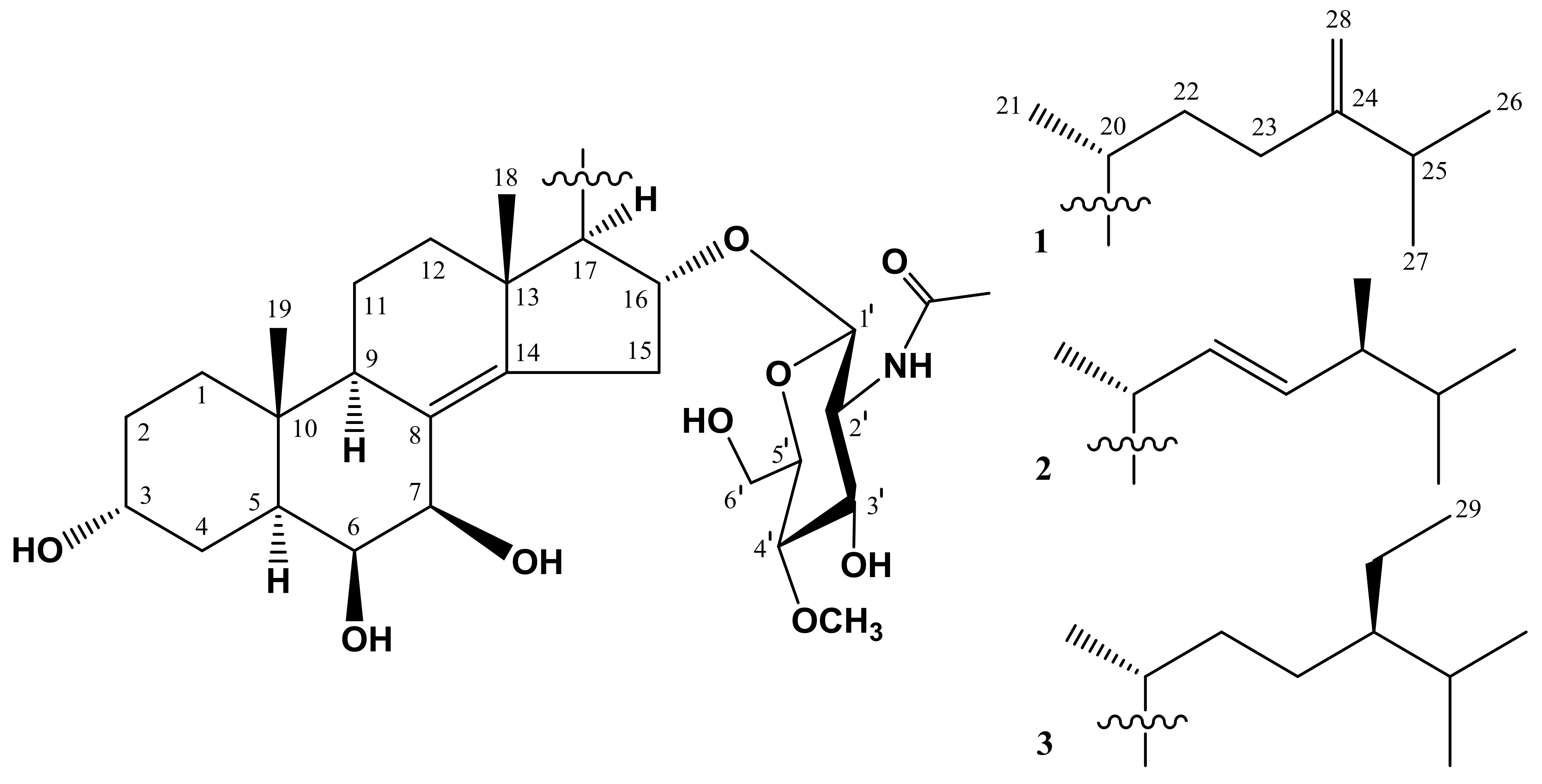
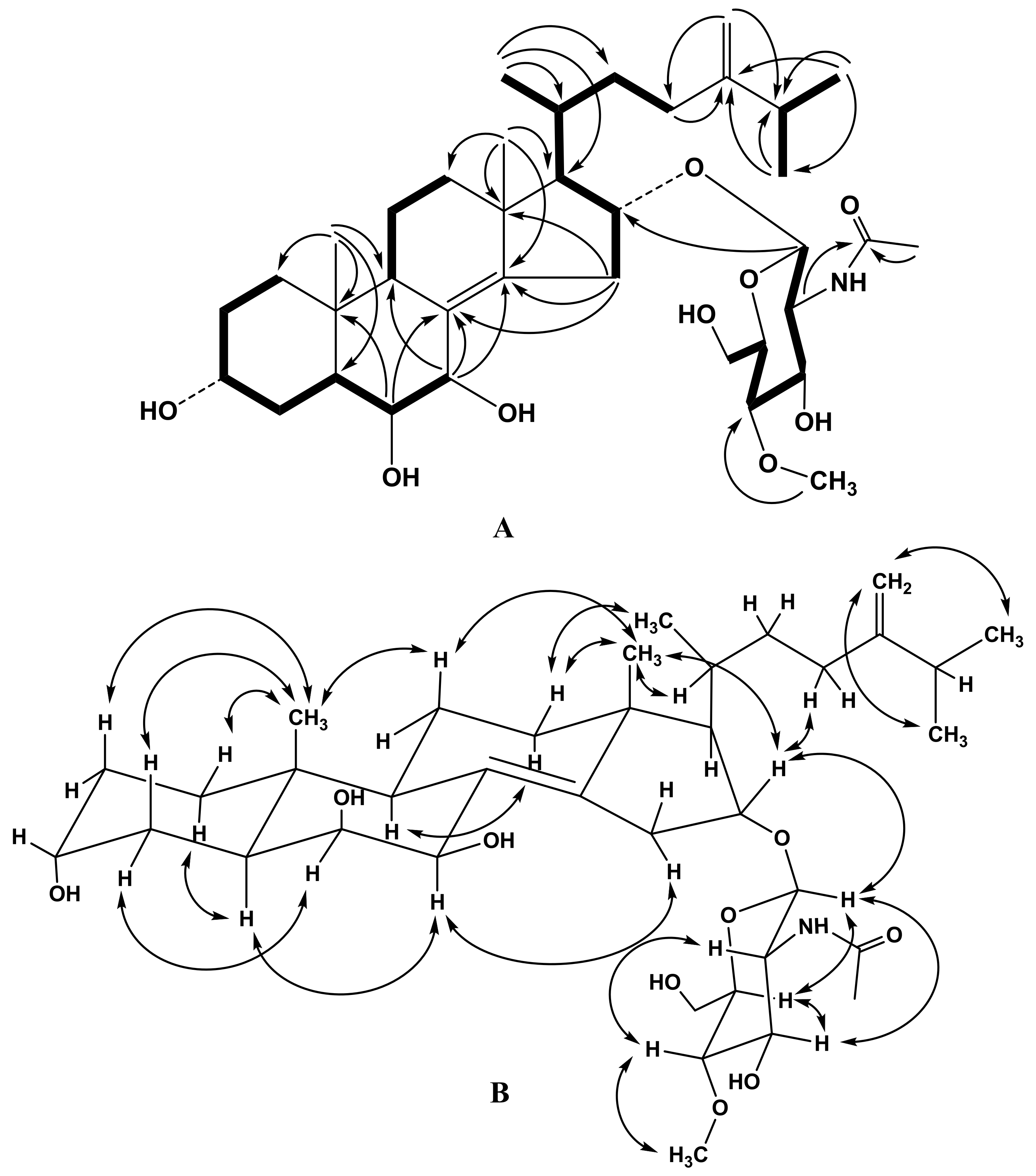
2.1. Structure Elucidation of Compounds 1–3
2.2. Biological Evaluation
2.2.1. The Effect of Compounds 1–3 on Cancer Cells’ Viability
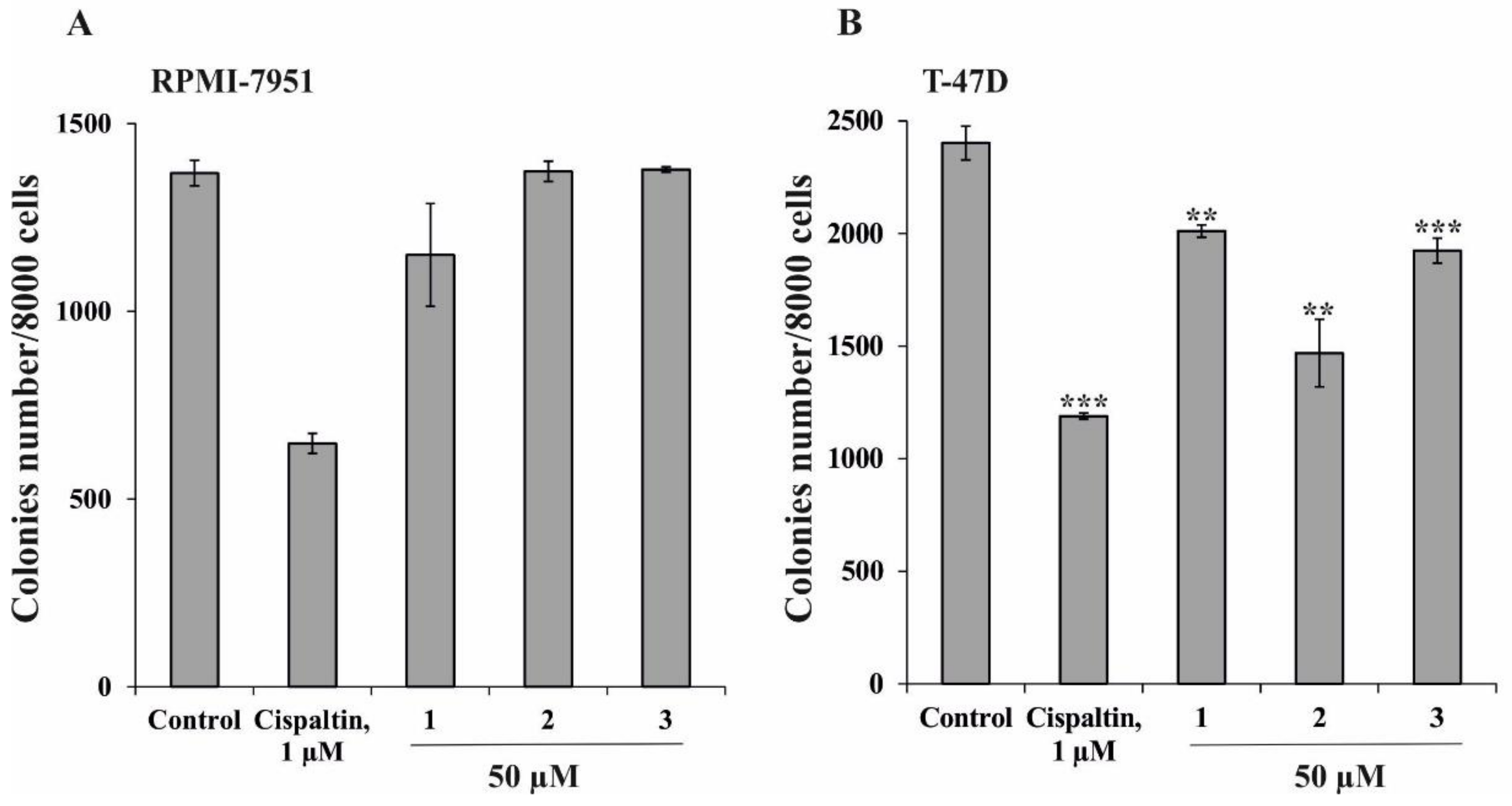
2.2.2. Effect of Compounds 1–3 on Colony Formation
3. Experimental Section
3.1. General Procedures
3.2. Animal Material
3.3. Extraction and Isolation
3.4. Compound Characterization Data
3.5. Acid Hydrolysis and Sugar Analysis
3.6. Bioactivity Assay
3.6.1. Reagents and Antibodies
3.6.2. Cell Lines and Culture Conditions
3.6.3. MTS Assay
3.6.4. Soft Agar Assay
3.6.5. Statistical Analysis
4. Conclusions
Supplementary Materials
Author Contributions
Acknowledgments
Conflicts of Interest
References
- Minale, L.; Riccio, R.; Zollo, F. Steroidal oligoglycosides and polyhydroxysteroids from Echinoderms. Fortschr. Chem. Org. Naturstoffe 1993, 62, 75–308. [Google Scholar]
- Stonik, V.A. Marine polar steroids. Russ. Chem. Rev. 2001, 70, 673–715. [Google Scholar] [CrossRef]
- Iorizzi, M.; De Marino, S.; Zollo, F. Steroidal oligoglycosides from the Asteroidea. Curr. Org. Chem. 2001, 5, 951–973. [Google Scholar] [CrossRef]
- Stonik, V.A.; Ivanchina, N.V.; Kicha, A.A. New polar steroids from starfish. Nat. Prod. Commun. 2008, 3, 1587–1610. [Google Scholar]
- Dong, G.; Xu, T.H.; Yang, B.; Lin, X.P.; Zhou, X.F.; Yang, X.W.; Liu, Y.H. Chemical constituents and bioactivities of starfish. Chem. Biodivers. 2011, 8, 740–791. [Google Scholar] [CrossRef] [PubMed]
- Ivanchina, N.V.; Kicha, A.A.; Stonik, V.A. Steroid glycosides from marine organisms. Steroids 2011, 76, 425–454. [Google Scholar] [CrossRef] [PubMed]
- Ivanchina, N.V.; Kicha, A.A.; Malyarenko, T.V.; Stonik, V.A. Advances in Natural Products Discovery; Gomes, A.R., Rocha-Santos, T., Duarte, A., Eds.; Nova Science Publishers: New York, NY, USA, 2017; Volume 6, pp. 191–224. [Google Scholar]
- Ma, N.; Tang, H.F.; Qiu, F.; Lin, H.W.; Tian, X.R.; Yao, M.N. Polyhydroxysteroidal glycosides from the starfish Anthenea chinensis. J. Nat. Prod. 2010, 73, 590–597. [Google Scholar] [CrossRef] [PubMed]
- Malyarenko, T.V.; Kharchenko, S.D.; Kicha, A.A.; Ivanchina, N.V.; Dmitrenok, P.S.; Chingizova, E.A.; Pislyagin, E.A.; Evtushenko, E.V.; Antokhina, T.I.; Minh, Ch.V.; et al. Anthenosides L‒U, steroidal glycosides with unusual structural features from the starfish Anthenea aspera. J. Nat. Prod. 2016, 79, 3047–3056. [Google Scholar] [CrossRef] [PubMed]
- Kicha, A.A.; Ha, D.T.; Ivanchina, N.V.; Malyarenko, T.V.; Kalinovsky, A.I.; Dmitrenok, P.S.; Ermakova, S.P.; Malyarenko, O.S.; Hung, N.A.; Thuy, T.T.T.; et al. Six new polyhydroxysteroidal glycosides, anthenosides S1–S6, from the starfish Anthenea sibogae. Chem. Biodivers. 2018, 15, e1700553. [Google Scholar] [CrossRef] [PubMed]
- Dai, H.F.; Edrada, R.A.; Ebel, R.; Nimtz, M.; Wray, V.; Proksch, P. Norlanostane triterpenoidal saponins from the marine sponge Melophlus sarasinorum. J. Nat. Prod. 2005, 68, 1231–1237. [Google Scholar] [CrossRef] [PubMed]
- Antonov, A.S.; Kalinovsky, A.I.; Stonik, V.A.; Afiyatullov, S.S.; Aminin, D.L.; Dmitrenok, P.S.; Mollo, E.; Cimino, G. Isolation and structures of erylosides from the Carribean sponge Erylus formosus. J. Nat. Prod. 2007, 70, 169–178. [Google Scholar] [CrossRef] [PubMed]
- Smirnova, G.P. Gangliosides from the starfish Evasterias echinosoma: Identification of a disialoganglioside containing 8-O-methyl-N-acetylneuraminic acid and N-formylgalactosamine. Russ. Chem. Bull. 2000, 49, 159–164. [Google Scholar] [CrossRef]
- Smirnova, G.P. Structure of gangliosides from gonads of the starfish Evasterias retifera. Russ. Chem. Bull. 2003, 52, 2270–2275. [Google Scholar] [CrossRef]
- Kalinovsky, A.I.; Antonov, A.S.; Afiyatullov, S.S.; Dimitrenok, P.S.; Evtuschenko, E.V.; Stonik, V.A. Mycaloside A, a new steroid oligoglycoside with an unprecedented structure from the Caribbean sponge Mycale laxissima. Tetrahedron Lett. 2002, 43, 523–525. [Google Scholar] [CrossRef]
- Afiyatullov, S.S.; Antonov, A.S.; Kalinovsky, A.I.; Dmitrenok, P.S. Two new steroidal oligoglycosides from the Caribbean sponge Mycale laxissima. Nat. Prod. Commun. 2008, 3, 1581–1586. [Google Scholar]
- Ma, N.; Tang, H.F.; Qiu, F.; Lin, H.W.; Tian, X.R.; Zhang, W. A new polyhydroxysteroidal glycoside from the starfish Anthenea chinensis. Chin. Chem. Lett. 2009, 20, 1231–1234. [Google Scholar] [CrossRef]
- Dasari, S.; Tchounwou, P.B. Cisplatin in cancer therapy: Molecular mechanisms of action. Eur. J. Pharmacol. 2014, 740, 364–378. [Google Scholar] [CrossRef] [PubMed]
- Malyarenko, T.V.; Kicha, A.A.; Ivanchina, N.V.; Kalinovsky, A.I.; Popov, R.S.; Vishchuk, O.S.; Stonik, V.A. Asterosaponins from the Far Eastern starfish Leptasterias ochotensis and their anticancer activity. Steroids 2014, 87, 119–127. [Google Scholar] [CrossRef] [PubMed]
- Malyarenko, T.V.; Malyarenko (Vishchuk), O.S.; Ivanchina, N.V.; Kalinovsky, A.I.; Popov, R.S.; Kicha, A.A. Four new sulfated polar steroids from the Far Eastern starfish Leptasterias ochotensis: Structures and activities. Mar. Drugs 2015, 13, 4418–4435. [Google Scholar] [CrossRef] [PubMed]
- Evtushenko, E.V.; Plisova, E.Y.; Ovodov, Y.S. Synthesis of methyl esters of methyl-2-acetamido-2-deoxy-alpha-D-glucopyranoside. Chem. Nat. Comp. 1987, 23, 651–653. [Google Scholar] [CrossRef]
Samples of all compounds in the manuscripts are available from the authors. |


| Position | 1 | 2 | ||||
|---|---|---|---|---|---|---|
| DEPT | δH | δC | DEPT | δH | δC | |
| 1β 1α | CH2 | 1.30, m 1.53, m | 34.5 | CH2 | 1.29, m 1.54, m | 34.5 |
| 2 | CH2 | 1.62, m | 29.5 | CH2 | 1.62, m | 29.5 |
| 3 | CH | 4.08, m | 67.4 | CH | 4.09, m | 67.4 |
| 4β 4α | CH2 | 1.96, m 1.37, m | 33.4 | CH2 | 1.95, td (13.8, 3.1) 1.37, br. d (13.8) | 33.4 |
| 5 | CH | 2.17, dt (13.0, 2.7) | 37.7 | CH | 2.16, dt (13.8, 3.1) | 37.7 |
| 6 | CH | 3.53, m | 77.3 | CH | 3.53, m | 77.3 |
| 7 | CH | 4.26, d (2.7) | 72.2 | CH | 4.24, d (2.5) | 72.2 |
| 8 | C | 128.0 | C | 128.1 | ||
| 9 | CH | 2.24, m | 45.5 | CH | 2.24, m | 45.7 |
| 10 | C | 38.6 | C | 38.7 | ||
| 11β 11α | CH2 | 1.53, m 1.65, m | 19.5 | CH2 | 1.51, m 1.65, m | 19.3 |
| 12β 12α | CH2 | 1.83, dt (12.7, 3.7) 1.27, m | 37.3 | CH2 | 1.78, dt (12.0, 3.4) 1.21, m | 36.8 |
| 13 | C | 45.1 | C | 45.0 | ||
| 14 | C | 147.4 | C | 147.0 | ||
| 15β 15α | CH2 | 2.90, ddd (17.1, 9.1, 3.0) 2.36, ddd (17.1, 5.1, 2.1) | 34.4 | CH2 | 2.89, ddd (17.0, 8.6, 3.2) 2.31, ddd (17.0, 5.7, 2.0) | 34.2 |
| 16 | CH | 4.55, td (9.1, 5.1) | 79.8 | CH | 4.57, m | 79.7 |
| 17 | CH | 1.45, dd (9.1, 5.6) | 62.5 | CH | 1.46, dd (9.6, 2.8) | 62.5 |
| 18 | CH3 | 0.92, s | 20.2 | CH3 | 0.89, s | 20.6 |
| 19 | CH3 | 0.83, s | 15.4 | CH3 | 0.83, s | 15.3 |
| 20 | CH | 1.69, m | 33.3 | CH | 2.38, m | 37.1 |
| 21 | CH3 | 1.02, d (6.9) | 21.0 | CH3 | 1.08, d (7.1) | 24.4 |
| 22 | CH2 | 1.81, m 1.45, m | 33.9 | CH | 5.77, ddd (15.4, 9.2, 1.0) | 135.9 |
| 23 | CH2 | 2.21, m 1.91, m | 33.4 | CH | 5.24, dd (15.4, 8.0) | 134.9 |
| 24 | C | 158.3 | CH | 1.98, m | 44.4 | |
| 25 | CH | 2.28, dsept (1.0, 6.8) | 34.9 | CH | 1.50, m | 34.6 |
| 26 | CH3 | 1.04, d (6.8) | 22.4 | CH3 | 0.87, d (6.8) | 20.6 |
| 27 | CH3 | 1.04, d (6.8) | 22.6 | CH3 | 0.85, d (6.8) | 20.2 |
| 28 | CH2 | 4.74, br. d (1.0) 4.73, br. s | 106.7 | CH3 | 0.96, d (6.8) | 18.3 |
| GlcNAc | ||||||
| 1′ | CH | 4.51, d (8.3) | 100.9 | CH | 4.50, d (8.2) | 101.3 |
| 2′ | CH | 3.52, dd (10.4, 8.3) | 58.4 | CH | 3.56, dd (10.4, 8.2) | 58.3 |
| 3′ | CH | 3.62, dd (10.4, 8.8) | 76.1 | CH | 3.62, dd (10.4, 8.6) | 76.1 |
| 4′ | CH | 3.05, dd (9.8, 8.8) | 81.8 | CH | 3.16, dd (9.7, 8.6) | 81.5 |
| 5′ | CH | 3.24, ddd (9.8, 5.4, 2.1) | 77.3 | CH | 3.25, ddd (9.7, 4.5, 2.2) | 77.2 |
| 6′ | CH2 | 3.83, dd (11.5, 2.1) 3.67, dd (11.5, 5.4) | 62.9 | CH2 | 3.87, dd (11.6, 2.2) 3.74, dd (11.6, 4.5) | 62.6 |
| C=O | C | 173.9 | C | 173.8 | ||
| CH3-CO | CH3 | 1.99, s | 23.2 | CH3 | 1.99, s | 23.2 |
| 4′-OCH3 | CH3 | 3.55, s | 60.9 | CH3 | 3.57, s | 60.9 |
© 2018 by the authors. Licensee MDPI, Basel, Switzerland. This article is an open access article distributed under the terms and conditions of the Creative Commons Attribution (CC BY) license (http://creativecommons.org/licenses/by/4.0/).
Share and Cite
Malyarenko, T.V.; Ivanchina, N.V.; Malyarenko, O.S.; Kalinovsky, A.I.; Dmitrenok, P.S.; Evtushenko, E.V.; Minh, C.V.; Kicha, A.A. Two New Steroidal Monoglycosides, Anthenosides A1 and A2, and Revision of the Structure of Known Anthenoside A with Unusual Monosaccharide Residue from the Starfish Anthenea aspera. Molecules 2018, 23, 1077. https://doi.org/10.3390/molecules23051077
Malyarenko TV, Ivanchina NV, Malyarenko OS, Kalinovsky AI, Dmitrenok PS, Evtushenko EV, Minh CV, Kicha AA. Two New Steroidal Monoglycosides, Anthenosides A1 and A2, and Revision of the Structure of Known Anthenoside A with Unusual Monosaccharide Residue from the Starfish Anthenea aspera. Molecules. 2018; 23(5):1077. https://doi.org/10.3390/molecules23051077
Chicago/Turabian StyleMalyarenko, Timofey V., Natalia V. Ivanchina, Olesya S. Malyarenko, Anatoly I. Kalinovsky, Pavel S. Dmitrenok, Evgeny V. Evtushenko, Chau Van Minh, and Alla A. Kicha. 2018. "Two New Steroidal Monoglycosides, Anthenosides A1 and A2, and Revision of the Structure of Known Anthenoside A with Unusual Monosaccharide Residue from the Starfish Anthenea aspera" Molecules 23, no. 5: 1077. https://doi.org/10.3390/molecules23051077