Stereoselective and Simultaneous Analysis of Ginsenosides from Ginseng Berry Extract in Rat Plasma by UPLC-MS/MS: Application to a Pharmacokinetic Study of Ginseng Berry Extract


Abstract
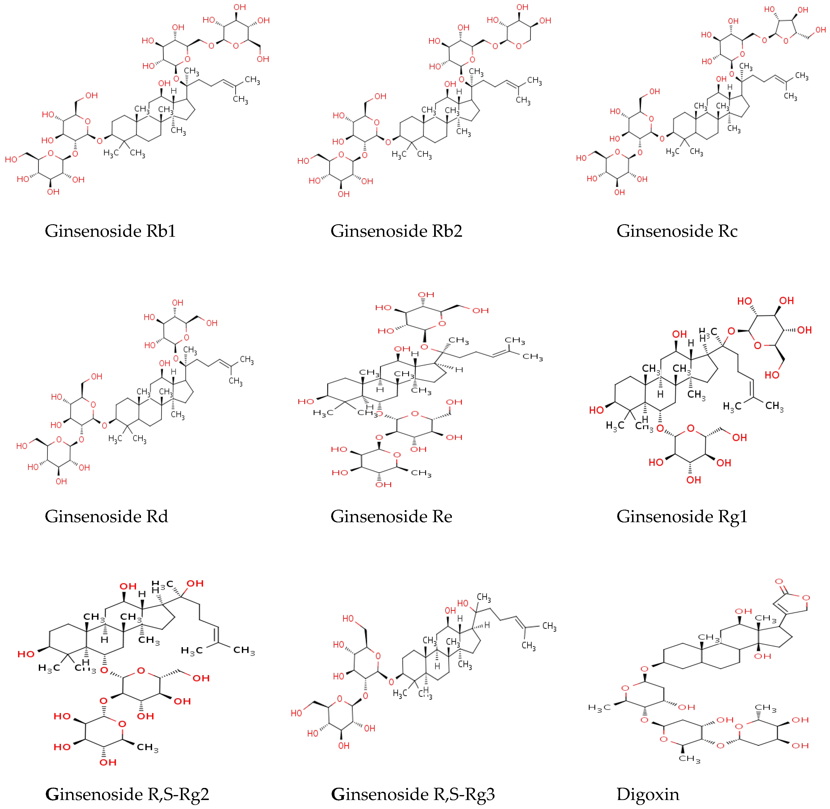
:1. Introduction
2. Results
2.1. UPLC-MS/MS Method Validation
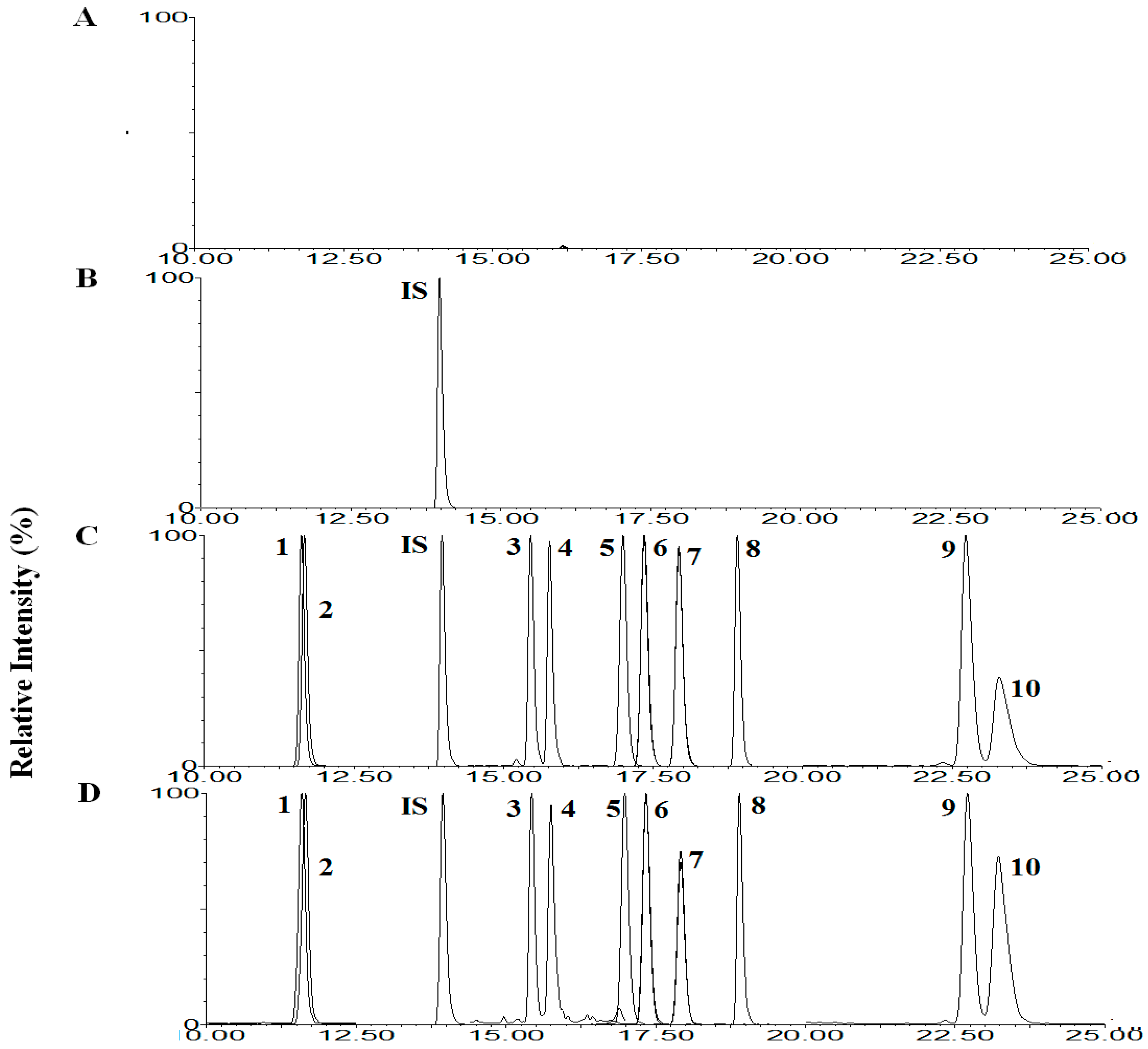
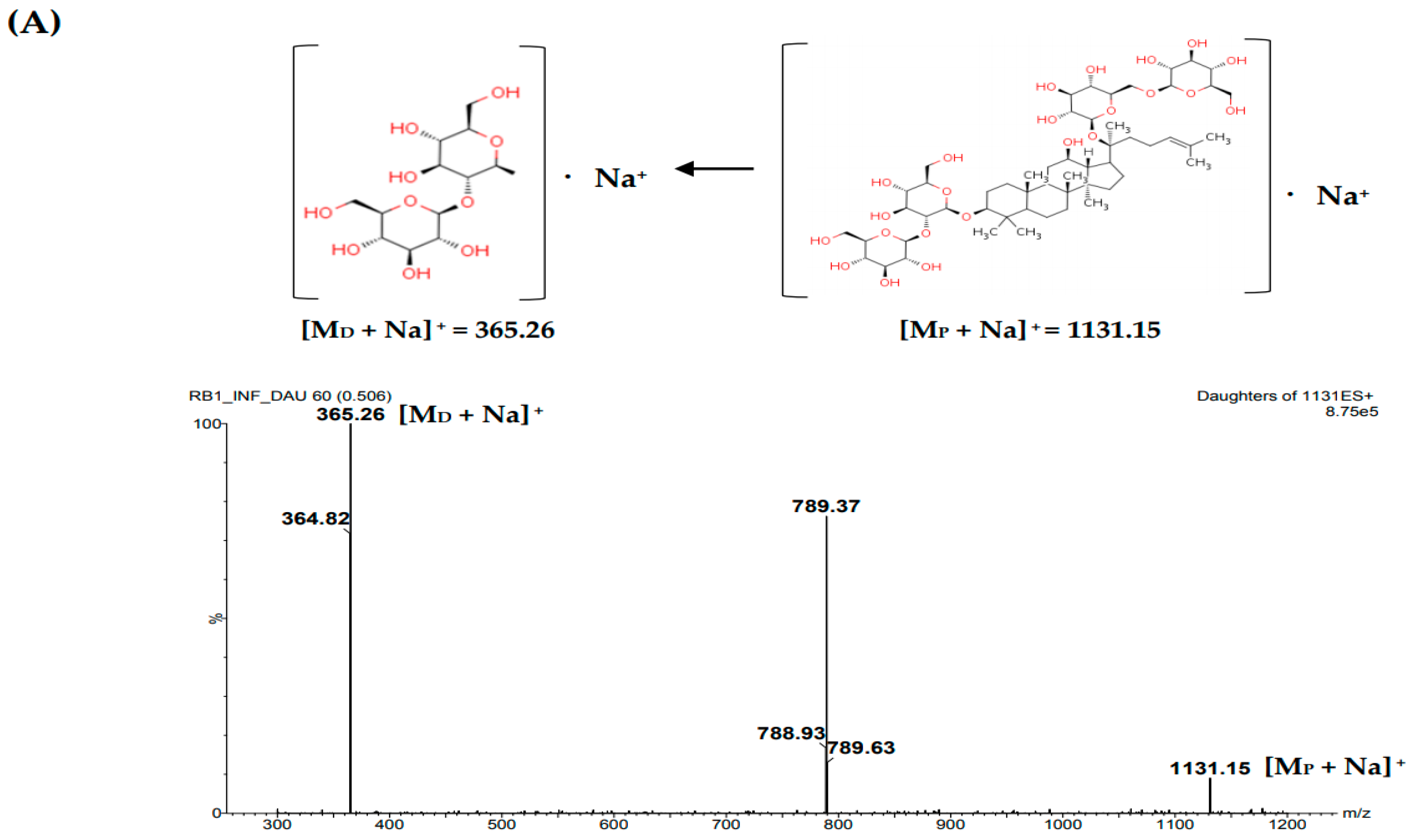
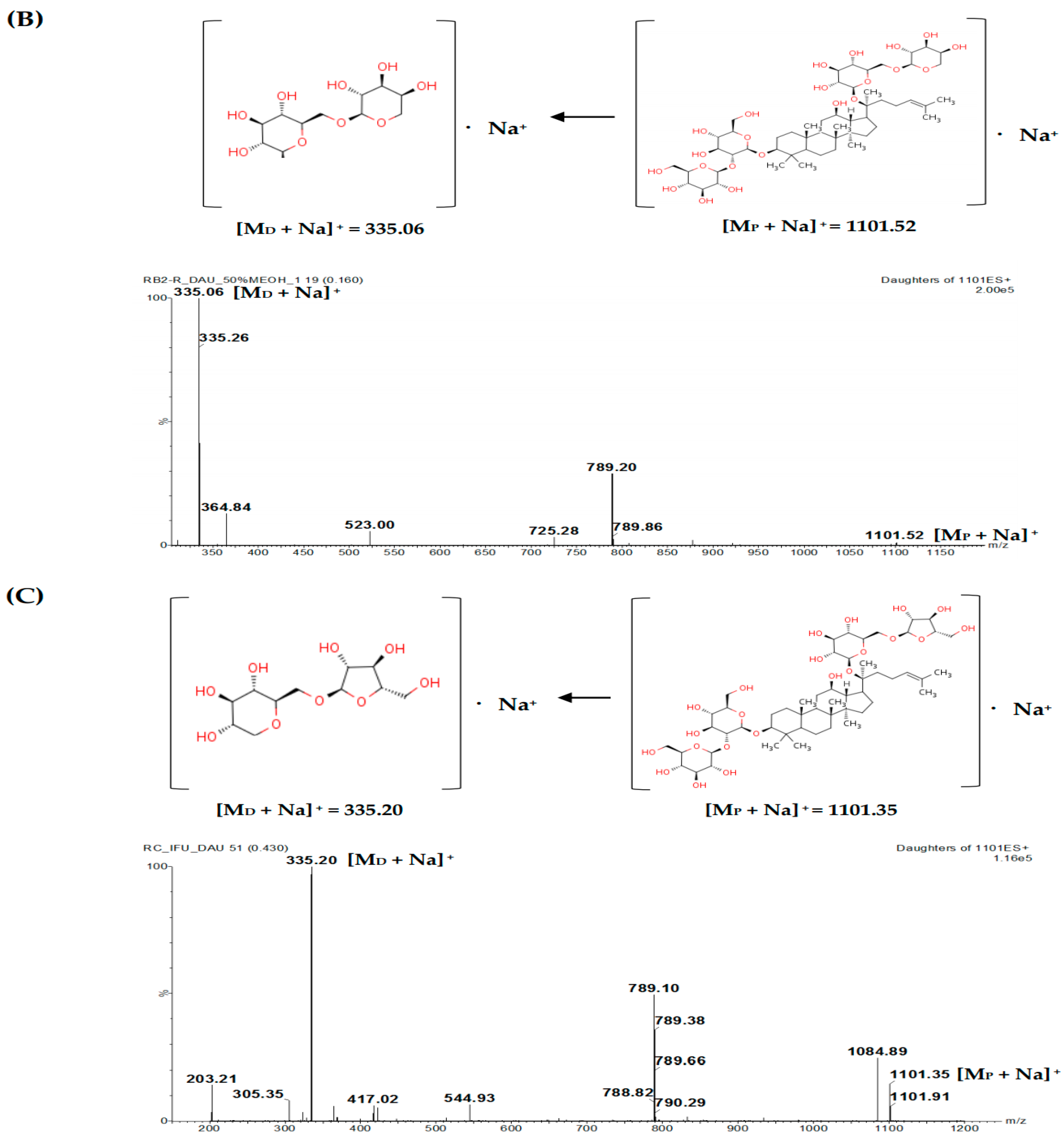
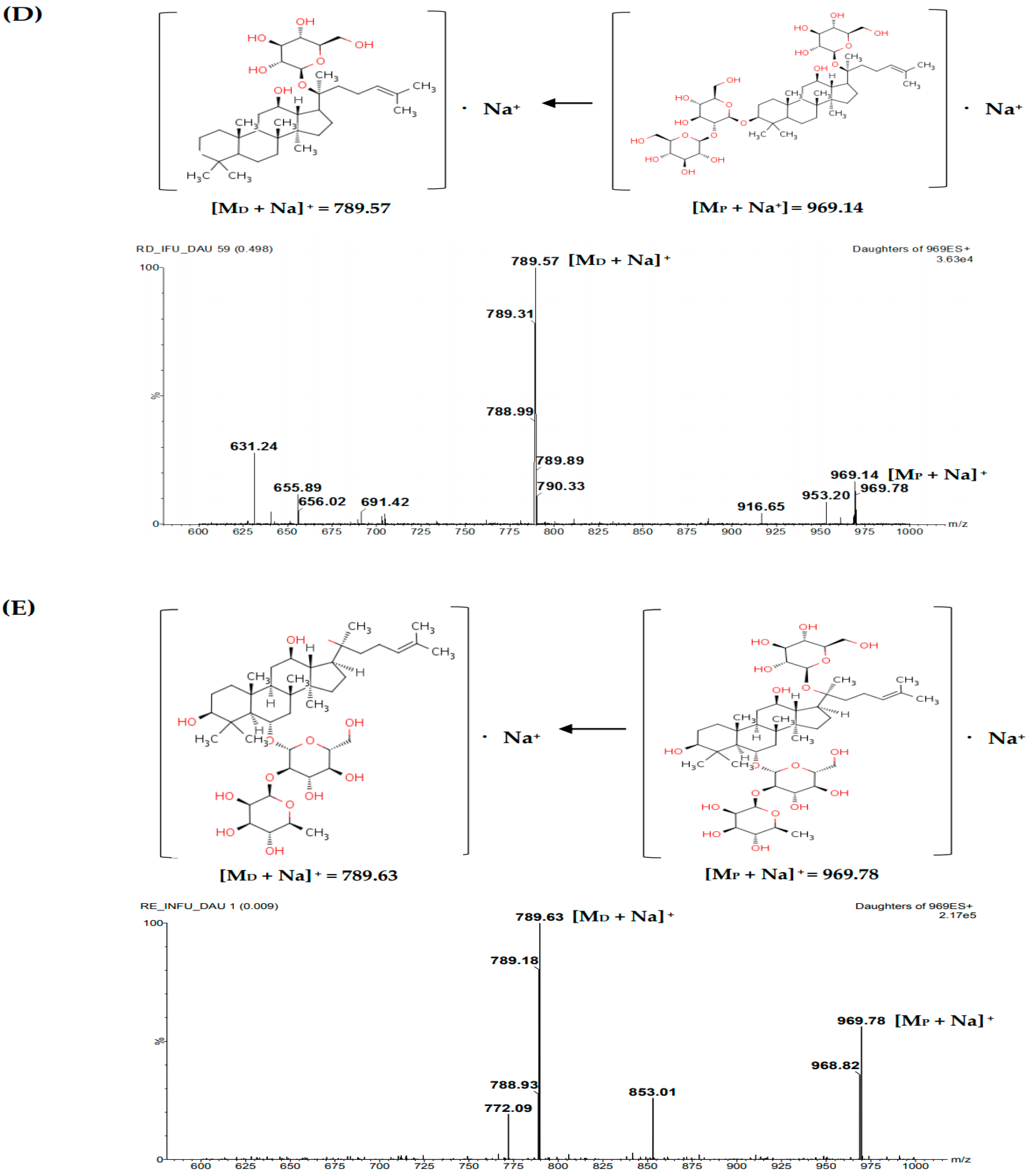
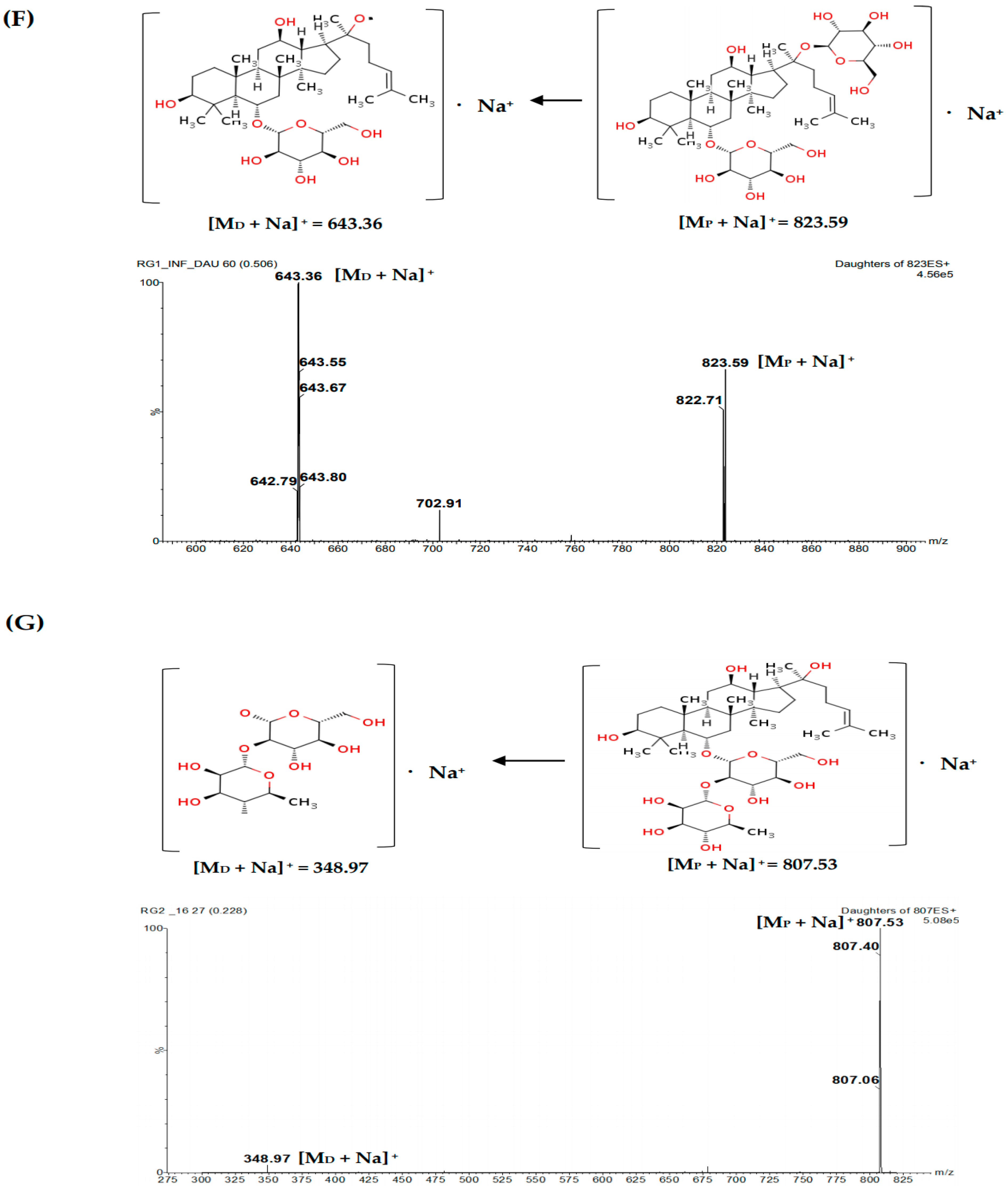
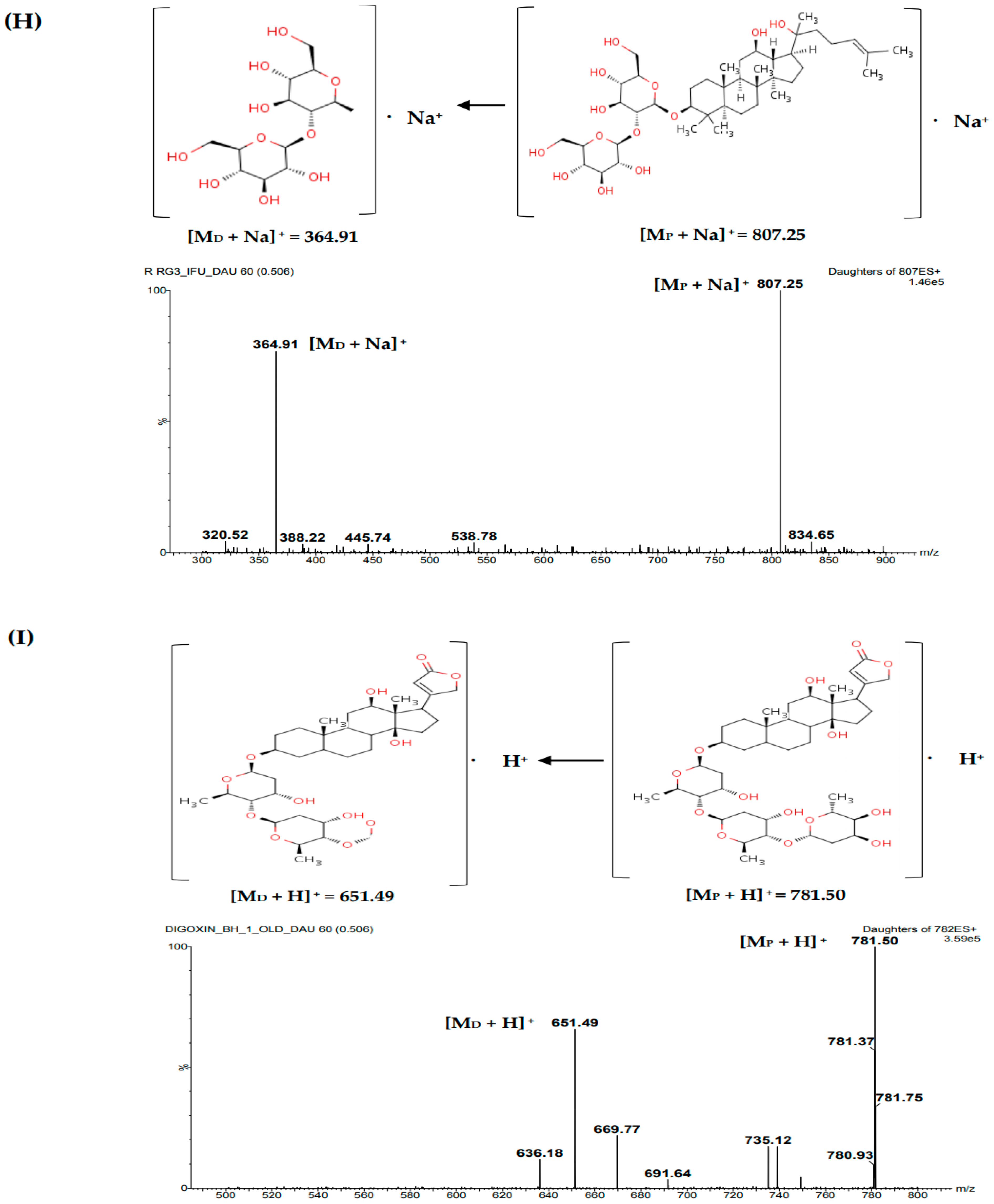
2.1.1. Selectivity
2.1.2. Linearity and Sensitivity
2.1.3. Precision and Accuracy
2.1.4. Matrix Effect
2.1.5. Stability
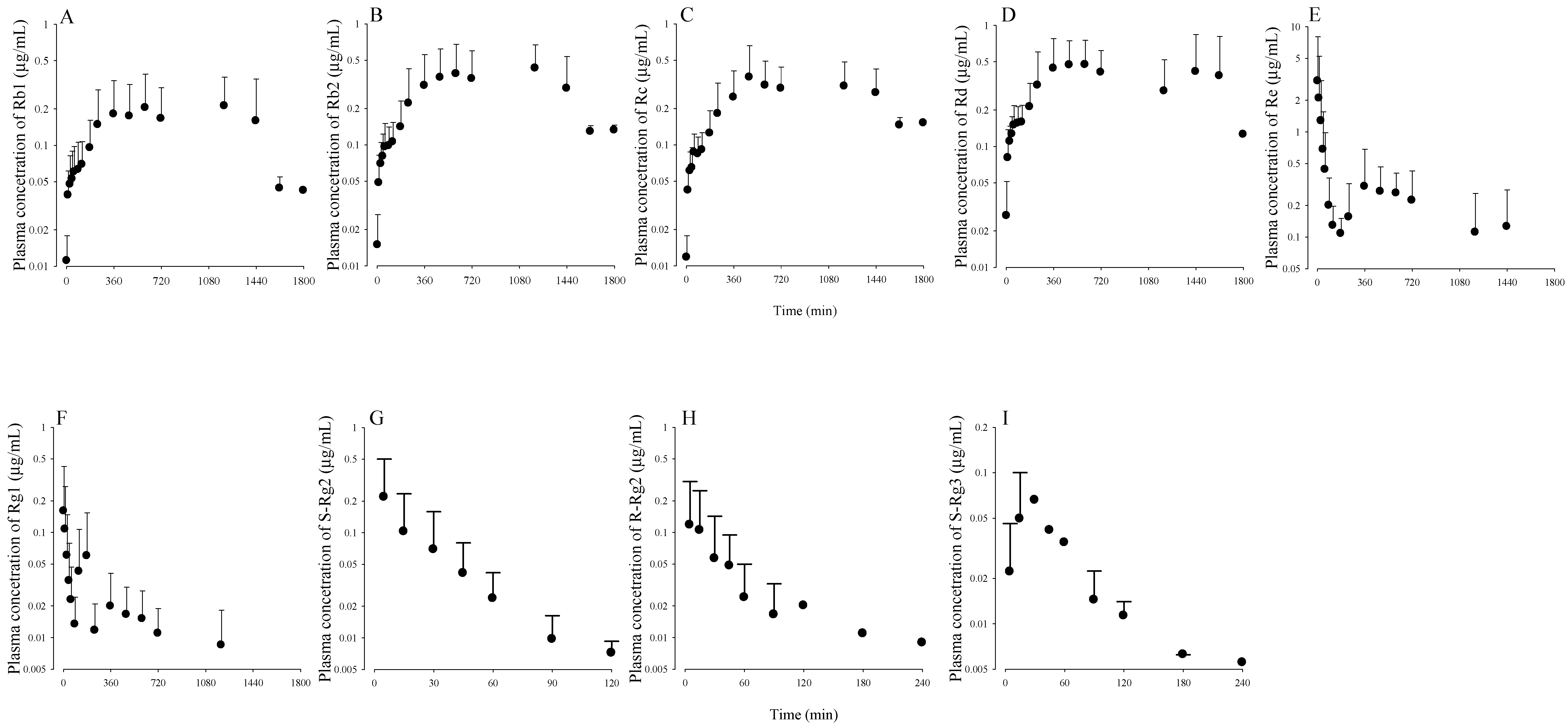
2.2. Pharmacokinetic Studies in Rats
3. Discussion
4. Materials and Methods
4.1. Chemicals and Reagents
4.2. Animals
4.3. Preparation of Stock Solutions, Plasma Samples and Quality Control Samples
4.4. Sample Preparations
4.5. UPLC-MS/MS Conditions
4.6. UPLS-MS/MS Analytical Validation Assays
4.7. Pharmacokinetic Study in Rats
Author Contributions
Funding
Conflicts of Interest
References
- Attele, A.S.; Wu, J.A.; Yuan, C.S. Ginseng pharmacology: Multiple constituents and multiple actions. Biochem. Pharmacol. 1999, 58, 1685–1693. [Google Scholar] [CrossRef]
- Attele, A.S.; Zhou, Y.P.; Xie, J.T.; Wu, J.A.; Zhang, L.; Dey, L.; Pugh, W.; Rue, P.A.; Polonsky, K.S.; Yuan, C.S. Antidiabetic effects of Panax ginseng berry extract and the identification of an effective component. Diabetes 2002, 51, 1851–1858. [Google Scholar] [CrossRef] [PubMed]
- Huo, Y.S. Anti-senility action of saponin in Panax ginseng fruit in 327 cases. Zhong Xi Yi Jie He Za Zhi 1984, 4, 593–596. [Google Scholar] [PubMed]
- Park, E.Y.; Kim, H.J.; Kim, Y.K.; Park, U.; Choi, J.E.; Cha, J.Y.; Jun, H.S. Increase in insulin secretion induced by panax ginseng berry extracts contributes to the amelioration of hyperglycemia in streptozotocin-induced diabetic mice. J. Ginseng Res. 2012, 36, 153–160. [Google Scholar] [CrossRef] [PubMed]
- Yang, H.T.; Zhang, J.R. Treatment of systemic lupus erythematosus with saponin of ginseng fruit (SPGF): An immunological study. Zhong Xi Yi Jie He Za Zhi 1986, 6, 157–159. [Google Scholar] [PubMed]
- Zhang, S.C.; Jiang, X.L. The anti-stress effect of saponins extracted from panax ginseng fruit and the hypophyseal-adrenal system (author’s transl). Yao Xue Xue Bao 1981, 16, 860–863. [Google Scholar] [PubMed]
- Zhang, S.C.; Ni, G.C.; Hu, Z.H. Therapeutic and preventive effects of saponin of ginseng fruit on experimental gastric ulcers. J. Tradit. Chin. Med. 1984, 4, 45–50. [Google Scholar] [PubMed]
- Choi, H.S.; Kim, S.M.; Kim, M.J.; Kim, M.S.; Kim, J.W.; Park, C.W.; Seo, D.B.; Shin, S.S.; Oh, S.W. Efficacy and safety of Panax ginseng berry extract on glycemic control: A 12-wk randomized, double-blind, and placebo-controlled clinical trial. J. Ginseng Res. 2017, 42, 1–8. [Google Scholar] [CrossRef] [PubMed]
- Leung, K.W.; Wong, A.S. Pharmacology of ginsenosides: A literature review. Chin. Med. 2010, 5, 20. [Google Scholar] [CrossRef] [PubMed] [Green Version]
- Cho, W.C.; Chung, W.S.; Lee, S.K.; Leung, A.W.; Cheng, C.H.; Yue, K.K. Ginsenoside Re of Panax ginseng possesses significant antioxidant and antihyperlipidemic efficacies in streptozotocin-induced diabetic rats. Eur. J. Pharmacol. 2006, 550, 173–179. [Google Scholar] [CrossRef] [PubMed]
- Kim, J.J.; Xiao, H.; Tan, Y.; Wang, Z.Z.; Paul Seale, J.; Qu, X. The effects and mechanism of saponins of Panax notoginseng on glucose metabolism in 3T3-L1 cells. Am. J. Chin. Med. 2009, 37, 1179–1189. [Google Scholar] [CrossRef] [PubMed]
- Kim, Y.K.; Yoo, D.S.; Xu, H.; Park, N.I.; Kim, H.H.; Choi, J.E.; Park, S.U. Ginsenoside content of berries and roots of three typical Korean ginseng (Panax ginseng) cultivars. Nat. Prod. Commun. 2009, 4, 903–906. [Google Scholar] [CrossRef] [PubMed]
- Ko, S.K.; Bae, H.M.; Cho, O.S.; Im, B.O.; Chung, S.H.; Lee, B.Y. Analysis of ginsenoside composition of ginseng berry and seed. Food Sci. Biotechnol. 2008, 17, 1379–1382. [Google Scholar]
- Lee, S.Y.; Kim, Y.K.; Park, N.I.; Kim, C.S.; Lee, C.Y.; Park, S.U. Chemical constituents and biological activities of the berry of Panax ginseng. J. Med. Plants Res. 2010, 4, 349–353. [Google Scholar]
- Wang, X.; Zhao, T.; Gao, X.; Dan, M.; Zhou, M.; Jia, W. Simultaneous determination of 17 ginsenosides in rat urine by UPLC-MS with SPE. Anal. Chim. Acta 2007, 594, 265–273. [Google Scholar] [CrossRef] [PubMed]
- Xie, J.T.; Zhou, Y.P.; Dey, L.; Attele, A.S.; Wu, J.A.; Gu, M.; Polonsky, K.S.; Yuan, C.S. Ginseng berry reduces blood glucose and body weight in db/db mice. Phytomedicine 2002, 9, 254–258. [Google Scholar] [CrossRef] [PubMed]
- Xie, J.T.; Mehendele, S.R.; Li, X.; Quigg, R.; Wang, X.; Wang, C.Z.; Wu, J.A.; Aung, H.H.; Reu, P.A.; Bell, G.I.; et al. Anti-diabetic effect of ginsenoside Re in ob/ob mice. Biochim. Biophys. Acta 2005, 1740, 319–325. [Google Scholar] [CrossRef] [PubMed] [Green Version]
- Yahara, S.; Tanaka, O. Further study on dammarane-type saponins of roots, leaves, flower-buds, and fruits of Panax ginseng C.A. Meyer. Chem. Pharm. Bull. (Tokyo) 1979, 27, 88–92. [Google Scholar] [CrossRef]
- Yang, C.Y.; Wang, J.; Zhao, Y.; Shen, L.; Jiang, X.; Xie, Z.G.; Liang, N.; Zhang, L.; Chen, Z.H. Anti-diabetic effects of Panax notoginseng saponins and its major anti-hyperglycemic components. J. Ethnopharmacol. 2010, 130, 231–236. [Google Scholar] [CrossRef] [PubMed]
- Joo, K.M.; Lee, J.H.; Jeon, H.Y.; Park, C.W.; Hong, D.K.; Jeong, H.J.; Lee, S.J.; Lee, S.Y.; Lim, K.M. Pharmacokinetic study of ginsenoside Re with pure ginsenoside Re and ginseng berry extracts in mouse using ultra performance liquid chromatography/mass spectrometric method. J. Pharm. Biomed. Anal. 2010, 51, 278–283. [Google Scholar] [CrossRef] [PubMed]
- Dong, H.; Bai, L.P.; Wong, V.K.; Zhou, H.; Wang, J.R.; Liu, Y.; Jiang, Z.H.; Liu, L. The in vitro structure-related anti-cancer activity of ginsenosides and their derivatives. Molecules 2011, 16, 10619–10630. [Google Scholar] [CrossRef] [PubMed]
- Esimone, C.O.; Nwafor, S.V.; Okoli, C.O.; Chah, K.F.; Uzuegbu, D.B.; Chibundu, C.; Eche, M.A.; Adikwu, M.U. In vivo evaluation of interaction between aqueous seed extract of Garcinia kola Heckel and ciprofloxacin hydrochloride. Am. J. Ther. 2002, 9, 275–280. [Google Scholar] [CrossRef] [PubMed]
- Gao, W.J.; Wang, X.; Ma, C.J.; Dai, R.H.; Bi, K.S.; Chen, X.H. Comparative study on pharmacokinetics of senkyunolide I after administration of simple recipe and compound recipe in rats. Zhongguo Zhong Yao Za Zhi 2013, 38, 427–431. [Google Scholar] [PubMed]
- Hussain, K.; Ismail, Z.; Sadikun, A.; Ibrahim, P. Bioactive markers based pharmacokinetic evaluation of extracts of a traditional medicinal plant, Piper sarmentosum. Evid. Based Complement Altern. Med. 2011, 980760. [Google Scholar] [CrossRef] [PubMed]
- Nahrstedt, A.; Butterweck, V. Lessons learned from herbal medicinal products: The example of St. John′s Wort (perpendicular). J. Nat. Prod. 2010, 73, 1015–1021. [Google Scholar] [CrossRef] [PubMed]
- Peng, W.W.; Li, W.; Li, J.S.; Cui, X.B.; Zhang, Y.X.; Yang, G.M.; Wen, H.M.; Cai, B.C. The effects of Rhizoma Zingiberis on pharmacokinetics of six Aconitum alkaloids in herb couple of Radix Aconiti Lateralis-Rhizoma Zingiberis. J. Ethnopharmacol. 2013, 148, 579–586. [Google Scholar] [CrossRef] [PubMed]
- Ma, L.Y.; Zhang, Y.B.; Zhou, Q.L.; Yang, Y.F.; Yang, X.W. Simultaneous determination of eight ginsenosides in rat plasma by liquid chromatography-electrospray ionization tandem mass spectrometry: Application to their pharmacokinetics. Molecules 2015, 20, 21597–21608. [Google Scholar] [CrossRef] [PubMed]
- Singh, S.S. Preclinical pharmacokinetics: An approach towards safer and efficacious drugs. Curr. Drug Metab. 2006, 7, 165–182. [Google Scholar] [CrossRef] [PubMed]
- Wang, C.Z.; Zhang, B.; Song, W.X.; Wang, A.; Ni, M.; Luo, X. Steamed American ginseng berry: Ginsenoside analyses and anticancer activities. J. Agric. Food Chem. 2006, 54, 9936–9942. [Google Scholar] [CrossRef] [PubMed]
- Zhou, Q.L.; Zhu, D.N.; Yang, Y.F.; Xu, W.; Yang, X.W. Simultaneous quantification of twenty-one ginsenosides and their three aglycones in rat plasma by a developed UFLC-MS/MS assay: Application to a pharmacokinetic study of red ginseng. J. Pharm. Biomed. Anal. 2017, 137, 1–12. [Google Scholar] [CrossRef] [PubMed]
- FDA. Guidance for Industry: Bioanalytical Method Validation. 2018. Available online: https://www.accessdata.fda.gov/drugsatfda_docs/nda/2015/205747Orig1s000SumR.pdf (accessed on 23 July 2018).
- Lu, T.; Yang, J.; Gao, X.; Chen, P.; Du, F.; Sun, Y.; Wang, F.; Xu, F.; Shang, H.; Huang, Y.; et al. Plasma and urinary tanshinol from Salvia miltiorrhiza (Danshen) can be used as pharmacokinetic markers for cardiotonic pills, a cardiovascular herbal medicine. Drug Metab. Dispos. 2008, 36, 1578–1586. [Google Scholar] [CrossRef] [PubMed]
- Liu, H.; Yang, J.; Du, F.; Gao, X.; Ma, X.; Huang, Y.; Xu, F.; Niu, W.; Wang, F.; Mao, Y.; et al. Absorption and disposition of ginsenosides after oral administration of Panax notoginseng extract to rats. Drug Metab. Dispos. 2009, 37, 2290–2298. [Google Scholar] [CrossRef] [PubMed]
- Lee, Y.K.; Chin, Y.W.; Bae, J.K.; Seo, J.S.; Choi, Y.H. Pharmacokinetics of isoliquiritigenin and its metabolites in rats: Low bioavailability is primarily due to the hepatic and intestinal metabolism. Planta. Med. 2016, 79, 1656–1665. [Google Scholar] [CrossRef] [PubMed]
- Gibaldi, M.; Perrier, D. General derivation of the equation for time to reach a certain fraction of steady state. J. Pharm. Sci. 1982, 71, 474–475. [Google Scholar]
Sample Availability: Samples of the compounds are not available from the authors. |


| Analytes | Regression Equation | r2 | Linear Range (µg/mL) | LLOQ (µg/mL) |
|---|---|---|---|---|
| Rb1 | y = 0.1661x + 0.000064 | 0.9999 | 0.01–10 | 0.01 |
| Rb2 | y = 0.2190x − 0.00010 | 0.9999 | 0.01–10 | 0.01 |
| Rc | y = 0.2850x − 0.00040 | 0.9993 | 0.01–10 | 0.01 |
| Rd | y = 0.2185x + 0.00062 | 0.9993 | 0.01–10 | 0.01 |
| Re | y = 0.07290x + 0.0027 | 0.9987 | 0.01–10 | 0.01 |
| Rg1 | y = 0.1432x + 0.021 | 0.9824 | 0.01–10 | 0.01 |
| R-Rg2 | y = 0.05100x + 0.0026 | 0.9978 | 0.01–10 | 0.01 |
| S-Rg2 | y = 0.05420x + 0.0012 | 0.9996 | 0.01–10 | 0.01 |
| R-Rg3 | y = 0.1792x − 0.00020 | 1.000 | 0.01–10 | 0.01 |
| S-Rg3 | y = 0.1285x + 0.0006 | 1.000 | 0.01–10 | 0.01 |
| Spiked Concentration (µg/mL) | Intra-Day | Inter-Day | ||||
|---|---|---|---|---|---|---|
| Precision | Accuracy (%) | Precision | Accuracy (%) | |||
| Mean ± SD | RSD a) (%) | Mean ± SD | RSD a) (%) | |||
| Rb1 | ||||||
| 0.01 | 0.161 ± 0.0080 | 4.78 | 95.0 | 0.164 ± 0.0067 | 4.09 | 97.0 |
| 0.05 | 0.166 ± 0.0040 | 2.44 | 99.0 | 0.168 ± 0.00090 | 0.543 | 99.5 |
| 0.5 | 0.164 ± 0.0080 | 4.86 | 98.7 | 0.169 ± 0.0018 | 1.05 | 100 |
| 5 | 0.167 ± 0.0011 | 0.664 | 101 | 0.167 ± 0.00030 | 0.175 | 99.1 |
| Rb2 | ||||||
| 0.01 | 0.211 ± 0.0056 | 2.64 | 98.9 | 0.217 ± 0.0037 | 1.72 | 109 |
| 0.05 | 0.218 ± 0.0058 | 2.64 | 101 | 0.220 ± 0.0021 | 0.970 | 104 |
| 0.5 | 0.216 ± 0.0033 | 1.54 | 98.7 | 0.217 ± 0.0028 | 1.30 | 98.6 |
| 5 | 0.221 ± 0.0078 | 3.54 | 101 | 0.223 ± 0.0047 | 2.09 | 101 |
| Rc | ||||||
| 0.01 | 0.250 ± 0.023 | 9.20 | 95.0 | 0.283 ± 0.0096 | 3.40 | 111 |
| 0.05 | 0.272 ± 0.012 | 4.27 | 98.3 | 0.278 ± 0.014 | 4.99 | 102 |
| 0.5 | 0.289 ± 0.032 | 11.0 | 102 | 0.276 ± 0.015 | 5.28 | 98.1 |
| 5 | 0.278 ± 0.010 | 3.63 | 97.4 | 0.284 ± 0.0085 | 2.99 | 100 |
| Rd | ||||||
| 0.01 | 0.237 ± 0.017 | 6.99 | 94.3 | 0.243 ± 0.025 | 10.2 | 98.9 |
| 0.05 | 0.223 ± 0.0080 | 3.61 | 96.3 | 0.220 ± 0.0040 | 1.82 | 95.1 |
| 0.5 | 0.213 ± 0.0069 | 3.25 | 97.1 | 0.218 ± 0.0066 | 3.04 | 97.8 |
| 5 | 0.213 ± 0.0079 | 3.71 | 97.3 | 0.218 ± 0.0050 | 2.29 | 98.3 |
| Re | ||||||
| 0.01 | 0.0791 ± 0.00019 | 0.0237 | 102 | 0.0777 ± 0.011 | 14.9 | 86.0 |
| 0.05 | 0.0794 ± 0.00052 | 0.659 | 101 | 0.0797 ± 0.0055 | 6.88 | 96.5 |
| 0.5 | 0.0784 ± 0.00075 | 0.959 | 98.8 | 0.0818 ± 0.0052 | 6.31 | 100 |
| 5 | 0.0739 ± 0.0025 | 3.41 | 93.1 | 0.0796 ± 0.0069 | 8.78 | 90.4 |
| Rg1 | ||||||
| 0.01 | 0.196 ± 0.0034 | 1.74 | 102 | 0.188 ± 0.0203 | 10.8 | 94.3 |
| 0.05 | 0.196 ± 0.0011 | 0.570 | 101 | 0.189 ± 0.012 | 6.36 | 98.4 |
| 0.5 | 0.196 ± 0.0029 | 1.47 | 100 | 0.193 ± 0.013 | 6.85 | 101 |
| 5 | 0.195 ± 0.0065 | 3.39 | 93.9 | 0.192 ± 0.0122 | 6.51 | 96.2 |
| R-Rg2 | ||||||
| 0.01 | 0.0579 ± 0.0028 | 4.91 | 103 | 0.0589 ± 0.0020 | 3.47 | 103 |
| 0.05 | 0.0585 ± 0.0026 | 4.41 | 103 | 0.0585 ± 0.00046 | 0.793 | 102 |
| 0.5 | 0.0568 ± 0.0010 | 1.80 | 98.6 | 0.0572 ± 0.00121 | 2.05 | 99.2 |
| 5 | 0.0508 ± 0.0011 | 2.16 | 88.1 | 0.0562 ± 0.00162 | 2.97 | 88.3 |
| S-Rg2 | ||||||
| 0.01 | 0.0574 ± 0.0045 | 7.79 | 89.7 | 0.0618 ± 0.00068 | 1.10 | 110 |
| 0.05 | 0.0594 ± 0.0038 | 6.34 | 99.8 | 0.0578 ± 0.0021 | 3.60 | 99.4 |
| 0.5 | 0.0584 ± 0.0013 | 2.25 | 102 | 0.0600 ± 0.00066 | 1.10 | 101 |
| 5 | 0.0542 ± 0.0038 | 7.03 | 94.9 | 0.0539 ± 0.0018 | 3.39 | 90.4 |
| R-Rg3 | ||||||
| 0.01 | 0.179 ± 0.0022 | 11.5 | 105 | 0.172 ± 0.016 | 9.07 | 89.3 |
| 0.05 | 0.179 ± 0.00061 | 0.342 | 102 | 0.178 ± 0.00092 | 0.519 | 95.2 |
| 0.5 | 0.179 ± 0.0016 | 0.876 | 100 | 0.177 ± 0.00072 | 0.408 | 99.4 |
| 5 | 0.178 ± 0.0012 | 0.686 | 99.5 | 0.177 ± 0.0026 | 1.48 | 99.8 |
| S-Rg3 | ||||||
| 0.01 | 0.144 ± 0.0152 | 10.8 | 107 | 0.122 ± 0.0123 | 10.4 | 97.6 |
| 0.05 | 0.130 ± 0.00079 | 0.610 | 95.6 | 0.129 ± 0.00061 | 0.474 | 98.3 |
| 0.5 | 0.130 ± 0.0034 | 2.65 | 100 | 0.130 ± 0.0029 | 2.21 | 101 |
| 5 | 0.128 ± 0.0013 | 1.01 | 99.5 | 0.129 ± 0.00074 | 0.572 | 100 |
| Spiked Concentration (µg/mL) | Short-Term Storage (25 °C) | Three-Thaw Cycles | Post-Treatment (25 °C) | Long-Term Storage (−80 °C) | |
|---|---|---|---|---|---|
| Rb1 | 0.05 | 109 | 96.9 | 88.2 | 98.1 |
| 0.5 | 97.4 | 91.4 | 93.6 | 99.6 | |
| 5 | 103 | 103.5 | 95.3 | 103 | |
| Rb2 | 0.05 | 97.5 | 97.3 | 94.6 | 95.4 |
| 0.5 | 93.6 | 94.5 | 94.9 | 101 | |
| 5 | 93.6 | 102 | 99.5 | 107 | |
| Rc | 0.05 | 109 | 102 | 107 | 90.8 |
| 0.5 | 107 | 103 | 100 | 97.9 | |
| 5 | 105 | 106 | 92.2 | 103 | |
| Rd | 0.05 | 96.2 | 93.5 | 74.3 | 88.7 |
| 0.5 | 100 | 94.4 | 78.4 | 99.0 | |
| 5 | 96.3 | 94.1 | 72.9 | 93.0 | |
| Re | 0.05 | 104 | 109 | 102 | 98.6 |
| 0.5 | 107 | 99.3 | 106 | 97.3 | |
| 5 | 99.8 | 102 | 109 | 94.8 | |
| Rg1 | 0.05 | 98.5 | 104 | 103 | 87.3 |
| 0.5 | 97.2 | 104 | 109 | 91.0 | |
| 5 | 97.6 | 104 | 101 | 95.0 | |
| R-Rg2 | 0.05 | 103 | 101 | 96.1 | 92.9 |
| 0.5 | 98.9 | 97.3 | 90.4 | 90.5 | |
| 5 | 105 | 108 | 95.5 | 94.0 | |
| S-Rg2 | 0.05 | 99.6 | 103 | 96.6 | 97.9 |
| 0.5 | 110 | 101 | 94.1 | 93.5 | |
| 5 | 108 | 101 | 95.9 | 101 | |
| R-Rg3 | 0.05 | 104 | 109 | 103 | 99.7 |
| 0.5 | 101 | 106 | 107 | 95.5 | |
| 5 | 103 | 109 | 101 | 106 | |
| S-Rg3 | 0.05 | 95.8 | 105 | 108 | 108 |
| 0.5 | 97.8 | 107 | 105 | 93.4 | |
| 5 | 96.1 | 101 | 101 | 102 | |
| Rb1 | Rb2 | Rc | |||||||
| AUClast (µg·min/mL) | 202 | ± | 168 | 376 | ± | 214 | 353 | ± | 190 |
| Normalized AUClast (µg·min/mL) | 26.1 | ± | 21.7 | 24.5 | ± | 13.9 | 68.5 | ± | 36.9 |
| Cmax (µg/mL) | 0.210 | ± | 0.183 | 0.395 | ± | 0.285 | 0.387 | ± | 0.292 |
| Normalized Cmax (µg/mL) | 0.0272 | ± | 0.0237 | 0.0257 | ± | 0.0185 | 0.0751 | ± | 0.0566 |
| Tmax (h) | 8 (4–12) | 10 (8–12) | 10 (8–12) | ||||||
| t1/2 (h) | 16.5 | ± | 2.71 | 15.9 | ± | 1.35 | 14.9 | ± | 1.84 |
| Rd | Re | Rg1 | |||||||
| AUClast (µg·min/mL) | 543 | ± | 384 | 349 | ± | 68.0 | 22.8 | ± | 5.83 |
| Normalized AUClast (µg·min/mL) | 27.3 | ± | 19.3 | 8.96 | ± | 1.74 | 15.8 | ± | 4.05 |
| Cmax (µg/mL) | 0.519 | ± | 0.293 | 3.32 | ± | 4.74 | 0.225 | ± | 0.216 |
| Normalized Cmax (µg/mL) | 0.0261 | ± | 0.0148 | 0.0852 | ± | 0.121 | 0.156 | ± | 0.150 |
| Tmax (h) | 480 (15–600) | 360 (5–720) | 3 (0.083–6) | ||||||
| t1/2 (h) | 12.9 | ± | 1.28 | 10.8 | ± | 5.73 | 10.3 | ± | 3.11 |
| S-Rg2 | R-Rg2 | S-Rg3 | |||||||
| AUClast (µg·min/mL) | 9.98 | ± | 3.36 | 4.21 | ± | 1.02 | 3.57 | ± | 2.03 |
| Normalized AUClast (µg·min/mL) | 2.79 | ± | 1.66 | 1.49 | ± | 1.02 | 4.25 | ± | 2.42 |
| Cmax (µg/mL) | 0.284 | ± | 0.163 | 0.108 | ± | 0.0153 | 0.0470 | ± | 0.0537 |
| Normalized Cmax (µg/mL) | 0.0777 | ± | 0.0495 | 0.0383 | ± | 0.00452 | 0.0560 | ± | 0.0640 |
| Tmax (h) | 0.25 (0.083–0.25) | 0.25 (0.083–0.25) | 1 (0.25–1.5) | ||||||
| t1/2 (h) | 2.38 | ± | 1.67 | 1.54 | ± | 0.353 | 3.12 | ± | 1.22 |
© 2018 by the authors. Licensee MDPI, Basel, Switzerland. This article is an open access article distributed under the terms and conditions of the Creative Commons Attribution (CC BY) license (http://creativecommons.org/licenses/by/4.0/).
Share and Cite
Han, S.Y.; Bae, M.G.; Choi, Y.H. Stereoselective and Simultaneous Analysis of Ginsenosides from Ginseng Berry Extract in Rat Plasma by UPLC-MS/MS: Application to a Pharmacokinetic Study of Ginseng Berry Extract. Molecules 2018, 23, 1835. https://doi.org/10.3390/molecules23071835
Han SY, Bae MG, Choi YH. Stereoselective and Simultaneous Analysis of Ginsenosides from Ginseng Berry Extract in Rat Plasma by UPLC-MS/MS: Application to a Pharmacokinetic Study of Ginseng Berry Extract. Molecules. 2018; 23(7):1835. https://doi.org/10.3390/molecules23071835
Chicago/Turabian StyleHan, Seong Yon, Min Goo Bae, and Young Hee Choi. 2018. "Stereoselective and Simultaneous Analysis of Ginsenosides from Ginseng Berry Extract in Rat Plasma by UPLC-MS/MS: Application to a Pharmacokinetic Study of Ginseng Berry Extract" Molecules 23, no. 7: 1835. https://doi.org/10.3390/molecules23071835