
A 7-Hydroxybenzoxazinone-Containing Fluorescence Turn-On Probe for Biothiols and Its Bioimaging Applications

School of Chemistry and Pharmaceutical Engineering, Qilu University of Technology (Shandong Academy of Sciences), 3501 Daxue Road, Jinan 250353, China
*
Author to whom correspondence should be addressed.
Molecules 2019, 24(17), 3102; https://doi.org/10.3390/molecules24173102
Submission received: 5 August 2019
/
Revised: 22 August 2019
/
Accepted: 26 August 2019
/
Published: 27 August 2019
(This article belongs to the Section Analytical Chemistry)
Abstract
:In this work, a novel 7-hydroxybenzoxazinone-based fluorescent probe (PBD) for the selective sensing of biothiols is reported. Upon treatment with biothiols, PBD shows a strong fluorescence enhancement (up to 70-fold) and a large Stokes shift (155 nm). Meanwhile, this probe exhibits high resistance to interference from other amino acids and competing species. PBD features good linearity ranges with a low detection limit of 14.5 nM for glutathione (GSH), 17.5 nM for cysteine (Cys), and 80.0 nM for homocysteine (Hcy), respectively. Finally, the potential utility of this probe for biothiol sensing in living HeLa cells is demonstrated.
1. Introduction
Sulfhydryl-containing amino acids, such as cysteine (Cys), glutathione (GSH), and homocysteine (Hcy), play key roles in maintaining appropriate physiological and biological processes [1,2,3,4,5]. Malfunction of Cys levels has been associated with hair depigmentation, slowed growth rate, liver damage, edema, and so on [6]. Elevated levels of Hcy result in cardiovascular and Alzheimer’s disease [7,8]. GSH is the most abundant intracellular non-protein thiol and its concentration is within millimolar range. GSH deficiency is linked with cancer, liver damage, and neurodegenerative disease [9,10,11]. Owing to their significant roles, it is of great importance to develop rapid, simple and reliable methods for monitoring the levels of biothiols in biological systems.
Among various detection methods, fluorescent-probe-based detection has been accepted as one of the most efficient methods due to its advantages of having operational simplicity, being low cost, working under in situ real time conditions and involving no invasive bioimaging [12,13,14,15,16].
Great efforts have been directed to the development of fluorescent probes toward biothiols (Cys, Hcy, and GSH) [17,18,19,20,21,22,23]. Many probes have been constructed based on traditional fluorescent dyes, such as fluorescein [24], naphthalimide [25], coumarin [26], cyanin [27], BODIPY (boron-dipyrromethene) [23], xanthene [28], 2-(2′-hydroxyphenyl)benzothiazole [29], and dicyanomethylene-4H-pyran derivatives [30]. In spite of some sensors with good sensing performance, many of these probes still suffer from poor solubility, low sensitivity, low quantum yield, and a time-consuming detection process. Thus, it would be beneficial to develop fluorophores with a new structural skeleton. Up until now, fluorescent dyes with a 7-hydroxybenzoxazinone skeleton have received little attention [31,32,33]. We prepared 7-hydroxy-3-phenylbenzoxazinone (PBOH) and evaluated its optical property. PBOH exhibits strong yellow-green fluorescence and a large Stokes shift (155 nm). We envisioned that masking the hydroxyl group in PBOH with a 2,4-dinitrophenylsulfonyl group (DNBS) could generate a new fluorescent probe (PBD) for the selective detection of biothiols (Scheme 1). As they are well-known, arenesulfonates and arenesulphonamides can be readily cleaved by thiolate anions, and, therefore, the 2,4-dintrobenzenesulfonyl (DNBS) group has often been chosen as the trigger group for thiols [24,34,35,36,37,38]. More importantly, the DNBS group can totally quench fluorescence, thus reducing background interference, because of its strong electron-withdrawing ability. We speculate that biothiols induce cleavage of the ester bond in the DNBS-PBOH conjugates, which can release free PBOH and thereby restore the strong emission, thus realizing a selective fluorescence off-on recognition of biothiols. The new probe PBD indeed exhibits a significant off-on response to biothiols. The investigation of PBD suggested that it possesses a strong anti-interference ability while sensing biothiols, with a low detection limit of 14.5 nM for GSH, 17.5 nM for Cys, and 80.0 nM for Hcy, respectively. In addition, PBD was successfully applied for biothiol detection in living HeLa cells.
2. Results and Discussion
PBOH showed strong fluorescence (Φ = 0.60) with a maximum at 540 nm and an absorption band centered at 385 nm in EtOH-PBS buffer (Phosphate Buffered Saline) solution (10 mM, pH = 7.4, 4:6, v/v) (Stokes shift = 155 nm). Owing to the quenching effect of the DNBS group, PBD alone exhibited extremely weak fluorescence with a low quantum yield at 540 nm (Φ = 0.011), and its absorption maximum was blue-shifted to 340 nm under the same aqueous conditions (Figure S1). As expected, upon addition of biothiols, a remarkable fluorescence intensity enhancement at 540 nm was observed. As such, we constructed a turn-on fluorescent probe for biothiols.
2.1. Time-Dependence and pH Effect
The sensory effect of PBD is exemplified by its reaction with GSH. The emission intensity was recorded at different time points to evaluate the response time between PBD and various amounts of GSH. As shown in Figure 1, after addition of GSH, the fluorescence emission peak at 540 nm reached a plateau in about 16 min and maintained stability for a long time.
Kinetic analysis was conducted next. Ten equivalents of GSH, Cys, and Hcy were added into the solutions of PBD (10 µM), respectively. Through monitoring the fluorescence emission at 540 nm, the reactions were found to obey a typical pseudo-first-order and the rate constants were determined to be 0.25 min−1, 0.26 min−1, and 0.20 min−1 for GSH, Cys, and Hcy respectively (Figure S5).
Furthermore, we evaluated the pH effect on the sensory response of PBD (Figure S3). PBD itself was inert to pH change, indicative of the excellent stability of the probe over a broad pH range. By contrast, the probe exhibited a marked fluorescence response to GSH within the pH range 7 to 9, thus enabling detection of biothiols across a relatively wide pH range.
The following experiments of PBD were performed in EtOH-PBS buffer (10 mM, pH 7.4, 4:6, v/v) and 405 nm was chosen as the excitation wavelength in the fluorescent measurements. For convenience, the fluorescent intensity was recorded at 16 min after the addition of GSH.
2.2. Sensitivity and Selectivity Studies
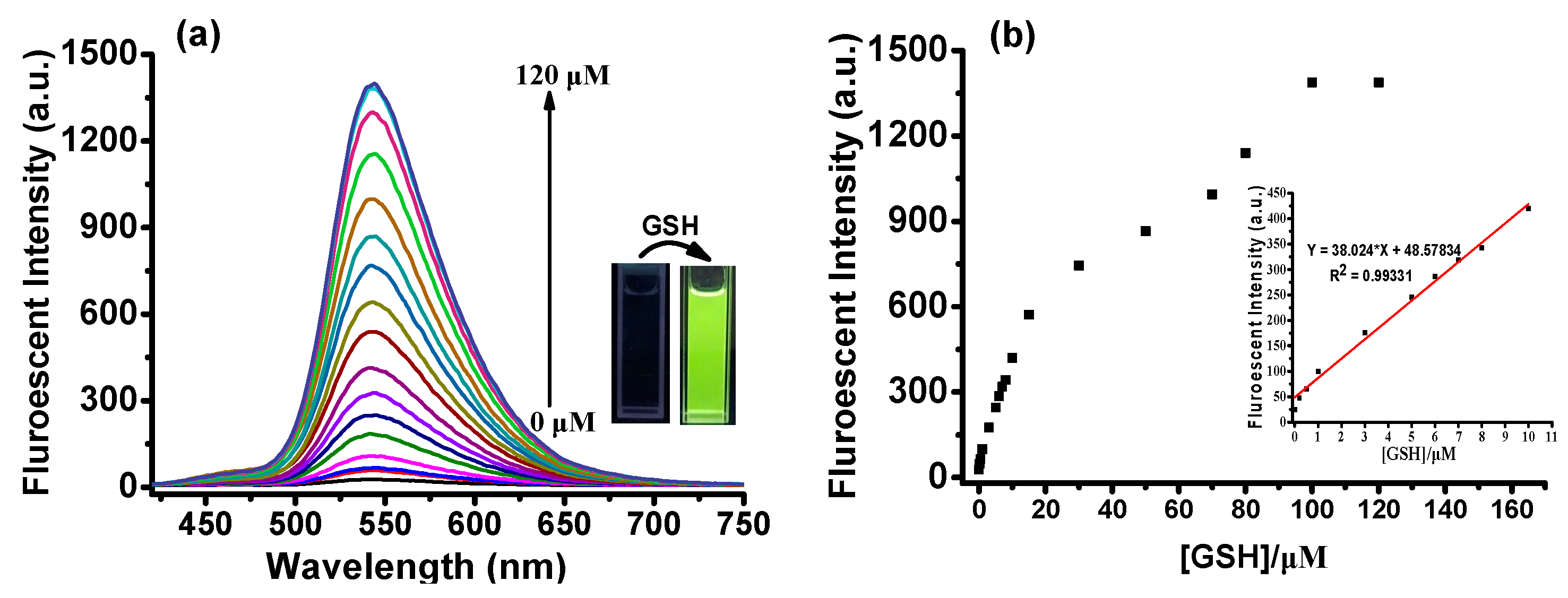
Concentration-dependent tests of PBD with GSH showed that the fluorescence intensity at 540 nm increased gradually with increasing GSH amounts until a stable signal was reached at 100 µM (Figure 2). The fluorescence enhancement could be about 70-fold when 10 equivalents of GSH were added to the solution of PBD. As shown in Figure 2, upon treatment with GSH, the testing solution gave rise to a strong yellow-green fluorescence which is consistent with the emission spectrum of dye PBOH. A calibration plot of the signal to the GSH concentrations from 0 µM to 10 µM showed good linearity (R2 = 0.9933), indicating that PBD can quantitatively detect GSH within this range. The detection limit of PBD toward GSH was found to be 14.5 nM based on the 3σ/slope method [39,40], thus enabling a highly sensitive detection of GSH. We also evaluated the response effect of Cys and Hcy to PBD. Under the same conditions, PBD exhibited similar optical behavior to Cys and Hcy with the detection limits being 17.5 nM and 80.0 nM, respectively (Figure S4).
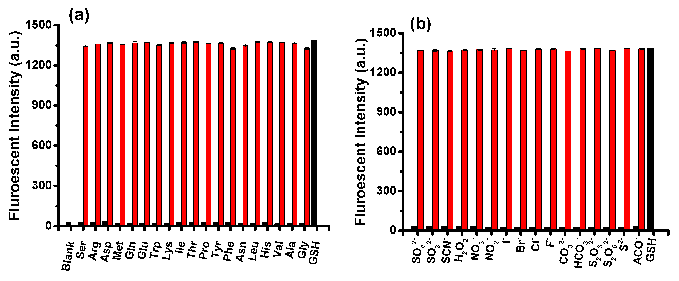
The selectivity of PBD for biothiols (100 μM) was evaluated by screening its response to potential competing species (200 μM), including various natural amino acids (Val, Glu, Leu, Met, Asn, Ala, His, Trp, Phe, Ser, Arg, Lys, Gln, Asp, Ile, Thr, Tyr, Pro, and Gly), sulfur species (Na2S, KSCN, Na2SO3, Na2SO4, Na2S2O3, and Na2S2O4), oxygen species (H2O2), and common salts (KF, NaCl, KBr, KI, Na2CO3, NaHCO3, NaNO2, NaNO3, and AcONa). Figure 3 illustrates that treatment of PBD solution with 10 equivalents of GSH resulted in a 70-fold enhancement at 540 nm. Cys gave rise to a similar fluorescence increase. Hcy induced an increment half that of GSH. High concentrations (200 µM) of interfering species induced negligible changes. Following on from this, competition studies were investigated by treating PBD with GSH (100 μM) in the presence of 20 equivalents of other analytes. As shown in Figure 4a,b, addition of GSH to the probe solution resulted in a robust signal enhancement in spite of the coexistence of competing species, demonstrating that probe PBD displays strong immunity to interference while sensing biothiols.
2.3. Sensing Mechanism Study
The fluorescence turn-on response of PBD to biothiols may be attributed to a thiol-anion-mediated SNAr process which led to the cleavage of 2,4-dinitrophenylsulfonate (Scheme 2). A comparison of the fluorescence spectral profiles of the reaction solution and the precursor compound provides evidence for the proposed mechanism. Upon addition of GSH to non-fluorescent PBD, a turn-on fluorescent signal at 540 nm was observed and the emission intensity nearly reached the intensity of PBOH. In addition, we successfully isolated the fluorescent product of the reaction by column chromatography. As expected, the 1H-NMR spectrum of the isolated fluorescent product is consistent with that of PBOH. Thus, the above results confirmed that dye PBOH was released via the cleavage of sulfonate in PBD mediated by GSH.
To confirm the sensing mechanism, we also carried out 1H-NMR titration experiments to track product formation. After the addition of excessive Cys to PBD in DMSO-d6/D2O (8/2, v/v), the signals of PBD (Ha and Hb) gradually declined. At the same time, three peaks corresponding to the protons (Ha’, Hb’, and Hc’) attributable to PBOH began to appear, indicating that PBOH had been released. With the continuation of this chemical reaction, the signals of PBOH (Ha’, Hb’, and Hc’) gradually increased. The signals of PBD (Ha and Hb) had almost completely disappeared at 20 min. These results demonstrate the cleavage of the DNBS group and the production of fluorophore PBOH (Figure 5).
To elucidate further the off-on sensing mechanism of probe PBD towards biothiols, DFT (Density functional theory) calculations were performed. The frontier orbital diagram indicates that the LUMO energy level of DNBS (−3.930 eV) is much lower than that of fluorophore the PBOH (−1.021 eV) (Figure 6), which implied that the photo-induced electron transfer (PET) process can happen from the fluorophore moiety to the DNBS group. Owing to the effective fluorescence quenching effect of DNBS, PBD is essentially nonfluorescent. After removal of the DNBS group, the fluorescence is recovered.
2.4. Live Cell Imaging
Having demonstrated its excellent responsiveness and anti-interference in sensing biothiols, we next examined whether PBD can be used to detect intracellular thiols in living cells. As shown in Figure 7, after incubating HeLa cells with PBD for 20 min, strong yellow-green fluorescence could be observed inside the cells under excitation at 405 nm. In the control experiments, cells were pretreated with N-ethylmaleimide (NEM) for 30 min to remove free thiols, and subsequently incubated with PBD for another 20 min. After being washed with PBS buffer three times, the cells showed almost no fluorescence under a confocal fluorescence microscope. The results showed that PBD is cell-permeable and can sense intracellular biothiols in living cells.
3. Materials and Methods
3.1. Materials.
All chemicals used were of analytical grade and were obtained from commercial suppliers. TLC (Thin-layer chromatography) silica gel plates (GF-254) and silica gel (200-300 mesh) for column chromatography were obtained from Qingdao Marine Chemicals, China. Deionized water was employed throughout all experiments. 1H-NMRand 13C-NMRspectra were acquired on a Bruker AVANCE II 400 spectrometer (Bruker, Switzerland), respectively. HRMS (High Resolution Mass Spectrometry) spectra were measured with a 6510-Q-TOF spectrometer (Agilent, USA). UV–Visible absorption spectra were recorded by a TU-1901 spectrometer (Beijing, China). Fluorescent measurements were performed on a Lengguang F97Pro FL Spectrophotometer (Shanghai, China). The pH measurement was carried out on a Leici PHS-3C pH meter (Shanghai, China). All spectra were recorded at room temperature.
3.2. Synthesis and Characterization of Compound Information
The four-step synthetic route is outlined in Scheme 3. The details of the synthesis and structural characterization are shown in the Supporting Information (Figures S8–S14).
4. Conclusions
In sum, we have developed a new off-on fluorescent probe PBD for biothiols by modifying a fluorescent dye 7-hydroxy-3-phenyl-benzoxazinone with a DNBS unit as a biothiol-specific recognition moiety. In response to biothiols, PBD displays significant fluorescence enhancement (up to 70-fold) and a large Stokes shift (155 nm). The probe exhibits a low detection limit as well as excellent selectivity and anti-interference ability toward biothiols over competing species. Finally, this probe has been used to sense biothiols in live HeLa cells.
Supplementary Materials
The supplementary materials are available online.
Author Contributions
B.L. and D.Z. conceived and designed experiments; B.L. performed experiments; B.L. and R.A. analyzed the data; Y.Z. conceived experiments and analyzed the data.
Funding
This study was supported by the Self-Innovation Project for Universities and Institutes of Jinan City (No. 201202035).
Conflicts of Interest
There are no conflicts of interest to declare.
References
- Nadeau, P.J.; Charette, S.J.; Toledano, M.B.; Landry, J. Disulfide Bond-mediated Multimerization of Ask1 and Its Reduction by Thioredoxin-1 Regulate H2O2-induced c-Jun NH2-terminal Kinase Activation and Apoptosis. Mol. Biol. Cell. 2007, 18, 3903–3913. [Google Scholar] [CrossRef]
- Requejo, R.; Hurd, T.R.; Costa, N.J.; Murphy, M.P. Cysteine residues exposed on protein surfaces are the dominant intramitochondrial thiol and may protect against oxidative damage. FEBS J. 2010, 277, 1465–1480. [Google Scholar] [CrossRef] [Green Version]
- Ghezzi, P.; Bonetto, V.; Fratelli, M. Thiol–Disulfide Balance: From the Concept of Oxidative Stress to that of Redox Regulation. Antioxid. Redox. Signal. 2005, 7, 964–972. [Google Scholar] [CrossRef]
- Cheng, Z.; Zhang, J.; Ballou, D.P.; Williams, C.H., Jr. Reactivity of Thioredoxin as a Protein Thiol-Disulfide Oxidoreductase. Chem. Rev. 2011, 111, 5768–5783. [Google Scholar] [CrossRef] [Green Version]
- Cross, J.V.; Templeton, D.J. Thiol oxidation of cell signaling proteins: Controlling an apoptotic equilibrium. J. Cell. Biochem. 2004, 93, 104–111. [Google Scholar] [CrossRef]
- Shahrokhian, S. Lead phthalocyanine as a selective carrier for preparation of a cysteine-selective electrode. Anal. Chem. 2001, 73, 5972–5978. [Google Scholar] [CrossRef]
- Seshadri, S.; Beiser, A.; Selhub, J.; Jacques, P.F.; Rosenberg, I.H.; Wilson, W.F.; Wolf, P.A. Plasma homocysteine as a risk factor for dementia and Alzheimer’s disease. N. Engl. J. Med. 2002, 346, 476–483. [Google Scholar] [CrossRef]
- Refsum, H.; Ueland, P.M.; Nygard, O.; Vollset, S.E. Homocysteine and cardiovascular disease. Annu. Rev. Med. 1998, 49, 31–62. [Google Scholar] [CrossRef]
- Lee, J.H.; Sharma, A.; Jang, J.H. Real time OFF–ON monitoring of gluthathione (GSH) in living cell. J. Incl. Phenom. Macro. 2015, 82, 117–122. [Google Scholar] [CrossRef]
- Aoyama, K.; Suh, S.W.; Hamby, A.M.; Liu, J.; Chan, W.Y.; Chen, Y.; Swanson, R.A. Neuronal glutathione deficiency and age-dependent neurodegeneration in the EAAC1 deficient mouse. Nat. Neurosci. 2006, 9, 119–126. [Google Scholar] [CrossRef]
- Jung, H.S.; Chen, X.; Peng, X. Recent progress in luminescent and colorimetric chemosensors for detection of thiols. Chem. Soc. Rev. 2013, 42, 6019–6031. [Google Scholar] [CrossRef]
- Lim, S.Y.; Hong, K.H.; Kim, D.I.; Kwon, H.; Kim, H.J. Tunable Heptamethine–Azo Dye Conjugate as an NIR Fluorescent Probe for the Selective Detection of Mitochondrial Glutathione over Cysteine and Homocysteine. J. Am. Chem. Soc. 2014, 136, 7018–7025. [Google Scholar] [CrossRef]
- Zamfir, L.G.; Rotariu, L.; Bala, C. Sensing of sulfhydryl based compounds by a simple electrochemical approach. Sens. Actuators B Chem. 2015, 206, 65–73. [Google Scholar] [CrossRef]
- Yin, J.; Hu, Y.; Yoon, J. Fluorescent probes and bioimaging: Alkali metals, alkaline earth metals and pH. Chem. Soc. Rev. 2015, 44, 4619–4644. [Google Scholar] [CrossRef]
- Farhadi, K.; Forough, M.; Pourhossein, A.; Molaei, R. Highly sensitive and selective colorimetric probe for determination of l-cysteine in aqueous media based on Ag/Pd bimetallic nanoparticles. Sens. Actuators B Chem. 2014, 202, 993–1001. [Google Scholar] [CrossRef]
- Lee, M.H.; Yang, Z.; Lim, C.W.; Lee, Y.H. Disulfide-cleavage-triggered chemosensors and their biological applications. Chem. Rev. 2013, 113, 5071–5109. [Google Scholar] [CrossRef]
- Shao, J.H.; Sun, H.; Guo, S.; Ji, J.; Zhao, W. A highly selective red-emitting FRET fluorescent molecular probe derived from BODIPY for the detection of cysteine and homocysteine: An experimental and theoretical study. Chem. Sci. 2012, 3, 1049–1061. [Google Scholar] [CrossRef]
- Ji, W.; Ji, Y.; Jin, Q.; Tong, Q.; Tang, X. Heavy atom quenched coumarin probes for sensitive and selective detection of biothiols in living cells. Analyst. 2015, 140, 4379–4383. [Google Scholar] [CrossRef]
- Peng, L.; Zhou, Z.; Wei, R.; Li, K.; Song, K.; Tong, A. A fluorescent probe for thiols based on aggregation-induced emission and its application in live-cell imaging. Dyes Pigment. 2014, 108, 24–31. [Google Scholar] [CrossRef]
- Shi, J.; Wang, Y.; Tang, X.; Liu, W.; Jiang, H.; Dou, W.; Liu, W. A colorimetric and fluorescent probe for thiols based on 1, 8-naphthalimide and its application for bioimaging. Dyes Pigment. 2014, 100, 255–260. [Google Scholar] [CrossRef]
- Liu, J.; Sun, Y.Q.; Zhang, H.; Huo, Y.; Shi, Y.; Shi, H.; Guo, W. A carboxylic acid-functionalized coumarin-hemicyanine fluorescent dye and its application to construct a fluorescent probe for selective detection of cysteine over homocysteine and glutathione. RSC Adv. 2014, 4, 64542–64550. [Google Scholar] [CrossRef]
- Yang, X.F.; Huang, Q.; Zhong, Y.; Li, Z.; Li, H.; Lowry, M.; Strongin, R.M. A dual emission fluorescent probe enables simultaneous detection of glutathione and cysteine/homocysteine. Chem. Sci. 2014, 5, 2177–2183. [Google Scholar] [CrossRef] [Green Version]
- Niu, L.Y.; Guan, Y.S.; Chen, Y.Z.; Wu, L.Z.; Tung, C.H.; Yang, Q. BODIPY-based ratiometric fluorescent sensor for highly selective detection of glutathione over cysteine and homocysteine. J. Am. Chem. Soc. 2012, 134, 18928–18931. [Google Scholar] [CrossRef]
- Liu, Y.C.; Xiang, K.Q.; Tian, B.Z. A fluorescein-based fluorescence probe for the fast detection of thiol. Tetra. Lett. 2016, 3, 2478–2483. [Google Scholar] [CrossRef]
- Zhu, X.Y.; Gao, H.; Zan, W.Y.; Li, Y. A rational designed thiols fluorescence probe: The positional isomer in PET. Tetrahedron. 2016, 72, 2048–2056. [Google Scholar] [CrossRef]
- Liao, Y.C.; Venkatesan, P.; Wei, L.F.; Wu, S.P. A coumarin-based fluorescent probe for thiols and its application in cell imaging. Sens. Actuators B Chem. 2016, 232, 732–737. [Google Scholar] [CrossRef]
- Guo, Z.; Nam, S.; Park, S.; Yoon, J. A highly selective ratiometric near-infrared fluorescent cyanine sensor for cysteine with remarkable shift and its application in bioimaging. Chem. Sci. 2012, 3, 2760–2765. [Google Scholar] [CrossRef]
- Rusin, O.; Luce NN, S.; Agbaria, R. Visual Detection of Cysteine and Homocysteine. J. Am. Chem. Soc. 2004, 2, 438–439. [Google Scholar] [CrossRef]
- Yang, X.; Guo, Y.; Strongin, R.M. Conjugate Addition/Cyclization Sequence Enables Selective and Simultaneous Fluorescence Detection of Cysteine and Homocysteine. Angew. Chem. Int. Ed 2011, 50, 10690–10693. [Google Scholar] [CrossRef] [Green Version]
- Lv, H.; Yang, X.F.; Zhong, Y.; Guo, Y.; Li, Z.; Li, H. Native Chemical Ligation Combined with Spirocyclization of Benzopyrylium Dyes for the Ratiometric and Selective Fluorescence Detection of Cysteine and Homocysteine. Anal. Chem. 2014, 3, 1800–1807. [Google Scholar] [CrossRef]
- Azuma, K.; Suzuki, S.; Uchiyama, S.; Kajiro, T.; Santa, T. A study on the thermal decomposition of KClO4and NaClO4by acoustic emission thermal analysis. Photobiol. Sci. 2003, 2, 443–449. [Google Scholar] [CrossRef]
- An, R.B.; Wei, P.; Zhang, D.T.; Su, N. A highly selective 7-hydroxy-3-methyl-benzoxazinone based fluorescent probe for instant detection of thiophenols in environmental samples. Tetra. Lett. 2016, 57, 3039–3042. [Google Scholar] [CrossRef]
- Manna, S.; Karmakar, P.; Ali, S.S.; Guria, U.N.; Mahapatra, A.K. Michael addition-cyclization-based switch-on fluorescent chemodosimeter for Cysteine and its application in living cell imaging. New. J. Chem. 2018, 42, 4951–4958. [Google Scholar] [CrossRef]
- Wang, J.; Zhou, C.; Zhang, J.; Zhu, X.; Liu, X.; Wang, Q.; Zhang, H. A new fluorescence turn-on probe for biothiols based on photoinduced electron transfer and its application in living cells. Spectrochim. Acta. A. 2016, 166, 31–37. [Google Scholar] [CrossRef] [Green Version]
- Dai, X.; Zhang, T.; Miao, J.Y.; Zhao, B.X. A ratiometric fluorescent probe with DNBS group for biothiols in aqueous solution. Sens. Actuators B Chem. 2016, 223, 274–279. [Google Scholar] [CrossRef]
- Dong, C.; Zhou, C.Q.; Yang, J.W.; Liao, T.C.; Chen, J.X.; Yin, C.X.; Chen, W.H. A novel 3,6-diamino-1,8-naphthalimide derivative as a highly selective fluorescent “turn-on” probe for thiols. RSC Adv. 2015, 5, 32990–32993. [Google Scholar] [CrossRef]
- Hu, Q.H.; Yu, C.M.; Xi, X.; Wu, S.Z. A fluorescent probe for simultaneous discrimination of GSH and Cys/Hcy in human serum samples via distinctly-separated emissions with independent excitations. Biosen. Bioelectron. 2016, 81, 341–348. [Google Scholar] [CrossRef]
- Zhang, J.J.; Yu, B.F.; Ning, L.; Zhu, X.Y.; Wang, J.X.; Chen, Z.J.; Liu, X.Y.; Yao, X.J.; Zhang, X.Y.; Zhang, H.X. A Near-Infrared Fluorescence Probe for Thiols Based on Analyte-Specific Cleavage of Carbamate and Its Application in Bioimaging. Eur. J. Org. Chem. 2015, 8, 1711–1718. [Google Scholar] [CrossRef]
- Chen, W.; Luo, H.; Liu, X.; Foley, J.W.; Song, X. Broadly Applicable Strategy for the Fluorescence Based Detection and Differentiation of Glutathione and Cysteine/Homocysteine: Demonstration in Vitro and in Vivo. Anal. Chem. 2016, 16, 3638–3646. [Google Scholar] [CrossRef]
- Liu, T.; Huo, F.; Yin, C.; Li, J.; Chao, J.; Zhang, Y. A triphenylamine as a fluorophore and maleimide as a bonding group selective turn-on fluorescent imaging probe for thiols. Dyes Pigment. 2016, 128, 209–214. [Google Scholar] [CrossRef]
Sample Availability: Samples of the compounds PBOH and PBD are not available from the authors. |
Scheme 1.
Design diagram of fluorescent probe PBD (2,4-dintrobenzenesulfonate of 7-hydroxy-3-phenylbenzoxazinone) for biothiol detection.
Scheme 1.
Design diagram of fluorescent probe PBD (2,4-dintrobenzenesulfonate of 7-hydroxy-3-phenylbenzoxazinone) for biothiol detection.

Figure 1.
Time-dependent course of the interaction of PBD (10 µM) with GSH in EtOH-PBS buffer (Phosphate Buffered Saline) (10 mM, pH 7.4, 4:6, v/v). Fluorescence intensity was recorded at 540 nm with λex = 405 nm.
Figure 1.
Time-dependent course of the interaction of PBD (10 µM) with GSH in EtOH-PBS buffer (Phosphate Buffered Saline) (10 mM, pH 7.4, 4:6, v/v). Fluorescence intensity was recorded at 540 nm with λex = 405 nm.

Figure 2.
(a) Fluorescence response of PBD (10 μM) upon the addition of GSH (0–120 μM) in EtOH-PBS buffer (10 mM, pH 7.4, 4:6, v/v). Inset: Fluorescence images of PBD (10 μM) in the absence (left) and presence (right) of GSH under a 365 nm UV lamp. (b) Fluorescence intensity of PDB (10 μM) at 540 nm as a function of GSH concentration (0–120 μM) in EtOH-PBS buffer (10 mM, pH 7.4, 4:6, v:v). Inset: the linear relationship between fluorescence intensity and GSH at low concentrations.

Figure 3.
Fluorescence spectra of PBD (10 µM) with biothiols (100 µM) and various tested analytes (200 µM). λex = 405 nm.
Figure 3.
Fluorescence spectra of PBD (10 µM) with biothiols (100 µM) and various tested analytes (200 µM). λex = 405 nm.

Figure 4.
(a,b) Selectivity and competition of PBD (10 µM) to GSH (100 µM) and other analytes (200 μM). Black bars represent the fluorescence intensity (540 nm) of a single analyte with probe PBD. Red bars represent the fluorescence intensity (540 nm) of PBD to GSH in the presence of various tested analytes. λex = 405 nm.

Scheme 2.
Schematic sensing mechanism of PBD toward biothiols.

Figure 5.
Partial 1H-NMR spectra of PBD before the addition (a) and the change after addition of Cys, 2 min (b), 5 min (c), and 20 min (d), and of PBOH (e) in DMSO-d6/D2O (8/2, v/v).
Figure 5.
Partial 1H-NMR spectra of PBD before the addition (a) and the change after addition of Cys, 2 min (b), 5 min (c), and 20 min (d), and of PBOH (e) in DMSO-d6/D2O (8/2, v/v).

Figure 6.
Frontier orbital diagrams of PBOH and 2,4-dinitrophenylsulfonyl (DNBS). Orbital energies were calculated using Gaussian 03 program at B3LYP/6-31 g (d, p) level.
Figure 6.
Frontier orbital diagrams of PBOH and 2,4-dinitrophenylsulfonyl (DNBS). Orbital energies were calculated using Gaussian 03 program at B3LYP/6-31 g (d, p) level.

Figure 7.
Confocal fluorescence images of HeLa cells. (Left) bright-field image; (middle) fluorescence image; and (right) overlayed image. (a) Cells treated with N-ethylmaleimide (NEM) (1 mM) for 30 min followed by incubation with PBD (10 μM) for 20 min. (b) Cells incubated with PBD (10 μM) for 20 min at 37 °C. (c) Untreated cells.

Scheme 3.
Synthesis of PBD. Reagents and conditions: (i) fuming HNO3, CHCl3, and CH3COOH, r.t.; (ii) Pd/C, H2, and CH3COOCH2CH3, r.t.; (iii) PhCOCOOH, EtOH, and CH3COOH, r.t; (iv) 2,4-Dinitrobenzenesulfonyl chloride, Et3N, and THF, r.t.
Scheme 3.
Synthesis of PBD. Reagents and conditions: (i) fuming HNO3, CHCl3, and CH3COOH, r.t.; (ii) Pd/C, H2, and CH3COOCH2CH3, r.t.; (iii) PhCOCOOH, EtOH, and CH3COOH, r.t; (iv) 2,4-Dinitrobenzenesulfonyl chloride, Et3N, and THF, r.t.

© 2019 by the authors. Licensee MDPI, Basel, Switzerland. This article is an open access article distributed under the terms and conditions of the Creative Commons Attribution (CC BY) license (http://creativecommons.org/licenses/by/4.0/).
Share and Cite
MDPI and ACS Style
Li, B.; Zhang, D.; An, R.; Zhu, Y. A 7-Hydroxybenzoxazinone-Containing Fluorescence Turn-On Probe for Biothiols and Its Bioimaging Applications. Molecules 2019, 24, 3102. https://doi.org/10.3390/molecules24173102
AMA Style
Li B, Zhang D, An R, Zhu Y. A 7-Hydroxybenzoxazinone-Containing Fluorescence Turn-On Probe for Biothiols and Its Bioimaging Applications. Molecules. 2019; 24(17):3102. https://doi.org/10.3390/molecules24173102
Chicago/Turabian StyleLi, Bin, Datong Zhang, Ruibing An, and Yaling Zhu. 2019. "A 7-Hydroxybenzoxazinone-Containing Fluorescence Turn-On Probe for Biothiols and Its Bioimaging Applications" Molecules 24, no. 17: 3102. https://doi.org/10.3390/molecules24173102