Article

Design and Synthesis of Novel Breast Cancer Therapeutic Drug Candidates Based upon the Hydrophobic Feedback Approach of Antiestrogens

1
School of Pharmacy, Showa University, 1-5-8, Hatanodai, Shinagawa-ku, Tokyo 142-8555, Japan
2
Faculty of Pharmaceutical Sciences, Tohoku Medical and Pharmaceutical University, 4-4-1 Komatsushima, Aoba-ku, Sendai 981-8558, Japan
*
Authors to whom correspondence should be addressed.
Molecules 2019, 24(21), 3966; https://doi.org/10.3390/molecules24213966
Submission received: 29 September 2019 / Revised: 23 October 2019 / Accepted: 30 October 2019 / Published: 1 November 2019
(This article belongs to the Special Issue Design and Synthesis of Organic Molecules as Antineoplastic Agents)

Abstract

:
Based upon hydrophobic feedback approaches, we designed and synthesized novel sulfur-containing ERα modulators (4 and 5) as breast cancer therapeutic drug candidates. The tetrahydrothiepine derivative 5a showed the highest binding affinity toward ERα because of its high hydrophobicity, and it acted as an agonist toward MCF-7 cell proliferation. The corresponding alkylamino derivative 5d maintained high binding affinity to ERα and potently inhibited MCF-7 cell proliferation (IC50: 0.09 μM). Docking simulation studies of compound 5d with the ERα BD revealed that the large hydrophobic moiety of compound 5d fit well into the hydrophobic pocket of the ERα LBD and that the sulfur atom of compound 5d formed a sulfur–π interaction with the amino acid residue His524 of the ERα LBD. These interactions play important roles for the binding affinity of compound 5d to the ERα LBD.

Graphical Abstract

1. Introduction

17β-Estradiol (E2) is an endogenous female hormone and plays an important role in the female reproductive system. Estrogens including E2 influence the growth, differentiation, and functioning of many target tissues mediated by the binding to estrogen receptor α (ERα) [1,2,3]. Although activation of ERα is essential for the maintenance of homeostasis of female functions, they can sometimes induce and cause progression of estrogen-dependent breast cancers [4,5,6]. Several antiestrogens have been developed to prevent and control hormone-responsive breast cancer [7,8,9,10]. Tamoxifen has been used worldwide for more than 40 years for the treatment of estrogen-dependent breast cancer (Figure 1) [11,12,13]. Interestingly, tamoxifen acts as either an agonist or an antagonist depending on tissue type; tamoxifen exhibits antiestrogenic action in breast cancer and hot flashes, and estrogenic action in bone and cholesterol metabolism, and is a selective estrogen receptor modulator (SERM) [11,12,13]. The dimethylaminoethyl chain of tamoxifen is attached to a benzene ring, where it plays a strategic role in the expression of antiestrogenic activity. The triphenylethylene moiety of tamoxifen plays an important role in controlling ERα binding affinity. The triphenylethylene moiety is a promising structure for the development of an ER ligand. The triphenylethylene structure exhibits geometric isomers E and Z that are caused by the key alkylamino chain, and the asymmetric hydrophobic part and the isomers easily isomerize between the E and Z forms. Therefore, the isomerization often become a big problem in the synthesis, purification, and preservation of an either isomer. Indeed, 4-hydroxytamoxifen, an active metabolite of tamoxifen, easily isomerizes from the active Z form to the inactive E form in solution and in in vitro studies [14,15,16,17]. In this regard, novel ER antagonists comprised of other skeleton structures, such as benzothiophene [18], dihydronaphthalene [19], benzopyrane [20], and steroid [21] structures, have been developed as ER antagonists.
On the other hand, we found that a simple 1,2-bis(4-hydroxyphenyl)-o-carborane, BE360 (1) exhibited high binding affinity toward ERα and estrogenic action in bone without showing estrogenic action in the uterus of ovariectomized (OVX) and orchidectomized (ORX) mice (Figure 1) [22]. That is, compound 1 acted as an agonist in bone and an antagonist in female reproductive tissues, despite lack of an alkylamino chain. Based upon the hydrophobic feedback approaches, we focused on three-dimensional hydrocarbon units as new hydrophobic pharmacophores to design and synthesize BE1060 (2) and BE1054 (3) (Figure 1) [23]. Although the tetramethylcyclohexene-based bisphenol compound 3 acted as a partial agonist, similar to compound 1, the bicyclo[2,2,2]octene-based bisphenol compound 2 showed potent estrogenic activity. Unlike the triphenylethylene structure, a vicinal bisphenol structure does not have a geometric isomer, even with the key alkylamino chain. We were interested in the effects of ring size and sulfur atom in the hydrophobic structure on ER activity. Furthermore, to understand the effects of sulfur, sulfone, and sulfoxide on the biological activity, we designed dihydrothiophene and tetrahydrothiepine derivatives (4a4d and 5a5d) as novel ER antagonist candidates. Herein, we describe the facile synthesis and biological activities of the designated compounds 4a4d and 5a5d (Figure 1).

2. Results and Discussion

2.1. Chemistry

Synthesis of the dihydrothiophene derivatives (4a4d) is summarized in Scheme 1. Commercially available 2-bromo-4′-methoxyacetophenone (6) was reacted with sodium sulfide (Na2S) to form the sulfide compound 7 in 95% yield [24]. Intramolecular McMurry coupling of 7 resulted in formation of the dihydrothiophene derivative 8 in 60% yield, which was demethylated with BBr3 to form the target compound 4a in 97% yield [25]. Although the dihydrothiophene derivative 8 was treated with 1 equimolar amount of m-chloroperbenzoic acid (m-CPBA) to form the sulfoxide compound 9 in 70% yield, followed by demethylation with BBr3 to form the decomposition product of compound 9, it did not result in the formation of compound 4b. Thus, we carried out oxidation of compound 4a with m-CPBA to form the desired compound 4b in 73% yield. When compound 8 was treated with two equimolar amounts of m-CPBA, the corresponding sulfone compound 10 was obtained and was then demethylated with BBr3 to form compound 4c in quantitative yield over two steps. Mitsunobu reaction of compound 4a with 3-dimethylamino-1-propanol resulted in formation of compound 4d with a dimethylaminopropyl chain in 37% yield [26].
Scheme 2 summarizes the synthesis of tetrahydrothiepine derivatives (5a5d). A starting material (12) for the seven-membered ring derivative 5 was synthesized by Friedel-Craft acylation of anisole with acyl chloride 11, followed by sulfide formation with Na2S to form compound 13 in 84% yield over two steps [24]. The tetrahydrothiepine derivatives (5a5d) were synthesized in the same manner as those of compounds 4a4d. Briefly, intramolecular McMurry coupling of compound 13, followed by demethylation, resulted in formation of the sulfide compound 5a in a 66% yield [25]. Compound 5a was transformed into the sulfoxide compound 5b by oxidation with one equimolar amount of m-CPBA in a 30% yield. Moreover, compound 5a was reacted with 3-dimethylamino-1-propanol under Mitsunobu reaction conditions to form compound 5d in a 28% yield [26]. Treatment of compound 14 with two equimolar amounts of m-CPBA followed by demethylation resulted in the formation of the sulfone compound 5c in a 49% yield over two steps.

2.2. Biological Evaluation
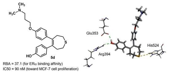
The binding affinities of the synthesized bisphenols (compounds 4a4c and 5a5c) toward ERα were evaluated by means of a competitive binding assay using [6,7-3H] 17β-estradiol and human recombinant ERα [27,28,29]. Figure 2A,B shows dose-response curves for competitive binding of the synthesized compounds 4a4c and 5a5c to ERα, respectively. Compound 4a, containing a sulfide group, moderately bound to ERα and the binding affinity was about 100 times lower than that of E2. The sulfoxide (4b) and sulfone (4c) compounds did not bind to the ERα ligand-binding domain (LBD), even at concentrations 1000 times higher than that of E2. The binding affinity of the tetrahydrothiepine derivative 5 toward ERα showed a similar tendency to that of compound 4, and the sulfide derivative 5a bound to ERα more potently than compound 4a. The tamoxifen-inspired derivatives 4d and 5d, both containing a dimethylaminopropyl group, showed similar binding affinities to compounds 4a and 5a, respectively. With the binding affinity of E2 to ERα taken as 100, the relative binding affinity (RBA) values of compounds 4d and 5d were 4.0 and 37.1, respectively (Table 1). It is suggested that the binding affinities of compounds 4 and 5 depend on the hydrophobicity of the ring structures and the polarity of the sulfur atoms, which can be understood from the CLogP values of compounds 4a4d and 5a5d in Table 1.
Next, estrogenic and antiestrogenic activities of compounds 4a4d and 5a5d, except for 4b and 5b (poor binding affinity toward ERa), were evaluated by means of a cell proliferation assay using the human breast cancer cell line MCF-7, which shows ER-dependent growth [27,28,29]. Table 1 summarizes the EC50, IC50, and Emax values estimated from the MCF-7 cell proliferation assays. Emax represents the maximal efficacy of each compound toward cell proliferation and is an index of ER partial agonistic activity. Emax was estimated relative to that of E2, which was taken as 100. Although the binding affinity of compound 4a was ~10 times higher than that of compound 4c, both compounds showed estrogenic activity with similar EC50 values. Based on the Emax values of compounds 4a and 4c, we suggest that the complex formed between compound 4c and the ERα LBD is more active for transcription than that formed between compound 4a and the ERα LBD. Compound 5a, which exhibited the most potent binding affinity to ERα LBD, also showed the most potent MCF-7 cell proliferation activity, with an EC50 value of 0.048 μM. As expected from the weak binding affinity to ERα LBD, compound 5c showed weak estrogenic activity. On the other hand, the tamoxifen-inspired derivatives 4d and 5d, containing alkylamino chains, potently inhibited cell proliferation of MCF-7 cells induced by 0.1 nM of E2, with IC50 values of 0.41 and 0.09 μM, respectively.

2.3. Docking Study

To understand the docking modes of the sulfur-containing ligands 4d and 5d with the ERα LBD, docking simulation studies of both compounds toward the human ERα LBD (PDB: 1ERR) were carried out using an automatic docking program (Discovery Studio 2018/CDOCKER) [30]. The docking mode of compound 4d was superimposed onto that of compound 5d at the ERα LBD (Figure 3A,B). The docking mode of compound 4d toward the ERα LBD was similar to that of compound 5d. Each phenolic hydroxy group of compounds 4d and 5d formed hydrogen bonds with the amino acid residues Glu353 and Arg394 (Figure 3C), and each dimethylaminopropyl chain of compounds 4d and 5d was found to be located within the same space of the ERα LBD (Figure 3A). The hydrophobic moiety of compound 5d was spread widely across the hydrophobic pocket of the ERα LBD and the sulfur atom of the tetrahydrothiepine ring formed a sulfur–π interaction with the amino acid residue His524 (Figure 3B,C). Docking scores (CDOCKER interaction energy) of compounds 4d and 5d toward the ERα LBD were 51.1 and 54.0, respectively, and the order of docking score was correlated with the results of the binding assay. Therefore, we attributed the potent binding affinity of compound 5d toward the ERα LBD to both hydrophobic interactions and sulfur–π interactions.

3. Materials and Methods

3.1. General Consideration

Melting points were determined with an MP-J3 micro melting point apparatus (Yanaco, Kyoto, Japan) and were not corrected. 1H NMR and 13C NMR spectra were recorded with JNM-LA-400 spectrometer (JEOL, Tokyo, Japan). Chemical shifts for 1H NMR spectra were referenced to tetramethylsilane (0.0 ppm) as an internal standard. Chemical shifts for 13C NMR spectra were referenced to residual 13C present in deuterated solvents. The splitting patterns are designated as follows: s (singlet), d (doublet), t (triplet), q (quartet) and m (multiplet). Mass spectra were recorded on a JEOL JMS-DX-303 spectrometer (JEOL, Tokyo, Japan). Elemental analyses were performed with a Perkin Elmer 2400 CHN spectrometer (PerkinElmer, Winter Street Waltham, MA, USA). Column chromatography was carried out using Merck silica gel 60 (0.063–0.200 μm) and TLC was performed on Merck silica gel F254.

3.2. Synthesis

1-(4-Methoxyphenyl)-2-[2-(4-methoxyphenyl)-2-oxo-ethylsulfanyl] ethanone (7). To a stirred solution of 6 (2.80 g, 12.2 mmol) in acetone (40 mL) was added a solution of Na2S 9H2O (1.50 g, 6.25 mmol) in H2O (12 mL) at 0 °C. After completion of the addition, the mixture was warmed to room temperature and stirred for 2.5 h. The solvents were removed, and the residue was recrystallized from MeOH to give 7 (1.91 g, 5.78 mmol, 95 %) as a colorless solid; 1H NMR (400 MHz, CDCl3) δ (ppm) 3.87 (6H, s), 3.93 (4H, s), 6.93 (4H, d, J = 8.8 Hz), 7.95 (4H, d, J = 8.8 Hz); 13C NMR (100 MHz, CDCl3) δ (ppm) 37.4, 55.5, 113.9, 128.4, 131.0, 163.8, 193.0; MS (EI) m/z = 330 (M+), 135 (100%).
3,4-Bis-(4-methoxyphenyl)-2,5-dihydrothiophene (8). To a suspension of Zn powder (1.57 g, 24.0 mmol) in dry THF (60 mL) was added TiCl4 (1.3 mL, 12.0 mmol) at −30 °C, and the mixture was refluxed for 2.5 h. A solution of 7 (1.0 g, 3.0 mmol) in dry THF (45mL) was slowly added to the reaction mixture at 0°C. The reaction mixture was refluxed for 2 h and then quenched with 10% K2CO3 aqueous solution. The insoluble materials were filtered through Celite, and the filtrate was extracted with AcOEt. The organic layer was washed with brine, dried over Na2SO4, and concentrated. Purification by silica gel column chromatography (eluent: AcOEt/n-hexane = 1/30) gave 8 as a colorless solid (0.55 g, 1.84 mmol, 61 %); 1H NMR (400 MHz, CDCl3) δ (ppm) 3.77 (6H, s), 4.22 (4H, s), 6.75 (4H, d, J = 8.8 Hz), 7.05 (4H, d, J = 8.8 Hz); 13C NMR (100 MHz, CDCl3) δ (ppm) 43.8, 55.1, 113.6, 129.4, 129.7, 134.3, 158.6; MS (EI) m/z = 298 (M+, 100%); Anal. Calcd for C18H18O2S: C, 72.45; H, 6.08, Found: C, 72.13; H, 6.20.
3,4-Bis-(4-hydroxyphenyl)-2,5-dihydrothiophene (4a). To a solution of 8 (407 mg, 1.36 mmol) in dry CH2Cl2 (9 mL) was added 1M of a solution of BBr3 in CH2Cl2 (3.4 mL, 3.4 mmol) at −80 °C, and the mixture was stirred at room temperature for 24 h. The mixture was poured onto ice and extracted with AcOEt. The organic layer was washed with brine, dried over MgSO4, and concentrated. Purification by silica gel column chromatography (eluent: AcOEt/n-hexane = 1/3) gave 4a as a colorless solid (358 mg, 1.33 mmol, 97 %); colorless needles (MeOH/CH2Cl2); mp 168.5–170.0 °C; 1H NMR (400 MHz, CD3OD) δ (ppm) 4.15 (4H, s), 6.62 (4H, d, J = 8.8 Hz), 6.95 (4H, d, J = 8.8 Hz); 13C NMR (100 MHz, CD3OD) δ (ppm) 44.3, 116.0, 129.8, 130.8, 135.5, 157.7; MS (EI) m/z = 270 (M+, 100%); HRMS Calcd for C16H14O2S: 270.0715, Found: 270.0707; Anal. Calcd for C16H14O2S 0.4 H2O: C, 69.23; H, 5.38, Found: C, 69.04; H, 5.66.
3,4-Bis-(4-methoxyphenyl)-2,5-dihydrothiophene-1-oxide (9). To a solution of 8 (406 mg, 1.36 mmol) in dry CH2Cl2 (15 mL) was portionwise added m-CPBA (65%, 349 mg, 1.41 mmol) at 0 °C. The reaction mixture was stirred at room temperature for 25 h and then quenched with saturated Na2SO3 aqueous solution. The mixture was extracted with AcOEt, washed with saturated NaHCO3 aqueous solution and brine, dried over MgSO4, and concentrated. Purification by silica gel column chromatography (eluent: AcOEt/n-hexane = 1/1) gave 9 as a colorless solid (302 mg, 0.96 mmol, 70%); colorless needles (CHCl3/n-hexane); mp 181.0–182.0 °C; 1H NMR (400 MHz, CDCl3) δ (ppm) 3.78 (6H, s), 4.00 (2H, d, J = 16.6 Hz), 4.34 (2H, d, J = 16.6 Hz), 6.77 (4H, d, J = 8.8 Hz), 7.10 (4H, d, J = 8.8 Hz); 13C NMR (100 MHz, CDCl3) δ (ppm) 55.2, 64.3, 113.9, 128.1, 129.9, 130.1, 159.1; MS (EI) m/z = 296 (M+, 100%); HRMS Calcd for C18H18O3S: 314.0977, Found: 314.0974; Anal. Calcd for C18H18O3S: C, 68.76; H, 5.77, Found: C, 68.42; H, 5.88.
3,4-Bis-(4-hydroxyphenyl)-2,5-dihydrothiophene-1-oxide (4b). To a solution of 4a (402 mg, 1.49 mmol) in dry CH2Cl2 (60 mL) was portion-wise added m-CPBA (65%, 368 mg, 1.49 mmol) at 0 °C. The reaction mixture was stirred at room temperature for 20 h and then quenched with saturated Na2SO3 aqueous solution. The mixture was extracted with AcOEt, washed with saturated NaHCO3 aqueous solution and brine, dried over MgSO4, and concentrated. Purification by silica gel column chromatography (eluent: AcOEt/n-hexane = 1/1) afforded 4b as a colorless solid (311 mg, 1.09 mmol, 73 %); colorless needles (MeOH/CH2Cl2); mp 219.0–220.0 °C; 1H NMR (400 MHz, CD3OD) δ (ppm) 3.94 (2H, d, J = 17.1 Hz), 4.49 (2H, d, J = 17.1 Hz), 6.67 (4H, d, J = 8.8 Hz), 7.05 (4H, d, J = 8.8 Hz); 13C NMR (100 MHz, CD3OD) δ (ppm) 64.9, 116.3, 128.4, 131.0, 131.1, 158.3; MS (EI) m/z = 286 (M+), 268 (100%); HRMS Calcd for C16H14O3S: 286.0664, Found: 286.0665; Anal. Calcd for C16H14O3S 0.1 H2O: C, 66.69; H, 4.97, Found: C, 66.47; H, 4.72.
3,4-Bis-(4-methoxyphenyl)-2,5-dihydrothiophene-1,1-dioxide (10). To a solution of 8 (601 mg, 2.02 mmol) in dry CH2Cl2 (30 mL) was portionwise added m-CPBA (65%, 1.11 g, 4.51 mmol) at 0 °C. The reaction mixture was stirred at room temperature for 3.5 h and then quenched with saturated Na2SO3 aqueous solution. The mixture was extracted with AcOEt, washed with saturated NaHCO3 aqueous solution and brine, dried over MgSO4, and concentrated. The residue was purified by recrystallization with CHCl3/n-hexane to give 10 (377 mg, 1.14 mmol, 57 %) as a colorless needles; mp 141.0–141.5 °C; 1H NMR (400 MHz, CDCl3) δ (ppm) 3.78 (6H, s), 4.26 (4H, s), 6.77 (4H, d, J = 9.3 Hz), 7.03 (4H, d, J = 9.3 Hz); 13C NMR (100 MHz, CDCl3) δ (ppm) 55.2, 61.0, 114.0, 127.2, 129.2, 129.7, 159.4; MS (EI) m/z = 330 (M+), 266 (100%); Anal. Calcd for C18H18O4S: C, 65.43; H, 5.49, Found: C, 65.15; H, 5.55.
3,4-Bis-(4-hydroxyphenyl)-2,5-dihydrothiophene-1,1-dioxide (4c). To a solution of 10 (203 mg, 0.61 mmol) in dry CH2Cl2 (10 mL) was added 1 M of a solution of BBr3 in CH2Cl2 (1.4 mL, 1.4 mmol) at −80 °C. The reaction mixture was stirred at room temperature for 24 h, poured onto ice, and then extracted with AcOEt. The organic layer was washed with brine, dried over MgSO4, and concentrated. Purification by silica gel column chromatography (eluent: AcOEt/n-hexane = 1/2) afforded 4c as a colorless solid (185 mg, 0.61 mmol, quant); colorless needles (AcOEt/n-hexane); mp 180.5–181.0°C; 1H NMR (400 MHz, CD3OD) δ (ppm) 4.27 (4H, s), 6.64 (4H, d, J = 8.8 Hz), 6.99 (4H, d, J = 8.8 Hz); 13C NMR (100 MHz, CD3OD) δ (ppm) 61.7, 116.2, 127.9, 130.5, 131.0, 158.6; MS (EI) m/z = 302 (M+), 238 (100%); HRMS Calcd for C16H14O4S: 302.0613, Found: 302.0618; Anal. Calcd for C16H14O4S: C, 63.56; H, 4.67, Found: C, 63.29; H, 4.75.
4-{4-[4-(3-dimethylaminopropoxy)phenyl]-2,5-dihydrothiophen-3-yl}phenol (4d). To a solution of 4a (100 mg, 0.37 mmol) in dry THF (5 mL), 3-dimethylamino-1-propanol (48 μL, 0.41 mmol), triphenylphosphine (97 mg, 0.37 mmol) was added a solution of diethyl azodicarboxylate in toluene (40%, 161 μL, 0.37 mmol) at room temperature, and the mixture was stirred for 22 h. The mixture was concentrated and purified by silica gel column chromatography (eluent: MeOH/CHCl3 = 1/5) gave 4d as a colorless solid (49 mg, 37%); colorless needles (AcOEt); mp 177.0–178.5°C; 1H NMR (400 MHz, DMSO-d6) δ (ppm) 1.79 (2H, quint, J = 6.5 Hz), 2.11 (6H, s), 2.30 (2H, t, J = 7.2 Hz), 3.92 (2H, t, J = 6.0 Hz), 4.14 (4H, s), 6.60 (2H, d, J = 8.2 Hz), 6.76 (2H, d, J = 8.7 Hz), 6.93 (2H, d, J = 7.7 Hz), 7.03 (2H, d, J = 8.2 Hz), 9.45 (1H, s); 13C NMR (100 MHz, DMSO-d6) δ (ppm) 26.9, 43.0, 45.2, 55.6, 65.6, 114.1, 115.1, 126.8, 128.2, 128.9, 129.5, 133.1, 134.1, 157.2, 157.6; MS (EI) m/z 355 (M+), 58 (100%); HRMS Calcd for C21H25NO2S: 355.1606, Found: 355.1607.
3-Chloro-1-(4-methoxyphenyl)-propan-1-one (12). To a soltion of anisol (8.5 mL, 78.2 mmol) and AlCl3 (11 g, 82.5 mmol) in dry 1,2-dichloroethane (30 mL) was added 3-chloropropionyl chloride 11 (5.5 mL, 57.61 mmol) at 0 °C, and the mixture was stirred at room temperature for 2 h. The mixture was poured onto ice and extracted with AcOEt. The organic layer was washed with brine, dried over MgSO4, and concentrated. The residue was purified by recrystallization from CH2Cl2/n-hexane to give 12 (9.3 g, 46.7 mmol, 81%) as a colorless solid; 1H NMR (400 MHz, CDCl3) δ (ppm) 3.40 (2H, t, J = 6.8 Hz), 3.87 (3H, s), 3.91 (2H, t, J = 6.8 Hz), 6.95 (2H, d, J = 8.8 Hz), 7.94 (2H, d, J = 8.8 Hz); 13C NMR (100 MHz, CDCl3) δ (ppm) 38.9, 40.9, 55.5, 113.9, 129.5, 130.3, 163.8, 195.2; MS (EI) m/z = 198 (M+), 135 (100%).
7-Methoxy-1-[3-(4-methoxy-phenyl)-3-oxo-propylsulfanyl]-4-methyl-hepta-4,6-dien-3-one (13). To a stirred solution of 12 (7.01 g, 35.4 mmol) in acetone (100 mL) was added a solution of Na2S 9H2O (4.12 g, 17.2 mmol) in H2O (12 mL) at 0 °C. After completion of the addition, the mixture was warmed to room temperature and stirred for 21 h. The solvents were removed, and the residue was recrystallized from MeOH to give 13 (6.79 g, 17.4 mmol, 98 %) as a colorless solid; 1H NMR (400 MHz, CDCl3) δ (ppm) 3.0 (4H, t, J = 7.3 Hz), 3.2 (4H, t, J = 7.3 Hz), 3.9 (6H, s), 6.9 (4H, d, J = 8.8 Hz), 7.9 (4H, d, J = 8.8 Hz); 13C NMR (100 MHz, CDCl3) δ (ppm) 26.7, 38.5, 55.5, 113.8, 129.7, 130.3, 163.6, 196.8. MS (EI) m/z = 358 (M+), 135 (100%).
4,5-Bis (4-methoxyphenyl)-2,3,6,7-tetrahydrothiepine (14). To a suspension of Zn powder (4.39 g, 67.1 mmol) in THF (90 mL) was added TiCl4 (3.8 mL, 34.6 mmol) at −30 °C, and the mixture was refluxed for 2.5 h. A solution of 13 (3.01 g, 8.4 mmol) in THF (120 mL) was slowly added to the reaction mixture at 0 °C. The reaction mixture was refluxed for 18 h and then quenched with 10% K2CO3 aqueous solution. The insoluble materials were filtered through Celite, and the filtrate was extracted with AcOEt. The organic layer was washed with brine, dried over MgSO4, and concentrated. The residue was purified by column chromatography on silica gel with n-hexane to give 14 (2.72 g, 8.34 mmol, 99%) as a colorless solid; 1H NMR (400 MHz, CDCl3) δ (ppm) 2.8 (4H, t, J = 5.3 Hz), 3.13 (4H, t, J = 5.3 Hz), 3.72 (6H, s), 6.65 (4H, d, J = 8.8 Hz), 6.88 (4H, d, J = 8.8 Hz); 13C NMR (100 MHz, CDCl3) δ (ppm) 27.3, 40.0, 55.1, 113.2, 130.0, 136.8, 139.2, 157.5; MS (EI) m/z = 326 (M+, 100%).
3,4-Bis (4-hydroxyphenyl)-2,3,6,7-tetrahydrothiepine (5a). To a solution of 14 (1.01 g, 3.08 mmol) in dry CH2Cl2 (15 mL) was added 1M of a solution of BBr3 in CH2Cl2 (7.6 mL, 7.6 mmol) at −80 °C, and the mixture was stirred at room temperature for 16 h. The mixture was poured onto ice and extracted with AcOEt. The organic layer was washed with brine, dried over MgSO4, and concentrated. The residue was purified by column chromatography on silica gel with 1:2 AcOEt:n-hexane to give 5a (413 mg, 2.02 mmol, 66 %) as a colorless solid; colorless needles (MeOH/CH2Cl2); mp 165.5–166.0 °C; 1H NMR (400 MHz, CD3OD) δ (ppm) 2.73 (4H, t, J = 5.4 Hz), 3.09 (4H, t, J = 5.4 Hz), 6.51 (4H, d, J = 8.8 Hz), 6.78 (4H, d, J = 8.8 Hz); 13C NMR (100 MHz, CD3OD) δ (ppm) 28.1, 41.0, 115.5, 131.2, 137.4, 140.5, 156.3; MS (EI) m/z = 298 (M+, 100%); HRMS Calcd for C18H18O2S: 298.1028, Found: 298.1034; Anal. Calcd for C18H18O2S: C, 72.45; H, 6.08, Found: C, 72.27; H, 6.15.
4,5-Bis (4-hydroxyphenyl)-2,3,6,7-tetrahydrothiepine-1-oxide (5b). To a solution of 5a (402 mg, 1.35 mmol) in CH2Cl2 (60 mL) was portionwise added m-CPBA (65%, 399 mg, 1.62 mmol) at 0 °C. The reaction mixture was stirred at room temperature for 9 h and then quenched with saturated Na2SO3 aqueous solution. The mixture was extracted with AcOEt, washed with saturated NaHCO3 aqueous solution and brine, dried over MgSO4, and concentrated. The residue was purified by column chromatography on silica gel with 3:1 AcOEt:n-hexane to give 5b (127 mg, 0.40 mmol, 30 %) as a colorless solid; colorless needles (MeOH/CH2Cl2); mp 190–191.5 °C; 1H NMR (400 MHz, CD3OD) δ (ppm) 2.51 (2H, dd, J = 15.8 Hz), 3.00 (2H, dd, J = 14.2 Hz), 3.10 (2H, t, J = 14.2 Hz), 3.65 (2H, dd, J = 15.6 Hz), 6.54 (4H, d, J = 8.3 Hz), 6.83 (4H, d, J = 8.3 Hz); 13C NMR (100 MHz, CD3OD) δ (ppm) 26.0, 46.8, 115.9, 131.4, 136.2, 140.4, 157.1; MS (EI) m/z = 314 (M+), 251 (100%); HRMS Calcd for C18H18O3S: 314.0977, Found: 314.0976.
4,5-Bis (4-methoxyphenyl)-2,3,6,7-tetrahydrothiepine-1,1-dioxide (15). To a solution of 14 (1.00 mg, 3.07 mmol) in dry CH2Cl2 (20 mL) was portion-wise added m-CPBA (65%, 1.75 g, 7.11 mmol) at 0 °C. The reaction mixture was stirred at room temperature for 22 h and then quenched with saturated Na2SO3 aqueous solution. The mixture was extracted with AcOEt, washed with saturated NaHCO3 aqueous solution and brine, dried over MgSO4, and concentrated. The residue was purified by column chromatography on silica gel with 1:3 AcOEt:n-hexane to give 15 (1.05 g, 2.93 mmol, 96 %) as colorless solid; 1H NMR (400 MHz, CDCl3) δ (ppm) 3.02 (4H, t, J = 5.1 Hz), 3.17 (4H, t, J = 5.1 Hz), 3.74 (6H, s), 6.68 (4H, d, J = 8.8 Hz), 6.88 (4H, d, J = 8.8 Hz); 13C NMR (100 MHz, CDCl3) δ (ppm) 30.4, 54.0, 55.3, 113.7, 130.1, 135.1, 138.6, 158.3. MS (EI) m/z = 358 (M+), 292 (100%).
4,5-Bis (4-hydroxyphenyl)-2,3,6,7-tetrahydrothiepine-1,1-dioxide (5c). To a solution of 15 (403 mg, 1.12 mmol) in dry CH2Cl2 (7 mL) was added 1M of a solution of BBr3 in CH2Cl2 (2.8 mL, 2.8 mmol) at −80 °C. The reaction mixture was stirred at room temperature for 31 h, poured onto ice, and then extracted with AcOEt. The organic layer was washed with brine, dried over MgSO4, and concentrated. The residue was purified by column chromatography on silica gel with 1 : 2 AcOEt : n-hexane to give 5c (195 mg, 0.20 mmol, 53%) as a colorless solid; colorless needles (AcOEt/n-hexane); mp 128.5–130.0 °C; 1H NMR (400 MHz, CD3OD) δ (ppm) 2.97 (4H, t, J = 5.3 Hz), 3.21 (4H, t, J = 5.3 Hz), 6.54 (4H, d, J = 8.8 Hz), 6.85 (4H, d, J = 8.8 Hz); 13C NMR (100 MHz, CD3OD) δ (ppm) 31.2, 54.5, 115.7, 131.4, 135.8, 139.8, 157.0; MS (EI) m/z = 330 (M+), 264 (100%); HRMS Calcd for C18H18O4S: 330.0926, Found: 330.0920; Anal. Calcd for C18H18O4S: C, 65.43; H, 5.49, Found: C, 65.40; H, 5.59.
4-{5-[4-(3-Dimethylaminopropoxy)phenyl]-2,3,6,7-tetrahydrothiepin-4-yl}phenol (5d). To a solution of 5a (200 mg, 0.67 mmol) in dry THF (10 mL), 3-dimethylamino-1-propanol (87 μL, 0.74 mmol), triphenylphosphine (176 mg, 0.67 mmol) was added a solution of diethyl azodicarboxylate in toluene (40%, 292 μL, 0.67 mmol) at room temperature, and the mixture was stirred for 22 h. After the solvent was removed, the residue was purified by column chromatography on silica gel with 1:5 MeOH:CHCl3 to give 5d (71 mg, 28%) as a colorless solid; Colorless needles (CH2Cl2/MeOH); mp 176.0–177.0 °C; 1H NMR (400 MHz, CDCl3) δ (ppm) 1.88 (2H, quint, J = 6.9 Hz), 2.25 (6H, s), 2.43 (2H, t, J = 7.7 Hz), 2.78 (4H, t, J = 4.8 Hz), 3.12 (4H, t, J = 5.1 Hz), 3.91 (2H, t, J = 6.3 Hz), 6.53 (2H, d, J = 8.7 Hz), 6.61 (2H, d, J = 8.7 Hz), 6.79 (2H, d, J = 8.7 Hz), 6.83 (2H, d, J = 8.7 Hz); 13C NMR (100 MHz, CDCl3) δ (ppm) 27.1, 27.3, 39.9, 45.2, 56.3, 66.0, 113.8, 115.0, 130.1, 130.2, 136.3, 137.0, 139.1, 139.5, 154.4, 156.7; MS (EI) m/z 383 (M+), 58 (100%); HRMS Calcd for C23H29NO2S: 383.1919, Found: 383.1931.

3.3. Competitive Binding Assay Using Human ER

The ligand binding activity of human estrogen receptor α (ER α) was determined by a nitrocellulose filter binding assay method. ER α was diluted with a binding assay buffer (20 mM Tris-HCl pH 8.0, 0.3 M NaCl, 1 mM EDTA pH 8.0, 10 mM 2-mercaptoethanol, 0.2 mM phenylmethylsulfonyl floride) and incubated with 4 nM [6, 7-3H]-17β-estradiol in the presence or absence of an unlabeled competitor at 4 °C for 18 h. The incubation mixture was absorbed by suction onto a nitrocellulose membrane that had been soaked in binding assay buffer. The membrane was washed two times with buffer (20 mM Tris-HCl pH 8.0, 0.15 M NaCl) and with 25% ethanol in distilled water. Radioactivity that remained in the membrane was measured in Atomlight by using a liquid scintillation counter.

3.4. MCF-7 Cell Proliferation Assay

The human breast adenocarcinoma cell line MCF-7 was routinely cultivated in DMEM supplemented with 10% FBS and 100 IU/mL penicillin and 100 mg/mL streptomycin at 37 °C in a 5% CO2 humidified incubator. Before an assay, MCF-7 cells were switched to DMEM (low glucose phenol red-free supplemented with 5% FBS, and 100 IU/mL penicillin and 100 mg/mL streptomycin). Cells were trypsinized from the maintenance dish with phenol red-free trypsin-EDTA and seeded in a 96-well plate at a density of 2000 cells per final volume of 100 μL DMEM supplemented with 5% stripped FBS and 100 IU/mL penicillin and 100 mg/mL streptomycin. After 24 h, the medium was removed and 90 μL of fresh medium and 10 μL of drug solution, supplemented with serial dilutions of test compounds or DMSO as dilute control in the presence or absence of 0.1 nM E2, were added to triplicate microcultures. Cells were incubated for four days. At the end of the incubation time, number of cells was counted by using the WST-8, which was added to microcultures 10 μL each, and they were incubated for 2–4 h. The absorbance at 450 nm was measured. This parameter relates to and number of living cells in the culture.

3.5. Docking Simulation Study

Three-dimensional (3D) structures of protein-ligand complexes were predicted using the Discovery Studio 2018/CDOCKER software (BIOVIA) with default settings. The 3D structures of ER used in this study were retrieved from the RCSB Protein Data Bank (PDB ID: 1ERR). Missing hydrogen atoms in the crystal structure were computationally added and the center of the active site was defined as the center of raloxifen in 1ERR. The conformations of 4d and 5d were optimized using the CHARMm force field, and the docking simulations of 4d and 5d with 1ERR were performed using the CDOCKER protocol. The docking poses for 4d and 5d with the highest CDOCKER ENERGY were selected for the discussions of 10 docking modes, respectively.

4. Conclusions

Novel sulfur-containing ERα modulators (compounds 4 and 5) as potential breast cancer therapeutic drug candidates were designed and synthesized based upon the hydrophobic feedback approach for the simple bisphenols 13 developed in our previous studies. Tetrahydrothiepine derivatives (compounds 5a5d) showed higher binding affinity toward ERα than the corresponding dihydrothiophene derivatives (compounds 4a4d) because of the hydrophobicity of the sulfur-containing ring structure. Although the bisphenol derivatives 4a, 4c, 5a, and 5c showed estrogenic activity toward the MCF-7 breast cancer cell line, the corresponding alkylamino derivatives (compounds 4d and 5d) acted as ER antagonists. In particular, compound 5d showed the most potent antiestrogenic activity among the tested compounds with an IC50 value of 0.09 μM. A sulfur-containing structure might be a promising scaffold for antiestrogen discovery, owing to ease of synthesis, binding modes toward the ERα LBD, and availability of various substituted derivatives.

Author Contributions

Conceptualization, K.O. and Y.E.; Data curation, A.K., F.T., S.A. and T.O.; Formal analysis, K.O., A.K. and T.O.; Funding acquisition, K.O. and Y.E.; Investigation, K.O., A.K., F.T. and S.A.; Methodology, K.O. and A.K.; Project administration, K.O.; Resources, K.O.; Software, A.K., F.T., S.A. and T.O.; Supervision, K.O. and Y.E.; Validation, A.K.; Visualization, K.O. and A.K.; Writing—original draft, K.O.; Writing—review & editing, K.O.

Funding

This research was supported by a grant-in-aid from the Strategic Research Program for Private Universities (2015–2019) and a grant-in-aid for Scientific Research (C) (26460151 and 15K08029) from the Japanese Ministry of Education, Culture, Sports, Science, and Technology (MEXT).

Conflicts of Interest

The authors declare no conflict of interest.

References

  1. Umetani, M.; Domoto, H.; Gormley, A.K.; Yuhanna, I.S.; Cummins, C.L.; Javitt, N.B.; Korach, K.S.; Shaul, P.W.; Mangelsdorf, D.J. 27-Hydroxycholesterol is an endogenous SERM that inhibits the cardiovascular effects of estrogen. Nat. Med. 2007, 13, 1185. [Google Scholar] [CrossRef] [PubMed]
  2. Takano-Yamamoto, T.; Rodan, G.A. Direct effects of 17 beta-estradiol on trabecular bone in ovariectomized rats. Proc. Natl. Acad. Sci. USA 1990, 87, 2172. [Google Scholar] [CrossRef] [PubMed]
  3. Ramirez, V.D.; Kipp, J.; Joe, I. Estradiol, in the CNS, targets several physiologically relevant membrane-associated proteins. Brain Res. Rev. 2001, 37, 141. [Google Scholar] [CrossRef]
  4. Green, S.; Walter, P.; Kumar, V.; Krust, A.; Bornert, J.M.; Argos, P.; Chambon, P. Human oestrogen receptor cDNA: Sequence, expression and homology to v-erb-A. Nature 1986, 320, 134. [Google Scholar] [CrossRef] [PubMed]
  5. Leng, X.H.; Bray, P.F. Hormone therapy and platelet function. Drug Discov. Today Disease Mechanisms 2005, 2, 85. [Google Scholar] [CrossRef]
  6. Motivala, A.; Pitt, B. Drospirenone for Oral Contraception and Hormone Replacement Therapy. Drugs 2007, 67, 647. [Google Scholar] [CrossRef]
  7. Labrie, F.; Labrie, C.; Belanger, A.; Simard, J. Selective Estrogen Receptor Modulators; Manni, A., Verderame, M., Eds.; Humana Press: Totowa, NJ, USA, 2002. [Google Scholar]
  8. Jameera, B.A.; Jubie, S.; Nanjan, M.J. Estrogen receptor agonists/antagonists in breast cancer therapy: A critical review. Bioorg. Chem. 2017, 71, 257. [Google Scholar] [CrossRef]
  9. Patel, H.K.; Bihani, T. Selective estrogen receptor modulators (SERMs) and selective estrogen receptor degraders (SERDs) in cancer treatment. Pharmacol. Ther. 2018, 186, 1. [Google Scholar] [CrossRef]
  10. Valera, M.C.; Fontaine, C.; Dupuis, M.; Noirrit-Esclassan, E.; Vinel, A.; Guillaume, M.; Gourdy, P.; Lenfant, F.; Arnal, J.F. Towards optimization of estrogen receptor modulation in medicine. Pharmacol. Ther. 2018, 189, 123. [Google Scholar] [CrossRef]
  11. Shiau, A.K.; Barstad, D.; Loria, P.M.; Cheng, L.; Kushner, P.J.; Agard, D.A.; Greene, G.L. The structural basis of estrogen receptor/coactivator recognition and the antagonism of this interaction by tamoxifen. Cell 1998, 95, 927. [Google Scholar] [CrossRef]
  12. Jordan, V.C. Tamoxifen: A most unlikely pioneering medicine. Nat. Rev. Drug Discov. 2003, 2, 205. [Google Scholar] [CrossRef] [PubMed]
  13. Shagufta; Ahmad, I. Tamoxifen a pioneering drug: An update on the therapeutic potential of tamoxifen derivatives. Eur. J. Med. Chem. 2018, 143, 515. [Google Scholar] [CrossRef] [PubMed]
  14. McCague, R.; Jarman, M.; Leung, O.T.; Foster, A.B.; Leclercq, G.; Stoessel, S.J. Inhibitors of steroid hormone biosynthesis and action. Steroid Biochem. 1988, 31, 545. [Google Scholar] [CrossRef]
  15. Katzenellenbogen, B.S.; Norman, M.J.; Eckert, R.L.; Peltz, S.W.; Mangel, W.F. Bioactivities, estrogen receptor interactions, and plasminogen activator-inducing activities of tamoxifen and hydroxy-tamoxifen isomers in MCF-7 human breast cancer cells. Cancer Res. 1984, 44, 112. [Google Scholar]
  16. Gauthier, S.; Mailhot, J.; Labrie, F. New Highly Stereoselective Synthesis of (Z)-4-Hydroxytamoxifen and (Z)-4-Hydroxytoremifene via McMurry Reaction. J. Org. Chem. 1996, 61, 3890. [Google Scholar] [CrossRef] [PubMed]
  17. Yu, D.D.; Forman, B.M. Simple and efficient production of (Z)-4-hydroxytamoxifen, a potent estrogen receptor modulator. J. Org. Chem. 2003, 68, 9489. [Google Scholar] [CrossRef]
  18. Mitlak, B.H.; Cohen, F.J. Selective estrogen receptor modulators: A look ahead. Drugs 1999, 57, 653. [Google Scholar] [CrossRef]
  19. Jones, C.D.; Blaszczak, L.C.; Goettel, M.E.; Suarez, T.; Crowell, T.A.; Mabry, T.E.; Ruenitz, P.C.; Srivatsan, V. Antiestrogens. 3. Estrogen receptor affinities and antiproliferative effects in MCF-7 cells of phenolic analogs of trioxifene, [3,4-dihydro-2-(4-methoxyphenyl)-1-naphthalenyl][4-[2-(1-pyrrolidinyl)ethoxy]phenyl] methanone. J. Med. Chem. 1992, 35, 931. [Google Scholar] [CrossRef]
  20. Gara, R.K.; Sundram, V.; Chauhan, S.C.; Jaggi, M. Anti-cancer potential of a novel SERM ormeloxifene. Curr. Med. Chem. 2013, 20, 4177. [Google Scholar] [CrossRef]
  21. Robertson, J.F.R.; Lindemann, J.; Garnett, S.; Anderson, E.; Nicholson, R.I.; Kuter, I.; Gee, J.M.W. A Good Drug Made Better: The Fulvestrant Dose-Response Story. Clin. Breast Cancer 2014, 14, 381. [Google Scholar] [CrossRef]
  22. Endo, Y.; Yoshimi, T.; Miyaura, C. Boron clusters for medicinal drug design: Selective estrogen receptor modulators bearing carborane. Pure Appl. Chem. 2003, 75, 1197. [Google Scholar] [CrossRef]
  23. Endo, Y.; Yoshimi, T.; Ohta, K.; Suzuki, T.; Ohta, S. Potent Estrogen Receptor Ligands Based on Bisphenols with a Globular Hydrophobic Core. J. Med. Chem. 2005, 48, 3941. [Google Scholar] [CrossRef] [PubMed]
  24. Gharpure, S.J.; Anuradha, D.; Prasad, J.V.K.; Srinivasa, R.P. Stereoselective Synthesis of cis-2,6-Disubstituted Morpholines and 1,4-Oxathianes by Intramolecular Reductive Etherification of 1,5-Diketones. Eur. J. Org. Chem. 2015, 86. [Google Scholar] [CrossRef]
  25. Schnapperelle, I.; Bach, T. Modular Synthesis of Phenanthro[9,10-c]thiophenes by a Sequence of C-H Activation, Suzuki Cross-Coupling and Photocyclization Reactions. Chem. Eur. J. 2014, 20, 9725. [Google Scholar] [CrossRef]
  26. Ohta, K.; Chiba, Y.; Kaise, A.; Endo, Y. Novel retinoid X receptor (RXR) antagonists having a dicarba-closo-dodecaborane as a hydrophobic moiety. Bioorg. Med. Chem. 2015, 23, 861. [Google Scholar] [CrossRef]
  27. Ohta, K.; Ogawa, T.; Kaise, A.; Endo, Y. Enhanced estrogen receptor beta (ERβ) selectivity of fluorinated carborane-containing ER modulators. Bioorg. Med. Chem. Lett. 2013, 23, 6555. [Google Scholar] [CrossRef]
  28. Ohta, K.; Ogawa, T.; Oda, A.; Kaise, A.; Endo, Y. Aliphatic Substitution of o-Carboranyl Phenols Enhances Estrogen Receptor Beta Selectivity. Chem. Pharm. Bull. 2014, 62, 386. [Google Scholar] [CrossRef]
  29. Ohta, K.; Ogawa, T.; Oda, A.; Kaise, A.; Endo, Y. Design and synthesis of carborane-containing estrogen receptor-beta (ERb)-selective ligands. Bioorg. Med. Chem. Lett. 2015, 25, 4174. [Google Scholar] [CrossRef]
  30. Jones, G.; Willett, P.; Glen, R.C. Molecular recognition of receptor sites using a genetic algorithm with a description of desolvation. J. Mol. Biol. 1995, 245, 43. [Google Scholar] [CrossRef]
Sample Availability: Samples of all the compounds are available from the authors.
Figure 1. Structures of estrogen receptor (ER) modulators and newly designed sulfur-containing ER antagonist candidates (compounds 4a4d and 5a5d).
Molecules 24 03966 g001
Scheme 1. Synthesis of dihydrothiophene derivatives (compounds 4a4d); Reagents and conditions: (a) Na2S·9H2O, acetone, H2O, (b) Zn, TiCl4, THF, (c) BBr3, CH2Cl2, (d) m-CPBA, CH2Cl2, (e) 3-dimethylamino-1-propanol, Ph3P, DEAD, THF.
Molecules 24 03966 sch001
Scheme 2. Synthesis of tetrahydrothiepine derivatives (compounds 5a5d); Reagents and conditions: (a) anisol, AlCl3, 1,2-dichloroetane, (b) Na2S·9H2O, acetone, H2O, (c) Zn, TiCl4, THF, (d) BBr3, CH2Cl2, (e) m-CPBA, CH2Cl2, (f) 3-dimethylamino-1-propanol, Ph3P, DEAD, THF.
Molecules 24 03966 sch002
Figure 2. Dose-response curves obtained from a competitive binding assay of compounds 4a4c (A) and 5a5c (B) toward ERα. All binding assays were performed in duplicate (n = 2). IC50 value of E2 for ERa is 4 nM.
Molecules 24 03966 g002
Figure 3. Docking modes of compounds 4d and 5d at the ERα ligand-binding domain (LBD). (A) Superimposed models of compounds 4d and 5d; (B) Interaction modes of compounds 4d and 5d with the amino acid residues of the ERα LBD; (C) A sulfur–π interaction of 5d with the amino acid residue His524 of the ERα LBD.
Molecules 24 03966 g003
Table 1. Biological profiles of compounds 4a4d and 5a5d.
CompoundRingXRRBA (%) aEC50 (μM) bEmax (%) cIC50 (μM) bCLogP d
4a5SH1.20.391193.1
4b SOH0.003NT eNT eNT e0.8
4c SO2H0.040.301300.7
4d S(CH2)3N(CH3)24.00.414.2
5a7SH4.90.048993.9
5b SOH0.001NT fNT fNT f1.6
5c SO2H0.014.7891.5
5d S(CH2)3N(CH3)237.10.0904.9
a Relative binding affinity (RBA) values of each compound were estimated from the sigmoidal dose-response curves in Figure 2A,B. The binding affinity of E2 was taken as 100. b EC50 and IC50 values of the test compounds were estimated from the sigmoidal dose-response curves using GraphPad Prism 7 software. c Emax indicates the maximal efficacy of each compound relative to that of E2, which was taken as 100. d CLogP values of each compound were estimated using ChemBioDraw Ultra 12.0. e “NT” indicates not tested.

