Article

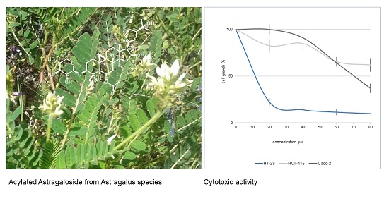
Spectroscopic Characterization and Cytotoxicity Assessment towards Human Colon Cancer Cell Lines of Acylated Cycloartane Glycosides from Astragalus boeticus L.

by
Vittoria Graziani
1,
Assunta Esposito
1,
Monica Scognamiglio
2,*,
Angela Chambery
1,
Rosita Russo
1,
Fortunato Ciardiello
3,
Teresa Troiani
3,
Nicoletta Potenza
1,
Antonio Fiorentino
1,4,* and
Brigida D’Abrosca
1,4
1
Dipartimento di Scienze e Tecnologie Ambientali Biologiche e Farmaceutiche (DiSTABiF), Università degli Studi della Campania “Luigi Vanvitelli”, via Vivaldi 43, I-81100 Caserta, Italy
2
Department of Biochemistry, Max Planck Institute for Chemical Ecology-Beutenberg Campus, Hans-Knöll-Straße, 8 D-07745 Jena, Germany
3
Dipartimento di Medicina di Precisione, Università degli Studi della Campania “Luigi Vanvitelli” - Via Pansini, 5, 80131 Napoli, Italy
4
Dipartimento di Biotecnologia Marina, Stazione Zoologica Anton Dohrn, Villa Comunale, 80121 Naples, Italy
*
Authors to whom correspondence should be addressed.
Molecules 2019, 24(9), 1725; https://doi.org/10.3390/molecules24091725
Submission received: 10 April 2019 / Revised: 29 April 2019 / Accepted: 1 May 2019 / Published: 3 May 2019
(This article belongs to the Special Issue Natural Products and Drug Discovery)

Abstract

:
In several European countries, especially in Sweden, the seeds of the species Astragalus boeticus L. were widely used as coffee substitutes during the 19th century. Nonetheless, data regarding the phytochemistry and the pharmacological properties of this species are currently extremely limited. Conversely, other species belonging to the Astragalus genus have already been extensively investigated, as they were used for millennia for treating various diseases, including cancer. The current work was addressed to characterize cycloartane glycosides from A. boeticus, and to evaluate their cytotoxicity towards human colorectal cancer (CRC) cell lines. The isolation of the metabolites was performed by using different chromatographic techniques, while their chemical structures were elucidated by nuclear magnetic resonance (NMR) (1D and 2D techniques) and electrospray-ionization quadrupole time-of-flight (ESI-QTOF) mass spectrometry. The cytotoxic assessment was performed in vitro by 3-(4,5-dimethylthiazol-2-yl)-2,5-diphenyltetrazolium bromide (MTT) assays in Caco-2, HT-29 and HCT-116 CRC cells. As a result, the targeted phytochemical study of A. boeticus enabled the isolation of three new cycloartane glycosides, 6-O-acetyl-3-O-(4-O-malonyl)-β-d-xylopyranosylcycloastragenol (1), 3-O-(4-O-malonyl)-β-d-xylopyranosylcycloastragenol (2), 6-O-acetyl-25-O-β-d-glucopyranosyl-3-O-β-d-xylopyranosylcycloastragenol (3) along with two known compounds, 6-O-acetyl-3-O-β-d-xylopyranosylcycloastragenol (4) and 3-O-β-d-xylopyranosylcycloastragenol (5). Importantly, this work demonstrated that the acetylated cycloartane glycosides 1 and 4 might preferentially inhibit cell growth in the CRC cell model resistant to epidermal growth factor receptor (EGFR) inhibitors.

Graphical Abstract

1. Introduction

Astragalus genus is the largest in the Fabaceae family and it is widely distributed throughout the cool, temperate, semiarid and arid regions of the world [1]. Astragalus boeticus L. is a Steno-Mediterranean species, which has represented an important cultivation in several countries of Europe, as its seeds have been widely used as coffee substitutes in times of poverty and coffee prohibition. In Sweden, during the 19th century, the monarchy introduced an extensive cultivation of the aforementioned species to produce the so-called Swedish coffee. After the beginning of the 20th century, its cultivation declined, and it was replaced by other substitutes [2]. In addition to this information, available literature data describing the phytochemistry and the bioactivities of A. boeticus are currently extremely limited.
On the contrary, a plethora of works regarding other species of the same genus exists. The Astragalus species were employed as forage for animals, albeit many species were found to be toxic, and responsible for causing locoism in cattle [3,4]. In both folk and modern medicine, several Astragalus spp. were considered medicinal plants of great importance, as these have been successfully used to cure a broad range of ailments [5]. In the Traditional Chinese Medicine “Astragali radix” (dried roots of Astragalus membranaceus Bunge and other Astragalus spp.) was a very well-known drug for its immune stimulant, hepato-protective, anti-diabetic, analgesic, expectorant and sedative properties [6].
Previous works investigated the chemical profile of Astragalus spp. in order to identify the active principles responsible for the bioactivity of the plant’s crude extracts. Results from these studies described imidazoline alkaloids, nitro toxins and selenium derivatives as toxic compounds, while polysaccharides, phenols and saponins as biologically active constituents [6]. Astragalus saponins include both oleanane and cycloartane-type glycosides, yet the former occur far less in nature, thus the Astragalus genus was especially employed as an ideal source to find cycloartane saponins [7].
These compounds were the most extensively studied secondary metabolites from Astragalus, as they exhibited a wide range of biological and pharmacological properties. Indeed, these molecules were found to exert immunomodulatory, anti-cancer, anti-fungal, hepato-, kidney-, neuro- and vascular-protective activities [7,8,9,10,11,12]. So far, the most well-characterized biological effects were those related to their immune stimulant properties, which made these compounds ideal vaccine adjuvant candidates [13]. Alongside the capacity to modulate key immunity pathways, recent evidence supported the effectiveness of Astragalus saponins as anti-tumor compounds and/or as adjuvants in combination with orthodox chemotherapeutic agents [14,15].
The anti-cancer activities of these compounds have been evaluated towards a wide range of human malignancies, and a large part of these works evidenced the effectiveness of Astragalus saponins against gastric and colorectal cancers [16]. Consistent with this, our group recently demonstrated the anti-proliferative effects of A. boeticus in human colorectal cancer (CRC) cells [17].
Colorectal cancer is one of the most frequently-diagnosed malignant diseases in Europe, and one of the leading causes of cancer-related deaths worldwide [18]. Even if the outcome of patients with metastatic colorectal cancer (mCRC) has clearly improved during the last years, the current therapies are still not entirely efficient. Nowadays, resistance to both chemotherapy and molecularly-targeted therapies represents a major problem for setting up effective treatment. The EGFR, which was found overexpressed in 60% to 80% of colorectal cancers, is a transmembrane tyrosine kinase receptor that, once activated, triggers two main signaling pathways. These include the RAS-RAF-MAPK axis, which is mainly involved in cell proliferation, and the PI3KPTEN-AKT pathway, which is especially involved in cell survival and motility [19]. Thus, EGFR inhibitors, such as Cetuximab and Panitumumab, have been developed to block specifically the abnormal activation of those pathways in wild-type KRAS CRC patients [20].
In this study, we aimed at providing a detailed chemical characterization of cycloartane glycosides from A. boeticus, and at assessing their anti-proliferative activity towards human colorectal cancer cells endowed with diverse mutation profiles and drug sensitiveness. The ultimate goal of this research is to contribute to the search for new effective agents against refractory CRCs. As a result, we isolated and characterized five cycloartane glycosides (15), identifying compound 4 as a strong inhibitor of proliferation in CRC cell models resistant to anti-EGFR therapies.

2. Results and Discussion

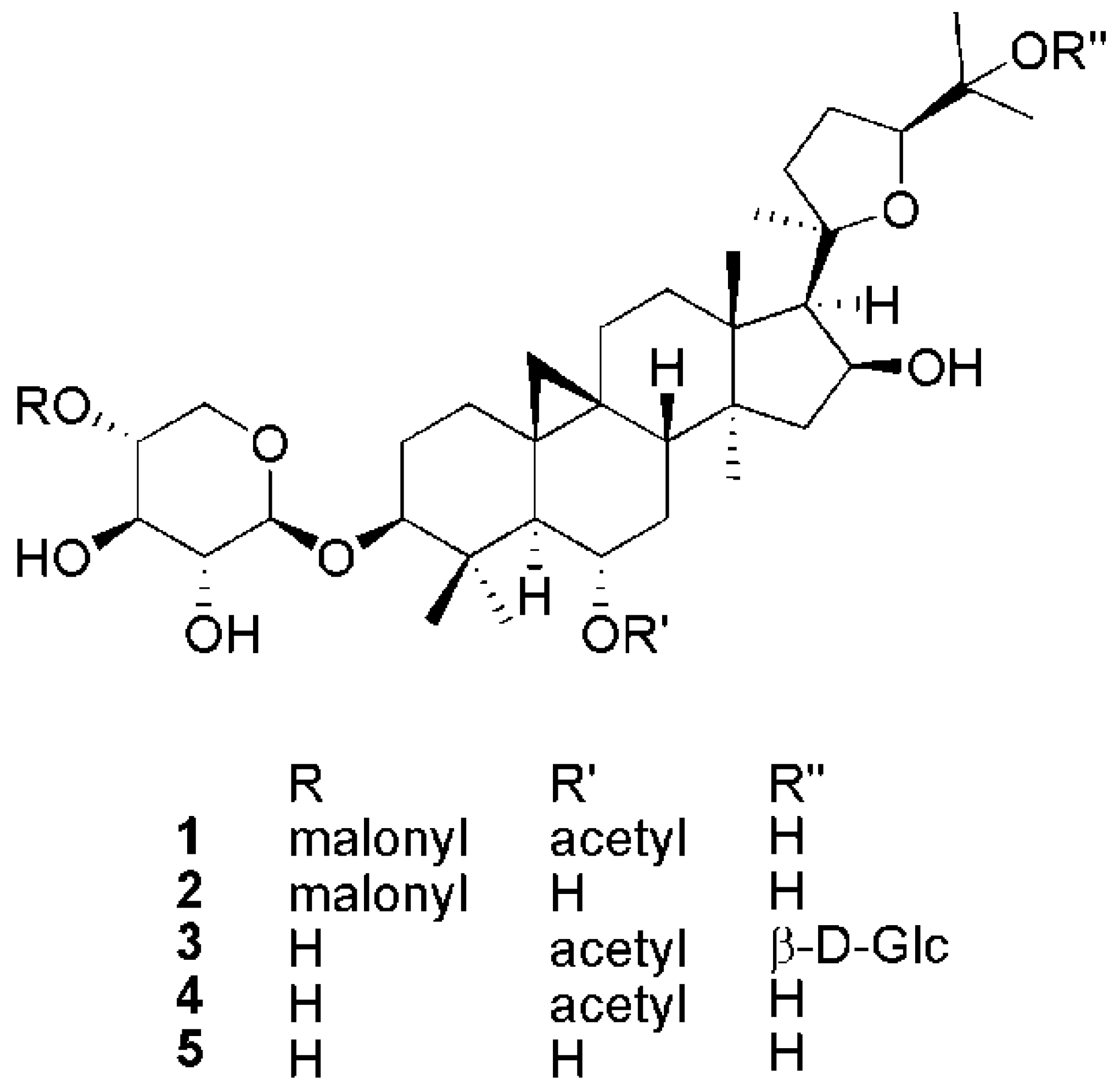
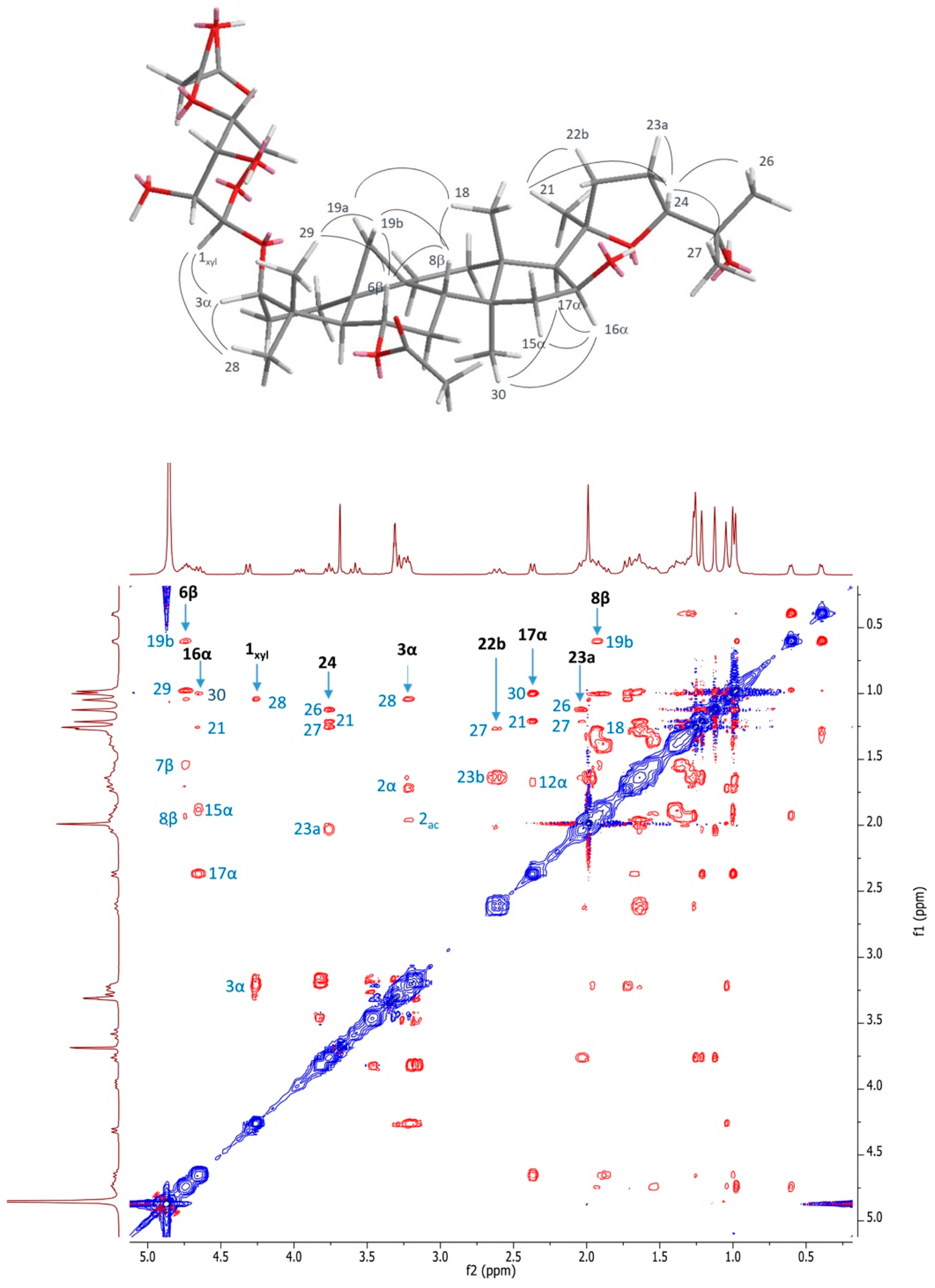
2.1. Structural Elucidation of Cycloartane Glycosides from Astragalus boeticus L.

A crude hydro-alcoholic extract of A. boeticus leaves was partitioned between EtOAc and H2O. The purification process, which was performed by using different chromatographic techniques, enabled the isolation of compounds 1, 2, 4 from the organic phase, while we also obtained 3 and 5 from the aqueous fraction (Figure 1). The structures of these metabolites were elucidated through a combination of NMR spectroscopy (1D and 2D techniques) and ESI-QTOF mass spectrometry.
Compound 1 showed a molecular formula C40H62O13 on the basis of the NMR data and ESI-QTOF mass spectrum. In fact, the 13C NMR displayed 40 signals, which were identified using the HSQC experiment as eight methyls (—CH3), eleven methylenes (=CH2), eleven methines (=CH−), and ten quaternary carbons. The ESI-QTOF spectrum displayed the sodiated adduct of the quasimolecular ion at m/z 773.49, and a strong peak at m/z 687.46, which indicated the easy loss of an 86 Da fragment. In the 1H NMR spectrum (Table 1), two methylene protons at δH 0.40 and δH 0.61 (δC 30.1), along with six singlet methyls at δH 0.98, 1.01, 1.05, 1.13, 1.22, 1.26, and 1.27 allowed compound 1 to be identified as a cycloartane triterpene. The doublet at δH 4.32 (δC 105.9), as well as other protons that resonated in the range between 3.18 and 4.85 ppm, suggested the presence of a sugar unit. Meanwhile, a methyl singlet at δH 1.99 supported the presence of an acetate group in the molecule. The above-mentioned methylene signals (δH 0.40 and δH 0.61) were assigned to H-19 protons; these, in the CIGAR-HMBC experiment (Figure 2), showed cross peaks with the C-9 (δC 21.8), C-10 (δC 29.6), C-1 (δC 32.8) C-11 (δC 26.8), C-5 (δC 51.2) and C-8 (δC 46.7). In the same experiment, the H-5 proton correlated with the C-1 methylene, the C-4 quaternary carbon at δC 42.8, and with two carbinols at δC 89.1 and δC 72.0. Thanks to the long-range heterocorrelations between the H-6 proton (δH 4.75) and the C-4, C-5, and C-8 carbons, it was feasible to assign the first carbinol to the C-3 methine, and then the second to the C-6 carbon. Moreover, the H-6 proton also had cross peaks with the carbonyl at δC 171.7, which in turn correlated with the methyl at δH 1.99. These data allowed the acetoxy (CH3-COO-) group to be located at position 6. On the other hand, the H-3 proton correlated with the anomeric carbon at δC 105.9, suggesting that the glycosylation site was located on the hydroxyl at the C-3 carbon. Furthermore, the C-4 carbon showed correlations with the methyls at δH 0.98 and δH 1.05, values that were consequently assigned to the H-29 and H-28 protons, respectively. In addition, the methyls at δH 1.27 (δC 21.4) and 1.01 (δC 20.3) were attributed to the H-18 and H-30 protons, respectively, on the basis of the long range heterocorrelations between the C-18 carbon with the proton at δH 2.38 (H-17), which in turn correlated with the H-21 methyl at δH 1.22 (δC 28.4).
In the COSY experiment, the H-17 proton had cross peaks with a proton geminal to oxygen at δH 4.65 (H-16), which homocorrelated with the methylene protons at δH 1.87 and δH 1.45 (H-15), suggesting the presence of another hydroxyl at the C-16 carbon. Consequently, the remaining methyls at δH 1.13 and δH 1.26 were attributed to the H-26 and H-27 protons, respectively. In the CIGAR-HMBC experiment, these latter protons displayed heterocorrelations with a quaternary carbinol carbon at δC 71.2 (C-25), and with a carbinol methine at δC 82.6, which was bound to a proton at δH 3.76. This latter signal evidenced heterocorrelations with the diasterotopic protons at δH 2.02 and δH 1.73, while homocorrelations were with the methylene protons at δH 2.62 and 1.87 (δC 35.5). All these data supported the presence of a tetrahydrofuran moiety formed by an oxygen bridge among the C-20 and C-24 carbons of the side chain. Besides the ester carbonyl of the acetate group, two additional carbonyls at δC 170.6 and 171.7 were also evident in the 13C NMR spectrum. In the CIGAR-HMBC experiment, both of them revealed cross peaks with the methylene at δH 3.69 (δH 51.8), while the carbon at δC 170.6 was further correlated with a proton at δC 4.72. These data were in agreement with the presence of a malonyl that was bound to the saccharide unit. In the HSQCTOXY experiment, the anomeric carbon at δC 105.9 showed cross peaks with the signals at δC 75.4, 75.2, 73.6 and δC 63.3. These carbons in the HSQC experiment correlated with the methines at δH 3.26, 3.57, 4.72 and to the methylene protons at δH 3.96 and 3.28, respectively. The H2BC experiment displayed the following correlations: Starting from the anomeric proton at δC 4.32 (H-1′)→75.4 (C-2′)→3.57 (H3′)→73.6 (C-4′)→3.96/3.28 (H-5′); starting from the anomeric carbon at δC 105.9 (C-1′)→3.26 (H-2′)→75.2 (C-3′)→4.72 (H-4′)→63.3 (C-5′). These data further supported the presence of a pentose that was bound to a malonyl group at the C-4′ carbon.
The stereostructure of the molecule was assigned on the basis of the nOe observed in the NOESY experiment (Figure 3). The sugar was identified as xylose by the GC-MS analysis of the acetylated alditol, which was obtained from the hydrolysis, reduction and acetylation of compound 1. The coupling constant value of the anomeric proton allowed a β configuration for the anomeric carbon to be determined. The absolute configuration of the sugar was assigned by GC-MS, after a reaction of hydrolyzed saponins with l-cisteine methyl ester and acetylation [21].
The nOe evidenced in the NOESY experiments (Figure 3) allowed the configuration at the C-6 and C-16 carbons to be assigned. Based on these data, compound 1 was unequivocally identified as 6-O-acetyl-3-O-(4-O-malonyl)-β-d-xylopyranosylcycloastragenol (Figures S1–S9).
Compound 2 showed the molecular formula C38H60O12 on the basis of the NMR data, and the presence of a quasimolecular peak at m/z 731.41 in the ESI-QTOF MS spectrum. The loss of an 86 Da fragment was demonstrated by the peak at m/z 645.39, indicating the presence of a malonyl moiety also in this molecule.
The 1H NMR spectrum showed the H-19 protons at δH 0.37 and 0.53, and seven singlet methyls at δH 0.96 (H-30), 0.98 (H-28), 1.15 (H-26), 1.24 (H-27), 1.26 (H-29 and H-21). The absence of the characteristic signal associated with the methyl group of the acetate moiety demonstrated that compound 2 was the deacetylated form of compound 1, and all the NMR data confirmed this hypothesis. In addition, the C3-sugar was characterized as xylose, and the malonyl was positioned to the C-4′ carbon of this saccharide moiety. Based on these data, compound 2 was unequivocally identified as 3-O-β-d (4-O-malonyl)xylopyranosylcycloastragenol.
Compound 3 showed the molecular formula C43H70O15, calculated on the basis of its spectro-scopic features. In the 1H-NMR spectrum, the signals of the aglycone, and those belonging to the C3-xylopyranose, were in good agreement with the previous metabolite. However, a further anomeric doublet at δC 4.51 suggested the presence of a second sugar. The heterocorrelation between this anomeric signal and the C-25 carbon at δH 79.9 allowed the second site of glycosylation to be located at position 25. The HSQCTOCSY experiment revealed the presence of an additional spin system due to this sugar, in which the anomeric proton (δC 4.51) correlated with the carbons at δc 99.6, 75.0, 78.2, 71.2, 77.5 and 62.7. These data were in agreement with the presence of a glucopyranose, a hypothesis that was confirmed by GC-MS analysis. Moreover, the coupling constant value indicated a β configuration for the anomeric carbon, while the D-series was established by GC-MS after the reaction of hydrolyzed saponins with L-cisteine methyl ester and acetylation. Based on these data, compound 3 was unequivocally identified as 6-O-acetyl-25-O-β-d-glucopyranosyl-3-O-β-d-xylo-pyranosylcycloastragenol.
Compound 4 showed a molecular formula C37H60O10, calculated on the basis of its spectroscopic data. Of interest, this compound was also obtained by the mild acidic hydrolysis of compound 1. In fact, when this compound was dissolved in water, after one day at room temperature, it was quantitatively converted in 4 by loss of the C4′-malonyl. All the NMR data confirmed this hypothesis, and allowed the identification of compound 4 as 6-O-acetyl-3-O-β-d-xylopyranosylcycloastragenol [22].
Compound 5 showed a molecular formula C35H58O9, calculated on the basis of its spectroscopic data. The 1H-NMR and 13C-NMR signals of the aglycone and those belonging to the C3-xylopyranose were superimposable with the previous metabolites. Conversely, the lack of the singlet peak at δH 1.99 proved the absence of the C6-acetyl. All of these data enabled the identification of compound 5 as 3-O-β-d-xylopyranosylcycloastragenol [23].

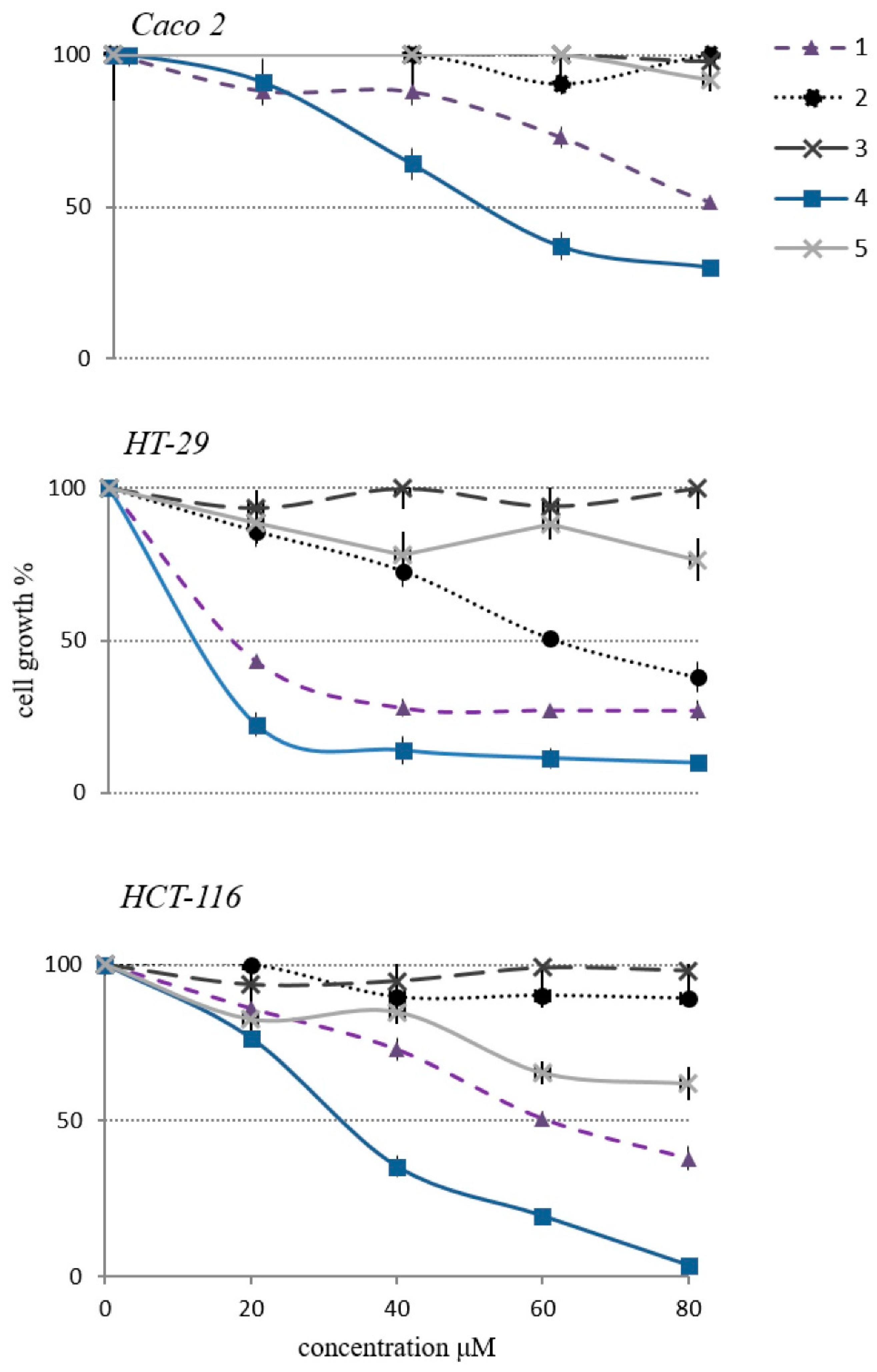
2.2. Cytotoxicity of Cycloartane Glycosides from Astragalus boeticus Against Human Colorectal Cancer Cells

The cytotoxic activity of the isolated compounds (15) was assessed on three human colorectal cancer cell lines (Caco-2, HT-29 and HCT-116), using MTT (3-[4,5-dimethylthiazol-2-yl]-2,5-diphenyltetrazolium bromide) tetrazolium salt colorimetric assay (Figure 4). Results from these experiments demonstrated that cell proliferation was reduced by the treatment with compounds 1 and 4 in a dose-dependent manner, while 2, 3 and 5 did not exert any significant effect. To our knowledge, no mechanistic studies regarding the proliferation reducing-effect of compounds 1 and 4 are available in literature. Yet, several previous works shed the light on the anticancer activity of other cycloartane glycosides (most of them isolated from A. membranaceus), which act by inducing apoptosis and modulating crucial cellular signaling pathways [24,25,26,27]. Importantly, a recent investigation pointed out that certain semisynthetic cycloastragenol derivatives impair inflammation-carcinogenesis by regulating the NF-KB signaling pathway [15].
Consistently, compound 4, which has already been purified from the leaf extract of A. membranaceus, was described as a potentially anti-inflammatory molecule, because it was found to exert an inhibitory activity on the nitric oxide production in macrophages [21]. Recently, our group identified compound 4 as a metabolite responsible for the cytotoxicity of the A. boeticus extract. Here, this species underwent a further phytochemical investigation to understand whether analogues of compound 4 exert cytotoxicity as well. As it was already anticipated above, we demonstrated that compounds 1 and 4 were the active cycloartane glycosides isolated from A. boeticus.
In an attempt to further validate our findings, we compared the cytotoxicity of 4 (active compound) and 3 (inactive compound) with astragaloside IV (AS-IV), a commercially-available cycloartane glycoside extensively recognized as one of the main active components of several Astragalus spp. Results from these experiments confirmed the notable cytotoxicity of compound 4, while no effect was found for AS-IV, at least in our experimental conditions (Figure S10).
From a structural point of view, these insights proved that the C3-xylopyranosyl, along with the C6-acetoxy group and the C25-free hydroxyl function, were essential structural requirements for the cytotoxic activity of these triterpenoids. As already discussed, when compound 1 was in water solutions, such as a cellular environment, it converted into compound 4 by losing the C4’ malonyl. Thereby, it was demonstrated that compound 4 was the real bioactive structure. Compounds 2, 3, 5 and AS-IV present the C3-xylopyranosyl, while none of them has either the C6-acetoxy group or the C25-free hydroxyl function. In accordance with previous investigations, acylation of the C-3 and C-6 secondary alcohols of diverse cycloartane derivatives resulted in a higher cytotoxic activity. This evidence led researchers to hypotheses that acylated cycloartane glycosides could lose the acyl substituents to modify proteins that play a key role in cellular signaling, whose deregulation is involved in carcinogenesis. Specifically, the acyl groups can be covalently attached to the amino acid side chains regulating the protein functions and impairing cancer progression [15].
In our study, a detailed analysis of the cytotoxic effect unveiled a differential response of Caco-2, HT-29 and HCT-116 to the treatment with compound 4. These insights could be interpreted by describing the human colon cancer cells employed in our experimental setting. Caco-2 cell line was the wild type for the KRAS, NRAS, BRAF, PIK3CA genes; thus representing an ideal cellular model to study metastatic CRCs sensitive to anti-EGFR agents. By contrast, HT-29 and HCT-116 harbored a BRAF and some KRAS/PIK3CA mutations, respectively—therefore identifying metastatic CRCs with intrinsic resistance to the anti-EGFR treatments [28,29].
Herein, the in vitro cytotoxic screening revealed as compound 4 was clearly more effective in treating HT-29 (3 µM) than HCT-116 (40 µM) and Caco-2 (50 µM) cell lines (Figure 4).
In clinics, the BRAF mutation status is a strong indicator of a very poor prognosis for mCRC patients; indeed, they showed a worse outcome for the therapies compared with those whose tumors were wild type [30,31,32]. Of interest, the inhibition of BRAF oncoprotein by the small-molecule drug PLX4032 (Vemurafenib) is highly effective in the treatment of melanoma [33], whilst metastatic CRC patients associated with the same mutation showed a very limited response to this drug [34]. In fact, Vemurafenib treatment induces EGFR feedback activation, causing continued proliferation in the presence of BRAF (V600E) inhibition. Unlike CRCs, melanomas express low levels of EGFR, and thus, they are not subject to this kind of feedback activation [35].

3. Materials and Methods

3.1. General Experimental Procedures

Analytical TLC was performed on Merck Kieselgel (Darmstadt, Germany) 60 F254 or RP-8 F254 plates with a 0.2 mm film thickness. Spots were visualized by UV light, or by spraying with H2SO4/AcOH/H2O (1:20:4), and then heating at 120 °C for 5 min. Preparative TLC was performed on Merck Kieselgel 60 F254 plates with a 0.5 or 1.0 mm film thickness.
Column chromatography (CC) was performed on Fluka (Seelze, Germany) Amberlite XAD-4 and XAD-7, on Pharmacia (Stockholm, Sweden) Sephadex LH-20, on Merck Kieselgel 60 (70–240 mesh), or on Baker (Deventer, Netherlands) RP-8. Nuclear magnetic resonance (NMR) spectra were recorded at 300 (1H) and 75 MHz (13C) on a Varian Mercury 300 FT-NMR spectrometer in CD3OD or pyridine-d5 solutions at 25 °C. Chemical shifts are reported in δ (ppm), and referenced to the residual solvent signal; J (coupling constant) are given in Hz. Standard pulse sequences and phase cycling from Varian library were used for 1H, 13C, DEPT, DQF-COSY, COSY, TOCSY, NOESY, HSQC, H2BC, HMBC and CIGAR–HMBC experiments. 1H NMR spectra were acquired over a spectral window from 14 to −2 ppm, with 1.0 s relaxation delay, 1.70 s acquisition time (AQ), and 90° pulse width = 13.8 μs. The initial matrix was zero-filled to 64 K. 13C NMR spectra were recorded in 1H broadband decoupling mode, over a spectral window from 235 to −15 ppm, 1.5 s relaxation delay, 90° pulse width = 9.50 μs, and AQ = 0.9 s. The number of scans for both 1H and 13C NMR experiments were chosen, depending on the concentration of the samples. With regards to the homonuclear and heteronuclear 2D-NMR experiments, the data points, number of scans and increments were adjusted according to the sample concentrations. Correlation spectroscopy (COSY) and double quantum filtered COSY (DQF-COSY) spectra were recorded with gradient-enhanced sequence at spectral widths of 3000 Hz in both f2 and f1 domains; the relaxation delays were of 1.0 s. The total correlation spectroscopy (TOCSY) experiments were performed in the phase-sensitive mode with a mixing time of 90 ms. The spectral width was 3000 Hz. Nuclear Overhauser effect spectroscopy (NOESY) experiments were performed in the phase-sensitive mode. The mixing time was 500 ms, and the spectral width was 3000 Hz. For all the homonuclear experiments, the initial matrix of 512 × 512 data points was zero-filled to give a final matrix of 1 k × 1 k points. Proton-detected heteronuclear correlations were also measured. Heteronuclear single-quantum coherence (HSQC) experiments (optimized for 1J(H,C) = 140 Hz) were performed in the phase sensitive mode with field gradient. The spectral width was 12,000 Hz in f1 (13C) and 3000 Hz in f2 (1H) and 1.0 s of relaxation delay; the matrix of 1 k × 1 k data points was zero-filled to give a final matrix of 2 k × 2 k points. Heteronuclear 2 bond correlation (H2BC) spectra were obtained with T = 30.0 ms, and a relaxation delay of 1.0 s; the third order low-pass filter was set for 130 < 1J(C,H) < 165 Hz. A heteronuclear multiple bond coherence (HMBC) experiment (optimized for 1J(H,C) = 8 Hz) was performed in the absolute value mode with field gradient; typically, 1H–13C gHMBC were acquired with spectral width of 18,000 Hz in f1 (13C) and 3000 Hz in f2 (1H) and 1.0 s of relaxation delay; the matrix of 1 k × 1 k data points was zero-filled to give a final matrix of 4 k × 4 k points. Constant time inverse-detected gradient accordion rescaled heteronuclear multiple bond correlation spectroscopy (CIGAR–HMBC) spectra (8 > nJ(H,C) > 5) were acquired with the same spectral width used for HMBC. Heteronuclear single quantum coherence-total correlation spectroscopy (HSQC-TOCSY) experiments were optimized for nJ(H,C) = 8 Hz, with a mixing time of 90 ms. For accurate mass measurements, the purified compounds were analyzed by an electrospray hybrid quadrupole orthogonal acceleration time-of-flight mass spectrometer (Q-TOF), fitted with a Z-spray electrospray ion source (Waters S.p.A.). All analyses were carried out in positive ion mode. The capillary source voltage and the cone voltage were set at 3500 V and 35 V, respectively. The source temperature was kept at 80 °C, and nitrogen was used as a drying gas (flow rate about 50 l/h). The time-of-flight analyzer of the mass spectrometer was externally calibrated, with GFP from m/z 50–1600. Accurate mass data were collected by directly infusing samples (1.5 pmol/μL in CH3CN/H2O, 1:1) into the system at a flow rate of 15 μL/min. The acquisition and processing of data were performed with the MassLynx 4.1 software (Waters S.p.A., Manchester, UK). GC-MS analyses were carried out using an HP 6890 GC instrument (Zebron ZB-5MS column, He flow 1.0 mL/min), coupled with a 5973 N mass spectrometer, equipped with an electron ionization source (EIMS), and operating with an electron energy of 70 eV. Full-scan mass spectra were collected between 0 and 600 amu at 2 scan/s. The MS was operated in the electron impact (EI) ionization mode, with an electron energy of 70 eV. The ion source and quadrupole temperatures were maintained at 230 and 150 °C, respectively. For the analyses of the acetylated alditols, the column head pressure was set at 7.41 p.s.i.
AS-IV was purchased from Shanghai Tauto Biotech Co., LTD. (Shanghai, China).

3.2. GC–MS Analysis of the Sugar Moieties

The GC-MS analysis of the sugar moieties has been previously described by Scognamiglio et al. [31]. Briefly, each metabolite (0.5 mg) was subjected to an acid hydrolysis with 2 N TFA (150 µL) at 120 °C for 1 h, obtaining the sugar moiety. This was dried under N2 flow, and reduced by adding MeOH (150 µL) and NaBH4 (1.0 mg). The solution was incubated at room temperature for 1 h and then dried under N2 flow after treatment with glacial AcOH and MeOH. The obtained alditol was acetylated by using anhydrous pyridine (200 µL) and Ac2O (200 µL). This mixture was incubated for 20 min at 120 °C. Then, 500 µL of H2O was added, and the product was extracted with CH2Cl2 (500 µL) following centrifugation at 3500 rpm for 5 min. The organic phase was dried under N2 flow, dissolved in CH2Cl2 (500 µL) and analyzed by GC-MS. Temperature conditions were as follows: Injector port at 250 °C; the initial oven temperature was 160 °C for 50 s, then linearly increased to 200 °C at 10 °C/min. A further linear increase at 2.5 C/min was performed to 300 °C, and held for 40 min. Sample solutions were injected using the split mode.

3.3. Determination of Absolute Configuration of Monosaccharides of Compound 1 and 5

Compound 1 and 5 (2 mg each) were hydrolyzed with 2 N TFA (250 µL) at 120 °C for 1 h. The reaction mixture was then dried under N2 flow and dissolved in dry pyridine (100 µL). 100 µL of pyridine solution of l-cysteine methyl ester hydrochloride (0.06 mol/L) were added to pyridine solutions of the hydrolyzed compounds 1 and 5 and pure d-xylose, and l-xylose (0.04 mol/L). These mixtures were warmed at 60 °C for 1 h, afterwards, acetic anhydride (150 µL) was added at 120 °C for 20 min. The products were dried under N2, dissolved in 500 µL of H2O, and extracted with CH2Cl2 (500 µL) following centrifugation at 3500 rpm for 5 min. The organic phase was dried under N2 flow, dissolved in CH2Cl2 (500 µL), and analyzed by GC-MS. Temperature conditions were as follows: Injector port at 250 °C; initial oven temperature 45 °C, then increased linearly to 300 °C at 20 °C/min, and then held for 25 min. Sample solutions were injected using the split mode. The retention times were: d-xylose 13.89 min, l-xylose 14.17 min.

3.4. Plant Material

Leaf samples of A. boeticus were collected at vegetative state, in April 2014 in “Castel Volturno Nature Reserve” (40°57.587′N, 14°00.105′E; southern Italy). Samples were harvested, frozen in liquid nitrogen and lyophilized. A Voucher specimen CE000016 has been deposited at the Herbarium of the Dipartimento di Scienze e Tecnologie Ambientali Biologiche e Farmaceutiche of Università degli Studi della Campania “Luigi Vanvitelli”.

3.5. Extraction and Isolation of Compound 15

Dried leaves (24.0 g) of A. boeticus were powdered, and underwent three cycles of an ultrasound-assisted extraction with an MeOH/H2O (1:1) solution (720 mL) [36], finally obtaining a crude extract (7.1 g). This was dissolved in H2O and separated by liquid–liquid extraction, by using EtOAc as our extracting solvent. As a result, an organic and a water fraction were obtained. The former (1g) was chromatographed by SiO2 CC, and eluted using a solution with an increasing degree of polarity (CHCl3, Me2CO/CHCl3, MeOH/CHCl3). Thus, 21 fractions have been collected. Of these, number 13 was chromatographed by C18 CC, and eluted with H2O/MeOH (3:2) to give compound 1 (18.6 mg) and 2 (7.1 mg). Meanwhile, number 12 was purified by Flash-CC eluting with MeOH/ CHCl3 (3:100) to obtain compound 4 (28 mg). On the other hand, the water fraction was chromatographed by XAD-4 (20–50 mesh; Fluka) and XAD-7 (20–50 mesh; Fluka) CC, obtaining an alcoholic eluate (900 mg) that was in turn purified by Sephadex LH-20. Subsequently, 17 fractions were given. Of these, fraction 13 was purified through RP-18 CC by using H2O/MeOH (4:1) as the eluting system, consequently obtaining 30 fractions. One of these was chromatographed by TLC (0,5 mm), and eluted with the organic phase of CHCl3/MeOH/H2O (13:7:2) to obtain compound 3 (11.8 mg). Finally, fraction 11 was chromatographed by TLC (0.5 mm) and eluted with the organic phase of CHCl3/MeOH/H2O (13:7:2) to give compound 5 (4.6 mg).
Compound 1: 6-O-acetyl-3-O-(4-O-malonyl)-β-d-xylopyranosylcycloastragenol. [α]D25 = +22.8 (c = 14.97 × 10⁻³, MeOH). 1H NMR (CD3OD) and 13C NMR (CD3OD) see Table 1; ESI/Q-TOF: m/z 773.49 [M + Na]+ (calcd.773.41 Da for C40H62O13Na);
Compound 2: 3-O-(4-O-malonyl)-β-d- xylopyranosylcycloastragenol. [α]D25 = +9.70 (c = 2.06 × 10⁻³, MeOH). 1H NMR (CD3OD) and 13C NMR (CD3OD) see Table 1; ESI/Q-TOF: m/z 731.41 [M+Na]+ (calcd. 731.40 Da for C38H60O12Na);
Compound 3: 6-O-acetyl-25-O-β-d-glucopyranosyl-3-O-β-d-xylopyranosylcycloastragenol. [α]D25 = +13.7 (c= 6.06 × 10⁻³, MeOH). 1H NMR (CD3OD) and 13C NMR (CD3OD) see Table 1; ESI/Q-TOF: m/z 849.46 [M + Na]+ (calcd.849.43 Da for C43H70O15Na);
Compound 4: 6-acetyl-3-O-(4-O-malonyl)-β-d-xylopyranosylcycloastragenol. [α]D25 = +7.65 (c = 5.1 × 10⁻³, MeOH/H2O, 2:1) [22];
Compound 5: 3-O-β-D-xylopyranosylcycloastragenol. [α]D25 = −340.1 (c = 3.74 × 10⁻³) MeOH/H2O, 1:1) [23].

3.6. Cell Lines

The human HCT-116, HT-29, Caco-2 colorectal cancer cell lines were obtained from the American Type Culture Collection (ATCC) (Manassas, VA). HCT-116, HT-29 cancer cells were cultured in RPMI 1640 medium (Lonza, Cologne, Germany) supplemented with 10% fetal bovine serum, 2 mM l-glutamine, 50 U/mL penicillin and 100 µg/mL streptomycin (Lonza, Cologne, Germany). The Caco-2 cell line was cultured in DMEM medium (Lonza, Cologne, Germany), supplemented with 10% fetal bovine serum, 2 mM l-glutamine, 1% non-essential amino acid, 50 U/mL penicillin and 100 µg/mL streptomycin (Lonza, Cologne, Germany).

3.7. Proliferation Assay

The cell proliferation assay was performed with a 3-(4,5-dimethylthiazol-2-yl)-2,5-diphenyl-tetrazolium bromide (MTT) assay. Briefly, cells in logarithmic growth phase were plated in 96-well plates and incubated for 24 h before exposure to increasing doses of DMSO-diluted compounds (20, 40, 60, and 80 µM). For compounds 1 and 4 that exerted a strong cytotoxic effect in the HT-29 cell line, a lower range of doses (2, 4, 6, and 10 µM) was also investigated. 48 h after treatment, 50 µL of 1 mg/mL (MTT) were mixed with 200 μL of medium and added to the well. 1 h after incubation at 37 °C, the medium was removed, and the purple formazan crystals produced in the viable cells were solubilized in 100 μL of dimethyl sulfoxide, and quantitated by measurement of absorbance at 570 nm with a plate reader. Results were reported as mean ± s.d. of % of cell growth, with respect to the control from six replicate analyses. The control was represented by a 0.08% DMSO treatment, which corresponded to the higher amount of DMSO used for the tests.

4. Conclusions

A plethora of previous investigation regarding the phytochemistry and the bioactive components of diverse species belonging to the Astragalus genus are available in literature. However, to our knowledge, there are no data regarding the phytochemical constituents of A. boeticus. In this study, we focused on the cycloartane derivatives present in A. boeticus, because of its putative cytotoxic activity.
Specifically, the targeted phytochemical study of A. boeticus led to the isolation of five cycloartane-type glycosides (15); of these, 1, 2 and 3 were isolated and characterized for the first time. The cytotoxic activity of compounds 15 was evaluated in vitro, disclosing a strong proliferation-reducing effect of compound 4 in the HT-29 human colon cancer cell line, which was employed as a preclinical model to study refractory mCRCs that are resistant to anti-EGFR therapies, as well as other chemotherapeutic drugs currently used in the clinical setting. Our results therefore provide a small molecule scaffold that might be potentially important for the development of new agents effective against refractory mCRCs.
Indeed, these insights pave the way to further investigations aimed to figure out whether compound 4 acts on CRC cell models resistant to EGFR inhibitors with high selectivity, and if so, to elucidate the mechanism by which the anti-proliferative activity occurs.
On the other hand, as several limitations are intrinsically associated with our in vitro experimental system, future experiments shall also be addressed to evaluate the pharmacokinetic properties, and the bioavailability of the active molecule in animal models.

Supplementary Materials

The following are available online at https://www.mdpi.com/1420-3049/24/9/1725/s1, Figure S1: 1H NMR spectrum of compound 1. Figure S2: 13C NMR spectrum of compound 1. Figure S3: COSY spectrum of compound 1. Figure S4: HSQC spectrum of compound 1. Figure S5: CIGAR-HMBC spectrum of compound 1. Figure S6: H2BC spectrum of compound 1. Figure S7: HSQCTOCSY spectrum of compound 1. Figure S8: ESI QTOF spectrum of compound 1. Figure S9: ESI TOF MS/MS spectrum of compound 1. Figure S10: cytotoxicity of AS-IV in comparison with compound 3 and 4.

Author Contributions

V.G. and M.S. designed the experiments, discussed and interpreted the results. A.E. selected and identified the plant species. V.G. and M.S. carried out the experiments. A.C. and R.R. carried out the M.S. analyses. V.G. and M.S. wrote the manuscript. A.E., B.D.A. and F.C. analyzed the experimental data. A.E., B.D.A. interpreted the results. T.T., N.P., A.F. designed the experiments, discussed and interpreted the results and reviewed the manuscript. All authors revised the manuscript.

Funding

This work was funded by Regione Campania, “Technology Platform per la Lotta alle Patologie Oncologiche” Grant: iCURE (CUP B21C17000030007–SURF17061BP000000008).

Conflicts of Interest

The authors declare no conflict of interest.

References

  1. Verotta, L.; El-Sebakhy, N. Cycloartane and oleanane saponins from Astragalus sp. Studies in Natural Products Chemistry; Elsevier: Amsterdam, The Netherlands, 2001; pp. 179–234. [Google Scholar]
  2. Prohens, J.; Andújar, I.; Vilanova, S.; Plazas, M.; Gramazio, P.; Prohens, R.; Herraiz, F.J.; De Ron, A.M. Swedish coffee (Astragalus boeticus L.), a neglected coffee substitute with a past and a potential future. Genet. Resour. Crop Evol. 2013, 61, 287–297. [Google Scholar] [CrossRef]
  3. Williams, M.C.; Davis, A.M. Nitro Compounds in Introduced Astragalus Species. J. Range Manage. 1982, 35. [Google Scholar] [CrossRef]
  4. Cook, D.; Ralphs, M.H.; Welch, K.D.; Stegelmeier, B.L. Locoweed Poisoning in Livestock. Rangelands 2009, 31, 16–21. [Google Scholar] [CrossRef]
  5. Ionkova, I.; Shkondrov, A.; Krasteva, I.; Ionkov, T. Recent progress in phytochemistry, pharmacology and biotechnology of Astragalus saponins. Phytochem. Rev. 2014, 13, 343–374. [Google Scholar] [CrossRef]
  6. Rios, J.L.; Waterman, P.G. A review of the pharmacology and toxicology of Astragalus. Phytother. Res. 1997, 11, 411–418. [Google Scholar] [CrossRef]
  7. Gülcemal, D.; Aslanipour, B.; Bedir, E. Secondary Metabolites from Turkish Astragalus Species. In Plant and Human Health, Volume 2: Phytochemistry and Molecular Aspects; Ozturk, M., Hakeem, K.R., Eds.; Springer International Publishing: Berlin, Germany, 2019; pp. 43–97. [Google Scholar]
  8. Wang, Y.; Auyeung, K.K.; Zhang, X.; Ko, J.K. Astragalus saponins modulates colon cancer development by regulating calpain-mediated glucose-regulated protein expression. BMC Complement. Altern. Med. 2014, 14, 401. [Google Scholar] [CrossRef]
  9. Yang, L.P.; Shen, J.G.; Xu, W.C.; Li, J.; Jiang, J.Q. Secondary metabolites of the genus Astragalus: Structure and biological-activity update. Chem. Biodivers. 2013, 10, 1004–1054. [Google Scholar] [CrossRef]
  10. Shkondrov, A.; Krasteva, I.; Bucar, F.; Kunert, O.; Kondeva-Burdina, M.; Ionkova, I. A new tetracyclic saponin from Astragalus glycyphyllos L. and its neuroprotective and hMAO-B inhibiting activity. Nat. Prod. Res. 2018, 23, 1–7. [Google Scholar] [CrossRef]
  11. Pistelli, L.; Bertoli, A.; Lepori, E.; Morelli, I.; Panizzi, L. Antimicrobial and antifungal activity of crude extracts and isolated saponins from Astragalus verrucosus. Fitoterapia 2002, 73, 336–339. [Google Scholar] [CrossRef]
  12. Yin, X.; Zhang, Y.; Yu, J.; Zhang, P.; Shen, J.; Qiu, J.; Wu, H.; Zhu, X. The antioxidative effects of astragalus saponin I protect against development of early diabetic nephropathy. J. Pharmacol. Sci. 2006, 101, 166–173. [Google Scholar] [CrossRef]
  13. Aslanipour, B.; Gulcemal, D.; Nalbantsoy, A.; Yusufoglu, H.; Bedir, E. Secondary metabolites from Astragalus karjaginii BORISS and the evaluation of their effects on cytokine release and hemolysis. Fitoterapia 2017, 122, 26–33. [Google Scholar] [CrossRef]
  14. Auyeung, K.K.; Law, P.C.; Ko, J.K. Combined therapeutic effects of vinblastine and Astragalus saponins in human colon cancer cells and tumor xenograft via inhibition of tumor growth and proangiogenic factors. Nutr. Cancer 2014, 66, 662–674. [Google Scholar] [CrossRef] [PubMed]
  15. Debelec-Butuner, B.; Ozturk, M.B.; Tag, O.; Akgun, I.H.; Yetik-Anacak, G.; Bedir, E.; Korkmaz, K.S. Cycloartane-type sapogenol derivatives inhibit NFkappaB activation as chemopreventive strategy for inflammation-induced prostate carcinogenesis. Steroids 2018, 135, 9–20. [Google Scholar] [CrossRef] [PubMed]
  16. Auyeung, K.K.; Han, Q.B.; Ko, J.K. Astragalus membranaceus: A Review of its Protection Against Inflammation and Gastrointestinal Cancers. Am. J. Chin. Med. 2016, 44, 1–22. [Google Scholar] [CrossRef]
  17. Graziani, V.; Scognamiglio, M.; Belli, V.; Esposito, A.; D’Abrosca, B.; Chambery, A.; Russo, R.; Panella, M.; Russo, A.; Ciardiello, F.; et al. Metabolomic approach for a rapid identification of natural products with cytotoxic activity against human colorectal cancer cells. Sci. Rep. 2018, 8, 5309. [Google Scholar] [CrossRef] [PubMed]
  18. Malvezzi, M.; Carioli, G.; Bertuccio, P.; Rosso, T.; Boffetta, P.; Levi, F.; La Vecchia, C.; Negri, E. European cancer mortality predictions for the year 2016 with focus on leukaemias. Ann. Oncol. 2016, 27, 725–731. [Google Scholar] [CrossRef] [PubMed]
  19. Baselga, J. The EGFR as a target for anticancer therapy - focus on cetuximab. Eur. J. Cancer 2001, 37, S16–S22. [Google Scholar] [CrossRef]
  20. Ciardiello, F.; Tortora, G. EGFR antagonists in cancer treatment. N. Engl. J. Med. 2008, 358, 1160–1174. [Google Scholar] [CrossRef]
  21. Scognamiglio, M.; D’Abrosca, B.; Fiumano, V.; Chambery, A.; Severino, V.; Tsafantakis, N.; Pacifico, S.; Esposito, A.; Fiorentino, A. Oleanane saponins from Bellis sylvestris Cyr. and evaluation of their phytotoxicity on Aegilops geniculata Roth. Phytochemistry 2012, 84, 125–134. [Google Scholar] [CrossRef]
  22. Wang, Z.B.; Zhai, Y.D.; Ma, Z.P.; Yang, C.J.; Pan, R.; Yu, J.L.; Wang, Q.H.; Yang, B.Y.; Kuang, H.X. Triterpenoids and Flavonoids from the Leaves of Astragalus membranaceus and Their Inhibitory Effects on Nitric Oxide Production. Chem. Biodivers. 2015, 12, 1575–1584. [Google Scholar] [CrossRef]
  23. Kitagawa, I.; Wang, H.; Saito, M.; Takagi, A.; Yoshikawa, M. Saponin and sapogenol. XXXV. Chemical constituents of astragali radix, the root of Astragalus membranaceus Bunge. (2). Astragalosides I, II and IV, acetylastragaloside I and isoastragalosides I and II. Chem. Pharm. Bull. 1983, 31, 698–708. [Google Scholar] [CrossRef]
  24. Tin, M.M.; Cho, C.H.; Chan, K.; James, A.E.; Ko, J.K. Astragalus saponins induce growth inhibition and apoptosis in human colon cancer cells and tumor xenograft. Carcinogenesis 2007, 28, 1347–1355. [Google Scholar] [CrossRef] [PubMed]
  25. Auyeung, K.K.; Cho, C.H.; Ko, J.K. A novel anticancer effect of Astragalus saponins: Transcriptional activation of NSAID-activated gene. Int. J. Cancer 2009, 125, 1082–1091. [Google Scholar] [CrossRef] [PubMed]
  26. Ionkova, I.; Momekov, G.; Proksch, P. Effects of cycloartane saponins from hairy roots of Astragalus membranaceus Bge., on human tumor cell targets. Fitoterapia 2010, 81, 447–451. [Google Scholar] [CrossRef]
  27. Auyeung, K.K.; Woo, P.K.; Law, P.C.; Ko, J.K. Astragalus saponins modulate cell invasiveness and angiogenesis in human gastric adenocarcinoma cells. J. Ethnopharmacol. 2012, 141, 635–641. [Google Scholar] [CrossRef] [PubMed]
  28. Saif, M.W. Colorectal cancer in review: The role of the EGFR pathway. Expert Opin Investig. Drugs 2010, 19, 357–369. [Google Scholar] [CrossRef] [PubMed]
  29. Veluchamy, J.P.; Spanholtz, J.; Tordoir, M.; Thijssen, V.L.; Heideman, D.A.; Verheul, H.M.; de Gruijl, T.D.; van der Vliet, H.J. Combination of NK Cells and Cetuximab to Enhance Anti-Tumor Responses in RAS Mutant Metastatic Colorectal Cancer. PLoS ONE 2016, 11, e0157830. [Google Scholar] [CrossRef] [PubMed]
  30. Richman, S.D.; Seymour, M.T.; Chambers, P.; Elliott, F.; Daly, C.L.; Meade, A.M.; Taylor, G.; Barrett, J.H.; Quirke, P. KRAS and BRAF Mutations in Advanced Colorectal Cancer Are Associated With Poor Prognosis but Do Not Preclude Benefit From Oxaliplatin or Irinotecan: Results From the MRC FOCUS Trial. J. Clin. Oncol. 2009, 27, 5931–5937. [Google Scholar] [CrossRef]
  31. Roth, A.D.; Tejpar, S.; Delorenzi, M.; Yan, P.; Fiocca, R.; Klingbiel, D.; Dietrich, D.; Biesmans, B.; Bodoky, G.; Barone, C.; et al. Prognostic role of KRAS and BRAF in stage II and III resected colon cancer: Results of the translational study on the PETACC-3, EORTC 40993, SAKK 60-00 trial. J. Clin. Oncol. 2010, 28, 466–474. [Google Scholar] [CrossRef]
  32. Van Cutsem, E.; Kohne, C.H.; Lang, I.; Folprecht, G.; Nowacki, M.P.; Cascinu, S.; Shchepotin, I.; Maurel, J.; Cunningham, D.; Tejpar, S.; et al. Cetuximab plus irinotecan, fluorouracil, and leucovorin as first-line treatment for metastatic colorectal cancer: Updated analysis of overall survival according to tumor KRAS and BRAF mutation status. J. Clin. Oncol. 2011, 29, 2011–2019. [Google Scholar] [CrossRef]
  33. Chapman, P.B.; Hauschild, A.; Robert, C.; Haanen, J.B.; Ascierto, P.; Larkin, J.; Dummer, R.; Garbe, C.; Testori, A.; Maio, M.; et al. Improved survival with vemurafenib in melanoma with BRAF V600E mutation. N. Engl. J. Med. 2011, 364, 2507–2516. [Google Scholar] [CrossRef] [PubMed]
  34. Kopetz, S.; Desai, J.; Chan, E.; Hecht, J.R.; O’Dwyer, P.J.; Lee, R.J.; Nolop, K.B.; Saltz, L. PLX4032 in metastatic colorectal cancer patients with mutant BRAF tumors. J. Clin. Oncol. 2010, 28, 3534. [Google Scholar] [CrossRef]
  35. Prahallad, A.; Sun, C.; Huang, S.D.; Di Nicolantonio, F.; Salazar, R.; Zecchin, D.; Beijersbergen, R.L.; Bardelli, A.; Bernards, R. Unresponsiveness of colon cancer to BRAF(V600E) inhibition through feedback activation of EGFR. Nature 2012, 483, 100–146. [Google Scholar] [CrossRef]
  36. Scognamiglio, M.; Fiumano, V.; D’Abrosca, B.; Esposito, A.; Choi, Y.H.; Verpoorte, R.; Fiorentino, A. Chemical interactions between plants in Mediterranean vegetation: The influence of selected plant extracts on Aegilops geniculata metabolome. Phytochemistry 2014, 106, 69–85. [Google Scholar] [CrossRef] [PubMed]
Sample Availability: Samples of the compounds are not available from the authors.
Figure 1. Structures of compounds 15.
Molecules 24 01725 g001
Figure 2. Selected H–H and H–C long range correlations of compound 1 evidenced in COSY and HMBC, respectively.
Molecules 24 01725 g002
Figure 3. Key nOe correlation of compound 1 (top); NOESY experiment of compound 1 in CD3OD (bottom).
Molecules 24 01725 g003
Figure 4. Cytotoxic activity of compounds 15 towards Caco-2, HT-29 and HCT-116 human colon cancer cell lines.
Molecules 24 01725 g004
Table 1. 1D and 2D nuclear magnetic resonance (NMR) data of compound 13 in CD3OD.
123
PositionδCTypeδH (J in Hz)HMBCaδC,TypeδH (J in Hz)HMBC aδC,TypeδH (J in Hz)HMBC a
132.8CH21.85 s
1.67 s
2, 10
2, 10
32.3CH21.84 s
1.65 s
2, 10
2, 10
32.8CH21.29 s
230.3CH21.98 ov
1.64 ov
3, 4
3, 4
30.0CH21.88 ov
1.69 ov
3, 4
3, 4
30.2CH21.99 ov3, 4
389.1CH3.21 d (J = 1.8)28, 29, 1xyl89.1CH3.29 ov28, 29, 1xyl89.1CH3.21 ov1xyl
442.8C 42.0C 42.8C
551.2CH1.71 s1, 4, 7, 10, 28, 2953.9CH1.38 s1, 4, 7, 10, 28, 2951.3CH1.72 s1, 4, 7, 10, 28, 29
672.0CH4.75 ov4, 5, 7, 8, 1Ac68.5CH3.48 ov4, 5, 7, 872.1CH4.75 ov4, 5, 1ac
734.0CH21.56 ov5, 633.4CH21.46 ov5, 634.1CH21.575, 6, 8, 9, 14
846.7CH1.95 s6, 9, 10, 13, 14, 19, 3048.0CH1.46 s6, 9, 10, 13, 14, 19, 3046.1CH1.94 s6, 10, 13, 14, 15, 19
921.8C 21.4C 21.6C
1029.6C 29.2C 29.2C
1126.8CH21.97 m 8, 12, 1926.4CH21.97 s8, 12, 1926.4CH21.97 s 12
1234.1CH21.64 ov
1.56 ov
14, 18
14, 18
33.4CH21.54 ov
1.37 ov
14, 18
14, 18
34.1CH21.64 ov
1.56 ov
1347.0C 44.7C 46.9C
1446.4C 47.0C 46.6C
1546.2CH21.87 d (J = 8.0)
1.39 d (J = 8.0)
8, 13, 17, 30
8, 13, 17, 30
46.4CH21.98 d (J = 4.4)
1.32 d (J = 6.8)
8, 13, 17, 30
8, 13, 17, 30
46.0CH21.86 d (J = 6.0)
1.42 d (J = 6.4)
8, 13, 14, 17, 30
13, 14, 30
1674.5CH4.65 m13, 14, 1573.9CH4.64 m13, 14, 1574.3CH4.65 m13, 14, 15
1758.9CH2.37 d (J = 7.8)13, 14, 16, 20, 21, 2258.2CH2.37 d (J = 7.7)13, 14, 16, 20, 21, 2258.7CH2.38 d (J=8.0)13, 14, 16, 20, 21, 22
1821.4CH31.27 s12, 15, 1721.4CH31.23 s12, 15, 1721.7CH31.27 s12, 15, 17
1930.1CH20.61 s
0.40 s
1, 5, 8, 9, 10
1, 5, 8, 9, 10, 11
31.4CH20.53 s
0.37 s
1, 5, 8, 9, 10
1, 5, 8, 9, 10, 11
30.2CH20.61 s
0.40 s
1, 5, 8, 9, 10,11
1, 5, 8, 9, 10, 11
2088.3C- 88.2C- 88.3C-
2128.4CH31.22 s17, 20, 2228.1CH31.25 s17, 20, 2228.0CH31.24 s
2235.5CH22.62 ov
1.87 ov
17, 20, 21
17, 20, 21
34.9CH21.69 ov17, 20, 21
17, 20, 21
35.6CH22.54 ov
1.84
17
17
2326.8CH22.02 ov
1.73
20, 24, 25
17, 24, 25
26.4CH22.02 ov20, 24, 25
17, 24, 25
26.8CH22.16 ov
1.72
24, 25
24, 25
2482.6CH3.76 m2581.4CH3.80 m2583.0CH3.82 m1glc
2571.2C- 71.2C- 79.9C-
2626.6CH31.13 s17, 24, 2526.5CH31.15 s17, 24, 2523.2CH31.22 s20, 23, 24, 25
2727.6CH31.26 s24, 25, 2627.1CH31.24 s24, 25, 2625.3CH31.38 s23, 24, 26
2827.2CH31.05 s3, 4, 5, 2916.1CH30.98 s3, 4, 5, 2916.5CH30.98 s3, 4, 5, 28
2916.6CH30.98 s3, 4, 5, 2827.1CH31.25 s3, 4, 5, 2827.3CH31.04 s3, 4, 5, 29
3020.3CH31.01 s9, 14, 1520.9CH30.96 s9, 14, 1520.2CH30.99 s14, 15
1xyl105.9CH4.32 d (J = 7.4)3, 5xyl105.9CH4.43 d (J = 7.4)3, 5xyl107.4CH4.26 d (J = 7.0)3
2xyl75.4CH3.26 ov4xyl75.4CH3.26 ov4xyl75.5CH3.19 ov
3xyl75.2CH3.57 ov4xyl, 5xyl75.2CH3.57 ov4xyl, 5xyl78.0CH3.29 ov
4xyl73.6CH4.72 m2xyl, 3xyl, 5xyl, 2mal73.6CH4.72 m2xyl, 3xyl, 5xyl, 2mal71.2CH3.46 m
5xyl63.3CH23.28 ov
3.96 ov
1xyl, 3xyl, 4xyl
1xyl, 3xyl, 4xyl
63.3CH23.28 ov
3.96 ov
1xyl, 3xyl, 4xyl
1xyl, 3xyl, 4xyl
66.7CH23.18 ov
3.82 ov
1glc--- - - 99.6CH4.51 d (J = 6.6)25
2glc--- - - 75.0CH3.18 ov5glc
3glc--- - - 78.2CH3.32 ov1glc, 5glc
4glc--- - - 71.2CH3.31 ov6glc
5glc--- - - 77.5CH3.24 ov1glc, 3glc
6glc--- - - 62.7CH23.65 ov
3.79 ov
4glc
4glc
1ac172.2C- - - 172.2C
2ac22.2CH31.99 s1ac- - 21.8CH31.99 s1ac
1mal170.6C- 170.6C- - -
2 mal51.8CH23.69 s1mal, 3mal51.8CH23.69 s1mal, 3mal- -
3 mal171.7C- 171.7C- - -
a HMBC correlations, optimized for 6 Hz, are from proton(s) stated to the indicated carbon; b obscured; d = doublet, m = multiplet, ov = overlapped, s = singlet, t = triplet.

