Article

NMR and Computational Studies as Analytical and High-Resolution Structural Tool for Complex Hydroperoxides and Diastereomeric Endo-Hydroperoxides of Fatty Acids in Solution-Exemplified by Methyl Linolenate

by
Raheel Ahmed
1,
Panayiotis C. Varras
2,
Michael G. Siskos
2,
Hina Siddiqui
1,*,
M. Iqbal Choudhary
1,3 and
Ioannis P. Gerothanassis
1,2,*
1
H.E.J. Research Institute of Chemistry, International Center for Chemical and Biological Sciences, University of Karachi, Karachi 75270, Pakistan
2
Section of Organic Chemistry and Biochemistry, Department of Chemistry, University of Ioannina, GR-45110 Ioannina, Greece
3
Department of Biochemistry, Faculty of Science, King Abdulaziz University, Jeddah 214412, Saudi Arabia
*
Authors to whom correspondence should be addressed.
Molecules 2020, 25(21), 4902; https://doi.org/10.3390/molecules25214902
Submission received: 29 September 2020 / Revised: 17 October 2020 / Accepted: 21 October 2020 / Published: 23 October 2020
(This article belongs to the Special Issue Theme Issue in Honor of Professor Atta-Ur-Rahman, FRS)

Abstract

:
A combination of selective 1D Total Correlation Spectroscopy (TOCSY) and 1H-13C Heteronuclear Multiple Bond Correlation (HMBC) NMR techniques has been employed for the identification of methyl linolenate primary oxidation products without the need for laborious isolation of the individual compounds. Complex hydroperoxides and diastereomeric endo-hydroperoxides were identified and quantified. Strongly deshielded C–O–O–H 1H-NMR resonances of diastereomeric endo-hydroperoxides in the region of 8.8 to 9.6 ppm were shown to be due to intramolecular hydrogen bonding interactions of the hydroperoxide proton with an oxygen atom of the five-member endo-peroxide ring. These strongly deshielded resonances were utilized as a new method to derive, for the first time, three-dimensional structures with an assignment of pairs of diastereomers in solution with the combined use of 1H-NMR chemical shifts, Density Functional Theory (DFT), and Our N-layered Integrated molecular Orbital and molecular Mechanics (ONIOM) calculations.

1. Introduction

Polyunsaturated fatty acids (PUFAs) are a broad and diverse group of naturally occurring biomolecules with numerous physicochemical and biological properties, and diverse roles in health and nutrition [1,2,3]. PUFAs are particularly susceptible to oxidation upon exposure of thermal stress, light, photosensitizing pigments, and metal ions. This results in primary oxidation products, such as hydroperoxides, endo-hydroperoxides, hydroperoxide bis cyclic peroxides, and polymeric hydroperoxides. Primary oxidation products degrade into secondary oxidation products such as aldehydes, ketones, free acids, and hydroxyl compounds that in high levels can be harmful to human health. Lipid oxidation products have cytotoxic and genotoxic properties. They are implicated in the disruption of membranes, inactivation of enzymes and proteins, the formation of age pigments in broken membranes, oxidative harm to lungs through atmospheric pollutants, and cancers [1,2,3,4,5,6,7].
The general mechanism of oxidation is a free radical chain reaction that entails hydrogen abstraction at the allylic carbon with the formation of allylic radicals in which electrons are delocalized by 3-carbon (oleate) or 5-carbon (linoleate and linolenate) systems. The rearrangement of the double bonds results in the formation of conjugated dienes; attack by molecular oxygen produces a peroxy radical, which can abstract a hydrogen atom from an adjacent carbon [7,8,9]. This results in complex isomeric hydroperoxides. The oxidation of unsaturated fatty acids containing more than two double bonds, such as in linolenate, results in the formation of a mixture of hydroperoxides, endo-hydroperoxides (diperoxides), and hydroperoxy bis cyclic peroxides, along with secondary oxidation products [10,11,12].
Numerous analytical methods have been developed to detect fatty acid oxidation and its level, including traditional chemical assaying methods (peroxide value, thiobarbituric acid (TBA) test, anisidine value, etc.) [13], headspace GC-MS to measure secondary oxidation products, such as aldehydes and ketones [14,15], and HPLC PDA to investigate non-volatile oxidation products [16,17]. With the above analytical methods, the sample has to be processed, and the progress of oxidation should be interrupted. 1H- and 13C-NMR have also been extensively utilized to elucidate the structures of primary and secondary oxidation products both in model compounds and in edible oils, which were subjected to a variety of degradative conditions [18,19,20,21,22,23,24]. More recently, 1H-NMR of hydroperoxide (-OOH) chemical shifts and 1D band selective Nuclear Overhauser Enhancement Spectroscopy (NOESY) and TOCSY experiments were utilized for the specific assignments of various hydroperoxide positions [25].
We report herein an extension of our previous study on the use of 1H-13C HMBC NMR experiments as a structural and analytical tool for the identification and quantification of hydroperoxide isomers [26,27]. Complex hydroperoxides and endo-hydroperoxides of methyl linolenate were identified and quantified, with the combination of selective 1D TOCSY and 1H-13C HMBC NMR techniques without laborious isolation of the individual components. Their three-dimensional (3D) structures with assignment of pairs of diastereomers in solution were elucidated, for the first time, with the combined use of 1H-NMR chemical shifts, Density Functional Theory (DFT) and Our N-layered Integrated molecular Orbital and molecular Mechanics (ONIOM) calculations.

2. Results and Discussion

2.1. NMR Studies (1H-13C HMBC and 1D TOCSY NMR)

Methyl linolenate was selected as a suitable representative of omega-3 fatty acids that contains three methylene intruded cis double bonds. Figure 1 illustrates the 1H-NMR spectrum of a sample of methyl linolenate, which was allowed to oxidize in atmospheric oxygen in a glass vial for 48 h in a conventional oven at 40 °C, and it was subsequently dissolved (20 mg of sample in CDCl3). A very significant number of sharp 1H-NMR resonances (Δν1/2 ≤ 2.0 Hz) of the hydroperoxide C–O–O–H protons in the region of 7.7 to 9.6 ppm was observed. The addition of 1 to 2 microdrops of D2O resulted in the elimination of the majority of these signals including those in the strongly deshielded region of 8.8 to 9.6 ppm (Figure 2), where resonances of saturated and unsaturated aldehydes, as secondary oxidation products, would be expected to appear.
Optimization of the 1H–13C HMBC experiments for an effective magnetization transfer from the hydroperoxide proton to the methine CH–O–O–H carbon would require the knowledge of the 3J(C–O–O–H) coupling constant which strongly depends upon the C–O–O–H torsion angle φ1. For hydroperoxides, a wide range of φ1 angles (100° ± 20°) were found crystallographically [28], which correspond to the minimum size of the 3J couplings in the Karplus equation. Furthermore, since the presence of electronegative atoms is expected to reduce the value of the 3J coupling constants, the 1H–13C HMBC experiment was optimized for an average 3J(C–O–O–H) coupling of ≈4 Hz.
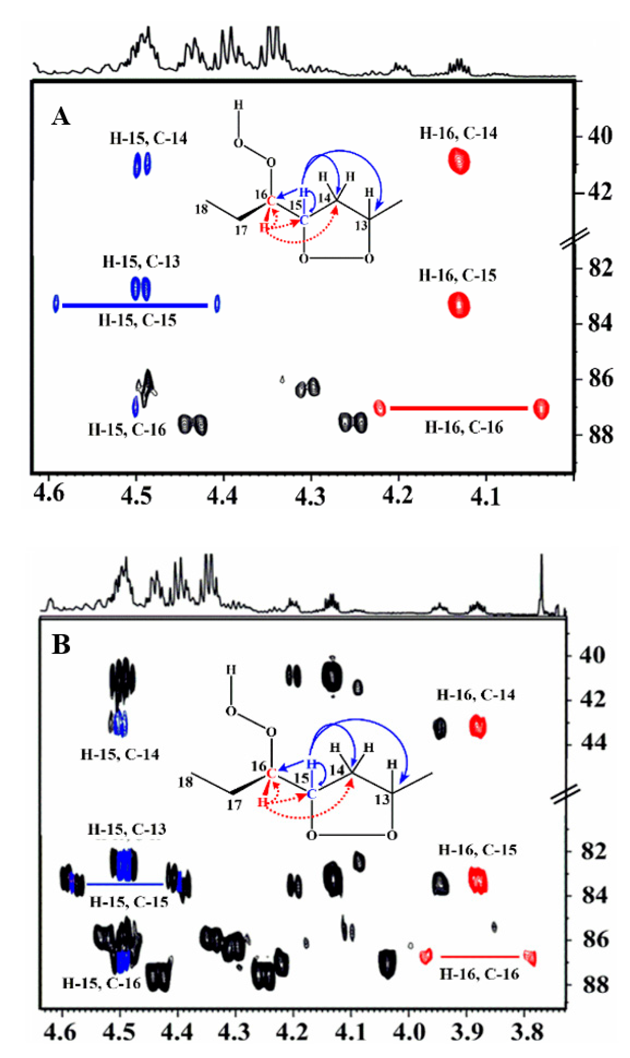
Figure 3 illustrates a selected region of the 1H-13C HMBC spectrum. Despite the low concentration of the hydroperoxides and endo-hydroperoxides (≈1.36–8.23%), the 1H-13C HMBC experiment revealed several 13C connectivities between δ 82 and 88 for the C–O–O–H carbons. Furthermore, the 1H-13C HMBC spectrum demonstrates very informative connectivities of the C–O–O–H carbon at δ of 87.5, with the terminal –CH3 (δ 0.9) and CH3–CH2 (δ 1.72, 1.55) protons, thus confirming the presence of the 16–OOH hydroperoxide. Of diagnostic importance are also the 1H-13C HMBC connectivities of the two deshielded terminal CH3 groups at δ 1.05 and 1.07 with the respective hydroperoxide carbons (δ 87.2, and 87.0) and C–O–O–H protons in the strongly deshielded region at δ 9.50 and 9.08, respectively (Figure 3). Furthermore, their CH–O–O–H protons at δ 4.13 and 3.87 demonstrate very informative additional connectivities in the region of the hydroperoxide carbons (Figure 4). This supported an unequivocal presence of two 16–OOH endo-hydroperoxide isomers of the oxidized fatty acid sample. Thus, the H-16 multiplet at δ 4.13 demonstrates connectivities with C-16 (δ 87.2), C-15 (δ 83.35), and C-14 (δ 40.7) (Figure 4A). The C-16 also shows connectivities with the H-15 multiplet (δ 4.49), which shows a characteristic doublet with C-15, due to 1J(13C,1H) of 146 Hz, and connectivities with C-13 (δ 82.8) and C-14. Similar connectivities were observed for the H-16 multiplet (δ 3.87) of the second diastereoisomer (Figure 4B).
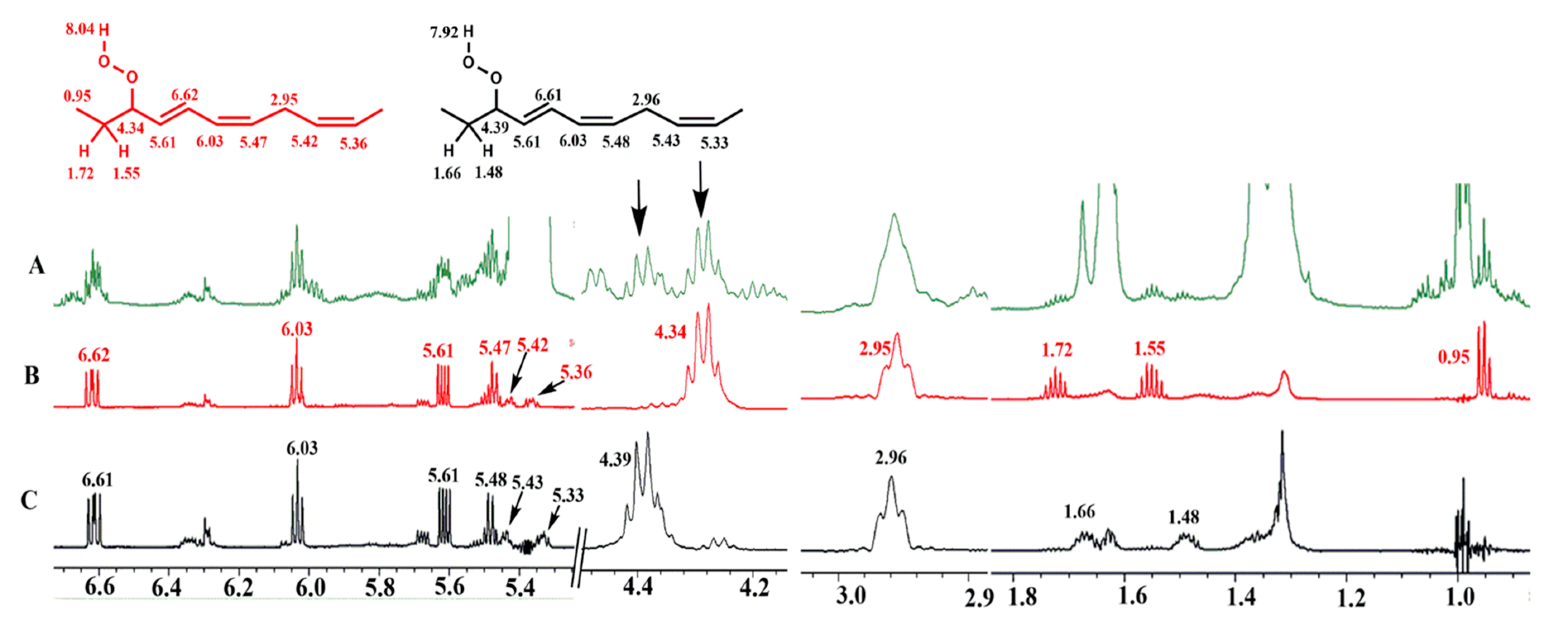
Furthermore, the identification of the above hydroperoxides was confirmed with the use of selective 1D TOCSY experiments [29]. An important application of the 1D experiment is the selective excitation of a suitable target resonance of the compound of interest, which reveals an extensive spin system belonging to a single chemical analyte, even in heavily overlapped resonances [30,31,32,33,34]. Selective excitation of the allylic CH–O–O–H proton of the 9-cis, 12-cis, 14-trans-16-OOH geometric hydroperoxide isomer at δ 4.34 (Figure 5B) shows connectivities with the terminal C-18 methyl group at δ 0.95, the H-17 (δ 1.72 and 1.55), the conjugated olefinic protons H-15 (δ 5.61), H-14 (δ 6.62), H-13 (δ 6.03), H-12 (δ 5.47), the H-11 bis allylic protons (δ 2.95), the olefinic protons H-10 (δ 5.42), and H-9 (δ 5.36). The selective excitation of the 10-trans, 12-cis, 15-cis-9-OOH isomer at δ 4.39 shows connectivities with H-8 (δ 1.66 and 1.48), the conjugated olefinic protons H-10 (δ 5.61), H-11 (δ 6.61), H-12 (δ 6.03), H-13 (δ 5.47), the H-14 bis allylic protons (δ 2.96), the olefinic H-15 (δ 5.43), and H-16 (δ 5.33) (Figure 5C).
The identification of the two diastereomeric 9-cis, 11-trans-16-OOH endo-hydroperoxides can be achieved by the selective excitation of the CH–O–O–H allylic protons at δ 4.13 and 3.87. Thus, selective excitation of the multiplet at δ 3.87 (Figure 6b) results in connectivities with the H-17 (δ 1.66 and 1.57), the terminal CH3 group (δ 1.07), H-15 (δ 4.40), H-14 (δ 2.88 and 2.23), H-13 (δ 4.79), and with the conjugated double bond protons of H-12 (δ 5.58), H-11 (δ 6.65), H10 (δ 6.00), and H-9 (δ 5.54). Similar long-range connectivities were observed with the selective excitation of the multiplet at δ 4.13 (Figure 6c).
Figure 7 illustrates schematically the unique advantages of the selective 1D TOCSY experiment. The method allows, through a careful selection of the spin system to be excited, the unambiguous identification of an extensive spin coupled system of minor abundance hydroperoxides and endo-hydroperoxides even in the cases of strongly overlapping resonances.
Similar experiments were performed for the strongly deshielded hydroperoxide protons at 9.55 and 9.12 ppm (Figure 2B), which were assigned to the two diastereomeric 13-trans, 15-cis-9-OOH endo-hydroperoxides. Of particular interest are the significant differences of the OOH chemical shifts of the two diastereomeric pairs of 9-cis, 11-trans-16-OOH, and 13-trans, 15-cis-9-OOH endo-hydroperoxides which allowed, through 1H-13C HMBC experiments, the identification of the respective CH-OOH protons that also have very significant chemical shift differences (Table S1). Figure S1A illustrates the 1H NMR chemical shift range of the endo-hydroperoxides, which shows sufficient resolution even when using a 400 MHz instrument. However, the proposed methodology implies 1H-13C HMBC connectivities of the -OOH with the CH-OOH protons. The achievable resolution of the CH-OOH protons using a 400 MHz instrument is not sufficient for identification due to strong signal overlapping (Figure S1B). However, the 1H-NMR spectrum using a 600 MHz instrument is nearly identical to that obtained at 800 MHz, which demonstrates that the proposed experiments can be performed using a 600 MHz instrument.
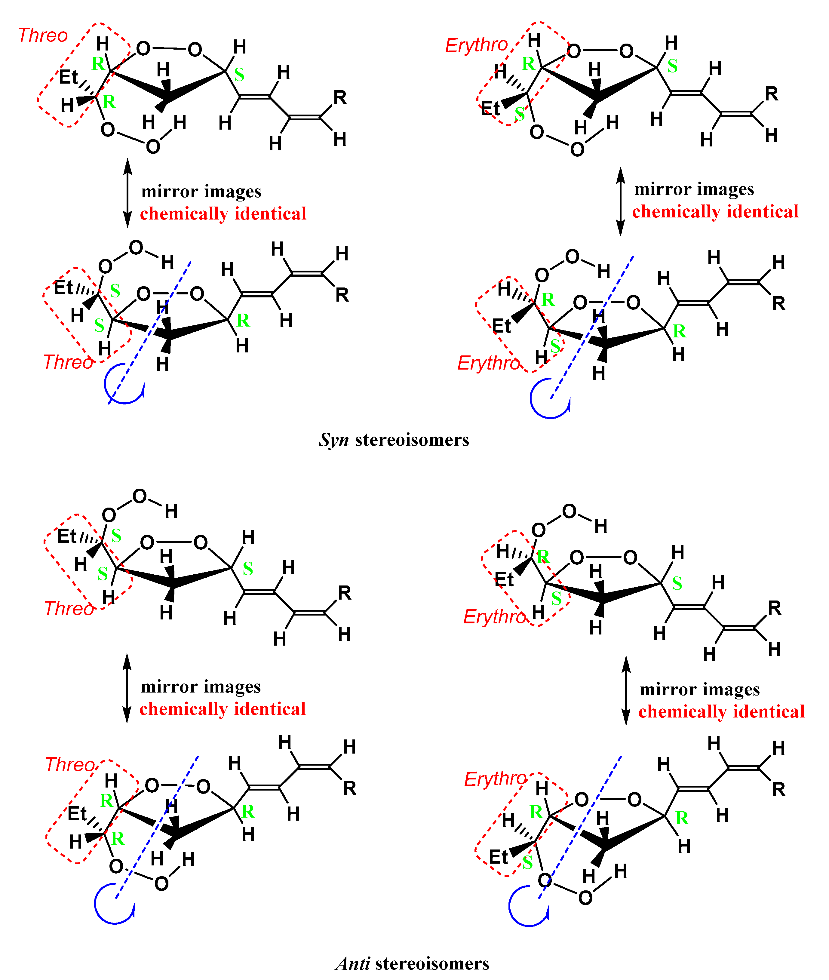
The formation of two pairs of 9-cis, 11-trans-16-OOH and 13-trans,15-cis-9-OOH endo-hydroperoxides of methyl linolenate has been attributed to hydrogen abstraction at carbons 11 and 14 (Figure S2), which results in the formation of two pentadienyl radicals. Reaction with oxygen at the end carbon positions produces a mixture of four peroxyl radicals leading to the corresponding conjugated dienoic 9-, 12-, 13-, and 16-hydroperoxides [10,12,35,36,37,38]. The peroxyl radicals derived from internal 12- and 13-hydroperoxides undergo rapid 1,5-cyclization to form two five-membered isomeric 9- and 16-hydroperoxy cyclo-endo-peroxides (Figure S2). In the case of e.g., 13-OOH hydroperoxide, the presence of three stereocenters C-13, C-15, and C-16 imply a number of 23 = 8 stereoisomers (23–1 = 4 pairs of enantiomers) of 9-cis, 11-trans-16-OOH linolenate endo-hydroperoxides (similar arguments also imply in the case of 13-trans, 15-cis-9-OOH hydroperoxides) (Figure 8). Only one pair of 16-OOH/9-OOH endo-hydroperoxides was identified by the use of preparative high-pressure liquid chromatography (HPLC) [4,35]. This aspect and the correct stereoisomerism of the resulting endo-hydroperoxides were investigated in detail with the use of DFT calculations (see discussion below).

2.2. Quantum Chemical Calculations

The development of quantum chemical methods for calculating NMR chemical shifts has led to a significant number of studies that combine experimental chemical shifts with computations [39,40,41,42]. Nevertheless, a few examples of organic molecules whose structures have been determined by both computations of NMR chemical shifts in solution and X-ray structures have been published [43,44,45,46,47,48,49]. Since no X-ray structures of the hydroperoxides of fatty acids have so far been reported, it would be of interest to investigate three-dimensional structures of hydroperoxides of the present work, which are based on the combined use of quantum chemical calculations and 1H-NMR chemical shifts.
Firstly, we investigated models of the two pairs of diastereoisomeric 9-cis, 11-trans-16-OOH endo-hydroperoxides (carbons 18 to 7) (Figure 9) and a model of 9-cis, 12-cis, 14-trans-16-OOH hydroperoxide (carbons 18 to 9) using the DFT quantum chemical method [50,51]. All optimizations were performed using the Austin-Frisch-Petersson functional with Dispersion (APFD) along with the 6-31+G(d) basis set: (i) in the gas phase, (ii) using the integral equation formalism polarizable continuum model (IEFPCM) in CHCl3, and (iii) with a discrete solvation molecule of CHCl3. Vibrational analysis was used to verify the nature of the stationary points. Secondly, we have also used the Our N-layered Integrated molecular Orbital and molecular Mechanics (ONIOM) method [52] to study the full-length compounds and compare the results with the model compounds. Furthermore, the NMR calculations were carried out at the optimized geometries, which were obtained with the DFT and ONIOM methods, using the B3LYP functional and the 6-311+G(2d,p) basis set. For comparison purposes, the NMR calculations were performed using the GIAO (Gauge-Independent Atomic Orbital) and the CSGT (Continuous Set of Gauge Transformations) methods [53].

2.2.1. DFT Studies of Model Compounds—Assigning the Stereochemistry of Pairs of Diastereoisomers

The minimum energy DFT structures of the two pairs of diastereoisomers of 9-cis, 11-trans-16-OOH endo-hydroperoxides are illustrated in Figure 9. Table 1 shows that the conformational and structural properties of the four stereoisomers remain essentially the same in the gas phase, in IEFPCM (CHCl3) and with a discrete solvation molecule of CHCl3 (Figure S3). The syn threo stereoisomer shows a hydrogen bond interaction of the hydroperoxide proton, H16b, with the O15 of the five-member endo-peroxide ring. The distance H16bO15 of 2.040 Å indicates the presence of a medium-strength hydrogen bond interaction. In the syn erythro stereoisomer, the interaction of the hydroperoxide proton H16b with the π-electrons of the conjugated double bond results in a very weak interaction with the oxygen O15 of the five-member endo-peroxide ring (H16bO15 = 2.431 Å), which is in the limits of the definition of hydrogen bond interaction (Figure 9).
The anti erythro and anti threo stereoisomers, with the -OOH and conjugated substituents in anti-position, show the hydrogen bond interaction of the hydroperoxide proton H16b with the O15 of the five-member endo-peroxide ring. The distances H16bO15 of 1.929 Å and 1.997 Å indicate the presence of stronger hydrogen bond interaction than in the syn threo and, especially, in syn erythro stereoisomer. However, in the region of strongly deshielded hydroperoxide protons (9.0 to 9.8 ppm, Figure 2B), only four prominent resonances were observed. These resonance were unequivocally assigned, with the combined use of 1H-13C HMBC and selected 1D-TOCSY experiments (see Section 2.1), to a diastereomeric pair of 9-cis, 11-trans-16-OOH endo-hydroperoxide (δ = 9.50 and 9.08 ppm) and a diastereomeric pair of 13-trans, 15-cis-9-OOH endo-hydroperoxide (δ = 9.55 and 9.12 ppm) (Figure 2B). Similarly, only two diastereomeric pairs were isolated with the use of preparative high-pressure liquid chromatography (HPLC) [4,35].
Table S2 shows a comparison of computational 1H-NMR chemical shifts of the four model diastereomeric endo-hydroperoxides with the experimental chemical shifts of the full-length molecules. The syn threo stereoisomer, both in PCM and with the inclusion of a discrete solvation molecule of CHCl3 in PCM, shows a strong magnetic non-equivalence of the C(14)Ha,b protons, which is in excellent agreement with the experimental data. On the contrary, the calculated C(14)Ha,b chemical shifts are very similar in the syn threo stereoisomer. This supports the conclusion that the stereoisomer with δ(OOH) = 9.50 ppm can be assigned to 9-cis, 11-trans, syn threo, 16-OOH endo-hydroperoxide. The syn erythro and anti erythro stereoisomers show strong magnetic non-equivalence of the C(14)Ha,b protons in both PCM and with the inclusion of a discrete solvation molecule of CHCl3 (Table S2); thus, they cannot be distinguished on the basis of this criterion. However, the syn erythro stereoisomer shows very weak interaction of the hydroperoxide proton with the O15 of the five-member endo-peroxide ring, contrary to the medium hydrogen bond interaction of the anti erythro stereoisomer (Figure 9 and Figure S2). Therefore, the resonance at 9.08 ppm, which is shielded relative to that at 9.50 ppm, may be assigned to 9-cis, 11-trans, syn erythro, 16-OOH endo-hydroperoxide.
Table S3 shows a comparison of experimental and computational 1H-NMR chemical shifts of the 10-cis, 13-cis, 15-trans-16-OOH hydroperoxide model with the chemical shifts of the full-length molecule. The inclusion of one solvation molecule of CHCl3 improves the agreement between computational and experimental 1H-NMR chemical shifts of the solvent-exposed hydroperoxide proton.

2.2.2. DFT and ONIOM Computational Studies of the Full Length 16-OOH Endo-Hydroperoxides

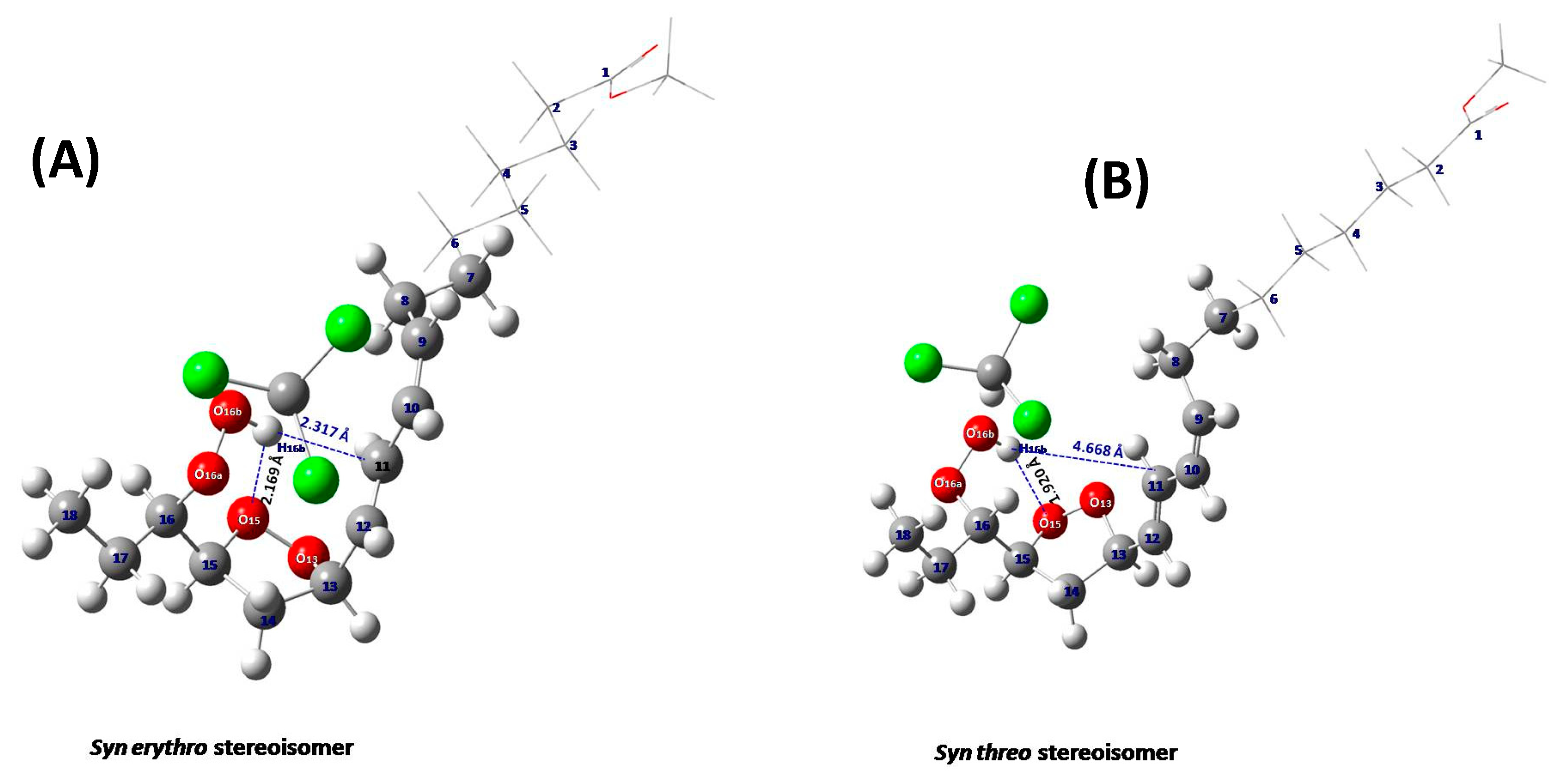
According to the ONIOM method, the molecule under study is divided into two layers, the high and the low layer, where the most important part of the molecule is placed in the high layer and treated with an accurate quantum chemical model such as the DFT method. The less important part is placed in the low layer and treated with a less expensive method, such as a semi-empirical (PM6). Figure 10 illustrates the structural comparison of the full-length syn erythro- (A) and syn threo- (B) endo-hydroperoxides with energy minimization using the APFD/6-31+G(d):PM6 method. Table S4 shows a comparison of experimental and computational 1H-NMR chemical shifts of the full-length (carbons 1 to 18) diastereomeric 9-cis, 11-trans-16-OOH endo-hydroperoxides with energy minimization at the APFD/6-31+G(d): PM6 level. The chemical shifts, structural, and conformational properties of the C–O–O–H hydroperoxide unit and the five-membered epidioxide ring are very similar to those of the model compounds of Figure S3 and Table S2. The computed and experimental 1H-NMR chemical shifts of the hydroperoxide proton are in agreement with a stronger hydrogen bond interaction of the hydroperoxide proton with the O15 oxygen of the five-member endo-peroxide ring in the major 15,16-threo than in the 15,16-erythro endo-hydroperoxide molecule (Figure 10, Table S5) in excellent agreement with the structural data of the model compounds (Figure 9, Table 1).
A large number of hydrogen bonds in crystal structures of alkyl hydroperoxides have been reported [28]. For some compounds, the formation of an intramolecular hydrogen bond would in principle have been feasible. Nevertheless, the number of examples in the literature so far is in favor of intermolecular rather than intramolecular hydrogen bonds. Therefore, no direct comparison with the structural data of Table S5 could be made.

2.3. Comparison with Literature Data

Table 2 presents a comparison of the 1H- and 13C-NMR experimental chemical shifts of endo-hydroperoxides of methyl linolenate of the present work with those reported in the literature for HPLC-isolated endo-hydroperoxides. The deviation of δ (1H) was found to be ≤0.02 ppm with the exception of the chemical shift of the hydroperoxide proton, which is expected to be very sensitive to inter- and intramolecular hydrogen bonds, temperature, and solution conditions. The deviation of δ(13C) was found to be ≤0.6 ppm. Therefore, it can be concluded that the agreement of the present NMR data with those of isolated analytes is excellent.
Table S6 presents the 1H NMR chemical shifts of 9-cis, 12-cis, 14-trans-16-OOH and 10-trans, 12-cis, 15-cis-9-OOH hydroperoxides of methyl linolenate. However, to the best of our knowledge, there are no literature data of 1H-NMR chemical shifts for comparison.
The excellent resolution achievable in the region of hydroperoxide C–O–O–H protons (δ 7.70 to 9.6, Figure 1 and Figure 2) allowed an accurate integration and, thus, quantification of primary oxidation products. Table 3 shows quantification of the hydroperoxide 1H-NMR resonances based on their integration, with respect to the total integral of the CH3- group at 0.86 ppm. A comparison with literature integration data [36] shows very good agreement, despite differences in experimental conditions. As pointed out [10,12,35,36,37,38], the peroxyl radicals derived from 12- and 13- hydroperoxides undergo rapid 1,5-cyclization to form two five-membered isomeric internal 9- and 16-hydroperoxy cyclo endo-peroxides. This rapid cyclization results in lower concentrations of the internal 12- and 13-hydroperoxides (14% and 15%, respectively) relative to the external 9- and 16- hydroperoxides (29% and 41%, respectively) (Table 3). Similar relative concentrations of hydroperoxides have been found in the literature under a wide range of oxidation conditions [10,12,35,36,37,38].

3. Materials and Methods

3.1. Materials

Methyl linolenate (methyl (9Z,12Z,15Z)-octadeca-9,12,15-trienoate) and deuterochloroform (CDCl3, 99.8 d-%,) were purchased from Sigma-Aldrich (St. Louis, MO, USA).

3.2. Oxidation Methodology

Methyl linolenate (200 mg) was oxidized in a glass vial (10 mL) at 40 °C for 48 h in the conventional oven (Labtech Windsor, Australia) circulating the atmospheric oxygen.

3.3. NMR Methodology

1H-NMR experiments were recorded at 298 K on a Bruker AVANCE III HD 800 spectrometer with 128 scans and a relaxation delay of 5 s. One-dimensional TOCSY experiments were recorded at 298 K on a Bruker AVANCE III HD 800 spectrometer, using the MLEV-17 (selmlgp) pulse program, 128 scans, and a relaxation delay of 5 s. Selective excitation, with a bandwidth of 12 Hz (≈0.015 ppm), was achieved with a gradient-based echo block that uses a soft Gaussian 180° selective refocusing pulse of 49 ms, with power attenuation as indicated in Figure S4. The mixing time for long-range connectivities was set in the range of 200 to 300 ms. 1H–13C HMBC experiments were recorded at 298 K on a Bruker AVANCE III HD 800 spectrometer. The magnitude mode gradient enhanced HMBC experiment (hmbcgndqf pulse program) was acquired in the quadrature mode, with data points set to 4 k × 256 (t2 × t1) (total experimental time 12 h) or 4 k × 512 (t2 × t1) (total experimental time 24 h), with 4 k × 1 k (t2 × t1) data points for transformation, relaxation delay of 5 s, number of scans 32, with a sine function for apodization. The long-range coupling time was set to 125 ms (optimized for nJCH ≈4 Hz), SW = 10,416.66 Hz (F1) and 46,285.23 Hz (F2).

3.4. DFT and ONIOM Calculations

The DFT computational study was performed by using the Gaussian 09 [54]. The structures were optimized by using the APFD functional and the 6-31+G(d) basis set in the gas phase, using the IEFPCM model in CHCl3, and with the inclusion of a discrete solvation molecule of CHCl3. The 1H-NMR chemical shifts were calculated with the GIAO and CSGT methods by using the B3LYP/6-311+G (2d, p) level with the PCM (polarizable continuum model) [55]. The optimized geometries were verified by performing frequency calculations at the same level (zero imaginary frequencies). TMS was used as a reference for the computed 1H-NMR chemical shift, and the optimization of TMS was calculated at the same level. The ONIOM optimizations were performed at the ONIOM(APFD/6-31+G(d):PM6) level of theory.

4. Conclusions

In this work, we have demonstrated that the combined use of 1D selective TOCSY and 1H-13C HMBC NMR techniques, DFT, and ONIOM computational studies is a powerful approach for obtaining detailed analytical and high-resolution structural information of hydroperoxides and diastereomeric endo-hydroperoxides in ω-3 fatty acid substrates. The proposed approach is of primary importance in hydroperoxide research because:
(a) It is rapid, selective, and does not require derivatization steps.
(b) It allows the unequivocal identification and quantification of minor analytes even in strongly overlapped spectral regions, and thus, it is of importance in the emerging field of lipidomics [56,57,58,59,60,61], provided that a spectrometer with 1H resonance frequency ≥600 MHz is used.
(c) It can provide an excellent method for obtaining three-dimensional structures with diastereomeric assignment in solution [62].
Further NMR and computational studies to establish the identities of hydroperoxide-OOH resonances are currently in progress with cis-5,8,11,14,17-eicosapentaenoic acid (EPA) and cis-4,7,10,13,16,19-docosahexaenoic acid (DHA) methyl esters.

Supplementary Materials

The following are available online, Figure S1: Selected 1H-NMR chemical shift ranges of the endo-hydroperoxide (OOH) region (A), and CH-OOH region (B), Figure S2: Proposed mechanism of the formation of two diastereomeric pairs of 9-cis, 11-trans-16-OOH, and 13-trans, 15-cis-9-OOH linolenate endo-hydroperoxides, Figure S3: Minimum energy structures of the two pairs of diastereomers of 9-cis, 11-trans-16-OOH endo-hydroperoxides with a discrete solvation molecule of CHCl3 in IEFPCM (CHCl3), Figure S4: Power attenuation details of the soft Gaussian 180o selective refocusing pulse of the selmlgp pulse program, Table S1: Critical hydroperoxide OOH and CH-OOH 1H-NMR chemical shifts for the identification of hydroperoxides and endo-hydroperoxides of methyl linolenate, Table S2: Comparison of computational [B3LYP/6-311G+d (2d, p)] 1H-NMR chemical shifts of the two pairs of diastereomeric 16-OOH endo-hydroperoxide models, with energy minimization at the APFD/6-31+G(d) level, with the experimental chemical shifts of the full length molecules, Table S3: Comparison of computational [B3LYP/6-311G+d (2d, p)] 1H-NMR chemical shifts of the 10-cis, 13-cis, 15-trans-16-OOH hydroperoxide model, with energy minimization at the APFD/6-31+G(d) level, with the experimental chemical shifts of the full length molecule, Table S4: Comparison of experimental and computational 1H-NMR chemical shifts of the full length diastereomeric 9-cis, 11-trans-l6-OOH endo-hydroperoxides with energy minimization at the APFD/6-31+G(d):PM6 level, Table S5: Conformational and structural properties of the full length diastereomeric 9-cis, 11-trans-16-OOH endo-hydroperoxides with energy minimization using the APFD/6-31+G(d):PM6 method, Table S6: 1H-NMR chemical shifts of hydroperoxides of methyl linolenate.

Author Contributions

R.A. performed NMR experiments, DFT and ONIOM computations, and analyzed the data; P.C.V. and M.G.S. performed quantum chemical calculations and contributed the writing of the manuscript; H.S., M.I.C., and I.P.G. conceived and designed the study, and contributed to the writing of the manuscript. All authors have read and agreed to the published version of the manuscript.

Funding

This research received no external funding.

Conflicts of Interest

The authors declare that they have no conflict of interest.

References

  1. Akoh, C.C.; Min, D.B. Food Lipids. Chemistry, Nutrition, and Biotechnology, 2nd ed.; Marcel Dekker: New York, NY, USA, 2002. [Google Scholar]
  2. Gunstone, F.D. Fatty Acid and Lipid Chemistry, 1st ed.; Springer: New York, NY, USA, 1996. [Google Scholar]
  3. Leray, C. Dietary Lipids for Healthy Brain Function; CRC Press: Boca Raton, FL, USA, 2017. [Google Scholar]
  4. Neff, W.E.; Frankel, E.N.; Weisleder, D. High pressure liquid chromatography of autoxidized lipids: II. Hydroperoxy-cyclic peroxides and other secondary products from methyl linolenate. Lipids 1981, 16, 439–448. [Google Scholar] [CrossRef]
  5. Gunstone, F.D. Chemical reactions of fatty acids with special reference to the carboxyl group. Eur. J. Lipid Sci. Technol. 2001, 103, 307–314. [Google Scholar] [CrossRef]
  6. Frankel, E. Lipid oxidation: Mechanisms, products and biological significance. J. Am. Oil Chem. Soc. 1984, 61, 1908–1917. [Google Scholar] [CrossRef]
  7. Frankel, E.N. Lipid oxidation. Prog. Lipid Res. 1980, 19, 1–22. [Google Scholar] [CrossRef]
  8. Pryor, W.A.; Stanley, J.P. Suggested mechanism for the production of malonaldehyde during the autoxidation of polyunsaturated fatty acids. Nonenzymic production of prostaglandin endoperoxides during autoxidation. J. Org. Chem. 1975, 40, 3615–3617. [Google Scholar] [CrossRef] [PubMed]
  9. Ernster, L.; Nordenbrand, K. Microsomal lipid peroxidation. Method. Enzymol. 1967, 10, 574–580. [Google Scholar]
  10. Frankel, E.N.; Garwood, R.F.; Khambay, B.P.; Moss, G.P.; Weedon, B.C. Stereochemistry of olefin and fatty acid oxidation. Part 3. The allylic hydroperoxides from the autoxidation of methyl oleate. Chem. Soc. Perkin Trans. I 1984, 2233–2240. [Google Scholar] [CrossRef]
  11. Frankel, E. Chemistry of free radical and singlet oxidation of lipids. Prog. Lipid Res. 1984, 23, 197–221. [Google Scholar] [CrossRef]
  12. Frankel, E.N.; Evans, C.D.; McConnell, D.G.; Selke, E.; Dutton, H.J. Autoxidation of methyl linolenate. Isolation and characterization of hydroperoxides. J. Org. Chem. 1961, 26, 4663–4669. [Google Scholar] [CrossRef]
  13. Shahidi, F.; Wanasundara, U.N. Methods for measuring oxidative rancidity in fats and oils. Food Lipids Chem. Nutr. Biotechnol. 2002, 17, 387–403. [Google Scholar]
  14. Tamura, H.; Shibamoto, T. Gas chromatographic analysis of malonaldehyde and 4-hydroxy-2-(E)-nonenal produced from arachidonic acid and linoleic acid in a lipid peroxidation model system. Lipids 1991, 26, 170–173. [Google Scholar] [CrossRef]
  15. Boyd, L.C.; Nwosu, V.C.; Young, C.L.; MacMillian, L. Monitoring lipid oxidation and antioxidant effects of phospholipids by headspace gas chromatographic analyses of rancimat trapped volatiles. Food Lipids 1998, 5, 269–282. [Google Scholar] [CrossRef]
  16. Kinter, M. Analytical technologies for lipid oxidation products analysis. J. Chromatogr. B 1995, 671, 223–236. [Google Scholar] [CrossRef]
  17. Bagchi, D.; Bagchi, M.; Hassoun, E.A.; Stohs, S.J. Detection of paraquat-induced in vivo lipid peroxidation by gas chromatography/mass spectrometry and high-pressure liquid chromatography. J. Anal. Toxicol. 1993, 17, 411–414. [Google Scholar] [CrossRef]
  18. Haywood, R.M.; Claxson, A.W.D.; Hawkes, G.E.; Richardson, D.P.; Naughton, D.P.; Coumbarides, G.; Hawkes, J.; Lynch, E.J.; Grootveld, M.C. Detection of aldehydes and their conjugated hydroperoxydiene precursors in thermally-stressed culinary oils and fats: Investigations using high resolution proton NMR spectroscopy. Free Radic. Res. 1995, 22, 441–482. [Google Scholar] [CrossRef]
  19. Silwood, C.J.L.; Grootveld, M. Application of high-resolution two-dimensional 1H and 13C nuclear magnetic resonance techniques to the characterization of lipid oxidation products in autoxidized linoleoyl/linolenoylglycerol. Lipids 1999, 34, 741–756. [Google Scholar] [CrossRef] [PubMed]
  20. Guillen, M.D.; Goicoechea, E. Oxidation of corn oil at room temperature: Primary and secondary oxidation products and determination of their concentration in the oil liquid matrix from 1H nuclear magnetic resonance data. Food Chem. 2009, 116, 183–192. [Google Scholar] [CrossRef]
  21. Guillén, M.D.; Uriarte, P.S. Study by 1H NMR spectroscopy of the evolution of extra virgin olive oil composition submitted to frying temperature in an industrial fryer for a prolonged period of time. Food Chem. 2012, 134, 162–172. [Google Scholar] [CrossRef]
  22. Goicoechea, E.; Guillen, M.D. Analysis of hydroperoxides, aldehydes and epoxides by 1H nuclear magnetic resonance in sunflower oil oxidized at 70 °C and 100 °C. J. Agric. Food Chem. 2010, 58, 6234–6245. [Google Scholar] [CrossRef]
  23. Martınez-Yusta, A.; Guillen, M.D. A study by 1H nuclear magnetic resonance of the influence on the frying medium composition of some soybean oil-food combinations in deep-frying. Food Res. 2014, 55, 347–355. [Google Scholar] [CrossRef]
  24. Gresley, A.L.; Ampem, G.; Grootveld, M.; Percival, B.C.; Naughton, D.P. Characterization of peroxidation products arising from culinary oils exposed to continuous and discontinuous thermal degradation. Food Funct. 2019, 10, 7952–7966. [Google Scholar] [CrossRef] [PubMed]
  25. Merkx, D.W.; Hong, G.S.; Ermacora, A.; Van Duynhoven, J.P. Rapid quantitative profiling of lipid oxidation products in a food emulsion by 1H NMR. Anal. Chem. 2018, 90, 4863–4870. [Google Scholar] [CrossRef] [PubMed]
  26. Alexandri, E.; Ahmed, R.; Siddiqui, H.; Choudhary, M.; Tsiafoulis, C.; Gerothanassis, I.P. High resolution NMR spectroscopy as a structural and analytical tool for unsaturated lipids in solution. Molecules 2017, 22, 1663. [Google Scholar] [CrossRef] [PubMed]
  27. Ahmed, R.; Siddiqui, H.; Choudhary, M.I.; Gerothanassis, I.P. 1H–13C HMBC NMR experiments as a structural and analytical tool for the characterization of elusive trans/cis hydroperoxide isomers from oxidized unsaturated fatty acids in solution. Magn. Reson. Chem. 2019, 57, S69–S74. [Google Scholar] [CrossRef] [PubMed]
  28. Rappoport, Z. The Chemistry of Peroxides, Part 1; John Wiley & Sons, Ltd.: West Sussex, UK, 2006; Volume 2. [Google Scholar]
  29. Parella, T. High-quality 1D spectra by implementing pulsed-field gradients as the coherence pathway selection procedure. Magn. Reson. Chem. 1996, 34, 329–347. [Google Scholar] [CrossRef]
  30. Sandusky, P.; Raftery, D. Use of selective TOCSY NMR experiments for quantifying minor components in complex mixtures: Application to the metabonomics of amino acids in honey. Anal. Chem. 2005, 77, 2455–2463. [Google Scholar] [CrossRef]
  31. Koda, M.; Furihata, K.; Wei, F.; Miyakawa, T.; Tanokura, M. Metabolic discrimination of mango juice from various cultivars by band-selective NMR spectroscopy. J. Agric. Food. Chem. 2012, 60, 1158–1166. [Google Scholar] [CrossRef]
  32. Papaemmanouil, C.; Tsiafoulis, C.G.; Alivertis, D.; Tzamaloukas, O.; Miltiadou, D.; Tzakos, A.; Gerothanassis, I.P. Selective 1D TOCSY NMR experiments for a rapid identification of minor components in the lipid fraction of milk and dairy products: Towards spin-chromatography. J. Agric. Food. Chem. 2015, 63, 5381–5387. [Google Scholar] [CrossRef]
  33. Kontogianni, V.G.; Tsiafoulis, C.G.; Roussis, I.G.; Gerothanassis, I.P. Selective 1D TOCSY NMR method for the determination of glutathione in white wine. Anal. Methods 2017, 9, 4464–4470. [Google Scholar] [CrossRef]
  34. Kontogianni, V.G.; Primikyri, A.; Sakka, M.; Gerothanassis, I.P. Simultaneous determination of artemisinin and its analogues and flavonoids in Artemisia annua crude extracts with the use of NMR spectroscopy. Magn. Reson. Chem. 2020, 58, 232–244. [Google Scholar] [CrossRef]
  35. Coxon, D.T.; Price, K.R.; Chan, H.W.-S. Formation, isolation and structure determination of methyl linolenate diperoxides. Chem. Phys. Lipids 1981, 28, 365–378. [Google Scholar] [CrossRef]
  36. Frankel, E.N. Lipid Oxidation, 2nd ed.; The Oily Press Lipid Library Series: Cambridge, UK, 2005. [Google Scholar]
  37. Chan, H.W.-S.; Levett, G. Autoxidation of methyl linolenate. Analysis of methylhydroxylinoleate isomers by high performance liquid chromatography. Lipids 1977, 12, 837–840. [Google Scholar] [CrossRef] [PubMed]
  38. Chan, H.W.-S.; Coxon, D.T. Lipid hydroperoxides. In Autoxidation of Unsaturated Lipids; Chan, H.W.-S., Ed.; Academic Press: London, UK, 1987; pp. 17–50. [Google Scholar]
  39. Rychnovsky, S.D. Predicting NMR spectra by computational methods: Structure revision of hexacyclinol. Org. Lett. 2006, 8, 2895–2898. [Google Scholar] [CrossRef] [PubMed]
  40. Smith, S.G.; Goodman, J.M. Assigning the stereochemistry of pairs of diastereoisomers using GIAO NMR shift calculation. J. Org. Chem. 2009, 74, 4597–4607. [Google Scholar] [CrossRef]
  41. Lodewyk, M.W.; Siebert, M.R.; Tantillo, D.J. Computational prediction of 1H and 13C chemical shifts: A useful tool for natural product, mechanistic, and synthetic organic chemistry. Chem. Rev. 2012, 112, 1839–1862. [Google Scholar] [CrossRef]
  42. Tarazona, G.; Benedit, G.; Fernández, R.; Pérez, M.; Rodriguez, J.; Jiménez, C.; Cuevas, C. Can stereoclusters separated by two methylene groups be related by DFT studies? The case of the cytotoxic meroditerpenes halioxepines. J. Nat. Prod. 2018, 81, 343–348. [Google Scholar] [CrossRef]
  43. Siskos, M.G.; Kontogianni, V.G.; Tsiafoulis, C.G.; Tzakos, A.G.; Gerothanassis, I.P. Investigation of solute–solvent interactions in phenol compounds: Accurate ab initio calculations of solvent effects on 1H NMR chemical shifts. Org. Biomol. Chem. 2013, 11, 7400–7411. [Google Scholar] [CrossRef]
  44. Siskos, M.G.; Tzakos, A.G.; Gerothanassis, I.P. Accurate ab initio calculations of O–HO and O–HO proton chemical shifts: Towards elucidation of the nature of the hydrogen bond and prediction of hydrogen bond distances. Org. Biomol. Chem. 2015, 13, 8852–8868. [Google Scholar] [CrossRef]
  45. Siskos, M.G.; Choudhary, M.I.; Tzakos, A.G.; Gerothanassis, I.P. 1H NMR chemical shift assignment, structure and conformational elucidation of hypericin with the use of DFT calculations–The challenge of accurate positions of labile hydrogens. Tetrahedron 2016, 72, 8287–8293. [Google Scholar] [CrossRef]
  46. Siskos, M.G.; Choudhary, M.I.; Gerothanassis, I.P. Hydrogen atomic positions of O–HO hydrogen bonds in solution and in the solid state: The synergy of quantum chemical calculations with 1H-NMR chemical shifts and X-ray diffraction methods. Molecules 2017, 22, 415. [Google Scholar] [CrossRef]
  47. Siskos, M.G.; Choudhary, M.I.; Gerothanassis, I.P. Refinement of labile hydrogen positions based on DFT calculations of 1H NMR chemical shifts: Comparison with X-ray and neutron diffraction methods. Org. Biomol. Chem. 2017, 15, 4655–4666. [Google Scholar] [CrossRef]
  48. Siskos, M.G.; Choudhary, M.I.; Gerothanassis, I.P. DFT-calculated structures based on 1H NMR chemical shifts in solution vs. structures solved by single-crystal X-ray and crystalline-sponge methods: Assessing specific sources of discrepancies. Tetrahedron 2018, 74, 4728–4737. [Google Scholar] [CrossRef]
  49. Mari, S.H.; Varras, P.C.; Wahab, A.-T.; Choudhary, I.M.; Siskos, M.G.; Gerothanassis, I.P. Solvent-dependent structures of natural products based on the combined use of DFT calculations and 1H-NMR chemical shifts. Molecules 2019, 24, 2290. [Google Scholar] [CrossRef] [PubMed] [Green Version]
  50. Cramer, C.J. Essentials of Computational Chemistry: Theories and Models, 2nd ed.; Wiley: Chichester, West Sussex, UK, 2008. [Google Scholar]
  51. Jensen, F. Introduction to Computational Chemistry, 2nd ed.; Wiley: Chichester, West Sussex, UK, 2007. [Google Scholar]
  52. Chung, L.W.; Sameera, W.M.C.; Ramozzi, R.; Page, A.J.; Hatanaka, M.; Petrova, G.P.; Harris, T.V.; Li, X.; Ke, Z.; Liu, F.; et al. The ONIOM method and its applications. Chem. Rev. 2015, 115, 5678–5796. [Google Scholar] [CrossRef] [Green Version]
  53. Trindle, C.; Shillady, D. Electronic Structure Modeling; CRC Press, Taylor and Francis Group: Boca Raton, FL, USA, 2008. [Google Scholar]
  54. Frisch, M.J.; Trucks, G.W.; Schlegel, H.B.; Scuseria, G.E.; Robb, M.A.; Cheeseman, J.R.; Scalmani, G.; Barone, V.; Mennucci, B.; Petersson, G.A.; et al. Gaussian 0.9, Revision. B.01; Gaussian, Inc.: Wallingford, UK, 2010. [Google Scholar]
  55. Klein, R.A.; Mennucci, B.; Tomasi, J. Ab initio calculations of 17O NMR-chemical shifts for water. The limits of PCM theory and the role of hydrogen-bond geometry and cooperativity. J. Phys. Chem. A 2004, 108, 5851–5863. [Google Scholar] [CrossRef]
  56. Massey, K.A.; Nicolaou, A. Lipidomics of oxidized polyunsaturated fatty acids. Free Rad. Biol. Med. 2013, 59, 45–55. [Google Scholar] [CrossRef]
  57. Li, J.; Voseqaard, T.; Guo, Z. Applications of nuclear magnetic resonance in lipid analyses: An emerging powerful tool for lipidomics studies. Prog. Lipid Res. 2017, 68, 37–56. [Google Scholar] [CrossRef]
  58. Tsiafoulis, C.G.; Papaemmanouil, C.; Alivertis, D.; Tzamaloukas, O.; Miltiadou, D.; Balayssac, S.; Malet-Martino, M.; Gerothanassis, I.P. NMR-Based metabolomics of the lipid fraction of organic and conventional bovine milk. Molecules 2019, 24, 1067. [Google Scholar] [CrossRef] [Green Version]
  59. Hatzakis, E. Nuclear magnetic resonance (NMR) spectroscopy in food science: A comprehensive review. Compr. Rev. Food Sci. Food Saf. 2019, 18, 189–220. [Google Scholar] [CrossRef] [Green Version]
  60. Wang, C.; Timári, I.; Zhang, B.; Li, D.-W.; Leggett, A.; Amer, A.O.; Bruschweiler-Li, L.; Kopec, R.E.; Brüschweiler, R. COLMAR lipids web server and ultrahigh-resolution methods for 2D NMR- and MS-based lipidomics. J. Proteome Res. 2020, 19, 1674–1683. [Google Scholar] [CrossRef]
  61. Boccia, A.C.; Cusano, E.; Scano, P.; Consonni, R. NMR lipid profile of milk from alpine goats with supplemented hempseed and linseed diets. Molecules 2020, 25, 1491. [Google Scholar] [CrossRef] [PubMed] [Green Version]
  62. Venianakis, T.; Oikonomaki, C.; Siskos, M.G.; Varras, P.C.; Primikyri, A.; Alexandri, E.; Gerothanassis, I.P. DFT calculations of 1H- and 13C-NMR chemical shifts of geometric isomers of conjugated linoleic acid (18:2 ω-7) and model compounds in solution. Molecules 2020, 25, 3660. [Google Scholar] [CrossRef] [PubMed]
Sample Availability: Sample of the compound methyl linolenate (methyl (9Z,12Z,15Z)-octadeca-9,12,15-trienoate) is available from the authors.
Publisher’s Note: MDPI stays neutral with regard to jurisdictional claims in published maps and institutional affiliations.
Figure 1. (a) 800 MHz 1H-NMR spectrum of 20 mg methyl linolenate in CDCl3 subjected to heating at 40 °C for 48 h, number of scans = 64, acquisition time = 1.02 s, experimental time = 6.5 min, relaxation delay = 5 s, T = 298 K; (b) selected region of the C–O–O–H resonances of its primary oxidation products.
Molecules 25 04902 g001
Figure 2. 800 MHz 1H-NMR spectra of the solution of Figure 1 before the addition (a), and after the addition (b) of 2 microdrops of D2O. (A,B) are the selected regions of the 7.86 to 8.14 and 8.96 to 9.82 ppm, respectively. For the method of assignment of the -OOH resonances, see text.
Molecules 25 04902 g002
Figure 3. Selected regions of 800 MHz 1H-13C HMBC NMR spectrum of 20 mg methyl linolenate in CDCl3, subjected to heating at 40 °C for 48 h, number of scans = 32, number of increments 256, total experimental time = 12 h, T = 298 K. The critical cross-peak connectivities of C-16 with H-1(OOH), H-16 (1J(13C, 1H)), H-17, and H-18 of the 9-cis, 12-cis, 14-trans-16-OOH hydroperoxide (A), and two diastereomeric 9-cis, 11-trans-16-OOH endo-hydroperoxides (in red) (B), are illustrated.
Molecules 25 04902 g003
Figure 4. 1H-13C HMBC correlations showing the assignments of the two diastereomeric 9-cis, 11-trans-16-OOH endo-hydroperoxides of methyl linolenate with OOH and CH-OOH resonances at 9.50 ppm and 4.3 ppm, respectively (A), and 9.08 ppm and 3.87 ppm, respectively (B). The critical connectivities of the H-15 with C-14, C-13, C-15 (1J(13C, 1H)), C-16, and connectivities of H-16 with C-14, C-15, and C-16 (1J(13C, 1H)) are illustrated.
Molecules 25 04902 g004
Figure 5. (A) 800 MHz 1D 1H-NMR spectrum of 20 mg methyl linolenate, subjected to heating at 40 °C for 48 h, in CDCl3 (acquisition time= 1.02 s, relaxation delay = 5 s, number of scans = 128, experimental time = 10 min), T = 298 K. (B,C) selective 1D TOCSY spectra of the same solution using a mixing time of τm = 300 ms. The arrows denote the selected resonances that were excited at δ 4.34 (B) of the 9-cis, 12-cis, 14-trans-16-OOH hydroperoxide and δ 4.39 (C) of the 10-trans, 12-cis, 15-cis-9-OOH hydroperoxide. Number of scans = 256, experimental time = 20 min.
Molecules 25 04902 g005
Figure 6. (a) 800 MHz 1D 1H-NMR spectrum of 20 mg of methyl linolenate, subjected to heating at 40 °C for 48 h, in CDCl3 (T = 298 K, acquisition time = 1.02 s, relaxation delay = 5 s, number of scans = 128, experimental time = 10 min). (b,c) selective 1D TOCSY spectra of the same solution using a mixing time of τm = 300 ms. The arrows denote the selected resonances of the two diastereomeric 9-cis, 11-trans-16-OOH endo-hydroperoxides that were excited at δ 3.87 (b) and δ 4.13 (c).
Molecules 25 04902 g006
Figure 7. Schematic presentation of the selective 1D TOCSY correlations which were observed in the case of the 9-cis, 12-cis, 14-trans-16-OOH hydroperoxide (a), and the 9-cis, 11-trans-16-OOH endo-hydroperoxide (b) of the methyl linolenate primary oxidation products.
Molecules 25 04902 g007
Figure 8. The 23 = 8 stereoisomers (23–1 = 4 pairs of enantiomers) of 9-cis, 11-trans, 16-OOH linolenate endo-hydroperoxides investigated in the present work.
Molecules 25 04902 g008
Figure 9. Minimum energy structures of two pairs of diastereomers of 9-cis, 11-trans-16-OOH endo-hydroperoxide models in integral equation formalism polarizable continuum model (IEFPCM) (CHCl3).
Molecules 25 04902 g009
Figure 10. Structural comparison of the full-length syn erythro (A) and syn threo (B) endo-hydroperoxides, with a discrete solvation molecule of CHCl3 (energy minimization using the APFD/6-31+G(d):PM6 method).
Molecules 25 04902 g010
Table 1. Conformational and structural properties of two pairs of diastereomers of the model 9-cis, 11-trans-16-OOH endo-hydroperoxides with energy minimization using the APFD/6-31+G(d) method in the gas phase and with a discrete solvation molecule of CHCl3.
CompoundC–O
(Å)
O–O
(Å)
O–H
(Å)
C(16) –O–O–H
(°)
C(17)–C(16)–O–O
(°)
C(15)–(16)
–O–O
(°)
(O)HO
(Å)
O(H)O
(Å)
O–HO
(°)
O(H)C11
(Å)
Syn erythro1.4221.4280.978−90.0−153.385.02.4312.882107.72.301
Syn erythro (PCM)1.4251.4300.979−88.9−155.583.02.4052.862107.92.275
Syn threo1.4221.4310.97678.2157.1−82.42.0392.761129.03.910
Syn threo (PCM)1.4271.4330.97873.9157.4−81.91.9902.743132.1°4.078
Anti erythro1.4221.4320.979−69.0−154.983.31.9812.730131.4
Anti erythro (PCM)1.4261.4340.980−66.7−156.182.11.9502.715133.1
Anti threo1.4221.4310.97773.2155.3−83.81.9972.752132.3
Anti threo (PCM)1.4271.4340.97971.1157.1−82.11.9442.724134.8
Syn erythro + CHCl31.4241.4300.979−79.6−153.484.62.1842.800119.52.307
Syn erythro + CHCl3 (PCM)1.4261.4310.979−78.9−154.383.82.1692.791120.02.317
Syn threo + CHCl31.4251.4310.97970.8158.4−80.81.9482.719133.84.591
Syn threo + CHCl3 (PCM)1.4291.4330.98068.9158.6−80.31.9212.707135.34.664
Anti erythro + CHCl31.4261.4330.981−69.0−154.583.31.9282.721136.0
Anti erythro + CHCl3 (PCM)1.4301.4330.982−62.8−154.782.91.9202.716136.3
Anti threo + CHCl31.4211.4340.97879.6159.4−79.51.9822.692127.5
Anti threo + CHCl3 (PCM)1.4261.4340.97977.4159.9−79.11.9582.685129.1
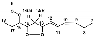
Table 2. Comparison of experimental 1H- and 13C-NMR chemical shifts of the 9-cis, 11-trans-16-OOH diastereomeric endo-hydroperoxides of methyl linolenate of the present work and with the reported literature data with the use of isolated endo-hydroperoxides.
Molecules 25 04902 i001
9-cis, 11-trans-16-OOH
endo-peroxy
(threo)
9-cis, 11-trans-16-OOH
endo-peroxy
(erythro)
9-cis, 11-trans-16-OOH
endo-peroxy
(threo)
Proton no.δ (1H),
ppm
δ (1H),
ppm a
δ (1H),
ppm b
δ (1H),
ppm
δ (1H),
ppm a
δ (13C),
ppm
δ (13C),
ppm b
181.051.041.031.071.0510.010.2
171.49---1.89−1.201.66---22.222.8
164.134.144.153.873.8687.287.4
154.494.494.494.494.4883.383.5
14(a)2.842.862.842.882.8840.741.3
14(b)2.482.462.472.232.23
134.814.804.804.794.7882.882.9
125.635.625.625.585.57126.2126.3
116.656.676.676.676.64131.6131.8
106.006.006.016.015.99127.3127.3
95.555.545.555.545.54135.4135.2
OOH9.509.529.389.089.04
a Ref [35]; b Ref [4].
Table 3. 1H-NMR quantification data of hydroperoxides and endo-hydroperoxides produced during the oxidation of methyl linolenate a.
Hydroperoxide δ (1H), ppmIntegration
Data (%)
Integration Data (%) of Total HydroperoxidesAssignment
7.925.78%29% (33.0%) b10-Trans, 12-cis, 15-cis-9-OOH
8.042.89%14% (10.8%) b9-Cis, 13-trans, 15-cis-12-OOH
8.058.23%41% (43.9%) b10-Cis, 13-cis, 15-trans-16-OOH
8.063.08%15% (12.3%) b9-Cis, 11-trans, 15-cis-13-OOH
9.081.80% 9-Cis, 11-trans, syn erythro, 16-OOH endo-hydroperoxide
9.121.36% 13-Trans, 15-cis, syn erythro, 9-OOH endo-hydroperoxide
9.502.40% 9-Cis, 11-trans, syn threo, 16-OOH endo-hydroperoxide
9.551.88% c 13-Trans, 15-cis, syn threo, 9-OOH endo-hydroperoxidec
a Heated at 40 °C for 48 h of the present work. b % Integration data from the literature at 40 °C [36]. c The concentration of the threo endo-hydroperoxide was estimated to be 1.68 mM in the NMR tube, which can be compared with the detection limit of 3 μM of the 800 MHz NMR instrument.