Article

Thermostable Tannase from Aspergillus Niger and Its Application in the Enzymatic Extraction of Green Tea

1
College of Food and Biological Engineering, Jimei University, Xiamen 361021, China
2
National R&D Center for Red Alga Processing Technology, Xiamen 361021, China
3
Fujian Provincial Engineering Technology Research Center of Marine Functional Food, Xiamen 361021, China
4
Xiamen Key Laboratory of Marine Functional Food, Xiamen 361021, China
*
Author to whom correspondence should be addressed.
These authors contributed equally to this work and share first authorship.
Molecules 2020, 25(4), 952; https://doi.org/10.3390/molecules25040952
Submission received: 30 January 2020 / Revised: 17 February 2020 / Accepted: 18 February 2020 / Published: 20 February 2020
(This article belongs to the Special Issue Protein Engineering: Different Biotechnology Applications)

Abstract

:
Tannase is widely used in tea beverage processing because of its ability to catalyze the hydrolysis of hydrolysable tannins or gallic acid esters and effectively improve the quality of tea extracts through enzymatic extraction. A new thermophilic tannase was cloned from Aspergillus niger FJ0118 and characterized. The tannase exhibited an optimal reaction temperature of 80 °C and retained 89.6% of the initial activity after incubation at 60 °C for 2 h. The enzymatic extraction of green tea at high temperature (70 °C) for a short time (40 min) was devised on the basis of the superior thermal stability of tannase. The enzymatic reaction significantly increased the total polyphenol content of green tea extract from 137 g·kg−1 to 291 g·kg−1. The enzymatic reaction effectively degraded the ester catechins into non-ester catechins compared with the water extraction method. Results suggested that the thermally stable tannase exhibited potential applications in the enzymatic extraction of green tea beverage.

1. Introduction

Green tea is receiving considerable interest as a distinctively flavored beverage because of its antioxidant, anticarcinogenic, and antimutagenic properties [1,2] and is consumed in most Asian countries, especially in China and Japan. Tea polyphenols, including epigallocatechin gallate (EGCG), epigallocatechin (EGC), epicatechin gallate (ECG), epicatechin (EC), gallocatechin, gallocatechin gallate, and catechin contribute to the biological functions of tea [2]. As a nonfermented tea, green tea contains higher amounts of polyphenols than oolong and black teas [3]. About 75–80% of tea polyphenols are found in 30% of the soluble ingredients of tea [2,4], and tea extract products differ because of extraction methods and conditions.
The extraction conditions during tea beverage manufacturing aim to achieve the maximum extraction yield. Attempts have been made to improve the extractability of polyphenols and the clarity of tea by using pectinase and tannase for enzymatic extraction. Treatment with tannase alone can maximally improve the quality of black tea because of the high amounts of polyphenols in extracted solids [5]. Various cell wall-digesting enzymes have been used during green tea extraction to improve its quality in terms of aroma, flavor, taste, cold water solubility, and extraction yield [6]. However, enzymatic extraction can only be performed at low temperatures (30–40 °C) at prolonged treatment time (over 2 h) because of the limitations in the ideal temperatures for the enzymatic reaction [7]. Prolonged treatment leads to quality deterioration during extraction [8] and is inconvenient for industrial production.
A thermally stable enzyme should be developed to improve the efficiency of high-temperature extraction. Tannin acyl hydrolase (EC 3.1.1.20), also known as tannase, is an enzyme that hydrolyzes ester and phenolic carboxy bonds in tannins to form glucose and gallic acid (GA) [9]. The ability of tannase to break ester catechins eliminates protein precipitation during tea infusion, thereby improving the quality of the tea beverage [10,11]. Several studies have characterized tannase from plants, animals, and microorganisms. Jana et al. [12] characterized a thermostable, solvent-tolerant, and cytosafe tannase from Bacillus subtilis PAB2, which exhibits a half-life (t1/2) of 4.5 h at 60 °C. Gonçalves et al. [13] have reported that Emericella nidulans produces tannase that has hypertolerance to temperature and organic solvents and t1/2 of about 72 h at 90 °C. However, most of the reported thermostable tannases are not of food grade. Aspergillus niger holds the Generally Recognized as Safe status from the Food and Drug Authority and is the primary filamentous fungi utilized for tannase production. The thermostability of tannase from A. niger is mainly between 30 °C and 50 °C [14,15,16]. Many studies have focused on the application of tannase in tea extract at 30–50 °C to reduce tea cream formation and improve the taste and color of green tea beverages [10,17,18]. The application of a thermally stable tannase derived from a food-grade microorganism in the enzymatic extraction of tea at high temperatures (≥ 70 °C) has not been studied yet.
In this work, rAntan1, a new thermophilic tannase, was cloned from A. niger FJ0118, expressed through a 5 L bioreactor fermentation, and purified using diethyl-aminoethyl anion exchange chromatography. The enzymatic and catalytic properties of rAntan1 were investigated. rAntan1 exhibited an optimal reaction temperature of 80 °C and retained 89.6% of its activity at 60 °C after 2 h. rAntan1 was applied in the enzymatic extraction of tea given its superior thermal stability to enhance the extraction yield and quality of green tea.

2. Results and Discussion

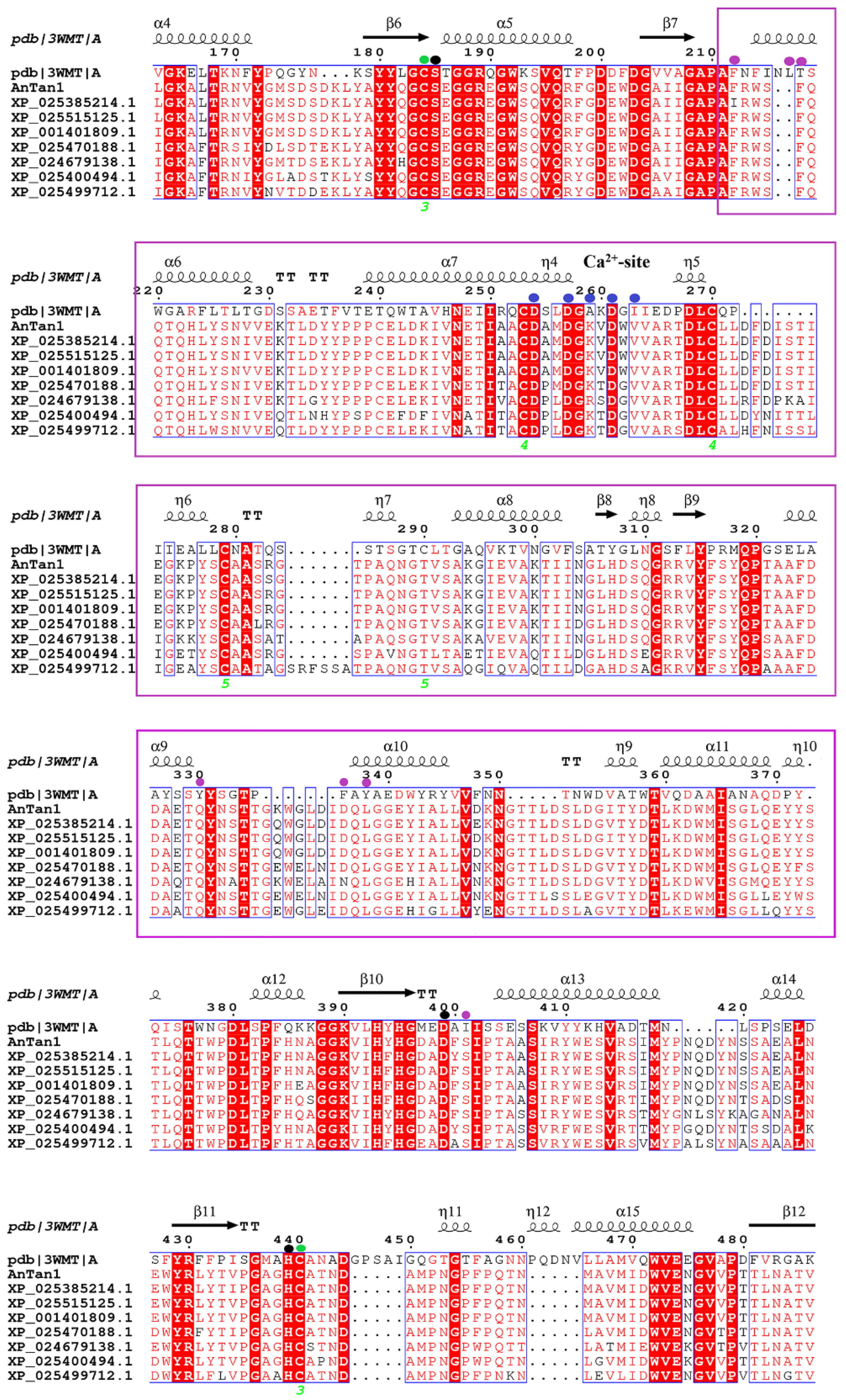
2.1. Analysis of Bioinformatics and Cloning of A. Niger Tannase

The tannase gene was amplified by PCR from A. niger FJ0118 genome in accordance with the sequence information of tannase (XM_001401772). The tannase gene, named Antan1, is 1725 bp in length without introns and codes 575 amino acids. The signal peptide of Antan1 was predicted using the SignalP (http://www.cbs.dtu.dk/services/SignalP-4.0/) as the N-terminal 20 amino acid. The amino acid sequences were aligned using the ClustalW (http://www.ebi.ac.uk/clustalw/), which showed that the Antan1 amino acid sequence had identity values of 98%, 95%, 91%, and 80% with tannases from Aspergillus luchuensis CBS 106.47 (GenBank accession number OJZ87444.1), Aspergillus brasiliensis CBS 101,740 (GenBank accession number OJJ71084.1), Aspergillus carbonarius ITEM 5010 (GenBank accession number OOF98052.1), and Aspergillus fischeri NRRL 181 (GenBank accession number XP_001261622.1), respectively. The phylogenetic tree was constructed for the assessment of the sequence relationship among the tannase family proteins (Figure 1). The protein functional domain of rAntan1 (Antan1 without signal peptide) was analyzed by comparing its sequence with the Pfam protein family database [19] (http://pfam.xfam.org/). Results indicated that amino acids 57–527 constituted a superfamily structure comprising a catalytic triad (serine–histidine–aspartic/glutamic acid). These characteristic sites were conserved in Antan1 and predicted to be Ser206, Asp439, and His485 (Figure 2, black circle) on the basis of the multiple sequence alignment of tannases [20,21]. In addition, rAntan1 has a “CS-D-HC motif” that is completely conserved among the biochemically distinct members of the tannase family [21]. In this CS-D-HC motif, two key residues in the catalytic triad, Ser206 and His485, are directly linked by the disulfide bonds of the adjacent cysteine residues (Cys205 and Cys486, Figure 2, green circle).

2.2. Tannase Production

The gene encoding rAntan1 was successfully expressed into Pichia pastoris and the verified transformant SMD-Antan1 was subjected to shake-flask fermentation. The rAntan1 activity reached a maximum of 1.55 U·mL−1 after 144 h of induction (data not shown). Fermentation was performed using a 5 L canister to increase the yield of rAntan1. The maximum biomass was 309 mg·mL−1, and the achieved rAntan1 activity was 390.4 U·mL−1 at 96 h (Supplementary Materials, Figure S1). The achieved rAntan1 activity was 252-fold of the yield of shake flask fermentation. The enzymatic activity of rAntan1 was higher than that of the tannases from A. niger Bdel4 (111.5 U·mL−1) [22], Pestalotiopsis guepinii (98.6 U·mL−1) [23] and Penicillium atramentosum (34.7 U·mL−1) [24], indicating the remarkable application potential of rAntan1 in the industry.

2.3. Enzymatic Characteristics

The molecular weight of pure rAntan1, as determined by sodium dodecyl sulfate (SDS)–polyacrylamide gel electrophoresis (PAGE), was 85 kDa (Figure 3), which was larger than the predicted 62.86 kDa gene product. In general, fungal tannases have a relatively high molecular weight (70–180 kDa), which is attributed to glycosylation [20,25].

2.3.1. Effects of Temperature and pH on Enzymatic Activity and Stability

As shown in Figure 4a, rAntan1 activity increased with increasing temperature (30–80 °C) and exhibited optimal reaction at 80 °C. rAntan1 retained more than 50% of its relative activity at 90 °C, showing superior heat-resistant ability. As shown in Figure 4b, rAntan1 retained 91.9% and 89.6% of its initial activity after incubation at 60 °C for 60 and 120 min, respectively. The half-life (t1/2) of rAntan1 at 60 °C was 5.4 h, suggesting its thermal stability. At 70 °C, rAntan1 retained 17.7% of its initial activity after 60 min of incubation. In addition, rAntan1 activity remained unchanged during incubation at 30–50 °C for 36 h (data not shown). As shown in Table 1, rAntan1 exhibited higher optimal reaction temperature (80 °C) than most reported microbially derived tannases (20–60 °C) [13,16,20,22,26,27,28,29,30]. The tannase from Aspergillus phoenicis [30] retained 10% of its initial activity after incubation at 60 °C for 60 min. Compared with the tannases reported in literature, rAntan1 is promising for thermal industrial applications.
As shown in Figure 4c, the optimal pH of rAntan1 was 6.0, and its capacity to hydrolyze the substrate was maintained by approximately 60%–100% under a wide pH range (pH 3–8). rAntan1 was stable at acidic to slightly basic pH conditions (3.0–8.0; Figure 4d). These results were comparable with those of the most reported fungal tannases [26,31]. However, other tannases, such as those from E. nidulans (pH 4.0–5.0) [13] and Aspergillus aculeatus (pH 5.0–6.0) [32], show a limited range of pH stability. The excellent stability of rAntan1 at high temperatures and a wide range of pH may pave the way for carrying out biotechnological processes with minimal risk of microbial contamination with reaction efficiency.

2.3.2. Effects of Metal Ions, Surfactants, Enzyme Inhibitors, and Solvents on Enzyme Activity

The effects of different metal ions on rAntan1 activity are shown in Table 2. Most metal ions (i.e., Na+, Mg2+, Cu2+, Ba2+, Zn2+, Cd2+, Al3+, and Mn2+) inhibited the activity of rAntan1 at 1.0 mM concentration, and Ca2+ had the highest inhibitory effect (32%). Cu2+ remarkably decreased enzymatic activity to 34% when its concentration increased to 10 mM. Heavy metal ions inhibited most tannases because the ions may have bound with sulfhydryl groups, tryptophan residues, and/or carboxyl groups at the enzyme active site [31].
As shown in Table 2, rAntan1 was slightly inhibited by SDS, ethylene diamine tetraacetic acid (EDTA), Tween-20, and Tween-80. β-mercaptoethanol had a strong inhibitory effect on the activity of rAntan1, which was possibly because of the reduction of the disulfide bond of the active center of tannase that deactivated the enzyme [29].
The effect of organic solvents on rAntan1 activity is shown in Table 3. At 40% concentration, the addition of n-hexane and cyclohexane resulted in enzyme activation by 56% and 157%, respectively, whereas dimethyl sulfoxide, n-butanol, n-propanol, and methanol showed strong enzymatic inhibition by 57%, 55%, 96%, and 77%, respectively. This result indicated that nonpolar and polar solvents can promote and inhibit, respectively, the activity of rAntan1. Similar reports have indicated that tannase can be activated by 60% benzene [33], whereas polar solvents, such as 20% acetone, inhibits tannase activity by 70% [13].

2.3.3. Substrate Specificity

The purified rAntan1 enzyme was incubated with different esters of phenolic acids to study its substrate spectrum. The results in Table 4 show that rAntan1 exhibited higher activity toward natural substrates (ECG and CG) than synthetic substrates (PG). The strong degradation of ECG and CG by rAntan1 as compared with other tannase suggests that rAntan1 can be used for tea treatment [20].

2.3.4. Kinetic Parameters

The kinetic parameters namely, the Michaelis–Menten constant (Km) and maximal velocity (Vmax), were calculated by the hydrolysis of PG (Supplementary Materials, Figure S2). The Lineweaver–Burk plot of rAntan1 gave a Km of 0.073 mM, which was lower than that of the tannases from Penicillium herquei (0.62 mM) [29] and A. phoenicis (0.6 mM) [30]. This result indicated that rAntan1 had higher substrate affinity than these tannases. rAntan1 exhibited Vmax, catalytic constant (kcat), and catalytic efficiency (kcat/Km) values of 0.29 U·mg−1, 1.06 × 10³ s−1 and 14.5 × 10³ mM−1 s−1, respectively. These values were higher than those for the tannase derived from E. nidulans (0.05 U·mg−1) [13] and Aspergillus oryzae (16.6 mM−1 s−1) when using PG as a substrate [28].

2.4. Application of Tannase in Tea Extraction

In general, the enzymatic treatment process for tea beverage can be performed in three stages, namely, tea pretreatment, extraction, and enzymatic hydrolysis [8]. Enzymatic extraction promotes extraction and improves the flavor of the extract [7], but prolonged treatment time leads to quality deterioration [8]. Several studies have reported the effects of extraction temperature, time, and method on the quality of tea infusion [34,35,36,37,38]. The tea infusion obtained using hot water showed better scavenging of free radicals than that obtained using cold water, and the extraction yield increased with increasing temperature and time. However, the active components (e.g., EGCG, EGC, and EC) decrease at temperatures higher than 80 °C because of degradation, oxidation, or epimerization [3]. Moreover, Yang et al. [39] compared the tea infusion content obtained using varying steeping temperatures (70 °C, 85 °C, and 100 °C). The tea steeped in water at 70 °C contains the highest caffeine, catechin, and GA contents.
Thus, the excellent thermostable rAntan1 was used in the enzyme-mediated extraction of green tea at 70 °C. The changes in catechin content in the tea infusions after different extraction methods (water and enzymatic extraction) were measured using high-performance liquid chromatography (HPLC). Figure 5a and Table 5 show that the enzymatic extraction remarkably improved the contents of non-ester catechins (EGC and EC) and decreased the content of ester catechins (EGCG and ECG). Ester catechins are the source of the bitter taste in tea infusion [40]. Thus, enzymatic extraction reduced the proportion of ester catechins in tea polyphenols, weakened the bitter taste, increased the sweet aftertaste, and improved the overall taste of the tea beverage. The amount of EGCG obtained through enzymatic extraction was almost impossible to detect (0 mg·L−1) compared with that obtained through water extraction. EGCG and caffeine simultaneously enhance each other’s adverse effects, such as astringency and bitterness, thereby inhibiting the development of sweet aftertaste [10]. As a nonfermented tea, green tea contains higher amounts of EGCG and ECG, which are associated with astringency and bitterness [36], compared with oolong (semifermented) and black (fully fermented) teas [40]. Therefore, enzymatic extraction can develop the sweet aftertaste of tea infusion.
In addition, a comparison of the main catechin variables and total polyphenol content showed that the enzymatic extraction of green tea improved the extract yield and remarkably changed its composition (Table 5). The polyphenol recoveries from the green tea used in the study under enzymatic and water extraction conditions were 291 and 137 g·kg−1, respectively. Enzymatic extraction showed an improvement by 2.1-fold. Chandini et al. [5] have reported an improvement in the polyphenol recovery through tannase-assisted extraction. This improvement may be because tannase cleaves some of the crosslinks existing between cell wall polymers that cause the degradation of plant cell wall, leading to an increase in polyphenol recovery [41]. Tea is valued for its polyphenols, which exhibit positive physiological and pharmacological effects, such as antioxidant [5], anticarcinogenic [2], and antimutagenic effects [1]. The enzymatic extraction time used in the present study achieved a significant reduction compared with other reports [5,6]. Preliminary results indicated that enzymatic extraction at high temperature is a promising approach to improve the quality of green tea extracts during the industrial production of tea beverages.

3. Materials and Methods

3.1. Microorganism

A. niger FJ0118 was purchased from the China Center for Type Culture Collection. Escherichia coli DH5α and P. pastoris strain SMD1168 were sourced from the Key Laboratory of Food Microbiology and Enzyme Engineering of Fujian Province (Jimei University, Xiamen, China). The extracellular expression vector pPIC9K was purchased from TaKaRa Bio (Dalian, China). Green tea (Xinyang Maojian, China) was purchased from a local market (Xiamen, China). The catechin standards (EGC, EGCG, ECG, and EC), GA, and caffeine were bought from Chengdu Biopurify Phytochemicals, Ltd. (Chengdu, China). All other reagents were of analytical grade and purchased from Sinopharm Chemical Reagent Co., Ltd. (Shanghai, China).

3.2. Construction of Plasmid

Primers Tan-F (5′-ATGCGCTCACCCACTCGAGTTTCC-3′) and Tan-R (5′- CTAGTACACAGGCATGGGAACCGCA-3′) were designed and compounded in accordance with the tannase gene sequence from A. niger CBS 513.88 (NCBI accession number XM_001401772) for the amplification of the tannase gene with the genomic DNA of A. niger FJ0118. The purified polymerase chain reaction (PCR) products were cloned into the pMD19-T vector (TaKaRa, Dalian, China), which constructed the plasmid pMD-Tan. The ligation mixture was transformed into E. coli DH5α. Positive clones were selected and confirmed by sequencing. For removing a signal peptide, Tan-F2 (5′-ACGACTCCTAGGGGGACTCCTTCCACGTTGGCGGA-3′; the AvrII site is underlined) and Tan-R2 (5′-ATAAGAATGCGGCCGCCTAGTACACAGGCATGGGAACCGCAT-3′; the NotI site is underlined) were designed and compounded to amplify the tannase gene with the plasmid DNA of the pMD-Tan as the template. The purified PCR products were digested with AvrII/NotI and ligated into the same enzyme-digested vector pPIC9K to generate the plasmid pPIC9K–Tan containing the tannase gene under the control of the alcohol oxidase 1 (AOX1) gene promoter. The recombinant plasmid pPIC9K–Tan was linearized with SalI and transformed into P. pastoris SMD1168 on the basis of the P. pastoris transformation protocol (Thermo Fisher Scientific, shanghai, China).

3.3. Production of Tannase

Shake flask fermentation was performed using buffered glycerol and buffered methanol complex media, as described by [20] to produce rAntan1.
The 5 L scale fermentation was performed in three phases:
(A) Glycerol fed-batch phase: Glycerol was added in batches in accordance with the change in dissolved oxygen until the biomass reached 180 mg·mL−1.
(B) Starvation phase: In this stage, no carbon source was added so that cell starvation for 30 min was ensured and the utilization of methanol by cells was not affected.
(C) Methanol fed-batch phase: Methanol containing 1.2% PTM1 was added at 1.5 mL h−1 L−1 during the first 12 h and increased to 3 mL h−1 L−1. This phase lasted for 96 h. The biomass and enzyme activity were measured at 8 h interval over the whole induction period.

3.4. Activity Assay of Tannase

rAntan1 activity was measured by examining the production of the GA released from propyl gallate. The amount of GA was estimated through a spectrophotometric method based on the formation of chromogen between GA and rhodamine [42]. The absorbance was recorded against the inactivated enzyme blank at 520 nm. One unit of enzyme activity was defined as the amount of enzyme required to produce 1 μmol of GA per minute at assay conditions.

3.5. Enzyme Purification

The crude enzyme was collected from the 5 L scale fermentation by centrifugation at 4000× g and 4 °C for 15 min. The supernatant was filtered through a 0.22 μm nylon membrane and applied to 1.6 cm × 20 cm DEAE–Sepharose fast flow column equilibrated with citrate buffer (10 mmol·L−1, pH 5.0). The enzyme was eluted with a stepwise increase in NaCl (0.05 and 0.1 M) in the same buffer at a flow rate of 1 mL·min−1. The fractions with tannase activities were combined and concentrated using a 10-kDa ultrafiltration membrane for SDS–PAGE.

3.6. Characterization of Tannase

3.6.1. Effects of Temperature and pH

The enzyme reaction was carried out within the temperature range of 30–90 °C to determine the optimum temperature. The thermal stability was investigated by treating rAntan1 at various temperatures (30–70 °C) for 20, 40, 60, 80, 100, and 120 min. The optimum pH was studied by performing the enzymatic reaction in citrate (0.05 M, pH 3.0–7.0), sodium phosphate (0.05 M, pH 7.0–9.0), and glycine-NaOH (0.05 M, pH 9.0–10.0) buffer solutions. The pH stability of the rAntan1 was determined by treating the enzyme in the same buffers at 4 °C for 24 h. The residual activity was detected under standard assay conditions and expressed as percentage of the initial activity.

3.6.2. Effect of Metal Ions and Other Additives on Enzyme Activity

rAntan1 was treated with metal ions (Na+, Mg2+, Fe3+, Cu2+, Ba2+, Zn2+, Cd2+, Ca2+, Mn2+, K+, and Al3+; 1 and 10 mM), surfactants (Tween-20, Tween-80, Triton X-100, SDS, and β-mercaptoethanol; 0.1% and 1% [v/v]), and metal chelators (EDTA; 1 and 10 mM) at 30 °C for 60 min. The enzyme activity without additives under the same measurement was determined as 100%.

3.6.3. Effects of Organic Solvents on Enzyme Activity

The effects of organic solvents including dimethyl sulfoxide, hexane, butanol, cyclohexane, propanol, isoamyl alcohol, benzene, methanol, and chloroform, on rAntan1 activity were estimated. The enzyme solution was treated at 30 °C for 60 min by using organic solvents at final concentrations of 20%, 40%, and 60%. The residual activity was detected under standard assay conditions and expressed as percentage of the initial activity.

3.6.4. Substrate Specificity and Kinetic Constants of Tannase

The hydrolytic activity of rAntan1 on EGCG, TA, PG, ECG, and CG was analyzed at a concentration of 0.01 M.
The values of the kinetic parameters, namely, Km, Vmax, kcat, and kcat/Km were estimated using PG (0.05–1.0 M) as substrate at standard assay condition. The calculation of kinetic parameters was based on the Lineweaver–Burk plots [43].

3.7. Application of Tannase

3.7.1. Enzymatic Extraction of Green Tea

Ground green tea leaves (40 mesh, 2 g) were steeped in water (leaf/water ratio of 1:50 [w/v]) and added to rAntan1 (10 U·g−1 green tea). The mixture was incubated in a closed system maintained at 70 °C for 40 min for tea extraction and catechin degradation. The enzyme reaction was inactivated by incubating the mixture in boiling water for 10 min. The samples were filtered prior to the analyses of catechin and total polyphenol contents.

3.7.2. Determination of Catechins and Caffeine by HPLC

The contents of catechins (ECG, EGC, EGCG, and EC), GA, and caffeine in the tea infusion samples were determined at 278 nm by using HPLC (Agilent Technologies, CA, USA). The samples were analyzed using a symmetry C18 column (3.0 mm × 250 mm, 5 μm). Elution was performed by using phases A (0.5% aqueous acetic acid solution) and B (acetonitrile). The elution was run for 45 min in a gradient as previously described [9]. The mobile phase flow rate was 0.5 mL·min−1.

3.7.3. Total Polyphenols Content

The content of total polyphenols in the extracts was determined using the Folin–Ciocalteu method [44]. A calibration curve was prepared using GA as the standard, and the results were expressed in g of GA equivalent per kg of green tea (g·kg−1).

4. Conclusions

rAntan1, a novel thermostable tannase, was cloned from A. niger FJ0118, and its enzymatic properties were characterized. This tannase exhibited a t1/2 of 5.4 h and 30 min at 60 °C and 70 °C, respectively, suggesting its remarkable thermal stability. A novel high-temperature enzymatic extraction was applied using the thermostable rAntan1 for polyphenol recovery and ester catechin degradation during green tea processing. The enzymatic extraction at high temperatures resulted in high non-ester catechin and total polyphenol contents than the traditional water extraction strategy. The enzymatic extraction time was reduced significantly and more effectively than those in other similar studies. In conclusion, high-temperature enzymatic extraction is a promising approach for improving the quality of green tea extracts during the industrial production of tea beverages.

Supplementary Materials

The following are available online at https://www.mdpi.com/1420-3049/25/4/952/s1, Figure S1: rAntan1 production process in 5 L fermentation tanks. Figure S2. Lineweaver–Burk plot of rAntan1.

Author Contributions

Y.-H.Z. and A.-F.X. designed the experiments. Y.S., F.Z., Q.-M.Y., and H.-F.W. conducted the experiments. Y.-H.Z., Y.S., Q.X., and A.-F.X. analyzed the data. Y.S., Y.-H.Z., and A.-F.X. wrote the main manuscript. All authors have read and agreed to the published version of the manuscript.

Funding

This work was supported by Projects of Science and Technology Program of Xiamen City (3502Z20183030) and Science and Technology Planning Project of Fujian Province, China (2016N0021).

Conflicts of Interest

The authors declare no competing financial interest.

References

  1. Zhang, Y.; Yin, J.; Chen, J.; Wang, F.; Du, Q.; Jiang, Y.; Xu, Y. Improving the sweet aftertaste of green tea infusion with tannase. Food Chem. 2016, 192, 470–476. [Google Scholar] [CrossRef] [PubMed]
  2. Lu, M.; Chu, S.; Yan, L.; Chen, C. Effect of tannase treatment on protein–tannin aggregation and sensory attributes of green tea infusion. Lwt - Food Sci. Technol. 2009, 42, 338–342. [Google Scholar] [CrossRef]
  3. Ananingsih, V.K.; Sharma, A.; Zhou, W. Green tea catechins during food processing and storage: A review on stability and detection. Food Res. Int. 2013, 50, 469–479. [Google Scholar] [CrossRef]
  4. Perva-Uzunalić, A.; Škerget, M.; Knez, Ž.; Weinreich, B.; Otto, F.; Grüner, S. Extraction of active ingredients from green tea (Camellia sinensis): Extraction efficiency of major catechins and caffeine. Food Chem. 2006, 96, 597–605. [Google Scholar] [CrossRef]
  5. Chandini, S.K.; Rao, L.J.; Gowthaman, M.K.; Haware, D.J.; Subramanian, R. Enzymatic treatment to improve the quality of black tea extracts. Food Chem. 2011, 127, 1039–1045. [Google Scholar] [CrossRef] [PubMed]
  6. Dubey, K.K.; Janve, M.; Ray, A.; Singhal, R.S. Ready-to-Drink Tea. In Trends in Non-alcoholic Beverages; Academic Press: Cambridge, MA, USA, 2020; pp. 101–140. [Google Scholar]
  7. Hong, Y.H.; Jung, E.Y.; Park, Y.; Shin, K.S.; Kim, T.Y.; Yu, K.W.; Chang, U.J.; Suh, H.J. Enzymatic improvement in the polyphenol extractability and antioxidant activity of green tea extracts. Biosci. Biotechnol. Biochem. 2013, 77, 22–29. [Google Scholar] [CrossRef]
  8. Murugesh, C.S.; Subramanian, R. Applications of Enzymes in Processing Green Tea Beverages: Impact on Antioxidants - Processing and Impact on Antioxidants in Beverages. In Processing and Impact on Antioxidants in Beverages; Academic Press: Cambridge, MA, USA, 2014; pp. 99–108. [Google Scholar]
  9. Ni, H.; Chen, F.; Jiang, Z.D.; Cai, M.Y.; Yang, Y.F.; Xiao, A.F.; Cai, H.N. Biotransformation of tea catechins using Aspergillus niger tannase prepared by solid state fermentation on tea byproduct. Lwt - Food Sci. Technol. 2015, 60, 1206–1213. [Google Scholar] [CrossRef]
  10. Cao, Q.; Zou, C.; Zhang, Y.; Du, Q.; Yin, J.; Shi, J.; Xue, S.; Xu, Y. Improving the taste of autumn green tea with tannase. Food Chem. 2019, 277, 432–437. [Google Scholar] [CrossRef]
  11. Baik, J.H.; Shin, K.; Park, Y.; Yu, K.; Suh, H.J.; Choi, H. Biotransformation of catechin and extraction of active polysaccharide from green tea leaves via simultaneous treatment with tannase and pectinase. J. Sci. Food Agr. 2015, 95, 2337–2344. [Google Scholar] [CrossRef]
  12. Jana, A.; Maity, C.; Halder, S.K.; Das, A.; Pati, B.R.; Mondal, K.C.; Das Mohapatra, P.K. Structural characterization of thermostable, solvent tolerant, cytosafe tannase from Bacillus subtilis PAB2. Biochem. Eng. J. 2013, 77, 161–170. [Google Scholar] [CrossRef]
  13. Gonçalves, H.B.; Riul, A.J.; Terenzi, H.F.; Jorge, J.A.; Guimarães, L.H.S. Extracellular tannase from Emericella nidulans showing hypertolerance to temperature and organic solvents. J. Mol. Catal. B: Enzym. 2011, 71, 29–35. [Google Scholar] [CrossRef]
  14. Mata-Gomez, M.; Rodriguez, L.V.; Ramos, E.L.; Renovato, J.; Cruz-Hernandez, M.A.; Rodriguez, R.; Contreras, J.; Aguilar, C.N. A novel tannase from the xerophilic fungus Aspergillus niger GH1. J. Microbiol. Biotechnol. 2009, 19, 987–996. [Google Scholar] [PubMed] [Green Version]
  15. Ramirez-Coronel, M.A.; Viniegra-Gonzalez, G.; Darvill, A.; Augur, C. A novel tannase from Aspergillus niger with beta-glucosidase activity. Microbiology 2003, 149, 2941–2946. [Google Scholar] [CrossRef] [PubMed] [Green Version]
  16. Sabu, A.; Kiran, G.S.; Pandey, A. Purification and Characterization of Tannin Acyl Hydrolase from Aspergillus niger ATCC 16620. Food Technol. Biotechnol. 2005, 43, 133–138. [Google Scholar]
  17. Ozturk, B.; Seyhan, F.; Ozdemir, I.S.; Karadeniz, B.; Bahar, B.; Ertas, E.; Ilgaz, S. Change of enzyme activity and quality during the processing of Turkish green tea. Lwt - Food Sci. Technol. 2016, 65, 318–324. [Google Scholar] [CrossRef]
  18. Lu, M.; Chen, C. Enzymatic modification by tannase increases the antioxidant activity of green tea. Food Res. Int. 2008, 41, 130–137. [Google Scholar] [CrossRef]
  19. Finn, R.D.; Coggill, P.; Eberhardt, R.Y.; Eddy, S.R.; Mistry, J.; Mitchell, A.L.; Potter, S.C.; Punta, M.; Qureshi, M.; Sangrador-Vegas, A. The Pfam protein families database: Towards a more sustainable future. Nucleic Acids Res. 2016, 44, D279–D285. [Google Scholar] [CrossRef] [PubMed]
  20. Koseki, T.; Ichikawa, K.; Sasaki, K.; Shiono, Y. Characterization of a novel Aspergillus oryzae tannase expressed in Pichia pastoris. J. Biosci. Bioeng. 2018, 126, 553–558. [Google Scholar] [CrossRef]
  21. Suzuki, K.; Hori, A.; Kawamoto, K.; Thangudu, R.R.; Ishida, T.; Igarashi, K.; Samejima, M.; Yamada, C.; Arakawa, T.; Wakagi, T.; et al. Crystal structure of a feruloyl esterase belonging to the tannase family: A disulfide bond near a catalytic triad. Proteins: Struct. Funct. Bioinform. 2014, 82, 2857–2867. [Google Scholar] [CrossRef]
  22. Liu, F.; Wang, B.; Ye, Y.; Pan, L. High level expression and characterization of tannase tan7 using Aspergillus niger SH-2 with low-background endogenous secretory proteins as the host. Protein Expres. Purif. 2018, 144, 71–75. [Google Scholar] [CrossRef]
  23. Amanda, R.D.S.; Gouveia, M.J.; Tonny, C.C.L.; Aparecida Moreira, K.; Sandra, A.D.A. Production, Characterization and Application of a Thermostable Tannase from Pestalotiopsis guepinii URM 7114. Food Technol. Biotechnol. 2014, 52, 459–467. [Google Scholar]
  24. Selwal, M.K.; Selwal, K.K. High-level tannase production by Penicillium atramentosum KM using agro residues under submerged fermentation. Ann. Microbiol. 2012, 62, 139–148. [Google Scholar] [CrossRef]
  25. Kumar, M.; Beniwal, V.; Salar, R.K. Purification and characterization of a thermophilic tannase from Klebsiella pneumoniae KP715242. Biocatal. Agric. Biotechnol. 2015, 4, 745–751. [Google Scholar] [CrossRef]
  26. Abdel-Naby, M.A.; El-Tanash, A.B.; Sherief, A.D.A. Structural characterization, catalytic, kinetic and thermodynamic properties of Aspergillus oryzae tannase. Int. J. Biol. Macromol. 2016, 92, 803–811. [Google Scholar] [CrossRef] [PubMed]
  27. Gayen, S.; Ghosh, U. Purification and Characterization of Tannin Acyl Hydrolase Produced by Mixed Solid State Fermentation of Wheat Bran and Marigold Flower by Penicillium notatum NCIM 923. Biomed. Res. Int. 2013, 2013, 1–6. [Google Scholar] [CrossRef] [PubMed] [Green Version]
  28. Mizuno, T.; Shiono, Y.; Koseki, T. Biochemical characterization of Aspergillus oryzae native tannase and the recombinant enzyme expressed in Pichia pastoris. J. Biosci. Bioeng. 2014, 118, 392–395. [Google Scholar] [CrossRef]
  29. Qiu, Y.; Niu, H.; Huang, W.; He, Y.; Wu, X. Properties and secondary structure of tannase from Penicillium herquei. Biotechnol. Bioproc. E 2011, 16, 858–866. [Google Scholar] [CrossRef]
  30. Riul, A.J.; Gonçalves, H.B.; Jorge, J.A.; Guimarães, L.H.S. Characterization of a glucose- and solvent-tolerant extracellular tannase from Aspergillus phoenicis. J. Mol. Catal. B: Enzym. 2013, 85, 126–133. [Google Scholar] [CrossRef]
  31. Mahmoud, A.E.; Fathy, S.A.; Rashad, M.M.; Ezz, M.K.; Mohammed, A.T. Purification and characterization of a novel tannase produced by Kluyveromyces marxianus using olive pomace as solid support, and its promising role in gallic acid production. Int. J. Biol. Macromol. 2018, 107, 2342–2350. [Google Scholar] [CrossRef]
  32. El-Tanash, A.B.; Sherief, A.A.; Nour, A. Catalytic properties of immobilized tannase produced from Aspergillus aculeatus compared with the free enzyme. Braz. J. Chem. Eng. 2011, 28, 381–391. [Google Scholar] [CrossRef]
  33. Chhokar, V.; Sangwan, M.; Beniwal, V.; Nehra, K.; Nehra, K.S. Effect of Additives on the Activity of Tannase from Aspergillus awamori MTCC9299. Appl. Biochem. Biotech. 2010, 160, 2256–2264. [Google Scholar] [CrossRef]
  34. Castiglioni, S.; Damiani, E.; Astolfi, P.; Carloni, P. Influence of steeping conditions (time, temperature, and particle size) on antioxidant properties and sensory attributes of some white and green teas. Int. J. Food Sci. Nutr. 2015, 66, 491–497. [Google Scholar] [CrossRef]
  35. Kim, E.S.; Liang, Y.R.; Jin, J.; Sun, Q.F.; Lu, J.L.; Du, Y.Y.; Lin, C. Impact of heating on chemical compositions of green tea liquor. Food Chem. 2007, 103, 1263–1267. [Google Scholar] [CrossRef]
  36. Xu, Y.; Ji, W.; Yu, P.; Chen, J.; Wang, F.; Yin, J. Effect of extraction methods on the chemical components and taste quality of green tea extract. Food Chem. 2018, 248, 146–154. [Google Scholar] [CrossRef] [PubMed]
  37. Venditti, E.; Bacchetti, T.; Tiano, L.; Carloni, P.; Greci, L.; Damiani, E. Hot vs. cold water steeping of different teas: Do they affect antioxidant activity? Food Chem. 2010, 119, 1597–1604. [Google Scholar] [CrossRef]
  38. Labbé, D.; Tremblay, A.; Bazinet, L. Effect of brewing temperature and duration on green tea catechin solubilization: Basis for production of EGC and EGCG-enriched fractions. Sep. Purif. Technol. 2006, 49, 1–9. [Google Scholar] [CrossRef]
  39. Yang, D.; Hwang, L.S.; Lin, J. Effects of different steeping methods and storage on caffeine, catechins and gallic acid in bag tea infusions. J. Chromatogr. A 2007, 1156, 312–320. [Google Scholar] [CrossRef] [PubMed]
  40. Li, J.; Xiao, Q.; Huang, Y.; Ni, H.; Wu, C.; Xiao, A. Tannase application in secondary enzymatic processing of inferior Tieguanyin oolong tea. Electron. J. Biotechn. 2017, 28, 87–94. [Google Scholar] [CrossRef]
  41. García-Conesa, M.T.; Østergaard, P.; Kauppinen, S.; Williamson, G. Hydrolysis of diethyl diferulates by a tannase from Aspergillus oryzae. Carbohyd. Polym. 2001, 44, 319–324. [Google Scholar] [CrossRef]
  42. Sharma, S.; Bhat, T.K.; Dawra, R.K. A Spectrophotometric Method for Assay of Tannase Using Rhodanine. Anal. Biochem. 2000, 279, 85–89. [Google Scholar] [CrossRef]
  43. Lineweaver, H.; Burk, D. The Determination of Enzyme Dissociation Constants. J. Am. Chem. Soc. 1934, 56, 658–666. [Google Scholar] [CrossRef]
  44. Swain, T.; Hillis, W.E. The phenolic constituents of Prunus domestica. I.—The quantitative analysis of phenolic constituents. J. Sci. Food Agr. 1959, 10, 63–68. [Google Scholar] [CrossRef]
Sample Availability: Samples of the compounds are not available from the authors.
Figure 1. Phylogenetic relationships among known tannase family proteins. Amino acid sequence alignment was performed using ClustalW, and the phylogenetic tree was constructed using molecular evolutionary genetics analysis software version 7.0 (MEGA7). The bar represents 0.05 amino acid substitutions per site.
Molecules 25 00952 g001
Figure 2. Alignment of multiple amino acid sequences of Antan1. The residues involved in formation of the substrate-binding pocket and the calcium-coordinating residues are indicated by black, green, magenta, and blue marks, respectively. The lid domain is boxed with magenta lines. Numbers below the sequences indicate disulfide bond pairs.
Molecules 25 00952 g002
Figure 3. SDS–PAGE results of rAntan1 from A. niger FJ0118, M: protein marker, lane 1: crude enzyme, lane 2: purified enzyme.
Molecules 25 00952 g003
Figure 4. Effects of temperature and pH on the activity and the stability of rAntan1. (a) The optimal temperature for rAntan1 was measured at pH 5.0 and different temperatures. (b) The thermal stability of rAntan1 was determined after incubating at 60 °C (■), 70 °C (▲) for 120 min. (c) The optimal pH for rAntan1 was determined by performing an activity assay in 50 mM citrate (pH 3.0–7.0, ■), 50 mM phosphate (pH 7.0–9.0, ▲), and 50 mM Gly-NaOH (pH 9.0–10.0, □) buffer solutions. (d) The pH stability of rAntan1 was determined by incubation at different pH values and 4 °C for 24 h. The residual activity was determined at the standard assay conditions.
Molecules 25 00952 g004
Figure 5. (a) Changes in catechins in the tea infusion after different treatment strategies. 1: GA (gallic acid), 2: EGC (epigallocatechin), 3: caffeine, 4: EGCG (epigallocatechin gallate), 5: EC (epicatechin), 6: ECG (epicatechin gallate). (b) Chromatograms of standards.
Molecules 25 00952 g005
Table 1. A summarized list of tannase properties.
MicroorganismMwKm
(1) Tannic Acid
(2) Methyl-Gallate
(3) Propyl-Gallate
pH
(1) Optima
(2) Stability
Temperature
(1) Optima
(2) Stability
(3) Residual Enzyme Activity at 60 °C for 1h
Ref.
Penicillium notatum97 kDa,
43 kDa (Dimeric)
3.3 mM (1)(1) 5.0
(2) 3.0–8.0
(1) 35–40 °C
(2) up to 60 °C
(3) 60%
Gayen and Ghosh 2013
Emericella nidulans302 kDa7.69 mM (3)(1) 5.0
(2) 4.0–5.0
(1) 45 °C
(2) 22–50 °C
Gonçalves et al. 2011
Aspergillus oryzae45–80 kDan.s.(1) 4.0–5.0(1) 40 °C
(2) up to 40 °C
Mizuno et al. 2014
Aspergillus phoenicis120 kDa,
93 kDa (Dimeric)
1.7 mM (1), 14.3 mM (2) and 0.6 mM (3)(1) 6.0
(2) 2.5–7.0
(1) 60 °C
(2) 40–60 °C
(3) 10%
Riul et al. 2013
Penicillium herquei72 kDa0.62 mM (3)(1) 6.0(1) 30 °CQiu et al. 2011
Aspergillus niger
SH-2
60–70 kDan.s.(1) 7.0(1) 40 °CLiu et al. 2018
Aspergillus oryzae90–120 kDa1.11 mM (2)(1) 6.0(1) 30–35 °C
(2) up to 40 °C
(3) 20%
Koseki et al. 2018
Aspergillus niger
ATTC 16620
168 kDan.s.(1) 6.0
(2) 4.0–8.0
(1) 40 °C
(2) 30–40 °C
Sabu et al. 2005
Aspergillus oryzae106 kDa3.13 mM (1)(1) 5.5
(2) 4.5–7.5
(1) 37 °CAbdel-Naby et al. 2016
Aspergillus niger85 kDa0.073 mM (3)(1) 6.0
(2) 3.0–8.0
(1) 80 °C
(2) 30–60 °C
(3) 91.9%
This work
n.s. means not available in the reports.
Table 2. Influence of metal ions, inhibitors, and surfactants on rAntan1.
Residual Activity (%) *
1 mM (0.1%)10 mM (1%)
Metal ionsControl100 ± 3 h100 ± 2 h
Na+86 ± 6 e,f82 ± 1 e,f
Mg2+89 ± 5 f,g76 ± 6 d,e
Cu2+74 ± 7 d,e34 ± 8 a
Ba2+84 ± 7 e,f83 ± 9 e,f
Zn2+85 ± 2 e,f86 ± 2 e,f
Cd2+78 ± 1 d,e,f88 ± 4 f,g
Ca2+67 ± 6 b,c,d75 ± 8 d,e
Mn2+88 ± 7 f,g61 ± 1 b,c
K+107 ± 10 h89 ± 2 f,g
Al3+70 ± 4 c,d58 ± 8 b
Surfactant and inhibitorControl100 ± 1 a100 ± 1 a
Tween-8089 ± 0 b,c88 ± 4 c
Tween-2086 ± 2 d98 ± 0 a
Triton X-10092 ± 1 b88 ± 2 c,d
β-Mercaptoethanol23 ± 1 f5 ± 0 g
SDS85 ± 1 d69 ± 3 e
EDTA89 ± 1 c91 ± 4 b,c
* Means with different superscript letters within the same column are considerably different (P < 0.05). Tests were carried out using 1 mM and 10 mM of metal ions, EDTA and SDS; 0.1% and 1% (v/v) of surfactants.
Table 3. Influence of solvent on rAntan1.
SolventResidual Activity (%) *
20%40%60%
Control100 ± 3 g,h100 ± 4 g,h100 ± 3 g,h
Dimethyl sulfoxide98 ± 1 g43 ± 1 c0 ± 0 a
N-hexane128 ± 10 k156 ± 5 l123 ± 9 j,k
N-butanol62 ± 4 e45 ± 2 c,d26 ± 0 b
Cyclohexane124 ± 13 j,k257 ± 2 n188 ± 27 m
N-propanol52 ± 3 c,d,e4 ± 2 a0 ± 0 a
Isoamyl alcohol106 ± 12 g,h,i85 ± 1 f56 ± 2 d,e
Benzene116 ± 4 i,j,k112 ± 4 h,I,j102 ± 2 g,h
Methanol52 ± 3 c,d,e23 ± 4 b11 ± 0 a
Trichloromethane86 ± 6 f85 ± 2 f85 ± 8 f
* Means with different superscript letters within the same column are considerably different (P < 0.05).
Table 4. Substrate specificity of rAntan1.
SubstrateTannase Activity (μmol·mL/min) *
PG219 ± 2 c
EGCG103 ± 13 a
TA159 ± 3 b
ECG221 ± 2 c
CG231 ± 6 c
* Means with different superscript letters within the same column are considerably different (P < 0.05). PG: propyl gallate; EGCG: epigallocatechin gallate; TA: tannic acid; ECG: epicatechin gallate; CG: catechin gallate.
Table 5. Characterization of green tea phenolic compounds.
TreatmentContents of Single Phenolic Compounds (mg·L−1) by HPLCTotal Phenolics Contents by Folin-Ciocalteu Method (g·kg−1–GT *)
GACaffeineEGCEGCGECECGNon-esterEster
Enzymatic extraction1906 ± 24 a 612 ± 8 a1951 ± 5 a0 ± 0 b878 ± 14 a0 ± 0 b2829 a0 b291 ± 5 a
Water
extraction
136 ± 2 b600 ± 9 a361 ± 5 b2687 ± 11 a130 ± 3 b980 ± 3 a491 b3666 a137 ± 1 b
* g·kg−1–GT: polyphenol content in green tea (GT); different letters in the same column indicate significant difference (P < 0.05); EGCG: epigallocatechin gallate; EGC: epigallocatechin; ECG: epicatechin gallate; EC: epicatechin; GA: gallic acid; content of non-ester catechins = EGC + EC; Content of ester catechins = EGCG + ECG.

