Article

Dihydroisocoumarins and Dihydroisoflavones from the Rhizomes of Dioscorea collettii with Cytotoxic Activity and Structural Revision of 2,2′-Oxybis(1,4-di-tert-butylbenzene)

1
Traditional Chinese Medicine Processing Technology Innovation Center of Hebei Province, Hebei University of Chinese Medicine, Shijiazhuang 050200, China
2
Tianjin Key Laboratory for Modern Drug Delivery & High-Efficiency, School of Pharmaceutical Science and Technology, Tianjin University, Tianjin 300072, China
3
School of Pharmacy, Ningxia Medical University, Yinchuan 750004, China
4
Hebei Chemical & Pharmaceutical College, Shijiazhuang 050200, China
*
Authors to whom correspondence should be addressed.
These authors contributed equally to this work.
Molecules 2021, 26(17), 5381; https://doi.org/10.3390/molecules26175381
Submission received: 11 August 2021 / Revised: 26 August 2021 / Accepted: 2 September 2021 / Published: 4 September 2021

Abstract

:
The investigation of the constituents of the rhizomes of Dioscorea collettii afforded one new dihydroisocoumarin, named (−)-montroumarin (1a), along with five known compounds—montroumarin (1b), 1,1′-oxybis(2,4-di-tert-butylbenzene) (2), (3R)-3′-O-methylviolanone (3a), (3S)-3′-O-methylviolanone (3b), and (RS)-sativanone (4). Their structures were elucidated using extensive spectroscopic methods. To the best of our knowledge, compound 1a is a new enantiomer of compound 1b. The NMR data of compound 2 had been reported but its structure was erroneous. The structure of compound 2 was revised on the basis of a reinterpretation of its NMR data (1D and 2D) and the assignment of the 1H and 13C NMR data was given rightly for the first time. Compounds 3a4, three dihydroisoflavones, were reported from the Dioscoreaceae family for the first time. The cytotoxic activities of all the compounds were tested against the NCI-H460 cell line. Two dihydroisocoumarins, compounds 1a and 1b, displayed moderate cytotoxic activities, while the other compounds showed no cytotoxicity.

Graphical Abstract

1. Introduction

Dioscorea collettii HK. f. (Dioscoreaceae) is a perennial herbaceous plant of the Dioscorea genus that is widely distributed in Southwest China, Myanmar, and India. The rhizomes of D. collettii have been used clinically in combination with its sister species D. collettii HK. f. var. hypoglauca in the treatment of gouty arthritis, hyperuricemia, hyperlipidemia, inflammation, and tumors in China as a traditional medicine [1,2,3,4,5]. Previous studies have revealed that the major active constituents of D. collettii are steroidal saponins [6,7]. An unusual tricyclic diarylheptanoid derivative with thirty-four compounds has been reported on the basis of the previous work of our group [8,9]. Pharmacological studies have demonstrated that steroidal saponins of the Dioscorea genus have significant tumor-suppressive activities in human cancer cells [10,11]. In our previous study, tsaokoarylone, a diarylheptanoid obtained from D. collettii, also showed strong cytotoxic activity against the NCI-H460 cell line [8]. However, to date, the chemical constituents of D. collettii and whether its other types of compounds have significant cytotoxic activities have remained unclear.
As part of our continuous search for structurally unique and biologically valuable natural products from the Dioscorea genus [8,9,12,13], this study aimed to investigate the phytochemical constituents and cytotoxic activity of D. collettii. In the present study, one new dihydroisocoumarin, named (−)-montroumarin (1a), together with five known compounds were obtained from the rhizomes of D. collettii (Figure 1). Compound 1a is the enantiomer of the known compound montroumarin (1b). Compound 2 was first discovered as a natural product from the marine mollusk Onchidium struma [14]. The originally proposed structure of compound 2 (2′) (Figure 1) possessed a phenyl ether skeleton bearing four tert-butyl substituents located in the aromatic rings at the positions C-1, C-4, C-1′, and C-4′. However, during our structure elucidation of compound 2, a closer inspection of its NMR data indicated that the originally proposed structure of compound 2 might not be correct. Herein, details of the isolation and structure elucidation of these compounds, the structural revision of compound 2, and their cytotoxic activities against the NCI-H460 cell line are described.

2. Results and Discussion

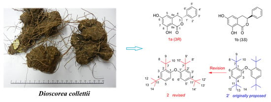
2.1. Structural Elucidation of the Compounds

Compound 1 (1a/1b) was obtained as a colorless solid. Its molecular formula was elucidated to be C15H12O4 by HR-ESI-MS at m/z 279.0626 for [M + Na]+ (calculated. for C15H12O4Na, 279.0633), with ten degrees of unsaturation. The 13C NMR data (Table 1) in combination with analysis of the HSQC spectrum for 1 showed the presence of one carbonyl carbon, nine olefinic or aromatic carbons, one oxymethine, and one methylene. In its 1H NMR spectrum, the five aromatic proton signals between δH 7.48 and 7.36 (δH 7.48 (2H, d, J = 7.5 Hz), 7.39 (2H, t, J = 7.5 Hz), and 7.36 (1H, t, J = 7.0 Hz)) indicated the presence of one mono-substituted benzene ring. Two meta-coupled aromatic signals at δH 6.26 (1H, br s) and 6.24 (1H, J = 1.8 Hz) suggested that 1 possessed one 1,2,3,5-tetrasubstituted aromatic ring. Additionally, one oxymethine proton at δH 5.56 (1H, dd, J = 12.0, 3.0 Hz) and one methylene group at δH 3.21 (1H, dd, J = 16.3, 12.2 Hz) and 3.06 (1H, dd, J = 16.4, 3.2 Hz) were observed in the 1H NMR spectrum, perfectly matching the above 13C NMR data. In addition to the two aromatic rings and one carbonyl, the remaining 1 degree of unsaturation suggested the existence of a one ring structure. All 1H and 13C chemical shifts for 1 were essentially identical to those observed for montroumarin [15]. Based on the interpretation of the HMBC and 1H-1H COSY spectra (Figure 2) as well as a comparison with the literature data, the planar structure of 1 was characterized as 6, 8-dihydroxy-3-phenyl-3, 4-dihydroisocoumarin.
Due to having a single C-3 stereogenic center, 1 might exist in two potential configurations, (3R)-1 and (3S)-1, and only the 3S-form (montroumarin) has been isolated previously [15]. Subsequent chiral HPLC separation on 1 afforded compounds 1a and 1b, a pair of enantiomers, in a ratio of 15:85 (Supplementary materials Figure S8), showing that 1 is a scalemic mixture. The opposite optical rotations and mirror-like electronic circular dichroism (ECD) spectra confirmed their enantiomeric relationship (Figure 3). The absolute configuration of 1a and 1b was determined by comparing their ECD spectra and optical rotations with those of montroumarin [15] and other similar dihydroisocoumarin derivatives [16]. The absolute configuration of (+)-1 (1b) was established as S on the basis of the strong positive Cotton effect (CE) at 233 nm in its ECD spectrum (Figure 3), which is consistent with that of montroumarin. However, (−)-1 (1a) showed a strong negative CE at 233 nm; thus, its absolute configuration was deduced to be R. A comparison of the optical rotations of 1a and 1b with that of montroumarin also supported the above conclusions. Thus, the structure of compound 1a was defined and named as (−)-montroumarin, and 1b was identified as the known compound montroumarin (see Figure 1).
Compound 2 was purified as a pale-yellow oil. The 1H NMR spectrum (Table 2) of 2 in CDCl3 showed signals attributable to an ABX-type aromatic ring at δH 7.54 (1H, d, J = 8.6 Hz, H-6),7.36 (1H, t, J = 2.5 Hz, H-3),and 7.13 (1H, dd, J = 8.6, 2.5 Hz, H-5), which indicated the presence of a 1,2,4-trisubstituted benzene ring [17,18]. In addition, two sets of non-equivalent signals each containing three methyl groups at δH 1.33 (9H, s, H-8/9/10) and 1.28 (9H, s, H-12/13/14) were also observed. The 13C NMR and HSQC spectra displayed 12 carbon resonances, which were classified as eight olefinic or aromatic carbons (δC 147.8, 147.8, 147.2, 138.7, 138.6, 124.6, 124.1, and 119.3), two quaternary carbons (δC 35.0 and 34.7), and two methyl carbons (δC 31.6 and 30.4). The 1H and 13C NMR data in CDCl3 (Figure S15) for compound 2 were highly consistent with the experimental data for 2, 2′-oxybis (1, 4-di-tert-butylbenzene) (2′), suggesting that 2 was the same substance isolated by Sun B-N and coworkers [14]. The molecular formula of compound 2′ is C28H42O and the structure of 2′ was originally identified as a phenyl ether derivative bearing four tert-butyl substituents located in the aromatic rings at the positions C-1, C-4, C-1′, and C-4′ (Figure 1). We subsequently performed 2D NMR experiments on 2. Careful examination of the HMBC spectrum (Figure 2) of 2 revealed a strong HMBC correlation from H-5 (δH 7.13) to C-1 (δC 147.8), which is apparently inconsistent with structure 2′ (from H-2 to C-5 of 2′) but consistent with 2. This key HMBC correlation indicated that the structure elucidation of 2′ is clearly incorrect. The 13C NMR data (δC 147.8, 147.8, 138.7, and 138.6) and the molecular formula of 2′ indicated that 2 was a dimer. In the HMBC spectrum, the correlation from the three equivalent methyl groups signals at δH 1.33 (9H, H-8/9/10) to the quaternary carbon signal at δC 35.0 (C-7) suggested a direct connection between C-8/9/10 (δC 30.4) and C-7, forming one tert-butyl substituent, which was further confirmed by the HMBC correlations from H-8 (δH 1.33, 9H) to C-9/10 (δC 30.4). This tert-butyl substituent attached to C-2 (δC 138.6) of the aromatic ring was clarified by the correlation from H-8/9/10 to C-2 in the HMBC spectrum. Similar, the HMBC correlations from H-12/13/14 (δH 1.28, 9H) to C-11 (δC 34.7) and C-4 (δC 147.2) suggested a connection between C-12/13/14 (δC 31.6) and C-11, forming another tert-butyl substituent attached to C-4 of the aromatic ring. The 1H-1H COSY correlation (Figure 2) of H-5/H-6 (δH 7.54) and the HMBC correlations of H-5/C-11 and H-6/C-4 elucidated the connectivity of C-4/C-5/C-6. The linkage between C-2 and C-3 was verified by the HMBC correlation from H-3 (δH 7.36) to C-7. The HMBC correlations from H-5 to C-3 (δC 124.6) and H-3 to C-5 (δC 124.1), together with the C-3 and C-5 located at the meta-position of the aromatic ring, indicated the connectivity of C-2/C-3/C-4/C-5/C-6. The established linkage of C-2/C-3/C-4/C-5/C-6 as well as the HMBC correlations from H-3 and H-5 to C-1 (δC 147.8) constructed the aromatic ring. Considering the downfield chemical shift of C-1, and the molecular formula required for 2, C-1 and C-1′ should be linked to the remaining one oxygen atom to form a dimer. The structure of 2, shown in Figure 1, was identified as 1,1′-oxybis(2,4-di-tert-butylbenzene).
This explained well the strong HMBC correlation from H-5 (δH 7.13) to C-1 (δC 147.8), which is apparently inconsistent with structure 2′. In addition, all the HMBC correlations reported for 2′ [14], including the weak 4JC,H long-range HMBC correlation from H-6 (δH 7.54) to C-7 (δC 35.0) [19,20], were also consistent with structure 2. Moreover, compound 2 might be a naturally occurring dimer of 2,4-Di-tert-butylphenol (2,4-DTBP), which is a common secondary metabolite produced by various groups of organisms [21]. Therefore, we compared the chemical shifts of compounds 2, 2′, and 2,4-DTBP and found that the structure elucidation of 2 was more reasonable than that of 2′ [22,23] (Figure S15). When running a 13C spectrum prediction for compounds 2,4-DTBP and 2′, it is trivial to see that the 13C-data of 2,4-DTBP are consistent with structure 2, whereas in the case of 2′ there is a massive inconsistency. By searching SciFinder, we found that the structure of 2 was not reported in any literature or patents but only had one commercial source. This was the first report of its detailed structure elucidation based on 1D and 2D NMR spectroscopy data.
Compound 3 (3a/3b) was obtained as a colorless needle. The results of a comparison between the 1H, 13C NMR data of 3 and those reported by Guimarães et al. [24] suggested that the planar structure of 3 was 3′-O-methylviolanone. Compound 3 was optically inactive, suggesting that it was a racemic mixture. Both of the two configurations (3R)-3 and (3S)-3 have been reported previously [25,26], but the ECD spectra of (3R/3S)-3 and the specific rotation of (3R)-3 have remained undefined. Subsequent chiral resolution of 3 (Figure S18) afforded a pair of enantiomers, 3a and 3b, and they displayed almost mirror-image ECD curves that showed opposite CEs at 193, 210, 230, 272, and 325 nm. The respective absolute configurations of (−)-3 and (+)-3 (3a and 3b) were defined as (3R) and (3S) via a comparison of their experimental and calculated ECD spectra (Figure 3).
Compound 4 was also obtained as a colorless needle. A comparison of the 1H and 13C NMR data of 4 with those of compound 3 showed that their structures are closely related, suggesting that 4 is also a dihydroisoflavone analogue. Through a comparison of its NMR data with those reported [27], the planar structure of 4 was identified as sativanone. Similarly to compound 3, 4 was also optically inactive, suggesting that it was a racemate. Both of the two configurations (3R)-4 and (3S)-4, including their ECD spectra and the specific rotations, have been reported [28,29]. Thus, compound 4 was identified as (RS)-sativanone.

2.2. Cytotoxic Activity against NCI-H460 Cell Line

All the isolated compounds 14 were further evaluated for their cytotoxicity against the human lung cancer NCI-H460 cell line (Table 3). The results indicated that compounds 1a and 1b, two dihydroisocoumarins, possess moderate cytotoxic activities, with IC50 values of 33.37 and 32.06 μM, respectively. Compounds 24 showed no activity, with an IC50 > 50 μM.

3. Materials and Methods

3.1. General Information

One-dimensional- and two-dimensional-NMR spectra were obtained from a Bruker Avance III 600 MHz spectrometer (Bruker BioSpin Inc., Zurich, Switzerland). Optical rotations were measured with an MCP 200 Modular Circular Polarimeter (Anton Paar, Graz, Austria). HRESIMS spectrum was obtained from a MicrOTOF-Q II ESI mass spectrometer (Bruker, Bremen, Germany). UV spectrum was obtained from a Hitachi U-3900 UV-visible spectrophotometer (Hitachi High-Tech Science Corporation, Tokyo, Japan). ECD spectra were recorded by a MOS-500 Circular Dichroism spectropolarimeter (Bio-Logic, Grenoble, France). Semi-preparative HPLC was performed on an LC-6A (Shimadzu, Kyoto, Japan) equipped with preparative YMC Pack ODS-A column (250 × 20 mm, 5 μm, YMC, Kyoto, Japan). A Daicel Chiralpak AD-H (250 × 4.6 mm, 5 μm, Daicel, Tokyo, Japan) was used for the chiral separations. Column chromatography (CC) was carried out over silica gel (100–200 and 200–300 mesh, Qingdao Haiyang Chemical Co., Ltd., Qingdao, China), Sephadex LH-20 (Amersham Pharmacia Biotech AB, Uppsala, Sweden), and ODS RP-C18 (40–63 μm, YMC, Kyoto, Japan). Thin Layer Chromatography (TLC) was performed on pre-coated silica gel GF-254 (Qingdao Marine Chemical Factory, Qingdao, China).

3.2. Materials

The rhizomes of D. collettii were collected from Mount Emei, Leshan City, Sichuan province, China, and authenticated by Prof. Wenyuan Gao (School of Pharmaceutical Science and Technology, Tianjin University). A voucher specimen (ID: 245020328) was deposited in the School of Pharmaceutical Science and Technology, Tianjin University, Tianjin, China.

3.3. Extraction and Isolation

The air-dried rhizomes of D. collettii (16.2 kg) were extracted three times with 90% aqueous ethanol and three times with 60% aqueous ethanol under reflux (30 L, each for 2 h). After removal of the solvent under reduced pressure, the residue was combined and suspended in water to a final volume of 10 L, and then sequentially partitioned with petroleum ether (PE, 60–90 °C), ethyl acetate (EtOAc), and n-butyl alcohol (n-BuOH).
The EtOAc extract (305.0 g) was subjected to silica gel column chromatography (CC) eluted with CH2Cl2-MeOH gradient (10:0 to 0:10, v/v) to afford 19 fractions (A–S). Fraction G was purified by Sephadex LH-20 CC eluted with CH2Cl2-MeOH (1:1) to yield 5 fractions (G1–G5). Fraction G2 was exposed to silica gel CC eluted with PE-EtOAc gradient (1:0 to 7:3, v/v) followed by Sephadex LH-20 CC eluted with CH2Cl2-MeOH (1:1), and finally purified by semi-preparative HPLC (YMC C18, 250 × 20 mm, 5 μm, 10 mL/min) eluted with 65% aqueous MeOH to yield compound 1 (15 mg, tR = 8 min). Fraction G4 was isolated by Sephadex LH-20 CC eluted with CH2Cl2-MeOH (1:1) to yield 51 subfractions (Fr.1–Fr.51). Fr.28–Fr.30 were submitted to silica gel CC eluted with PE-EtOAc (75:25, v/v) and then purified by Sephadex LH-20 CC eluted with CH2Cl2-MeOH (1:1) to obtain compound 3 (16 mg). Fr.31–Fr.39 was applied on silica gel CC eluted with PE-EtOAc (8:2, v/v) to obtain compound 4 (7 mg). Fraction L was chromatographed over silica gel CC with PE-EtOAc solvent system with increasing polarity to afford 3 fractions (L1–L3). Fraction L1 was isolated by Sephadex LH-20 CC eluted with CH2Cl2-MeOH (1:1) to obtain compound 2 (9 mg).
Compound 1 was separated using chiral-phase HPLC (AD-H column, n-hexane/ethanol, 70:30, flow rate: 1.0 mL/min) to afford 1b (tR = 10.0 min) and 1a (tR = 11.9 min). Compound 3 was further resolved using chiral-phase HPLC (AD-H column, n-hexane/ethanol, 85:15, flow rate: 1.0 mL/min) to obtain 3b (tR = 8.2 min) and 3a (tR = 9.2 min).

3.3.1. (−)-Montroumarin (1a)

Colorless solid; [ α ] D 20 − 93.65 (c 0.20, MeOH); UV (MeOH) λmax 195, 217, 271, 303 nm; 1H-NMR (600 MHz, CD3OD) and 13C-NMR (150 MHz, CD3OD) spectroscopic data, see Table 1; HR ESI-TOF MS m/z 279.0626 [M + Na]+ (calcd. for C15H12O4Na 279.0633).

3.3.2. Montroumarin (1b)

Colorless solid; [ α ] D 20 + 71.72 (c 0.20, MeOH); 1H-NMR (600 MHz, CD3OD) and 13C-NMR (150 MHz, CD3OD) spectroscopic data, see Table 1; HR ESI-TOF MS data, see compound 1a.

3.3.3. 1,1′-Oxybis(2,4-di-tert-butylbenzene) (2)

Pale-yellow oil; 1H-NMR (600 MHz, CDCl3) and 13C-NMR (150 MHz, CDCl3) spectroscopic data, see Table 2; HR ESI-TOF MS m/z 395.3373 [M + H]+ (calcd. for C28H43O 395.3314).

3.3.4. (3R)-3′-O-Methylviolanone (3a)

Colorless needle; [ α ] D 20 − 39.17 (c 0.20, MeOH); 1H-NMR (600 MHz, C5D5N) δ: 4.79 (1H, t, J = 11.3 Hz, H-2ax), 4.44 (1H, dd, J = 11.7 Hz, 5.4 Hz, H-3ax), 4.60 (1H, dd, J = 10.8 Hz, 5.5 Hz, H-2eq), 8.23 (1H, d, J = 8.6 Hz, H-5), 6.93 (1H, dd, J = 8.6 Hz, 2.2 Hz, H-6), 6.84 (1H, d, J = 2.2 Hz, H-8), 6.71 (1H, d, J = 8.5 Hz, H-5′), 7.03 (1H, d, J = 8.5 Hz, H-6′), 3.84 (1H, s, 2′-OMe), 3.92 (1H, s, 3′-OMe), 3.72 (1H, s, 4′-OMe). 13C-NMR (150 MHz, C5D5N) δ: 72.1 (C-2), 49.1 (C-3), 191.6 (C-4), 130.4 (C-5), 112.1 (C-6), 166.5 (C-7), 104.2 (C-8), 164.9 (C-8a), 115.6 (C-4a), 123.1 (C-1′), 153.1 (C-2′), 143.4 (C-3′), 154.5 (C-4′), 108.7 (C-5′),125.4 (C-6′), 61.3 (C-2′-OMe), 60.9 (C-4′-OMe), 56.4 (C-3′-OMe); HR ESI-TOF MS m/z 353.0998 [M + Na]+ (calcd. for C18H18O6Na 353.1001).

3.3.5. (3S)-3′-O-Methylviolanone (3b)

Colorless needle; [ α ] D 20 + 28.33 (c 0.20, MeOH); 1H-NMR (600 MHz, C5D5N), 13C-NMR (600 MHz, C5D5N) spectroscopic data and HR ESI-TOF MS data, see compound 3a.

3.3.6. (RS)-Sativanone (4)

Colorless solid; [ α ] D 20 − 0.06 (c 0.38, MeOH); 1H-NMR (600 MHz, C5D5N) δ: 4.76 (1H, t, J = 11.1 Hz, H-2ax), 4.50 (1H, dd, J = 11.3 Hz, 5.2 Hz, H-3ax), 4.59 (1H, dd, J = 10.8 Hz, 5.3 Hz, H-2eq), 8.25 (1H, d, J = 8.6 Hz, H-5), 6.92 (1H, dd, J = 8.6 Hz, 2.1 Hz, H-6), 6.84 (1H, d, J = 2.1 Hz, H-8), 6.67 (1H, d, J = 2.0 Hz, H-3′), 6.58 (1H, dd, J = 8.3 Hz, 2.0 Hz, H-5′), 7.24 (1H, d, J = 8.3 Hz, H-6′), 3.63 (1H, s, 2′-OMe), 3.69 (1H, s, 4′-OMe). 13C-NMR (150 MHz, C5D5N) δ: 71.8 (C-2), 48.2 (C-3), 191.6 (C-4), 130.4 (C-5), 112.0 (C-6), 166.5 (C-7), 104.0 (C-8), 164.9 (C-8a), 115.7 (C-4a), 117.5 (C-1′), 159.4 (C-2′), 99.8 (C-3′), 161.4 (C-4′), 105.7 (C-5′), 131.7 (C-6′), 55.9 (C-2′-OMe), 55.6 (C-4′-OMe); HR ESI-TOF MS m/z 301.1006 [M + H]+ (calcd. for C17H17O5 301.1076).

3.4. Cytotoxicity Assays

Compounds 14 were evaluated for their cytotoxic activities by the MTT method using NCI-H460 cell line. The NCI-H460 cells were seeded at a density of 1 × 10⁴/well in a complete growth medium in 96-well plates. The cells were incubated with the test compounds for 24 h before the MTT assay. Then, a fresh solution of MTT (0.5 mg/mL) was added to each single well of the 96-well plate. The plate was incubated in a CO2 incubator for another 4 h. Finally, the cells were dissolved with 100 μL of DMSO and then analyzed in a multiwall plate reader with a wavelength of 570 nm.

4. Conclusions

The chemical investigation of D. collettii led to the isolation of six compounds (14), including one new dihydroisocoumarin, (−)-montroumari (1a). The structure of 2,2′-oxybis(1,4-di-tert-butylbenzene) (2′) was revised to be 1,1′-oxybis(2,4-di-tert-butylbenzene) (2), assisted by a careful re-examination of the structural elucidation process. All the compounds 14 were isolated from D. collettii for the first time. The isolation of compounds 3a, 3b, and 4 from the Dioscorea species has not been reported yet [30,31]. Notably, to the best of our knowledge, dihydroisoflavones have not been described from any other species in the Dioscoreaceae family. Our discovery of these dihydroisoflavones (3a, 3b, and 4) enriches the structural diversity of the Dioscoreaceae family. Dihydroisocoumarins (1a1b) exhibited moderate cytotoxic activities against the NCI-H460 cell line, with IC50 values ranging from 32.06 to 33.37 μM, whereas the other compounds, including the three dihydroisoflavones (3a4), did not show any activities at the tested concentrations (IC50 > 50 μM). Mounting evidence has revealed that steroidal saponins exert strong cytotoxic activities against human cancer cells, which means they could be considered as promising cytotoxic agents against human cancer cells. Dihydroisocoumarins and diarylheptanoids showed moderate cytotoxic activities against the NCI-H460 cell line. However, more selectivity studies are needed to determine whether these two types of compounds in D. collettii have cytotoxic activities against human cancer cells.

Supplementary Materials

The following are available online. Figure S1: 1H-NMR (600 MHz, CD3OD) spectrum of compound 1a/1b, Figure S2: 13C-NMR (150 MHz, CD3OD) spectrum of compound 1a/1b, Figure S3: HSQC spectrum of compound 1a/1b, Figure S4: 1H-1H COSY spectrum of compound 1a/1b, Figure S5: HMBC spectrum of compound 1a/1b, Figure S6: HR-ESI-MS spectrum of compound 1a/1b, Figure S7: UV spectrum of compound 1a/1b, Figure S8: Chiral HPLC separation chromatogram of compound 1, Figure S9: 1H-NMR (600 MHz, CDCl3) spectrum of compound 2, Figure S10: 13C-NMR (150 MHz, CDCl3) spectrum of compound 2, Figure S11: HSQC spectrum of compound 2, Figure S12: 1H-1H COSY spectrum of compound 2, Figure S13: HMBC spectrum of compound 2, Figure S14: The enlarged HMBC spectrum of compound 2 (I, II, III, and IV), Figure S15: Structures and comparison of chemical shifts between compounds 2, 2′, and 2,4-DTBP in CDCl3, Figure S16: 1H-NMR (600 MHz, C5D5N) spectrum of compound 3a/3b, Figure S17: 13C-NMR (150 MHz, C5D5N) spectrum of compound 3a/3b, Figure S18: Chiral HPLC separation chromatogram of compound 3, Figure S19: 1H-NMR (600 MHz, C5D5N) spectrum of compound 4, Figure S20: 13C-NMR (150 MHz, C5D5N) spectrum of compound 4, Figure S21: ECD spectrum of compound 3, and ECD calculation of compound 3.

Author Contributions

Conceptualization, W.G. and S.J.; methodology, C.Z., X.L. and L.G.; investigation, S.J. and Z.Q.; data curation, L.G.; writing—original draft, S.J.; writing—review and editing, S.J., Z.Q. and Z.L.; project administration, W.G. and Y.Z.; funding acquisition, W.G., S.J., Z.Q. and Y.Z. All authors have read and agreed to the published version of the manuscript.

Funding

This research was funded by Natural Science Foundation of Hebei Province (H2019423126), Research Foundation of Hebei Province Education Department (QN2021107), Research Foundation of Hebei Provincial Administration of Traditional Chinese Medicine (2019086, 2019079), Innovation Team of Hebei Province Modern Agricultural Industry Technology System (HBCT2018060205), Key Research and Development Program of Ningxia (2021BEG03103), and West Light Foundation of the Chinese Academy of Sciences (XAB2020YW15).

Institutional Review Board Statement

Not applicable.

Informed Consent Statement

Not applicable.

Data Availability Statement

Data are contained within the article or supplementary material.

Conflicts of Interest

The authors declare no conflict of interest.

Sample Availability

Samples of the compounds 14 are available from the authors.

References

  1. Zhu, L.; Dong, Y.; Na, S.; Han, R.; Wei, C.; Chen, G. Saponins extracted from Dioscorea collettii rhizomes regulate the expression of urate transporters in chronic hyperuricemia rats. Biomed. Pharmacother. 2017, 93, 88–94. [Google Scholar] [CrossRef] [PubMed]
  2. Qi, Q.; He, X.; Wang, G.; Na, S.; Lv, L.; Chen, G. Anti-inflammatory and analgesic effects of total saponin of Dioscorea. Chin. J. Clin. Pharm. Therap. 2020, 25, 257–264. [Google Scholar] [CrossRef]
  3. Hu, K.; Yao, X. Methyl protogracillin (NSC-698792): The spectrum of cytotoxicity against 60 human cancer cell lines in the National Cancer Institute’s anticancer drug screen panel. Anti-Cancer Drugs 2001, 12, 541–547. [Google Scholar] [CrossRef]
  4. Tseng, S.-C.; Shen, T.-S.; Wu, C.-C.; Chang, I.-L.; Chen, H.-Y.; Hsieh, C.-P.; Cheng, C.-H.; Chen, C.-L. Methyl protodioscin induces apoptosis in human osteosarcoma cells by caspase-dependent and MAPK signaling pathways. J. Agric. Food Chem. 2017, 65, 2670–2676. [Google Scholar] [CrossRef] [PubMed]
  5. Wang, L.; Sha, N.; Chen, G. Effect of total saponin of Dioscorea on NALP3 inflammasome signaling pathway with acute gouty in rats. Chin. Pharmacol. Bull. 2017, 33, 354–360. [Google Scholar] [CrossRef]
  6. Yang, M.; Chen, Y. Steroidal sapogenins in Dioscorea collettii. Planta Med. 1983, 49, 38–42. [Google Scholar] [CrossRef]
  7. Liu, C.; Chen, Y.; Ge, S.; Li, B. Studies on the Constituents of Dioscorea Plants. II. Isolation and Identification of Steroidal Saponins from Dioscorea collettii Hook. F. Acta Pharm. Sin. 1983, 18, 597–606. [Google Scholar] [CrossRef]
  8. Jing, S.-S.; Wang, Y.; Yan, Y.-M.; Li, X.; Li, X.-J.; Zhao, C.-C.; Sun, J.-C.; Qiu, P.-Y.; Man, S.-L.; Gao, W.-Y. Diocollettines A, an unusual tricyclic diarylheptanoid derivative from the rhizomes of Dioscorea collettii. Tetrahedron Lett. 2016, 57, 3215–3217. [Google Scholar] [CrossRef]
  9. Jing, S.-S.; Wang, Y.; Li, X.-J.; Li, X.; Zhao, W.-S.; Zhou, B.; Zhao, C.-C.; Huang, L.-Q.; Gao, W.-Y. Phytochemical and chemotaxonomic studies on Dioscorea collettii. Biochem. Syst. Ecol. 2017, 71, 10–15. [Google Scholar] [CrossRef]
  10. Li, X.; Qu, Z.; Jing, S.; Li, X.; Zhao, C.; Man, S.; Wang, Y.; Gao, W. Dioscin-6′-O-acetate inhibits lung cancer cell proliferation via inducing cell cycle arrest and caspase-dependent apoptosis. Phytomedicine 2019, 53, 124–133. [Google Scholar] [CrossRef]
  11. Si, L.; Zheng, L.; Xu, L.; Yin, L.; Han, X.; Qi, Y.; Xu, Y.; Wang, C.; Peng, J. Dioscin suppresses human laryngeal cancer cells growth via induction of cell-cycle arrest and MAPK-mediated mitochondrial-derived apoptosis and inhibition of tumor invasion. Eur. J. Pharmacol. 2016, 774, 105–117. [Google Scholar] [CrossRef] [PubMed]
  12. Li, X.; Jing, S.; Man, S.; Li, X.; Zhao, C.; Wang, Y.; Gao, W. A new acetylated spirostanol saponin and other constituents from the rhizomes of Dioscorea althaeoides R. Knuth (Dioscoreaceae). Biochem. Syst. Ecol. 2016, 65, 17–22. [Google Scholar] [CrossRef]
  13. Li, X.; Zhao, C.; Jing, S.; Sun, J.; Li, X.; Man, S.; Wang, Y.; Gao, W. Novel phenanthrene and isocoumarin from the rhizomes of Dioscorea nipponica Makino subsp. rosthornii (Prain et Burkill) CT Ting (Dioscoreaceae). Bioorg. Med. Chem. Lett. 2017, 27, 3595–3601. [Google Scholar] [CrossRef] [PubMed]
  14. Sun, B.-N.; Shen, H.-D.; Wu, H.-X.; Cui, F.; Yao, L.-X.; Cheng, Z.-Q.; Diao, Y. Chemical Constituents of Onchidium struma from Chongming Island, Shanghai. Nat. Prod. Res. Dev. 2014, 26, 987–989. [Google Scholar] [CrossRef]
  15. Ito, C.; Mishina, Y.; Litaudon, M.; Cosson, J.-P.; Furukawa, H. Xanthone and dihydroisocoumarin from Montrouziera sphaeroidea. Phytochemistry 2000, 53, 1043–1046. [Google Scholar] [CrossRef]
  16. Paraschos, S.; Magiatis, P.; Kalpoutzakis, E.; Harvala, C.; Skaltsounis, A.-L. Three New Dihydroisocoumarins from the Greek Endemic Species Scorzonera cretica. J. Nat. Prod. 2001, 64, 1585–1587. [Google Scholar] [CrossRef]
  17. Yang, C.-J.; Wang, Z.-B.; Zhu, D.-L.; Yu, Y.; Lei, Y.-T.; Liu, Y. Two new cyanogenic glucosides from the leaves of Hydrangea macrophylla. Molecules 2012, 17, 5396–5403. [Google Scholar] [CrossRef] [PubMed]
  18. Long, Z.; Zhang, Y.; Guo, Z.; Wang, L.; Xue, X.; Zhang, X.; Wang, S.; Wang, Z.; Civelli, O.; Liang, X. Amide alkaloids from Scopolia tangutica. Planta Med. 2014, 80, 1124–1130. [Google Scholar] [CrossRef] [Green Version]
  19. Perlatti, B.; Lan, N.; Earp, C.E.; AghaAmiri, S.; Vargas, S.H.; Azhdarinia, A.; Bills, G.F.; Gloer, J.B. Arenicolins: C-glycosylated depsides from Penicillium arenicola. J. Nat. Prod. 2020, 83, 668–674. [Google Scholar] [CrossRef] [PubMed]
  20. Köck, M.; Lindel, T.; Junker, J. Incorporation of 4J-HMBC and NOE Data into Computer-Assisted Structure Elucidation with WEBCOCON. Molecules 2021, 26, 4846. [Google Scholar] [CrossRef]
  21. Zhao, F.; Wang, P.; Lucardi, R.D.; Su, Z.; Li, S. Natural sources and bioactivities of 2, 4-di-tert-butylphenol and its analogs. Toxins 2020, 12, 35. [Google Scholar] [CrossRef] [Green Version]
  22. Chu, C.; Gu, Q.; Fang, Y. Isolation and identification of a phenolic derivative from the ascidian. J. Ocean. Univ. Qingdao 1999, 29, 53–56. [Google Scholar] [CrossRef]
  23. Li, M.; Huang, X.-l.; Wang, L.; Li, Q.-h.; Zhang, J. Chemical Constituents from Sauropus spatulifolius. Chin. Med. Mater. 2019, 7, 1541–1545. [Google Scholar] [CrossRef]
  24. Guimarães, I.d.S.; Gottlieb, O.R.; Andrade, C.S.; Magalhaes, M.T. Flavonoids from Dalbergia cearensis. Phytochemistry 1975, 14, 1452–1453. [Google Scholar] [CrossRef]
  25. Doran, R.; Carroll, M.P.; Akula, R.; Hogan, B.F.; Martins, M.; Fanning, S.; Guiry, P.J. A stereoselective switch: Enantiodivergent approach to the synthesis of isoflavanones. Chem.-Eur. J. 2014, 20, 15354–15359. [Google Scholar] [CrossRef] [PubMed]
  26. Lee, C.; Lee, J.W.; Jin, Q.; Jang, D.S.; Lee, S.-J.; Lee, D.; Hong, J.T.; Kim, Y.; Lee, M.K.; Hwang, B.Y. Inhibitory constituents of the heartwood of Dalbergia odorifera on nitric oxide production in RAW 264.7 macrophages. Bioorg. Med. Chem. Lett. 2013, 23, 4263–4266. [Google Scholar] [CrossRef] [PubMed]
  27. Zhao, X.; Mei, W.; Gong, M.; Zuo, W.; Bai, H.; Dai, H. Antibacterial activity of the flavonoids from Dalbergia odorifera on Ralstonia solanacearum. Molecules 2011, 16, 9775–9782. [Google Scholar] [CrossRef] [PubMed] [Green Version]
  28. Umehara, K.; Nemoto, K.; Matsushita, A.; Terada, E.; Monthakantirat, O.; De-Eknamkul, W.; Miyase, T.; Warashina, T.; Degawa, M.; Noguchi, H. Flavonoids from the heartwood of the Thai medicinal plant Dalbergia parviflora and their effects on estrogenic-responsive human breast cancer cells. J. Nat. Prod. 2009, 72, 2163–2168. [Google Scholar] [CrossRef]
  29. Chan, S.-C.; Chang, Y.-S.; Wang, J.-P.; Chen, S.-C.; Kuo, S.-C. Three new flavonoids and antiallergic, anti-inflammatory constituents from the heartwood of Dalbergia odorifera. Planta Med. 1998, 64, 153–158. [Google Scholar] [CrossRef] [PubMed] [Green Version]
  30. Obidiegwu, J.E.; Lyons, J.B.; Chilaka, C.A. The Dioscorea Genus (Yam)—An appraisal of nutritional and therapeutic potentials. Foods 2020, 9, 1304. [Google Scholar] [CrossRef] [PubMed]
  31. Sautour, M.; Mitaine-Offer, A.-C.; Lacaille-Dubois, M.-A. The Dioscorea genus: A review of bioactive steroid saponins. J. Nat. Med.-Tokyo 2007, 61, 91–101. [Google Scholar] [CrossRef]
Figure 1. Chemical structures of compounds 14 isolated from rhizomes of Dioscorea collettii.
Molecules 26 05381 g001
Figure 2. 1H-1H COSY, key HMBC correlations of 12.
Molecules 26 05381 g002
Figure 3. ECD spectrum of compound 1 (a) and experimental and calculated ECD spectra of 3 (b).
Molecules 26 05381 g003
Table 1. 1H-NMR (600 MHz), 13C NMR (150 MHz) data, and HMBC correlations of compound 1 in CD3OD.
No.δH, mult (J in Hz)δCHMBC (H→C)
1 171.5, C
35.56, dd (12.0, 3.0)81.8, CHC-1, C-4, C-4a, C-1′, C-2′, C-6′
43.21, dd (16.3, 12.2)
3.06, dd (16.4, 3.2)
36.0, CH2C-3, C-4a, C-5, C-8a, C-1′
4a 143.4, C
56.26, br s108.0, CHC-4, C-6, C-7, C-8a
6 166.4, C
76.24, d (1.8)102.4, CHC-5, C-6, C-8, C-8a
8 165.7, C
8a 101.7, C
1′ 140.1, C
2′, 6′7.48, d (7.5)127.3, CHC-3, C-3′, C-4′, C-5′
3′, 5′7.41, t (7.5)129.7, CHC-1′, C-3′, C-5′
4′7.36, t (7.0)129.7, CHC-2′, C-6′
Table 2. 1H-NMR (600 MHz), 13C NMR (150 MHz) data, and HMBC correlations of compound 2 in CDCl3.
No.δH, mult (J in Hz)δCHMBC (H→C)
1 (1′) 147.8 (147.8), C
2 (2′) 138.6 (138.7), C
3 (3′)7.36, t (2.5)124.6, CHC-7, C-11, C-5, C-1
(C-7′, C-11′, C-5′, C-1′)
4 (4′) 147.2, C
5 (5′)7.13, dd (8.6, 2.5)124.1, CHC-11, C-6, C-3, C-1
(C-11′, C-6′, C-3′, C-1′)
6 (6′)7.54, d (8.6)119.3, CHC-2, C-4 (C-2′, C-4′)
7 (7′) 35.0, C
8/9/10 (8′/9′/10′)1.33, s30.4, CH3C-7, C-2 (C-7′, C-2′)
11 (11′) 34.7, C
12/13/14 (12′/13′/14′)1.28, s31.6, CH3C-11, C-4 (C-11′, C-4′)
Table 3. The IC50 value of compounds on NCI-H460 cells (μM).
SampleIC50 (μM)SampleIC50 (μM)
Compound 1a33.37Compound 1b32.06
Compound 2>100Compound 3a57.83
Compound 3b59.34Compound 464.01
Cisplatin1.27
Publisher’s Note: MDPI stays neutral with regard to jurisdictional claims in published maps and institutional affiliations.

