
The Antiproliferative and Apoptotic Effect of a Novel Synthesized S-Triazine Dipeptide Series, and Toxicity Screening in Zebrafish Embryos

 , ,
, ,  , , and
Abstract
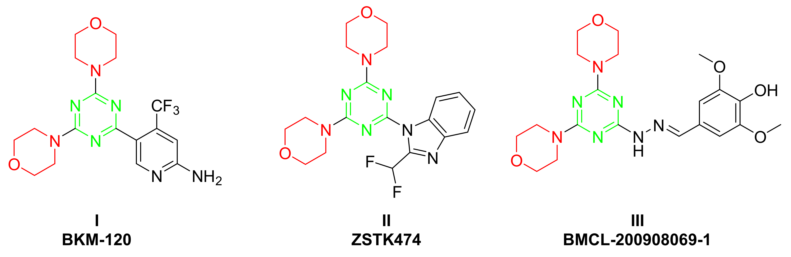
:1. Introduction
2. Results and Discussion
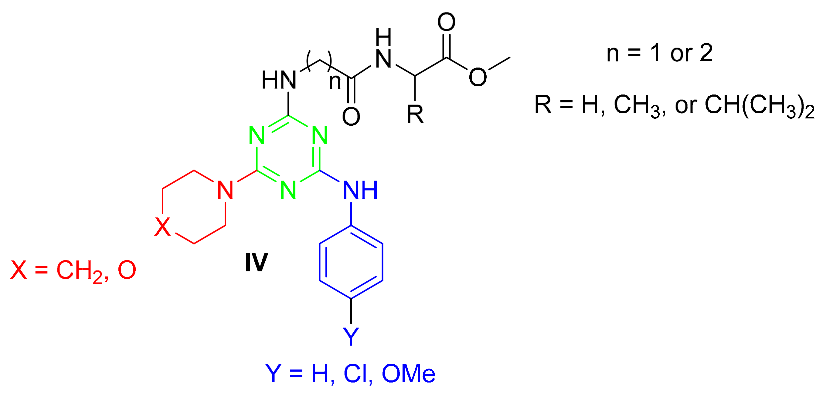
2.1. Chemistry
2.2. Biology
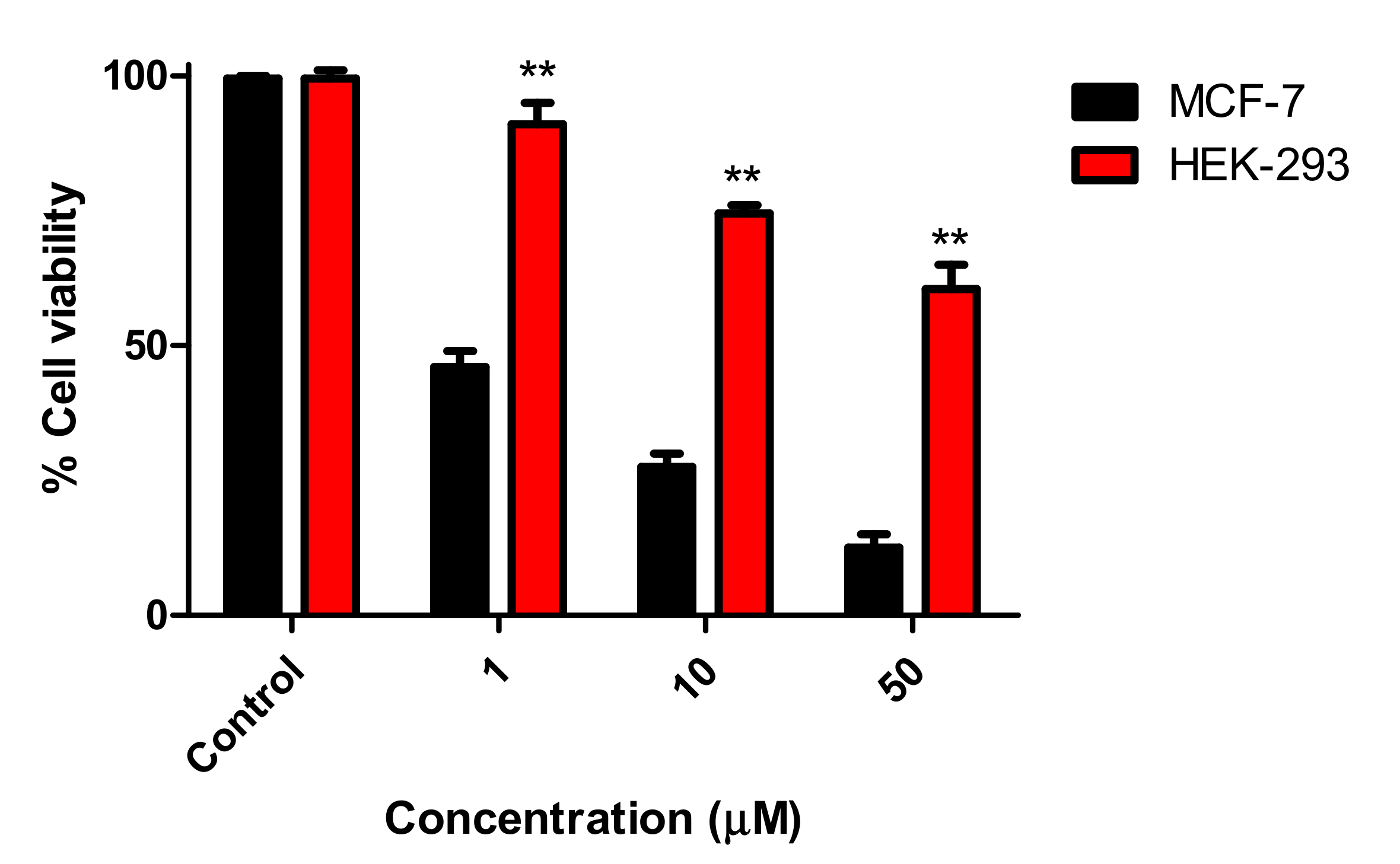
2.2.1. Antiproliferative Activity
2.2.2. Effects on the Cell Cycle Distribution and Apoptosis Assay
2.2.3. In Vivo Evaluation in Zebrafish
3. Materials and Methods
3.1. Materials and Methods
3.2. Chemistry
3.2.1. General Procedure for the Synthesis of the 4,6-Disubstituted s-Triazin-2-yl Amino Acid Derivatives
3.2.2. General Method for the Synthesis of the 4,6-Disubstituted s-Triazine Dipeptide Derivatives
3.3. Biology
3.3.1. MTT Cell Proliferation Assays
3.3.2. Cell Culture
3.3.3. Cell Viability Assay
3.3.4. Cell Cycle Analysis
3.3.5. Annexin V/PI Apoptotic Assay
3.3.6. Zebrafish Embryo Treatments
3.3.7. Imaging and Microscopy
4. Conclusions
Supplementary Materials
Author Contributions
Funding
Institutional Review Board Statement
Conflicts of Interest
Sample Availability
References
- Fisher, C.S.; Wachtel, M.S.; Margenthaler, J.A. Outcomes for Patients who Develop Both Breast and Colorectal Cancer. Ann. Surg. Oncol. 2011, 19, 242–248. [Google Scholar] [CrossRef]
- Jordan, V.C. Tamoxifen as the first targeted long-term adjuvant therapy for breast cancer. Endocrine-Related Cancer 2014, 21, R235–R246. [Google Scholar] [CrossRef] [Green Version]
- Katzenellenbogen, J.A.; Mayne, C.G.; Katzenellenbogen, B.S.; Greene, G.L.; Chandarlapaty, S. Structural underpinnings of oestrogen receptor mutations in endocrine therapy resistance. Nat. Rev. Cancer 2018, 18, 377–388. [Google Scholar] [CrossRef]
- Riggins, R.B.; Schrecengost, R.S.; Guerrero, M.S.; Bouton, A.H. Pathways to tamoxifen resistance. Cancer Lett. 2007, 256, 1–24. [Google Scholar] [CrossRef] [PubMed] [Green Version]
- Bhusnure, O.G.; Mane, J.M.; Gholve, S.B. Drug Target Screening and its Validation by Zebrafish as a Novel Tool. Pharm. Anal. Acta 2015, 6, 1–9. [Google Scholar] [CrossRef]
- Macrae, C.A.; Peterson, R.T. Zebrafish as tools for drug discovery. Nat. Rev. Drug Discov. 2015, 14, 721–731. [Google Scholar] [CrossRef] [PubMed]
- Sieber, S.; Grossen, P.; Bussmann, J.; Campbell, F.; Kros, A.; Witzigmann, D.; Huwyler, J. Zebrafish as a preclinical in vivo screening model for nanomedicines. Adv. Drug Deliv. Rev. 2019, 151–152, 152–168. [Google Scholar] [CrossRef]
- Katoch, S.; Patial, V. Zebrafish: An emerging model system to study liver diseases and related drug discovery. J. Appl. Toxicol. 2021, 41, 33–51. [Google Scholar] [CrossRef] [PubMed]
- Lieschke, G.J.; Currie, P.D. Animal models of human disease: zebrafish swim into view. Nat. Rev. Genet. 2007, 8, 353–367. [Google Scholar] [CrossRef]
- Shah, D.R.; Modh, R.P.; Chikhalia, K.H. Privileged s-triazines: structure and pharmacological applications. Futur. Med. Chem. 2014, 6, 463–477. [Google Scholar] [CrossRef]
- Singla, P.; Luxami, V.; Paul, K. Triazine as a promising scaffold for its versatile biological behavior. Eur. J. Med. Chem. 2015, 102, 39–57. [Google Scholar] [CrossRef]
- Sharma, A.; El-Faham, A.; De La Torre, B.G.; Albericio, F. Exploring the Orthogonal Chemoselectivity of 2,4,6-Trichloro-1,3,5-Triazine (TCT) as a Trifunctional Linker With Different Nucleophiles: Rules of the Game. Front. Chem. 2018, 6, 516. [Google Scholar] [CrossRef] [Green Version]
- Sharma, A.; Sheyi, R.; Kumar, A.; El-Faham, A.; De La Torre, B.G.; Albericio, F. Investigating Triorthogonal Chemoselectivity. Effect of Azide Substitution on the Triazine Core. Org. Lett. 2019, 21, 7888–7892. [Google Scholar] [CrossRef]
- Abd Alhameed, R.; Almarhoon, Z.; Sholkamy, N.E.; Ali Khan, S.; Ul-Hag, Z.; Sharma, A.; de la Torre, B.G.; Albericio, F.; El-Faham, A. Novel 4,6-Disubstituted s-Triazin-2-yl Amino Acid Derivatives as Promising Antifungal Agents. J. Fungi 2020, 6, 237. [Google Scholar] [CrossRef]
- El-Faham, A.; Farooq, M.; Almarhoon, Z.; Alhameed, R.A.; Wadaan, M.A.; de la Torre, B.G.; Albericio, F. Di- and tri-substituted s-triazine derivatives: Synthesis, characterization, anticancer activity in human breast-cancer cell lines, and developmental toxicity in zebrafish embryos. Bioorganic Chem. 2020, 94, 103397. [Google Scholar] [CrossRef] [PubMed]
- Barakat, A.; El-Senduny, F.F.; Almarhoon, Z.; Al-Rasheed, H.H.; Badria, F.A.; Al-Majid, A.M.; Ghabbour, H.A.; El-Faham, A. Synthesis, X-Ray Crystal Structures, and Preliminary Antiproliferative Activities of New s-Triazine-hydroxybenzylidene Hydrazone Derivatives. J. Chem. 2019, 2019, 1–10. [Google Scholar] [CrossRef] [Green Version]
- Sunduru, N.; Gupta, L.; Chaturvedi, V.; Dwivedi, R.; Sinha, S.; Chauhan, P.M. Discovery of new 1,3,5-triazine scaffolds with potent activity against Mycobacterium tuberculosis H37Rv. Eur. J. Med. Chem. 2010, 45, 3335–3345. [Google Scholar] [CrossRef] [PubMed]
- Patel, R.V.; Kumari, P.; Rajani, D.P.; Pannecouque, C.; de Clercq, E.; Chikhalia, K.H. Antimicrobial, anti-TB, anticancer and anti-HIV evaluation of new s-triazine-based heterocycles. Future Med. Chem. 2012, 4, 1053–1065. [Google Scholar] [CrossRef] [PubMed]
- Khattab, S.N.; Khalil, H.H.; Bekhit, A.A.; El-Rahman, M.M.A.; El-Faham, A.; Albericio, F. Synthesis and Preliminary Biological Evaluation of 1,3,5-Triazine Amino Acid Derivatives to Study Their MAO Inhibitors. Molecules 2015, 20, 15976–15988. [Google Scholar] [CrossRef] [Green Version]
- Maira, S.-M.; Pecchi, S.; Huang, A.; Burger, M.; Knapp, M.; Sterker, D.; Schnell, C.; Guthy, D.; Nagel, T.; Wiesmann, M.; et al. Identification and Characterization of NVP-BKM120, an Orally Available Pan-Class I PI3-Kinase Inhibitor. Mol. Cancer Ther. 2012, 11, 317–328. [Google Scholar] [CrossRef] [Green Version]
- Kong, D.; Yamori, T. ZSTK474 is an ATP-competitive inhibitor of class I phosphatidylinositol 3 kinase isoforms. Cancer Sci. 2007, 98, 1638–1642. [Google Scholar] [CrossRef]
- Zheng, M.; Xu, C.; Ma, J.; Sun, Y.; Du, F.; Liu, H.; Lin, L.; Li, C.; Ding, J.; Chen, K.; et al. Synthesis and antitumor evaluation of a novel series of triaminotriazine derivatives. Bioorg. Med. Chem. 2007, 15, 1815–1827. [Google Scholar] [CrossRef] [PubMed]
- Al Rasheed, H.H.; Malebari, A.M.; Dahlous Kh., A.; El-Faham, A. Synthesis and characterization of new series of 1,3-5-triazine hydrazone derivatives with promising anti-proliferative activity. Molecules 2020, 25, 2708. [Google Scholar] [CrossRef] [PubMed]
- El-Faham, A.; Al Marhoon, Z.; Abdel-Megeed, A.; Albericio, F. OxymaPure/DIC: An Efficient Reagent for the Synthesis of a Novel Series of 4-[2-(2-Acetylaminophenyl)-2-oxo-acetylamino] Benzoyl Amino Acid Ester Derivatives. Molecules 2013, 18, 14747–14759. [Google Scholar] [CrossRef] [PubMed] [Green Version]
- Aggelis, V.; Johnston, S.R.D. Advances in Endocrine-Based Therapies for Estrogen Receptor-Positive Metastatic Breast Cancer. Drugs 2019, 79, 1849–1866. [Google Scholar] [CrossRef] [PubMed]
- Jordan, V.C. The SERM Saga, Something from Nothing: American Cancer Society/SSO Basic Science Lecture. Ann. Surg. Oncol. 2019, 26, 1981–1990. [Google Scholar] [CrossRef]
- Xia, L.; Zheng, L.; Zhou, J.L. Transcriptional and morphological effects of tamoxifen on the early development of zebrafish (Danio rerio). J. Appl. Toxicol. 2016, 36, 853–862. [Google Scholar] [CrossRef]
- Yu, Q.; Huo, J.; Zhang, Y.; Liu, K.; Cai, Y.; Xiang, T.; Jiang, Z.; Zhang, L. Tamoxifen-induced hepatotoxicity via lipid accumulation and inflammation in zebrafish. Chemosphere 2020, 239, 124705. [Google Scholar] [CrossRef]
- Farooq, M.; Al Marhoon, Z.M.; Taha, N.A.; Baabbad, A.A.; Al-Wadaan, M.A.; El-Faham, A. Synthesis of Novel Class of N-Alkyl-isatin-3-iminobenzoic Acid Derivatives and Their Biological Activity in Zebrafish Embryos and Human Cancer Cell Lines. Biol. Pharm. Bull. 2018, 41, 350–359. [Google Scholar] [CrossRef] [PubMed] [Green Version]
- Strähle, U.; Scholz, S.; Geisler, R.; Greiner, P.; Hollert, H.; Rastegar, S.; Schumacher, A.; Selderslaghs, I.; Weiss, C.; Witters, H.; et al. Zebrafish embryos as an alternative to animal experiments—A commentary on the definition of the onset of protected life stages in animal welfare regulations. Reprod. Toxicol. 2012, 33, 128–132. [Google Scholar] [CrossRef]


| Compound | IC50 (μM) | ||
|---|---|---|---|
| MCF-7 | MDA-MB-231 | HCT-116 | |
| 3a | 0.82 ± 0.37 | 9.36 ± 1.15 | 17.89 ± 3.74 |
| 3b | 6.18 ± 0.46 | >50 | >50 |
| 3c | 13.50 ± 1.01 | >50 | >50 |
| 3d | 3.57 ± 0.81 | 16.25 ± 0.63 | >50 |
| 3e | 4.35 ± 2.86 | 8.20 ± 1.68 | >50 |
| 3f | 8.02± 0.50 | 28.07 ±1.07 | >50 |
| 3g | 13.24 ± 0.60 | 24.13 ± 2.61 | 12.19 ± 1.74 |
| 3h | 19.64 ± 0.08 | >50 | 15.22 ± 2.04 |
| 3i | 8.37 ± 0.77 | 22.26 ± 1.87 | 18.46 ± 1.45 |
| 3j | 25.29 ± 2.75 | >50 | 23.36 ± 3.43 |
| 3k | 15.31 ± 2.49 | 13.26 ± 1.12 | >50 |
| 3l | 1.46 ± 0.69 | 8.31 ± 0.69 | 7.06 ± 0.27 |
| 3m | 19.73 ± 1.48 | 26.66 ± 1.32 | >50 |
| 3n | 2.35 ± 1.06 | 8.48 ± 1.71 | 14.38 ± 1.37 |
| 3o | 4.64 ± 1.58 | 15.71 ± 2.21 | 18.93 ± 2.82 |
| Tamoxifen | 5.12 ± 0.36 | 15.01± 0.12 | 26.41 ± 4.11 |
| Compound | * LC50 (µM) | Compound | * LC50 (µM) |
|---|---|---|---|
| 3a | NA | 3i | 6.22 ± 0.39 |
| 3b | NA | 3j | 80.15 ± 0.59 |
| 3c | NA | 3k | 5.98 ± 0.53 |
| 3d | NA | 3l | 4.25 ± 0.38 |
| 3e | 2.37 ± 0.27 | 3m | 5.58 ± 0.95 |
| 3f | 28.36 ± 1.18 | 3n | 5.84 ± 1.09 |
| 3g | 132.14 ± 0.86 | 3o | 1.97 ± 0.27 |
| 3h | 10.08 ± 0.43 | Tamoxifen | 8.00 ± 0.95 |
Publisher’s Note: MDPI stays neutral with regard to jurisdictional claims in published maps and institutional affiliations. |
© 2021 by the authors. Licensee MDPI, Basel, Switzerland. This article is an open access article distributed under the terms and conditions of the Creative Commons Attribution (CC BY) license (http://creativecommons.org/licenses/by/4.0/).
Share and Cite
Malebari, A.M.; Abd Alhameed, R.; Almarhoon, Z.; Farooq, M.; Wadaan, M.A.M.; Sharma, A.; de la Torre, B.G.; Albericio, F.; El-Faham, A. The Antiproliferative and Apoptotic Effect of a Novel Synthesized S-Triazine Dipeptide Series, and Toxicity Screening in Zebrafish Embryos. Molecules 2021, 26, 1170. https://doi.org/10.3390/molecules26041170
Malebari AM, Abd Alhameed R, Almarhoon Z, Farooq M, Wadaan MAM, Sharma A, de la Torre BG, Albericio F, El-Faham A. The Antiproliferative and Apoptotic Effect of a Novel Synthesized S-Triazine Dipeptide Series, and Toxicity Screening in Zebrafish Embryos. Molecules. 2021; 26(4):1170. https://doi.org/10.3390/molecules26041170
Chicago/Turabian StyleMalebari, Azizah M., Rakia Abd Alhameed, Zainab Almarhoon, Muhammad Farooq, Mohammad A. M. Wadaan, Anamika Sharma, Beatriz G. de la Torre, Fernando Albericio, and Ayman El-Faham. 2021. "The Antiproliferative and Apoptotic Effect of a Novel Synthesized S-Triazine Dipeptide Series, and Toxicity Screening in Zebrafish Embryos" Molecules 26, no. 4: 1170. https://doi.org/10.3390/molecules26041170