Article

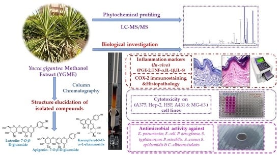
Elucidation of the Metabolite Profile of Yucca gigantea and Assessment of Its Cytotoxic, Antimicrobial, and Anti-Inflammatory Activities

by
Nashwah G. M. Attallah
1,†,
Suzy A. El-Sherbeni
2,*,†,
Aya H. El-Kadem
3,
Engy Elekhnawy
4,*,
Thanaa A. El-Masry
3,
Elshaymaa I. Elmongy
1,
Najla Altwaijry
1 and
Walaa A. Negm
2,*
1
Department of Pharmaceutical Science, College of Pharmacy, Princess Nourah bint Abdulrahman University, P.O. Box 84428, Riyadh 11671, Saudi Arabia
2
Pharmacognosy Department, Faculty of Pharmacy, Tanta University, Tanta 31111, Egypt
3
Department of Pharmacology and Toxicology, Faculty of Pharmacy, Tanta University, Tanta 31111, Egypt
4
Pharmaceutical Microbiology Department, Faculty of Pharmacy, Tanta University, Tanta 31111, Egypt
*
Authors to whom correspondence should be addressed.
These authors contributed equally to this work.
Molecules 2022, 27(4), 1329; https://doi.org/10.3390/molecules27041329
Submission received: 26 January 2022 / Revised: 8 February 2022 / Accepted: 14 February 2022 / Published: 16 February 2022

Abstract

:
The acute inflammation process is explained by numerous hypotheses, including oxidative stress, enzyme stimulation, and the generation of pro-inflammatory cytokines. The anti-inflammatory activity of Yucca gigantea methanol extract (YGME) against carrageenan-induced acute inflammation and possible underlying mechanisms was investigated. The phytochemical profile, cytotoxic, and antimicrobial activities were also explored. LC-MS/MS was utilized to investigate the chemical composition of YGME, and 29 compounds were tentatively identified. In addition, the isolation of luteolin-7-O-β-d-glucoside, apigenin-7-O-β-d-glucoside, and kaempferol-3-O-α-l-rhamnoside was performed for the first time from the studied plant. Inflammation was induced by subcutaneous injection of 100 μL of 1% carrageenan sodium. Rats were treated orally with YGME 100, 200 mg/kg, celecoxib (50 mg/kg), and saline, respectively, one hour before carrageenan injection. The average volume of paws edema and weight were measured at several time intervals. Levels of NO, GSH, TNF-α, PGE-2, serum IL-1β, IL-6 were measured. In additionally, COX-2 immunostaining and histopathological examination of paw tissue were performed. YGME displayed a potent anti-inflammatory influence by reducing paws edema, PGE-2, TNF-α, NO production, serum IL-6, IL-1β, and COX-2 immunostaining. Furthermore, it replenished the diminished paw GSH contents and improved the histopathological findings. The best cytotoxic effect of YGME was against human melanoma cell line (A365) and osteosarcoma cell line (MG-63). Moreover, the antimicrobial potential of the extract was evaluated against bacterial and fungal isolates. It showed potent activity against Gram-negative, Gram-positive, and fungal Candida albicans isolates. The promoting multiple effects of YGME could be beneficial in the treatment of different ailments based on its anti-inflammatory, antimicrobial, and cytotoxic effects.

Graphical Abstract

1. Introduction

Yucca is a genus of perennial trees and shrubs that belongs to the Asparagaceae family and the Agavoideae subfamily [1]. Yucca contains approximately 50 species that can be distributed from northern Canada to the southern and southwestern United States, Mexico, and the Caribbean [2]. Furthermore, certain species, as well as various cultivars, have flooded the global horticultural market, as several Yuccas can withstand relatively severe temperate climates [2,3]. The Yucca genus has long been utilized in folk medicine. Native Americans valued the ethnobotanical significance of these native flowering plants [4,5]. Yucca gigantea Lem. (a synonym of Yucca elephantipes) has many traditional uses as a source of fibers for textiles, basketry, and cordage, as well as food [6]. In addition to the functional food potential of Yucca species, antioxidant, anti-cancer, antidiabetic, antibacterial, anti-arthritic, and hypocholesterolemic characteristics have also been reported [4]. El Sayed et al [7] reported that LC-ESI-MS/MS of Y. gigantea (synonym Y. elephantipes) tentatively revealed the presence of 11 compounds. Spirostanol saponins comprised the majority of the tentatively recognized compounds: spirostan-3-ol-3-O-[β-d-galactopyranosyl-(1→4)-β-d-glucopyranoside, gallic acid, hecogenin-rhamnoside, (3β, 5α, 25R)-Spirostan-3-ol-3-O-[β-d-glucopyranosyl-(1→2)-β-dglucopyranoside, (3β, 5β, 25R)-Spirostan-3-ol-3-O-[β-d-glucopyranosyl-(1→2)-β-dglucopyranoside], (3β, 5α, 25S)-Spirostan-3-ol-3-O-[β-d-glucopyranosyl-(1→2)-β-dglucopyranoside], (3β, 5β, 25S)-Spirostan-3-ol-3-O-[β-d-glucopyranosyl-(1→2)-β-dglucopyranoside], 25R-Spirostan-diol-dihexoside isomer, 25S-Spirostan-diol-dihexoside isomer, chlorogenic acid and cinnamic acid. In our study, we investigated the chemical composition of Y. gigantea in detail using the negative ion mode of LC-ESI-MS/MS, which tentatively revealed the presence of 29 different compounds. Zhang et al. [8] reported the isolation of elephanosides G and H, new spirostanol saponins, from the leaves of Yucca elephantipes. Two known furostanol saponins and six known flavonoid glycosides were also separated. The isolated flavonoid glycosides were kaempferol 3-{O-β-d-glucopyranosyl-(1→2)-O-[α-l-rhamnopyranosyl-(1→6)]-β-d-galactopyranoside}, 6,8-bis(β-d-glucopyranosyl)apigenin, 8-(2-O-α-l-rhamnopyranoyl-β-d-glucopyranosyl) apigenin, (rutin), 4′-O-methylquercetin 3-(6-O-α-l-rhamnopyranosyl-β-d-glucopyranoside), quercetin 3-{O-β-d-glucopyranosyl-(1→2)-O-[α-l-rhamnopyranosyl-(1→6)]-β-d-galactopyranoside}. In the present study, we isolated other known flavonoid glycosides.
Inflammation is a protective body response to a variety of boosts, including microbial infection, chemical irritants, and tissue injury. By being a defense mechanism, many reactions can be induced or aggravated by the vast array of events and mediators involved in the inflammatory response [9]. Numerous proinflammatory mediators, such as interleukin-6 (IL-6) and tumor necrosis factor-alpha (TNF-α), are created throughout the inflammatory process [10] as they trigger and amplify the course of inflammation [11]. In addition, various signaling molecules and enzymatic pathways participate in driving the inflammation, including cyclooxygenase 2 (COX-2), a crucial enzyme that regulates the synthesis of prostaglandins (PGs) during inflammation [12]. In addition, TNF-α and interleukin 1 beta (IL-1β) were investigated as the essential main mediators that contribute to acute and chronic inflammation [13]. Reactive oxygen species (ROS) play a critical role in cellular defense systems as they are secreted by the inflammatory cells and exacerbate the oxidative stress process [14]. ROS can also activate the intracellular signaling pathways and boost pro-inflammatory cytokine gene production [9,15].
Finding safe and efficacious drugs to limit inflammation has proven to be difficult; hence, numerous animal models have been developed for evaluating anti-inflammatory medication effects. The most extensively used model to explore the anti-inflammatory potential of several natural and synthetic compounds is the carrageenan-induced acute inflammation model [16]. Although numerous drugs are available to treat inflammatory diseases, their long-term use induces severe adverse effects. Nonsteroidal anti-inflammatory drugs are commonly used to decrease inflammation. However, their long-term usage is linked to cardiac, renal, and gastrointestinal toxicity [17]. The development of safer anti-inflammatory agents is an important subject. The sensible and effective technique for treating inflammatory illnesses is the discovery of anti-inflammatory drugs obtained from natural sources [9].
Natural products have recently become a key source of pharmaceuticals and are currently being investigated as a medical candidate for their anti-inflammatory and antimicrobial effects. They have many advantages of being safe, effective, biocompatible, and cost-efficient alternatives. Other Yucca species were medicinally known to treat osteoarthritis, inflammation, and infections [18]. In addition, they protect the skin and treat skin sores and infections [19]. Few studies have investigated the different Yucca species introduced to Egypt [7]. Therefore, the antimicrobial potential of Y. gigantea methanol extract of leaves (YGME) was tested against certain pathogenic microbial isolates by the agar well diffusion method. We also investigated the anti-inflammatory and cytotoxic effects of Y. gigantea methanol extract.

2. Materials and Methods

2.1. Plant Materials, Extraction, and Isolation of Pure Compounds

Yucca gigantea leaves were gathered from a nursery at Al Qanatir Al Khayriyyah, El-Qalyubia Governorate on 13th December 2020. The plant was recognized by Dr. Esraa Ammar, Plant Ecology lecturer, Botany Department, Faculty of Science, Tanta University. A voucher sample (PG-M-0069) was deposited at the Herbarium of the Pharmacognosy Department, Tanta University. The plant was dried at room temperature, then powdered. The Y. gigantea powder (1.2 kg) was extracted by 95% methanol (4 L × 3 times) using the cold maceration method to yield 90.3 g of YGME.
Total methanolic extract (50 g) was suspended in deionized water and applied to the Diaion HP-20 column. The column was initially eluted with deionized water, followed by 100% methanol (MeOH). MeOH fraction (21.5 g) was resuspended in aqueous MeOH (50%) and successively partitioned with dichloromethane (CH2Cl2), ethyl acetate (EtOAc), and n-butanol (n-BuOH). Based on thin-layer chromatography (Merck, Darmstad, Germany), EtOAc fraction (9.1 g) was subjected to further investigation and chromatographed over vacuum liquid chromatography (silica gel 50 g, ϕ 4 cm × 11 cm) eluted with CH2Cl2 before adding MeOH in 1% increments. After TLC observation using Camag UV lamp at 254 and 366 nm, fractions were divided into four groups, from YE-1 to YE-4. Fraction YE-2 (518 mg) was rechromatographed over (silica gel 20 g, ϕ 1 cm × 25 cm) column, then purified using Sephadex LH-20 (Merck, Darmstad, Germany) with methanol to afford compounds I and II (11 and 9 mg, respectively). Isocratic CC on silica gel with (95:5) CH2Cl2: MeOH was performed on fraction YE-3 (260 mg) to give compound III (13 mg). A High-Performance Digital FT-Avance III NMR spectrometer (Bruker, Karlsruhe, Germany) was used to record NMR spectra. The 1H and 13C-NMR samples were examined at 400 MHz and 100 MHz, respectively. DMSO-d6 was used to dissolve the samples.

2.2. Animals

The animal house at Cairo University’s College of Veterinary Medicine provided a total of 40 male Wistar albino rats with a weight of 170–210 g. All rats were housed in normal rat cages with ad libitum access to standard pellet food and filtered water under controlled temperature (25 ± 2 °C) and illumination (12 h-light/dark cycle). Before being used in experiments, all rats were given a one-week acclimatization period. The Research Ethical Committee authorized all methods and experimental protocols in conformity with the rules for the care and use of laboratory animals (Faculty of Pharmacy, Tanta University, Egypt).

2.3. Materials, Drugs and Chemicals

Celecoxib was purchased from Amoun Pharma (Cairo, Egypt). All other chemicals and solvents used were bought from Merck (Kenilworth, NJ, USA) and were of a high analytical grade.

2.4. LC-MS/MS for Metabolite Analysis

2.4.1. Sample Preparations

The sample was prepared by macerating Yucca gigantea powder at room temperature in mild petroleum ether. The powder was extracted with MeOH after exhaustion, then the extract was evaporated under vacuum at 45 °C. A weighed amount of the dry residue (50 mg) was then added to a 1 mL solution of deionized water:methanol:acetonitrile (50:25:25). This mixture was vortexed for 2 min, ultra-sonicated for 10 min, then centrifuged for another 10 min at 1000 rpm. The sample solution was diluted with the reconstitution solvent and 10 L was injected at a concentration of 1 g/L [20].

2.4.2. Acquisition Method and Analytical Parameters

Analysis was carried out in the Proteomics and Metabolomics Unit, Children’s Cancer Hospital (57357), Basic Research Department, Cairo, Egypt. Adopting the methods previously reported by Negm et al. [21]. In-line filter disks (0.5 µm × 3.0 mm, Phenomenex®, Torrance, CA, USA) and X select HSS T3 (2.5 µm, 2.1 mm × 150 mm, Waters®, 40 °C, Milford, MA, USA) were used as a pre-column and analytical column, respectively. The mobile phases consisted of buffer A (5 mM ammonium formate buffer pH 3 containing 1% methanol), buffer B (5 mM ammonium formate buffer pH 8 containing 1% methanol), and buffer C (100 % acetonitrile). The flow rate was adjusted at 0.3 mL/min. The liquid chromatography (ExionLC -High flow LC, Sciex®) was programmed to use a mobile phase composition of buffer A and C in positive mode, and buffer B and C in negative mode. The mobile phase composition started with 90 (A or B): 10 (C) for the first 20 min, which was inversed from 21–25 min, and finally returning to the starting values for the last 3 min until the end of the protocol at 28 min. In addition, the instrument (ExionLC -High flow LC-, Sciex®, Framingham, MA, USA) was coupled with Triple TOF 5600+ (Sciex®) for IDA acquisition and Analyst TF 1.7.1 (Sciex®) for LC-Triple TOF control.

2.4.3. Data Processing

MasterView was used for feature (peaks) extraction from Total ion chromatogram (TIC) based on a signal-to-noise of greater than 5 (non-targeted analysis) and sample-to-blank intensities of greater than 3. In addition, Reifycs Abf (Analysis Base File) Converter (Reifycs®) was applied for Wiff file conversion and MS-DIAL 4.6 (RIKEN®) for data analysis. The ReSpect Database used possessed 1573 and 2737 records for negative mode, respectively. Metabolite’s annotation was conducted with the ReSpect Database and fragmentation pattern and retention times mentioned in previous reports for metabolites isolated from the investigated plant or others [7,22].

2.5. Cell Lines

Nawah Scientific Inc., (Mokatam, Cairo, Egypt) provided A375 (human melanoma), Hep-2 (head and neck cancer), A431 (human epidermoid skin carcinoma), MG-63 (osteosarcoma), and HSF (human skin fibroblast) cell lines. The cells were cultured in a humidified, 5% (v/v) CO2 atmosphere at 37 °C.

2.6. In Vitro Cytotoxicity

Sulforhodamine B (SRB) assay was carried out to determine the in vitro cytotoxicity of YGME. In 96-well plates, 100 μL cell suspension aliquots (5 × 10³ cells) were cultured in complete media for 24 h. Other aliquots of 100 μL media, containing various concentrations (0.03, 0.3, 3, 30, 300 µg/mL of YGME, were added to the cells. After 72 h of drug treatment, the cells were fixed by adding 150 μL of 10% trichloroacetic acid to the media and incubating for one hour at 4 °C. The trichloroacetic acid solution was taken out and distilled water was used to wash the cells five times. Then, 70 μL SRB solution (0.4 % w/v) was added in aliquots and incubated for 10 min in the dark. Using acetic acid (1%), plates were washed three times and 150 μL of TRIS (10 mM) was added to dissolve the protein-bound SRB stain. At 540 nm, the absorbance was measured using a microplate reader (BMGLABTECH® FLUO-star Omega, Ortenberg, Germany) [23].

2.7. In Vitro Antimicrobial Activity

2.7.1. Agar Well Diffusion Method

Using the agar well diffusion method, the antimicrobial activity of YGME extract was evaluated [24,25]. The tested bacterial and fungal species were Escherichia coli (ATCC 25922), Klebsiella pneumoniae (ATCC 13883), Pseudomonas aeruginosa (ATCC 27853), Proteus mirabilis (ATCC 35659), Salmonella typhimurium (ATCC 14028) as Gram-negative bacteria. In addition, Staphylococcus aureus (ATCC 25923) and Staphylococcus epidermidis (ATCC 6538) as Gram-positive bacteria. Candida albicans (ATCC 90028) was used as an example for fungi. On Muller–Hinton agar (MHA) plates, the bacterial isolates were streaked using sterile cotton swabs. A sterile cork borer was used to make 6 mm diameter wells in the agar plates, and 100 µL of 2 mg/mL (w/v) extract was applied to the wells. The agar plates were then incubated at 37 °C for 24 h using 0.1% chlorhexidine as a positive control and DMSO as a negative control, with a concentration of 10%. The appearance of clear zones indicated inhibition of bacterial growth and the zone diameters were measured in millimeters. Both 2% glucose and 5 µg/mL methylene blue were added to MHA to test the antifungal activity [26].

2.7.2. Determination of MIC Values

The broth microdilution method was employed to assess the MIC values [27]. One hundred microliters were taken from the stock solution of the extract (1000 µg/mL) and two-fold diluted with 100 µL of the test organism in Muller–Hinton broth (MHB). Each microtitration pate contained a negative growth control well (MHB only) and a positive control well (bacteria only). The microtitration plates were aerobically incubated for 24 h at 37 °C. The MIC of YGME is the lowest concentration at which observable bacteria growth is completely inhibited.

2.8. In Vivo Anti-Inflammatory Activity

2.8.1. Induction and Assessment of Carrageenan-Induced Paw Edema

In our study, rats were divided into five groups (8 rats/group). Group I: normal control group was administered 0.9% saline (10 mL/kg p.o). Groups II (positive control) and III (standard treatment) were orally treated with 0.9% saline and celecoxib (50 mg/kg) [11], respectively. Groups IV and V were orally treated with YGME (100 and 200 mg/kg, respectively). Doses of YGME were chosen based on the results of our pilot study and doses used in other Yucca species [28]. After 1 h, acute inflammation was triggered by subcutaneous injection of 100 μL of 1% carrageenan sodium in 0.9% saline into the plantar side of the right hind paw of rats in all groups except group I [29]. The thickness of the paws was measured at 0 h, and then 1, 2, 3 and 4 h later, to measure the increase in paw thickness [30]. At the end of the experiment, the weight of both the left and right paws was determined. The difference between the right and left paw weights was used to calculate the edema weight [31].

2.8.2. Sample Collection

Rats were anesthetized with diethyl ether, 4 h after receiving carrageenan, and blood was obtained through heart puncture into a syringe and centrifuged for 10 min at 3000 rpm. The serum was properly removed and stored at −20 °C to measure IL-6 and IL-1β. The rats were subsequently sacrificed by cervical dislocation under light ether anesthesia, and both paws were removed and weighed to determine the weight of the edema. The paw tissue was then separated, and one part was snap-frozen in liquid nitrogen to measure reduced glutathione (GSH), nitric oxide (NO), myeloperoxidase (MPO), TNF-α, and prostaglandin E2 (PGE2). The other part was used for immunohistochemical staining and histopathological examination.

2.8.3. Determination of Paw GSH Concentration

Reduced glutathione was spectrophotometrically measured in paws using the previously published method [32]. The absorbance of the yellow color, measured at 412 nm, is proportional to GSH concentration.

2.8.4. Determination of Paw NO Content

Stable metabolites nitrite and nitrate were measured in paw tissue homogenate to assess the NO level, as previously described [33], using the Griess reagent, which caliometrically detects these anions. At 540 nm, the absorbance was measured and, to determine NO concentration in each sample, a sodium nitrite standard curve was established.

2.8.5. Determination of Paw MPO Activity

The activity of MPO was measured using the previously reported method [34]. The kinetic measurement of yellowish-orange compounds for 5 min at 460 nm was used to develop this approach using a UV-Vis spectrophotometer (Shimadzu, Kyoto, Japan).

2.8.6. Determination of the Levels of Inflammatory Markers

According to the manufacturer’s protocol, the levels of the inflammatory mediators TNF-α and PGE2 in rat paw tissues were measured using commercial ELISA kits (Sun Red biotechnology Co., Ltd., Shanghai, China). In addition, the levels of IL-1β and IL-6 in the serum samples were measured using ELISA kits (Abcam corporation, Waltham, MA, USA).

2.8.7. Histopathologic Examination of the Paw Tissue

Paw tissues were fixed in formalin solution and embedded in paraffin wax, serially sectioned (5 μm), and stained with hematoxylin and eosin (H&E) to assess histopathologic changes. The stained sections were examined using a light microscope.

2.8.8. Immunohistochemical Determination of COX-2 Expression

The expression of COX-2 was detected by immunostaining tissue sections prepared from formalin-fixed, paraffin-embedded hind paws using a kit obtained from ABclonal Technology (Woburn, MA, USA). An immunoperoxidase (PAP, peroxidase/anti-peroxidase) technique was adopted. In this way, the cytoplasm of each COX-2 (+) cell was stained brown. The results were scored according to the percentage of positive staining for COX-2 as follows: score 0, no positive staining; mild, score 1+, from 1–10%; moderate, score 2++, from 11–50%; strong, score 3+++, more than 50% positive cells. In this study, scores of +, ++, and +++ were considered to be positive immunostaining, and a score of 0 was regarded as negative immunostaining [13].

2.9. Statistical Analysis

The measurements in the current study were perfumed three times and the obtained results were recorded in the form of mean ± standard deviation (SD). All standard curves were subjected to regression analysis and correlation coefficients were calculated. One-way analysis of variance (ANOVA) was used to compare distinct groups, followed by a Tukey–Kramer posthoc test. The significance level was established at (p < 0.05). Prism version 9 (GraphPad Software Inc., San Diego, CA, USA) was used to conduct the statistical analysis.

3. Results

3.1. LC-ESI-MS/MS Analysis of YGME

In negative mode, YGME components were tentatively identified using the liquid chromatography with mass spectrometry (LC-MS/MS) technique. Table 1 shows the presence of 29 compounds from different phytochemical subclasses and the total ion chromatogram (TIC) of YGME (negative mode) is represented in the Supplementary Materials, Figure S1. The MS/MS spectrums of the major identified compounds are displayed in Figure S2.

3.1.1. Characterization of Flavonols and Flavonols Glycosides

Most of the identified flavonols glycosides were found to be kaempferol derivatives. Compounds 7, 8 and 16 produced pseudo-molecular ion peaks [M − H] at m/z 593.152, 431.192, 593.519, respectively, which were ascribed to kaempferol-7-O-neohesperidoside, kaempferol-3-O-α-l-rhamnoside, kaempferol-3-O-(6-p-coumaroyl)-glucoside, respectively. The [M − H] of the aglycone ion (kaempferol) was recognized at m/z 285. Additionally, quercetin glycosides were identified in YGME. Quercetin-4′-O-glucoside showed a pseudomolecular ion peak [M − H] at m/z 463.085. The ion fragment of aglycone moiety (quercetin) was detected at m/z 301.041 [M − H-162] due to the neutral loss of glucose moiety. The chemical structures of these compounds are displayed in Figure 1a.

3.1.2. Characterization of Hydroxylated and/or Methoxylated Flavonols and Flavonols Glycosides

Compounds 13, 15, 18, 25, and 26 were tentatively identified as isorhamnetin, isorhamnetin-3-O-rutinoside, 4′,5,7-trihydroxyflavonol, 3,3′,4′,5-tetrahydroxy-7-methoxy flavone, and 3,5,7-trihydroxy-4′-methoxyflavone, respectively, which showed deprotonated molecular ion peaks [M − H] at m/z 315.135, 623.197, 285.077, 315.092 and 299.093, respectively. The MS/MS fragment ion at m/z 315.049 [M − H-308] of isorhamnetin-3-O-rutinoside was due to the neutral loss of sugar moiety (rhamno-glucoside) (Figure 1b).

3.1.3. Characterization of Flavones and Flavones Glycosides

The in-house database identified compounds 10, 21, 22 and 24 as Luteolin-7-O-β-d-glucoside, apigenin-7-O-β-d-glucoside, luteolin, and acacetin, respectively, based on pseudo-molecular ion peaks [M − H] at m/z 447.09, 431.17, 285.039, and 283.060, respectively (Figure 1c).

3.1.4. Characterization of Flavanones and Flavanone Glycosides

The deprotonated molecular ion peaks [M − H] of compounds 12, 17, 19 and 20 were exhibited at m/z 609.147, 593.265, 301.069, and 271.060, respectively. The in-house database deduced that they were hesperetin-7-O-neohesperidoside, poncirin, hesperetin, and naringenin, respectively. The MS/MS fragment ions of the aglycone of hesperetin-7-O-neohesperidoside and poncirin were detected at m/z 301.023 and 285.038, respectively. The neutral loss of sugar moiety of neohesperidoside produced the fragment ion of the aglycone (Figure 1d).

3.1.5. Characterization of Saponins

The four compounds 5, 6, 9 and 27, which are saponin derivatives, were tentatively identified. Compound 27 produced a deprotonated molecular ion peak [M − H + HCOOH] at m/z 461.26, which corresponded to the formic adduct of spirostan-3-ol, the main saponin aglycone in Yucca species [35]. Pseudo-molecular ion peaks [M − H] at m/z 739.79, attributed to spirostanol-3-ol-dihexose, were found in compounds 5 and 9. Due to the order of elution, compound 9 was thought to be the di-glucoside, while compound 5 is the glucoside-galactoside isomer [7]. Compound 6 was identified as hecogenin, an aglycone found in numerous Yucca species; the molecule produced a deprotonated molecular ion [M − H] at m/z 429.171 (Figure 1e).

3.1.6. Characterization of Organic and Phenolic Acids

The in-house database identified compounds 1, 3, 4 and 23 as maleic acid, citraconic acid, muconic acid, and cafeic acid, respectively, based on the deprotonated molecular ion peaks [M − H] at 115.001, 129.019, 141.020, and 179.033, respectively (Figure 1f).

3.1.7. Characterization of Other Compounds

Compound 2, a chalconoid derivative, at m/z 449.085, displays a pseudo-molecular ion peak [M-H], which was identified to be marein or okanin-4′-O-glucoside. Compounds 28 and 29 are 3-(4-hydroxy-3,5-dimethoxy phenyl)-2-propenoic acid and gamma-linolenic acid, respectively, with pseudo-molecular ion peaks [M − H] at m/z 223.171 and 277.197, respectively. Compound 11 is a flavonoid C-glycoside with a pseudo-molecular ion peak [M − H] at m/z 577.156 attributed to vitexin-2″-O-rhamnoside (Figure 1f).

3.2. Structure Elucidation of Compounds Isolated from YGME

1H-NMR (DMSO-d6, 400 MHz) and 13C-NMR (DMSO-d6, 100 MHz) for isolated compounds were displayed in Table S1. ESI-MS m/z [M − H] were 447.09, 431.17, and 431.192 for compounds I, II, and III, respectively (Figure 2).
The 1H-NMR spectrum of compound I exhibited two meta–coupled doublets (J = 2.4 Hz) at δH 6.78 and 6.44, each integrating with one proton, and were assigned to H-8 and H-6, respectively, of ring A of 5,7-dihydroxyflavonoids. The presence of an ABX system of C6′, C5′ and C2′ at δH 7.42 of (dd, J = 8.0, 2.4 Hz), 7.45 (d, J = 2.4 Hz) and 6.91 (d, J = 8.0 Hz), respectively, is known for the 1,2,4-trisubstituted phenyl unit [36]. A singlet resonating at δH 6.77, integrating with one proto, was characteristic for the C-3 proton of flavonoids [37]. The luteolin skeleton was cleared with these spectral data. In addition, the anomeric proton and carbon were located at δH 4.69 and δC 99.9, respectively. the 13C-NMR chemical shifts in the sugar carbons were shown at δC (99.9, 73.2, 77.2, 69.6, 76.5, 60.7), revealing the presence of β-O-glucoside unit in luteolin-7-O-glucoside. The 13C-NMR data showed the presence of a ketone carbonyl δC 181.9, two olefinic carbons (δC 164.6 and 103.3), and four hydroxyl carbons of C5, C7, C3′, and C4′ (δC 161.2, 163.0, 145.9 and 150.9). The ESI-MS m/z [M − H] was 447.09. The previous information confirmed the structure of luteolin-7-O-glucoside, as reported in the literature [38].
1H-NMR and 13C-NMR spectra confirmed the presence of apigenin and glucose moieties in the structure of compound II. The apigenin skeleton was deduced from two doublets δH at 6.45 (d, J = 2 Hz, H-6) and δH at 6.93 (d, J = 2 Hz, H-8) on the A-ring; A2B2-type aromatic δH at 7.95 (d, J = 8 Hz, H-2′, H-6′) and δH at 6.94 (d, J = 8 Hz, H-3′, H-5′); together with an olefinic δH at 6.83 (s, H-3) on a flavone C-ring. Besides this, glycosidic δH at 5.06 (d, J = 7.2 Hz, H-1″) and δC at 99.9 (C-1″) were evident in the 1H and 13C-NMR spectra. The multiplet δH at 3.27–3.47 (5H, m, H-3″–H-6″) could be assigned to the coupling between protons and methylene protons of the glucosyl ring. Proton δH at 3.71 (m, H-2″). The 13C-NMR displayed 21 carbon atoms for this compound. A glucose moiety was assigned at δC 99.9, 73.2, 76.5, 69.6, 77.3, and 60.7 for C-1″–C-6″, respectively. The flavone spectrum also displayed signals at δC (161.5, 99.6, 164.3 and 94.9) for C5–C8 of A-ring, respectively. δC (163.0, 103.2, 182.1, 161.5 and 105.4) for the C2–C4, C9, C10 of C-ring, respectively. Carbons C1′–C6′ of B-ring were displayed at δC (121.1, 128.7, 116.1 and 161.2). The ESI-MS m/z [M − H] was 431.17. The spectral data fit with the data that were previously reported in the literature. Therefore, the structure of the isolated compound was elucidated as apigenin- 7-O-β-d-glycoside [39].
1H-NMR spectrum of compound III showed four aromatic hydrogen signals. Two protons at δH 7.77 (2H, dd, J = 8.0, 1.6 Hz, H-2′ and H-6′) and two protons at δH 6.92 (2H, dd, J = 8.0, 1.6 Hz, H-3′and H-5′) showed a symmetric pattern with substitution at 1 and 4 positions of B-ring. Protons at H-2′ and H-3′ were ortho-coupled as well as at H-5′ and H-6′ (J = 8 Hz), while the proton at H-2′ meta-coupled with H-6′, and H-3′ with H-5′ (J = 1.6 Hz) of flavone skeleton. The other two aromatic proton signals of H-8 and H-6 were displayed at δH 6.43 and 6.21, respectively. The anomeric proton was located at δH 5.29 and δC 102.2. The 13C-NMR spectrum indicated 19 carbon signals. The spectrum displayed trihydroxy substitutions at δC of C-5 (161.7), C-7 (164.1), and C-4′ (160.4) in the flavone skeleton. The deoxy carbon C-6″ was resonating at δC 17.9, in addition to the other carbons C-1″–C-5″, which were resonating at 102.2, 70.6, 70.8, 71.5, and 71.1, respectively. This indicated the occurrence of the rhamnosyl unit in this compound. The ESI-MS m/z [M − H] was 431.192. Following the literature data, this compound was identified as kaempferol-3-O-α-l-rhamnoside (afzelin) [40]. Thin-layer chromatography was used to compare the Rf values of the three isolated flavonoid glycosides to authentic samples. This led to another confirmation of the elucidated structure.

3.3. In Vitro Cytotoxic Activity of YGME

Using SRB cell viability assay, IC50 (half-maximal concentration) of YGME was determined. The values of IC50 were 17.7 ± 0.43, 21.7 ± 0.556, 17.9 ± 1.059, 35.4 ± 0.984, and 30.7 ± 1.425 µg/mL against A375, Hep-2, MG-63, A431, and HSF cell lines, respectively. The best cytotoxic activity was recorded against A-375 and MG-63 cell lines. The results of the SRB cell viability assay are displayed in Figure 3 and the values of IC50 of YGME against the different investigated cell lines are presented in Figure 4.

3.4. In Vitro Antimicrobial Activity of YGME

In the current study, we investigated the antibacterial activity of Y. gigantea methanol extract of leaves (YGME) using the agar well diffusion method, as shown in Table 2, against Gram-negative standard isolates, including Escherichia coli (ATCC 25922), Klebsiella pneumoniae (ATCC 13883), Pseudomonas aeruginosa (ATCC 27853), Proteus mirabilis (ATCC 35659), Salmonella typhimurium (ATCC 14028), and Gram-positive standard isolates including Staphylococcus aureus (ATCC 25923), Staphylococcus epidermidis (ATCC 6538). In addition, an antifungal activity against Candida albicans (ATCC 90028) was found. Moreover, the minimum inhibitory concentrations of YGME were determined against the aforementioned isolates. The study performed by Mahjbeen et al. [41] determined an antibacterial activity against Gram-positive bacteria (S. aureus, B. subtilis) as well as Gram-negative bacteria (E. coli, P. aeruginosa) using the disc-diffusion method. In our study, we tested the antibacterial activity against a larger number of Gram-negative isolates and tested the antifungal activity of YGME in addition to measuring the MIC values.

3.5. In Vivo Anti-Inflammatory Activity

3.5.1. Effect of YGME on the Average Edema Volume

As shown in Table 3, the carrageenan group induced severe paw edema, which attained a peak level of 4 h after carrageenan administration. The edema volume (mm) was measured after different time intervals (1, 2, 3 and 4 h) in all studied groups. After 1 h, the average change in the edema volume showed a higher level in group II, while the edema volume in groups III, IV, and V had a lower paw volume. Similar results were observed after 2, 3 and 4 h of the experiment, with the highest paw volume was observed in group II, and a significant reduction in the edema volume seen in group V. These findings demonstrated that YGME produced a pronounced decline in the edema volume compared to the other treatments at p < 0.05.

3.5.2. Effect of YGME on the Average Paw Edema Weight

In comparison with group I, group II exhibited a significant increase in the paw edema weight, with a percentage of 1133.3%. Treatment with celecoxib (group III) presented a significant reduction in the edema weight compared to the carrageenan group (group II), with a percentage of 59.45%. Treatment with YGME 100 (group IV) and 200 (group V) significantly decreased the edema weight, with percentages of 54.05, and 81.08%, respectively, compared to group II. The decrease in the edema weight in group V was superior, as shown in Table 4 (p ˂ 0.05).

3.5.3. Effect of YGME on MPO Activity

In comparison with group I, group II presented a marked increase in MPO activity, with a percentage of 388.3%. Group III exhibited a pronounced decrease in MPO activity compared to group II, with a percentage of 66.46%, while groups IV and V showed a significantly suppressed MPO activity (55.31 and 75.91%, respectively) when compared to group II, with the results being superior in group V, as shown in Table 4 (p ˂ 0.05).

3.5.4. Effect of YGME on the Oxidative Stress Markers

By comparison to the group I, group II revealed massive oxidative damage, as evident by a considerable reduction in GSH content in the paw tissue (30.12%). Markedly increased GSH levels (7.6%) were found in group III compared to group II. In addition, groups IV and V showed significant restoration of the GSH levels (20.51, and 77.91%, respectively) compared to group II, with a more prominent effect in group V, as shown in Table 4 (p ˂ 0.05).
Group II exhibited a significant elevation in the paw NO content (90.56%) in comparison to group I. Group III revealed a significant reduction in the NO levels (46.53%) compared to the group II. In addition, treatment with YGME 100 and 200 (groups IV and V) induced a significant suppression of NO levels (32.67 and 48.01%, respectively) compared to group II, with a more significant result in group V, as shown in Table 4 (p ˂ 0.05).

3.5.5. Effect of YGME on the Levels of the Inflammation Markers

Group II showed a marked inflammation, demonstrated by the remarkable increase in TNF-α content (799%) in comparison to group I. Celecoxib treatment (group III) significantly reduced TNF-α (83.34%) compared to the group II, while treatment with YGME 100 and 200 (groups IV and V) induced a considerable reduction in TNF-α levels (51.13 and 80%, respectively) compared to group II, with a more pronounced effect in group V, as shown in Figure 5 (p ˂ 0.05).
Group II also displayed a marked increase in IL-1β and IL-6 serum levels (86.98 and 165.25%, respectively) in comparison to group I. Celecoxib treatment (group III) strongly reduced IL-1β and IL-6 levels (39.52 and 56.39%, respectively) compared to group II. In addition, treatment with YGME 100 and 200 (groups IV and V) induced a significant suppression of IL-1β levels (16.94, 19.54% and 32.65, 46.9%, respectively) in comparison with group II, with a more pronounced effect in group V, as shown in Figure 5 (p ˂ 0.05).

3.5.6. Effect of YGME on Paw PGE-2 Levels

The current study showed a marked-up regulation in paw PGE-2 levels (1522%) in group II compared to group I. Celecoxib treatment (group III) significantly reduced PGE-2 levels (92.89%), compared to group II. In addition, treatment with YGME 100 and 200 (groups IV and V) induced a marked decrease in PGE-2 levels (65.43 and 91.74%, respectively) in comparison to group II. The results were more significant in group V, as shown in Figure 5 (p ˂ 0.05).

3.5.7. Effect of YGME on the Histopathology of Paw Tissues

The section in the normal control group (group I) paw showed normal skin consisting of epidermis of average thickness lined with thick keratin and underlying normal dermis and normal muscles (Figure 6A). The section in the carrageenan group (group II) showed deep dense dermal infiltration, with chronic inflammatory cells with congested vessels (Figure 6B1). In addition, a severe interstitial inflammatory reaction could be seen between muscle bundles (Figure 6B2). Furthermore, the section in the celecoxib-treated group (group III) showed no inflammation: muscles were normal; however, the epidermis was thickened and covered with excessive keratosis with underlying excessive collagenosis (Figure 6C). The section in YGME-100-treated group (group IV) showed superficial dermal mild chronic inflammation with mild edema and mild vascular congestion. Moreover, the epidermis was covered with excessive keratosis (Figure 6D). However, the sections of the YGME-200-treated group (group V) showed normal skin without inflammation or collagenosis and normal epidermis lined with thick keratin and dermis (Figure 6E).

3.5.8. Effects of YGME on the Immunohistochemical Staining of COX-2 in Paw Tissues

The section in the normal control group showed negative COX2 immunostaining (Figure 7A). The section in carrageenan (positive control group) showed a positive strong COX-2 immunostaining score 3 (Figure 7B). The section in celecoxib 50 group showed a positive mild COX-2 immunostaining score 1 (Figure 7C).
The section in the YGME100-treated group showed a moderate, positive COX-2 immunostaining score of 2 (Figure 7D). The section in the YGME-200-treated group showed negative COX2 immunostaining (Figure 7E).

4. Discussion

The inflammation process is a cause of discomfort and a key driver of pathophysiological events that eventually induce disease progression [14]. The carrageenan-induced paw edema is a useful model for evaluating the anti-inflammatory action of a high number of natural and synthetic products [42,43]. This is a strong chemical that induces the release of several inflammatory and pro-inflammatory mediators.
Acute inflammation is a double-phase process. The initial stage begins with the liberation of numerous mediators after carrageenan injection, while the second one is associated with prostaglandins’ release in 2–3 h [11].
The major goal of this research is to alleviate the inflammatory response in a model of acute inflammation using natural products from medicinal plants such as Yucca species. Several studies have investigated the anti-inflammatory, anti-arthritic and anti-oxidant effects of numerous Yucca species [44,45]. Yucca polyphenolics have free-radical scavenging activity, which could suppress the ROS and halt inflammatory responses [46]. They have been investigated to suppress iNOS expression, and thus inhibit NO production [47]. They also inhibit NFkB, which, in turn, stimulates iNOS, which produces the inflammatory agent nitric oxide [47]. This may clarify the anti-inflammatory effects of Yucca species.
This is the premier study investifating the anti-inflammatory and antioxidant activities of Yucca gigantea methanol extract (YGME) in carrageenan-induced hind paw edema. The activation of the cyclooxygenase-2 pathway and the production of PGE2, which are the major regulators of many cell activities and the principal mediators in the acute inflammation process, is often related to the carrageenan model. COX-2 is activated by elevated NO levels, owing to severe inflammatory responses in a variety of chronic inflammatory diseases [48]. In the current study, the carrageenan induced a strong, positive immunostaining of COX-2 and up-regulated paw PGE-2 levels. Furthermore, this increase in PGE-2 levels was attenuated by celecoxib, which is a selective COX-2 inhibitor. YGME treatment significantly reduced COX-2 staining and PGE-2 levels in paw tissues in a dose-dependent manner. LC-ESI-MS/MS of YGME revealed that it contained several flavonoids, such as apigenin and quercetin, which strongly suppressed COX-2 and PGE-2 levels, and these results are consistent with References [49,50,51].
A histopathological study of paw tissue in a group treated by YGME 200 mg/kg exhibited normal skin without inflammation or collagenosis and normal epidermis lined with thick keratin and dermis.
In addition, the data from this study indicate that carrageenan induced the production of several inflammatory cytokines, such as TNF-α, IL-1β, and IL-6, which are dose-dependently attenuated by YGME 100 and 200, and the results were equivalent to celecoxib, confirming the anti-inflammatory results of YGME. In addition, carrageenan induced a marked neutrophil infiltration, shown by a significant increase in MPO activity, which was strongly attenuated by YGME, with promising potential for the use of YGME as an anti-inflammatory compound in different diseases. YGME flavonoids strongly suppressed several inflammatory mediators, which may have crucial value in the protection and treatment of various inflammatory diseases linked to the excessive production of chemical mediators [52], and these results are consistent with previous reports [50,53,54,55,56,57]
It is revealed that YGME contain apigenin and quercetin, which were found to inhibit PGE-2 [58] and NO production. The latter primarily regulated PGE-2 synthesis by preventing the production of ROS [52,59]. It was reported that quercetin suppressed the LPS-stimulated TNF-α release in RAW cells [60]. The luteolin flavonoid’s ability to inhibit protein tyrosine phosphorylation, NFĸB-mediated gene expression, and the release proinflammatory mediators in macrophages was also investivated [60]. In addition, luteolin decreased the expression of inflammatory mediators via blocking the Akt/NFκB pathway in mice with acute lung injury caused by LPS [61].
In this study, paw NO was elevated, whereas GSH was reduced, after carrageenan injection. Treatment with YGME 100 and 200 reduced paws’ NO production and restored depleted GSH levels in a dose-dependent manner. In addition, the celecoxib-treated group showed a marked decrease in paw NO content and replenished the paw’s depleting GSH levels.
Flavonoids and saponins were reported to inhibit NO production. YGME contained several flavonoids, such as luteolin and kaempferol derivatives, which were reported to have strong inhibitory effects on NO production, and these results are consistent with the literature, which reported that they inhibit NO in lipopolysaccharide (LPS)-stimulated macrophages [9,60,61,62]. Finally, these findings could explain the anti-inflammatory and antioxidant effects of YGME on the acute inflammation model.
Free radical generation at the inflammation site causes major tissue damage, accompanied by many inflammatory disorders [14]. NO produced by inducible nitric oxide synthase (iNOS) was a principle arbitrator in the inflammation process and played a pivotal role in aggravating inflammation and NO-mediated cytotoxicity [13]. Therefore, in this study, we assessed the cytotoxic effect of YGME using SRB cell viability assay to find this extract’s ability to prevent and treat cancer in addition to reducing inflammation. YGME was topically applied to protect skin and treat different skin infections and diseases [19].
The SRB assay revealed that the YGME exerted a cytotoxic effect against all tested cell lines. The best cytotoxic effect was against human melanoma cell line (A375) and osteosarcoma cell line (MG-63). The results showed that YGME can reduce inflammation, e.g., arthritis, and has anti-osteosarcoma as well as anti-melanoma effects. The selectivity index was calculated to be 1.73, 1.41, 1.72, and 0.87 of YGME against A375, Hep-2, MG-63, and A431, respectively. Nutraceuticals include active phytochemical components as flavonoids in plants, and are used as an integrative treatment in up to 82% of patients suffering from cancer. Flavonoids are the most important group of active constituents, composed of flavones, flavonols, flavanones, flavanols, and isoflavonols [63]. LC-ESI-MS/MS of Y. gigantea revealed the presence of hesperetin, naringenin, quercetin-4′-O-glucoside, luteolin, kaempferol glycosides, caffeic acid, and methoxylated flavones. It was reported that hesperetin exerted an apoptotic effect towards A431 epidermoid skin carcinoma through cyclin and MAPK regulation. Naringenin was found to be effective against A431 cancer. Hesperidin is reported to be more cytotoxic than neohesperdin and naringin against HepG2, as well as showing anti-lung-cancer activity against the A549 cell line [64]. The apoptotic effect of luteolin and quercetin was evidenced in TRAIL-resistant cancer cells. They showed, besides kaempferol, an antiproliferative effect against VCAR3 ovarian cancer [65]. Quercetin exerted cytotoxicity against different types of cancers [66]. Polyphenols such as caffeic acid are antioxidants; they showed an anticancer effect by inhibiting DNA methylation, cyclooxygenase, and DNA topoisomerase enzymes, in addition to influencing cell signaling [65]. Benzo(a)pyrene or B(a)P is a common cancer-promoter through its bioactivation to a form that eventually binds to DNA, causing cancer initiation. Methoxylated flavones, e.g., 3′,4′-dimethoxyflavone, and 5,7-dimethoxyflavone, were evidenced to inhibit the bioactivation of B(a)P in vitro. 5,7-dimethoxyflavone could strongly bind to DNA in cell culture, and inhibit the bioactivation of B(a)P in HepG2 cell line [67,68].
Previous studies on innate immunity suggested that intestinal inflammation could be caused by bacterial infections in genetically susceptible hosts [69]. Infectious agents such as Gram-positive and Gram-negative bacteria could activate inflammatory cells and trigger inflammatory signaling pathways [70]. At present, bacterial infections are a critical threat, owing to the spread of antibiotic resistance, which has been linked to the misuse and overuse of antibiotics alongside the lack of development of new medications [71]. The emergence and dissemination of antibiotic-resistant bacteria have reduced the effectiveness of the currently used antibiotics for treatment and raised the need for the exploration of new antimicrobials from natural sources [72]. This study provided evidence for the antimicrobial potential of YGME extract, which could be utilized as an alternative to antimicrobials, as it is obvious from our results that YGME extract has a broad spectrum of antimicrobial activity. As shown in Table 2, YGME exhibited a promising antimicrobial activity against bacteria (both Gram-positive and Gram-negative). In addition, it revealed antifungal activity against Candida albicans as a representative for fungi. Thus, the antimicrobial activity of YGME should be investigated in more depth in future studies to elucidate its mechanism of action.
The antimicrobial effect of YGME against Gram-positive, Gram-negative, and fungi could prevent inflammation caused by an infectious pathogen. As well as its anti-inflammatory effect, it could prevent various disorders and chronic health conditions, such as cancer and cardiovascular diseases. A low-grade inflammatory state is correlated with these ailments [73]. The multiple biological effects of YGME may help transnational industries in the development of new, curative drugs from extraordinary plants such as Y. gigantea.

5. Conclusions

The findings of this study indicate that methanol extract from the leaves of Yucca gigantea alleviated inflammation and oxidative stress in the carrageenan-induced acute inflammation model in rats. This was shown by a significant reduction in paw COX-2 staining, PGE-2 levels, inflammatory cytokines IL-1β, TNF- α, and IL-6 levels. It also strongly suppressed NO production and restored GSH antioxidant capacity. Using LC-MS/MS to investigate the chemical composition of YGME, 29 compounds were tentatively identified from different phytochemical classes, such as saponins, flavonoids, organic and phenolic compounds. Flavonoids were reported to have anti-microbial, anti-inflammatory, antioxidants, and cytotoxic effects, including apigenin, kaempferol, luteolin, and quercetin, which could explain the beneficial effects of YGME in infectious, inflammatory, and cancer conditions. YGME exhibited cytotoxic activity against all the investigated cell lines, especially against A-375 and MG-63 cell lines. YGME also presented a potent activity against Gram-negative, Gram-positive isolates, and the fungal isolate Candida albicans. The antimicrobial effect of YGME could prevent inflammation caused by an infectious pathogen. In addition, its ability to reduce anti-inflammatory effects could prevent cancer and cardiovascular diseases. Therefore, Yucca gigantea Lem. may have a promising future in the natural drug discovery of anti-inflammatory, antioxidants, antimicrobial, and cytotoxic drugs.

Supplementary Materials

The following supporting information can be downloaded. Figure S1: TIC and BPC of LC-ESI-MS/MS analysis of Yucca gigantea methanol extract in negative ionization mode, Figure S2: The MS/MS spectrum of the major identified compounds, Table S1: 1H-NMR (DMSO-d6, 400 MHz) and 13C NMR (DMSO-d6, 100 MHz) for the isolated pure compounds.

Author Contributions

Conceptualization, W.A.N., E.E., A.H.E.-K. and S.A.E.-S.; data curation, W.A.N., A.H.E.-K. and S.A.E.-S.; formal analysis, N.G.M.A. and N.A.; funding acquisition, N.G.M.A., T.A.E.-M. and N.A.; investigation, W.A.N., E.E., A.H.E.-K. and S.A.E.-S.; methodology, W.A.N., E.E., A.H.E.-K. and S.A.E.-S.; project administration, N.G.M.A., E.I.E. and T.A.E.-M.; resources, N.A.; software, N.G.M.A.; supervision, T.A.E.-M.; validation, W.A.N., E.E., A.H.E.-K. and S.A.E.-S.; visualization, W.A.N.; writing—original draft, W.A.N., E.E., A.H.E.-K. and S.A.E.-S.; writing—review and editing, W.A.N., E.E., A.H.E.-K. and S.A.E.-S. All authors have read and agreed to the published version of the manuscript.

Funding

This work was funded by Princess Nourah bint Abdulrahman University Researchers Supporting Project number (PNURSP2022R204), Princess Nourah bint Abdulrahman University, Riyadh, Saudi Arabia.

Institutional Review Board Statement

All procedures and experimental protocols were conducted following the guidelines for the care and use of laboratory animals was approved by the Research Ethical Committee (Faculty of Pharmacy, Tanta University, Tanta, Egypt) and the approval date was 24 July 2021, and the ethical code NO. PO-00101.

Informed Consent Statement

Not applicable.

Data Availability Statement

The data are available upon request.

Acknowledgments

This work was supported by Princess Nourah bint Abdulrahman University Researchers Supporting Project number (PNURSP2022R204), Princess Nourah bint Abdulrahman University, Riyadh, Saudi Arabia.

Conflicts of Interest

The authors declare no conflict of interest.

Sample Availability

Not available.

References

  1. Chase, M.W.; Reveal, J.L.; Fay, M.F. A subfamilial classification for the expanded asparagalean families Amaryllidaceae, Asparagaceae and Xanthorrhoeaceae. Bot. J. Linn. Soc. 2009, 161, 132–136. [Google Scholar] [CrossRef] [Green Version]
  2. Smith, C. Yuccas: Giants among the Lilies; National Council for the Conservation of Plants & Gardens: Guildford, UK, 2004. [Google Scholar]
  3. Smith, G.F.; Figueiredo, E. Widely cultivated, large-growing yuccas: Notes on Yucca elephantipes Regel ex Trel., Yucca gigantea Lem., and Yucca guatemalensis Baker (Agavaceae). Bradleya 2016, 2016, 28–37. [Google Scholar] [CrossRef]
  4. Patel, S. Yucca: A medicinally significant genus with manifold therapeutic attributes. Nat. Prod. Bioprospect. 2012, 2, 231–234. [Google Scholar] [CrossRef] [Green Version]
  5. Zubair, M.; Rasool, N.; Mansha, A.; Anjum, F.; Iqbal, M.; Mushtaq, M.; Shahid, M. Antioxidant, antibacterial, antifungal activities and phytochemical analysis of dagger (Yucca aloifolia) leaves extracts. J. Med. Plants Res. 2013, 7, 243–249. [Google Scholar]
  6. Pascual-Mendoza, S.; Saynes-Vásquez, A.; Pérez-Herrera, A. Traditional knowledge of edible plants in an indigenous community in the Sierra Norte of Oaxaca, Mexico. Plant Biosyst.-Int. J. Deal. All Asp. Plant Biol. 2021, 1–13. [Google Scholar] [CrossRef]
  7. El Sayed, A.M.; Basam, S.M.; Marzouk, H.S.; El-Hawary, S. LC–MS/MS and GC–MS profiling as well as the antimicrobial effect of leaves of selected Yucca species introduced to Egypt. Sci. Rep. 2020, 10, 17778. [Google Scholar] [CrossRef]
  8. Zhang, Y.; Yang, C.R.; Zhang, Y.J. New Steroidal Saponins from the Leaves of Yucca elephantipes. Helv. Chim. Acta 2013, 96, 1807–1813. [Google Scholar] [CrossRef]
  9. Patil, K.R.; Mahajan, U.B.; Unger, B.S.; Goyal, S.N.; Belemkar, S.; Surana, S.J.; Ojha, S.; Patil, C.R. Animal models of inflammation for screening of anti-inflammatory drugs: Implications for the discovery and development of phytopharmaceuticals. Int. J. Mol. Sci. 2019, 20, 4367. [Google Scholar] [CrossRef] [Green Version]
  10. Karim, N.; Khan, I.; Khan, W.; Khan, I.; Khan, A.; Halim, S.A.; Khan, H.; Hussain, J.; Al-Harrasi, A. Anti-nociceptive and anti-inflammatory activities of asparacosin a involve selective cyclooxygenase 2 and inflammatory cytokines inhibition: An in-vitro, in-vivo, and in-silico approach. Front. Immunol. 2019, 10, 581. [Google Scholar] [CrossRef]
  11. Zhang, H.; Shang, C.; Tian, Z.; Amin, H.K.; Kassab, R.B.; Abdel Moneim, A.E.; Zhang, Y. Diallyl disulfide suppresses inflammatory and oxidative machineries following carrageenan injection-induced paw edema in mice. Mediat. Inflamm. 2020, 2020, 8508906. [Google Scholar] [CrossRef] [Green Version]
  12. Alam, M.B.; Kwon, Y.-G.; Simu, S.Y.; Abrar Shahriyar, S.; Lee, S.H. Attenuation of Inflammatory Symptoms by Icariside B2 in Carrageenan and LPS-Induced Inflammation Models via Regulation of MAPK/NF-κB Signaling Cascades. Biomolecules 2020, 10, 1037. [Google Scholar] [CrossRef] [PubMed]
  13. Kim, K.H.; Im, H.-W.; Karmacharya, M.B.; Kim, S.; Min, B.-H.; Park, S.R.; Choi, B.H. Low-intensity ultrasound attenuates paw edema formation and decreases vascular permeability induced by carrageenan injection in rats. J. Inflamm. 2020, 17, 7. [Google Scholar] [CrossRef] [PubMed]
  14. Cordaro, M.; Siracusa, R.; Fusco, R.; D’Amico, R.; Peritore, A.F.; Gugliandolo, E.; Genovese, T.; Scuto, M.; Crupi, R.; Mandalari, G. Cashew (Anacardium occidentale L.) nuts counteract oxidative stress and inflammation in an acute experimental model of Carrageenan-induced Paw edema. Antioxidants 2020, 9, 660. [Google Scholar] [CrossRef] [PubMed]
  15. Biswas, S.K. Does the interdependence between oxidative stress and inflammation explain the antioxidant paradox? Oxidative Med. Cell. Longev. 2016, 2016, 5698931. [Google Scholar] [CrossRef] [PubMed] [Green Version]
  16. Sarkhel, S. Evaluation of the anti-inflammatory activities of Quillaja saponaria Mol. saponin extract in mice. Toxicol. Rep. 2016, 3, 1–3. [Google Scholar] [CrossRef] [Green Version]
  17. Sun, K.; Song, X.; Jia, R.; Yin, Z.; Zou, Y.; Li, L.; Yin, L.; He, C.; Liang, X.; Yue, G. Evaluation of analgesic and anti-inflammatory activities of water extract of Galla chinensis in vivo models. Evid.-Based Complement. Altern. Med. 2018, 2018, 6784032. [Google Scholar] [CrossRef] [Green Version]
  18. El Hawary, S.; El Sayed, A.; Helmy, M.W.; El Naggar, E.M.B.; Marzouk, H.S.; Bassam, S.M. DNA fingerprinting, biological and chemical investigation of certain Yucca species. Nat. Prod. Res. 2018, 32, 2617–2620. [Google Scholar] [CrossRef]
  19. GARCÍA-BORES, A.M.; Bello, C.; Campos, Y.; del Carmen Benitez, J.; Flores, S.; Canales, M.; Hernandez, T.; ACEVEDO, J.G.A. Photoprotective activity of Yucca periculosa polyphenols. Bol. Latinoam. Caribe Plantas Med. Aromát. 2010, 9, 100–108. [Google Scholar]
  20. Attallah, N.G.M.; Negm, W.A.; Elekhnawy, E.; Elmongy, E.I.; Altwaijry, N.; El-Haroun, H.; El-Masry, T.A.; El-Sherbeni, S.A. Elucidation of Phytochemical Content of Cupressus macrocarpa Leaves: In Vitro and In Vivo Antibacterial Effect against Methicillin-Resistant Staphylococcus aureus Clinical Isolates. Antibiotics 2021, 10, 890. [Google Scholar] [CrossRef]
  21. Negm, W.A.; El-Kadem, A.H.; Elekhnawy, E.; Attallah, N.G.; Al-Hamoud, G.A.; El-Masry, T.A.; Zayed, A. Wound-Healing Potential of Rhoifolin-Rich Fraction Isolated from Sanguisorba officinalis Roots Supported by Enhancing Re-Epithelization, Angiogenesis, Anti-Inflammatory, and Antimicrobial Effects. Pharmaceuticals 2022, 15, 178. [Google Scholar] [CrossRef]
  22. Alotaibi, B.; Mokhtar, F.A.; El-Masry, T.A.; Elekhnawy, E.; Mostafa, S.A.; Abdelkader, D.H.; Elharty, M.E.; Saleh, A.; Negm, W.A. Antimicrobial Activity of Brassica rapa L. Flowers Extract on Gastrointestinal Tract Infections and Antiulcer Potential Against Indomethacin-Induced Gastric Ulcer in Rats Supported by Metabolomics Profiling. J. Inflamm. Res. 2021, 14, 7411. [Google Scholar] [CrossRef]
  23. Attallah, N.G.; Negm, W.A.; Elekhnawy, E.; Altwaijry, N.; Elmongy, E.I.; El-Masry, T.A.; Alturki, E.A.; Yousef, D.A.; Y Shoukheba, M. Antibacterial Activity of Boswellia sacra Flueck. Oleoresin Extract against Porphyromonas gingivalis Periodontal Pathogen. Antibiotics 2021, 10, 859. [Google Scholar] [CrossRef] [PubMed]
  24. Clinical and Laboratory Standards Institute. Performance Standards for Antimicrobial Susceptibility Testing; 20th Informational Supplement; CLSI Document M100; CLSI: Wayne, PA, USA, 2017; pp. 1–50. [Google Scholar]
  25. Negm, W.A.; El-Aasr, M.; Kamer, A.A.; Elekhnawy, E. Investigation of the Antibacterial Activity and Efflux Pump Inhibitory Effect of Cycas thouarsii R. Br. Extract against Klebsiella pneumoniae Clinical Isolates. Pharmaceuticals 2021, 14, 756. [Google Scholar] [CrossRef] [PubMed]
  26. Khadka, S.; Sherchand, J.B.; Pokhrel, B.M.; Parajuli, K.; Mishra, S.K.; Sharma, S.; Shah, N.; Kattel, H.P.; Dhital, S.; Khatiwada, S. Isolation, speciation and antifungal susceptibility testing of Candida isolates from various clinical specimens at a tertiary care hospital, Nepal. BMC Res. Notes 2017, 10, 218. [Google Scholar] [CrossRef] [PubMed] [Green Version]
  27. Weinstein, M.P. Performance Standards for Antimicrobial Disk Susceptibility Tests. National Committee for Clinical Laboratory Standards: Wayne, PA, USA, 2018. [Google Scholar]
  28. Ince, S.; Kucukkurt, I.; Turkmen, R.; Demirel, H.H.; Sever, E. Dietary Yucca schidigera supplementation reduces arsenic-induced oxidative stress in Swiss albino mice. Toxicol. Ind. Health 2013, 29, 904–914. [Google Scholar] [CrossRef]
  29. Coura, C.O.; Souza, R.B.; Rodrigues, J.A.G.; Vanderlei, E.d.S.O.; de Araújo, I.W.F.; Ribeiro, N.A.; Frota, A.F.; Ribeiro, K.A.; Chaves, H.V.; Pereira, K.M.A. Mechanisms involved in the anti-inflammatory action of a polysulfated fraction from Gracilaria cornea in rats. PLoS ONE 2015, 10, e0119319. [Google Scholar]
  30. Amdekar, S.; Roy, P.; Singh, V.; Kumar, A.; Singh, R.; Sharma, P. Anti-inflammatory activity of lactobacillus on carrageenan-induced paw edema in male wistar rats. Int. J. Inflamm. 2012, 2012, 752015. [Google Scholar] [CrossRef] [Green Version]
  31. El-Shitany, N.A.; El-Bastawissy, E.A.; El-desoky, K. Ellagic acid protects against carrageenan-induced acute inflammation through inhibition of nuclear factor kappa B, inducible cyclooxygenase and proinflammatory cytokines and enhancement of interleukin-10 via an antioxidant mechanism. Int. Immunopharmacol. 2014, 19, 290–299. [Google Scholar] [CrossRef]
  32. El-Mahdy, N.A.; El-Sayad, M.E.S.; El-Kadem, A.H.; Abu-Risha, S.E.S. Targeting IL-10, ZO-1 gene expression and IL-6/STAT-3 trans-signaling by a combination of atorvastatin and mesalazine to enhance anti-inflammatory effects and attenuates progression of oxazolone-induced colitis. Fundam. Clin. Pharmacol. 2021, 35, 143–155. [Google Scholar] [CrossRef]
  33. El-Mahdy, N.A.; El-Sayad, M.E.-S.; El-Kadem, A.H. Combination of telmisartan with sildenafil ameliorate progression of diabetic nephropathy in streptozotocin-induced diabetic model. Biomed. Pharmacother. 2016, 81, 136–144. [Google Scholar] [CrossRef]
  34. El-Mahdy, N.A.; El-Sayad, M.E.-S.; El-Kadem, A.H.; Abu-Risha, S.E.-S. Metformin alleviates inflammation in oxazolone induced ulcerative colitis in rats: Plausible role of sphingosine kinase 1/sphingosine 1 phosphate signaling pathway. Immunopharmacol. Immunotoxicol. 2021, 43, 192–202. [Google Scholar] [CrossRef] [PubMed]
  35. Qu, L.; Wang, J.; Ruan, J.; Yao, X.; Huang, P.; Wang, Y.; Yu, H.; Han, L.; Zhang, Y.; Wang, T. Spirostane-type saponins obtained from Yucca schidigera. Molecules 2018, 23, 167. [Google Scholar] [CrossRef] [PubMed] [Green Version]
  36. Usia, T.; Iwata, H.; Hiratsuka, A.; Watabe, T.; Kadota, S.; Tezuka, Y. Sesquiterpenes and Flavonol Glycosides from Zingiber a romaticum and Their CYP3A4 and CYP2D6 Inhibitory Activities. J. Nat. Prod. 2004, 67, 1079–1083. [Google Scholar] [CrossRef] [PubMed]
  37. Mabry, T.; Markham, K.R.; Thomas, M.B. The Systematic Identification of Flavonoids; Springer Science & Business Media: Berlin/Heidelberg, Germany, 2012. [Google Scholar]
  38. Chiruvella, K.K.; Mohammed, A.; Dampuri, G.; Ghanta, R.G.; Raghavan, S.C. Phytochemical and antimicrobial studies of methyl angolensate and luteolin-7-O-glucoside isolated from callus cultures of Soymida febrifuga. Int. J. Biomed. Sci. IJBS 2007, 3, 269. [Google Scholar]
  39. SHEN, C.-C.; CHANG, Y.-S.; HO, L.-K. Nuclear-magnetic-resonance studies of 5,7-dihydroxyflavonoids. Phytochemistry 1993, 34, 843–845. [Google Scholar] [CrossRef]
  40. Utari, F.; Itam, A.; Syafrizayanti, S.; Putri, W.H.; Ninomiya, M.; Koketsu, M.; Tanaka, K.; Efdi, M. Isolation of flavonol rhamnosides from Pometia pinnata leaves and investigation of α-glucosidase inhibitory activity of flavonol derivatives. J. Appl. Pharm. Sci. 2019, 9, 53–65. [Google Scholar]
  41. Mahjbeen, S.; Amjad, Y.; Iqbal, K.; Hamid, I. Antibacterial and anti-oxidant potential of Yucca elephantipes and comparative safety profile of its various extracts. J. Toxicol. Pharm. Sci. 2018, 2, 28–36. [Google Scholar]
  42. Boominathan, R.; Parimaladevi, B.; Mandal, S.; Ghoshal, S. Anti-inflammatory evaluation of Ionidium suffruticosam Ging. in rats. J. Ethnopharmacol. 2004, 91, 367–370. [Google Scholar] [CrossRef]
  43. Panthong, A.; Norkaew, P.; Kanjanapothi, D.; Taesotikul, T.; Anantachoke, N.; Reutrakul, V. Anti-inflammatory, analgesic and antipyretic activities of the extract of gamboge from Garcinia hanburyi Hook f. J. Ethnopharmacol. 2007, 111, 335–340. [Google Scholar] [CrossRef]
  44. Alagawany, M.; Abd El-Hack, M.E.; El-Kholy, M.S. Productive performance, egg quality, blood constituents, immune functions, and antioxidant parameters in laying hens fed diets with different levels of Yucca schidigera extract. Environ. Sci. Pollut. Res. 2016, 23, 6774–6782. [Google Scholar] [CrossRef]
  45. Farag, M.R.; Alagawany, M.; Abd El-Hack, M.E.; El-Sayed, S.A.; Ahmed, S.Y.; Samak, D.H. Yucca schidigera extract modulates the lead-induced oxidative damage, nephropathy and altered inflammatory response and glucose homeostasis in Japanese quails. Ecotoxicol. Environ. Saf. 2018, 156, 311–321. [Google Scholar] [CrossRef]
  46. Cheeke, P.; Piacente, S.; Oleszek, W. Anti-inflammatory and anti-arthritic effects of Yucca schidigera: A review. J. Inflamm. 2006, 3, 1–7. [Google Scholar] [CrossRef] [PubMed] [Green Version]
  47. Marzocco, S.; Piacente, S.; Pizza, C.; Oleszek, W.; Stochmal, A.; Pinto, A.; Sorrentino, R.; Autore, G. Inhibition of inducible nitric oxide synthase expression by yuccaol C from Yucca schidigera roezl. Life Sci. 2004, 75, 1491–1501. [Google Scholar] [CrossRef]
  48. Abdelwahab, S.I.; Hassan, L.E.A.; Sirat, H.M.; Yagi, S.M.A.; Koko, W.S.; Mohan, S.; Taha, M.M.E.; Ahmad, S.; Chuen, C.S.; Narrima, P. Anti-inflammatory activities of cucurbitacin E isolated from Citrullus lanatus var. citroides: Role of reactive nitrogen species and cyclooxygenase enzyme inhibition. Fitoterapia 2011, 82, 1190–1197. [Google Scholar] [CrossRef]
  49. Kiraly, A.J.; Soliman, E.; Jenkins, A.; Van Dross, R.T. Apigenin inhibits COX-2, PGE2, and EP1 and also initiates terminal differentiation in the epidermis of tumor bearing mice. Prostaglandins Leukot. Essent. Fat. Acids 2016, 104, 44–53. [Google Scholar] [CrossRef] [PubMed]
  50. Okla, M.; Al Madani, J.O.; Chung, S.; Alfayez, M. Apigenin Reverses Interleukin-1β-Induced Suppression of Adipocyte Browning via COX2/PGE2 Signaling Pathway in Human Adipocytes. Mol. Nutr. Food Res. 2020, 64, 1900925. [Google Scholar] [CrossRef] [PubMed]
  51. Cai, S.-Q.; Zhang, Q.; Zhao, X.-H.; Shi, J. The In Vitro Anti-Inflammatory Activities of Galangin and Quercetin towards the LPS-Injured Rat Intestinal Epithelial (IEC-6) Cells as Affected by Heat Treatment. Molecules 2021, 26, 7495. [Google Scholar] [CrossRef]
  52. Ko, H.-H.; Weng, J.-R.; Tsao, L.-T.; Yen, M.-H.; Wang, J.-P.; Lin, C.-N. Anti-inflammatory flavonoids and pterocarpanoid from Crotalaria pallida and C. assamica. Bioorg. Med. Chem. Lett. 2004, 14, 1011–1014. [Google Scholar] [CrossRef]
  53. Gendrisch, F.; Esser, P.R.; Schempp, C.M.; Wölfle, U. Luteolin as a modulator of skin aging and inflammation. Biofactors 2021, 47, 170–180. [Google Scholar] [CrossRef]
  54. Aziz, N.; Kim, M.-Y.; Cho, J.Y. Anti-inflammatory effects of luteolin: A review of in vitro, in vivo, and in silico studies. J. Ethnopharmacol. 2018, 225, 342–358. [Google Scholar] [CrossRef]
  55. Sato, S.; Mukai, Y. Modulation of chronic inflammation by quercetin: The beneficial effects on obesity. J. Inflamm. Res. 2020, 13, 421. [Google Scholar] [CrossRef] [PubMed]
  56. Saeedi-Boroujeni, A.; Mahmoudian-Sani, M.-R. Anti-inflammatory potential of Quercetin in COVID-19 treatment. J. Inflamm. 2021, 18, 3. [Google Scholar] [CrossRef] [PubMed]
  57. De Stefano, A.; Caporali, S.; Daniele, N.D.; Rovella, V.; Cardillo, C.; Schinzari, F.; Minieri, M.; Pieri, M.; Candi, E.; Bernardini, S. Anti-inflammatory and proliferative properties of luteolin-7-O-glucoside. Int. J. Mol. Sci. 2021, 22, 1321. [Google Scholar] [CrossRef] [PubMed]
  58. Ruangnoo, S.; Jaiaree, N.; Makchuchit, S.; Panthong, S.; Thongdeeying, P.; Itharat, A. An in vitro inhibitory effect on RAW 264.7 cells by anti-inflammatory compounds from Smilax corbularia Kunth. Asian Pac. J. Allergy Immunol. 2012, 30, 268. [Google Scholar]
  59. Scuro, L.S.; Simioni, P.; Grabriel, D.; Saviani, E.E.; Modolo, L.V.; Tamashiro, W.M.; Salgado, I. Suppression of nitric oxide production in mouse macrophages by soybean flavonoids accumulated in response to nitroprusside and fungal elicitation. BMC Biochem. 2004, 5, 5. [Google Scholar] [CrossRef] [Green Version]
  60. Xagorari, A.; Papapetropoulos, A.; Mauromatis, A.; Economou, M.; Fotsis, T.; Roussos, C. Luteolin inhibits an endotoxin-stimulated phosphorylation cascade and proinflammatory cytokine production in macrophages. J. Pharmacol. Exp. Ther. 2001, 296, 181–187. [Google Scholar]
  61. Li, Y.-C.; Yeh, C.-H.; Yang, M.-L.; Kuan, Y.-H. Luteolin suppresses inflammatory mediator expression by blocking the Akt/NFκB pathway in acute lung injury induced by lipopolysaccharide in mice. Evid.-Based Complement. Altern. Med. 2012, 2012, 383608. [Google Scholar] [CrossRef] [Green Version]
  62. Wang, G.-J.; Chen, Y.-M.; Wang, T.-M.; Lee, C.-K.; Chen, K.-J.; Lee, T.-H. Flavonoids with iNOS inhibitory activity from Pogonatherum crinitum. J. Ethnopharmacol. 2008, 118, 71–78. [Google Scholar] [CrossRef]
  63. Nwanodi, O. Nutraceuticals: Curative integrative cancer treatment. Altern. Integr. Med. 2017, 6, 1000240. [Google Scholar] [CrossRef]
  64. Yi, L.; Ma, S.; Ren, D. Phytochemistry and bioactivity of Citrus flavonoids: A focus on antioxidant, anti-inflammatory, anticancer and cardiovascular protection activities. Phytochem. Rev. 2017, 16, 479–511. [Google Scholar] [CrossRef]
  65. Rais, J.; Jafri, A.; Siddiqui, S.; Tripathi, M.; Arshad, M. Phytochemicals in the treatment of ovarian cancer. Front. Biosci. (Elite Ed.) 2017, 9, 67–75. [Google Scholar] [CrossRef] [PubMed]
  66. Almatroodi, S.A.; Alsahli, M.A.; Almatroudi, A.; Verma, A.K.; Aloliqi, A.; Allemailem, K.S.; Khan, A.A.; Rahmani, A.H. Potential therapeutic targets of quercetin, a plant flavonol, and its role in the therapy of various types of cancer through the modulation of various cell signaling pathways. Molecules 2021, 26, 1315. [Google Scholar] [CrossRef] [PubMed]
  67. Tsuji, P.A.; Walle, T. Inhibition of benzo[a]pyrene-activating enzymes and DNA binding in human bronchial epithelial BEAS-2B cells by methoxylated flavonoids. Carcinogenesis 2006, 27, 1579–1585. [Google Scholar] [CrossRef]
  68. Wen, X.; Walle, U.K.; Walle, T. 5,7-Dimethoxyflavone downregulates CYP1A1 expression and benzo[a]pyrene-induced DNA binding in Hep G2 cells. Carcinogenesis 2005, 26, 803–809. [Google Scholar] [CrossRef] [Green Version]
  69. Ohkusa, T.; Nomura, T.; Sato, N. The role of bacterial infection in the pathogenesis of inflammatory bowel disease. Intern. Med. 2004, 43, 534–539. [Google Scholar] [CrossRef] [Green Version]
  70. Chen, L.; Deng, H.; Cui, H.; Fang, J.; Zuo, Z.; Deng, J.; Li, Y.; Wang, X.; Zhao, L. Inflammatory responses and inflammation-associated diseases in organs. Oncotarget 2018, 9, 7204. [Google Scholar] [CrossRef] [Green Version]
  71. El-Zayat, M.M.; Eraqi, M.M.; Alrefai, H.; El-Khateeb, A.Y.; Ibrahim, M.A.; Aljohani, H.M.; Aljohani, M.M.; Elshaer, M.M. The Antimicrobial, Antioxidant, and Anticancer Activity of Greenly Synthesized Selenium and Zinc Composite Nanoparticles Using Ephedra aphylla Extract. Biomolecules 2021, 11, 470. [Google Scholar] [CrossRef]
  72. Serwecińska, L. Antimicrobials and antibiotic-resistant bacteria: A risk to the environment and to public health. Water 2020, 12, 3313. [Google Scholar] [CrossRef]
  73. Donninelli, G.; Del Cornò, M.; Pierdominici, M.; Scazzocchio, B.; Varì, R.; Varano, B.; Pacella, I.; Piconese, S.; Barnaba, V.; D’Archivio, M. Distinct blood and visceral adipose tissue regulatory T cell and innate lymphocyte profiles characterize obesity and colorectal cancer. Front. Immunol. 2017, 8, 643. [Google Scholar] [CrossRef] [Green Version]
Figure 1. (a) Structures of flavonols and flavonol glycosides identified in YGME (Neo = neohesperidoside, Rha = rhamnose, Glu = glucose); (b) Structures of hydroxylated and/or methoxylated flavonols and flavonol glycosides identified in YGME (Rut = Rutinoside); (c) Structures of flavones and flavone glycosides identified in YGME; (d) Structures of flavanones and flavanone glycosides identified in YGME.; (e) Structures of saponin and saponin glycosides identified in YGME (5 R = Glu-Gal, 9 R = Glu-Glu, 27 R = OH).; (f) Structures of organic, phenolic acids, and other compounds identified in YGME.
Molecules 27 01329 g001aMolecules 27 01329 g001b
Figure 2. Chemical structures of the compounds isolated from YGME. Compound I: Luteolin-7-O-glucoside, II: Apigenin-7-O-β-d-glucoside, and III: Kaempferol-3-O-α-l-rhamnoside.
Molecules 27 01329 g002
Figure 3. SRB cell viability assay for determination of the cytotoxicity of YGME, at different concentrations, on (A) A375; (B) Hep-2; (C) HSF; (D) A431 and (E) MG-63 cell lines. Values are presented as mean ± SD of 3 independent experiments.
Molecules 27 01329 g003
Figure 4. The values of IC50 of YGME against different investigated cell lines.
Molecules 27 01329 g004
Figure 5. Drug treatment effects on (A) paw PGE-2 level, (B) paw TNF-α level, (C) serum IL-1β level, (D) serum IL-6 level. Data expressed as mean ± SD (n = 8/group). Significant difference vs. * respective control saline, # respective carrageenan group, $ respective YGME 100 group, each at p ˂ 0.05.
Molecules 27 01329 g005
Figure 6. Effects of different treatments on histopathological examination of paw tissues. (A): Section in paw skin of the normal control group showed normal skin consisting of epidermis of average thickness (red arrow) lined with thick keratin (green arrow) and underlying normal dermis (blue arrow) and normal muscles (black arrow) [H&E × 100]. (B1): section in paw tissue of Carrageenan group [positive control group] showed deep, dense dermal infiltration with chronic inflammatory cells (blue arrows) with congested vessel (red arrow) [H&E × 100]. (B2): section in paw tissue of Carrageenan group [positive control group] showed severe interstitial inflammatory reaction (blue arrows) between muscle bundles (red arrow) [H&E × 200]. (C): section in paw tissue of celecoxib-50-treated group showed no inflammation. Muscles are normal (green arrow); however, the epidermis was thickened and covered with excessive keratosis (red arrow) with underlying excessive collagenosis (blue arrow) [H&E × 200]. (D): Section in paw tissue of YGME 100 showed superficial dermal mild chronic inflammation (blue arrows) with mild edema (red arrows) and mild vascular congestion (black arrow); the epidermis was covered with excessive keratosis (green arrow) [H&E ×200]. (E): Section in paw tissue of YGME 200 Group showed normal skin without inflammation or collagenosis, with normal epidermis lined with thick keratin (blue arrow) and dermis (red arrow) [H&E × 200].
Molecules 27 01329 g006
Figure 7. Effects of different treatments on immune histochemical staining of COX-2 in paw tissues. (A): Section in paw tissue of normal control group showed negative COX2 immunostaining [×100]. (B): Section in paw tissue of Carrageenan group [positive control group] showed positive strong COX-2 immunostaining score 3 [×100]. (C): Section in paw tissue of Celecoxib-treated group showed positive mild COX-2 immunostaining score 1 [×100]. (D): Section in paw tissue of YGME 100-treated group showed moderate, positive COX-2 immunostaining score 2 [×100]. (E): Section in paw tissue of YGME-200-treated group showed negative COX2 immunostaining [×100].
Molecules 27 01329 g007
Table 1. Phytochemical profiling of YGME by LC-ESI-MS/MS in negative mode.
No.Rt (min.)[M − H]
m/z
MS2 m/z or FragmentsFormulaIdenification
11.128115.000115.001, 89.023, 71.012C4H4O4Maleic acid
21.348449.085449.088, 269.015, 113.019C21H22O11Okanin-4′-O-glucoside (Marein)
31.398129.019129.019C5H6O4Citraconic acid
41.411141.018141.020, 97.028C6H6O4Muconic acid
54.37739.790739.792, 577.415C39H61O13Spirostan-3-ol-glucoside-galactoside
64.77429.171429.171C27H42O4Hecogenin
74.901593.152593.156, 473.105, 341.111, 285,251C27H30O15Kaempferol-7-O-neohesperidoside
85.64431.192431.163, 392.918, 385.182, 341.164, 324.942C21H20O10Kaempferol-3-O-α-l-rhamnoside
96.22739.790739.792, 577.415C39H61O13Spirostan-3-ol-diglucoside
106.239447.09447.090, 285.214, 248.960C27H30O16Luteolin-7-O-β-d-glucoside
116.276577.156577.160, 413.081, 311.048, 293.046C27H30O14Vitexin-2″-O-rhamnoside
126.495609.147609.143, 301.023C28H34O15Hesperetin-7-O-neohesperidoside
136.659315.135315.197, 287.198C16H12O73′-Methoxy-4′,5,7-trihydroxyflavonol (Isorhamnetin)
146.817463.085463.089, 354.916, 326.930, 301.041, 286.936C21H20O12Quercetin-4′-O-glucoside
156.978623.197623.159, 577.163, 315.049C28H32O16Isorhamnetin-3-O-rutinoside
167.093593.519593.149, 285.030, 241.042C30H26O13Kaempferol-3-O-(6-p-coumaroyl)-
glucopyranoside
177.263593.265593.149, 570.246, 547.242, 285.038C28H34O14Isosakuranetin-7-O-neohesperidoside (Poncirin)
188.182285.077285.071, 179.037, 165.016, 119.049C15H10O64′,5,7-Trihydroxyflavonol
199.458301.069301.073, 273.072, 139.048C16H14O6Hesperetin
2010.199271.060271.064, 196.049, 165.024, 151.005C15H12O5Naringenin
2110.488431.17431.170, 269.040, 253.054C15H10O5Apigenin-7-O-β-d-glucoside
2210.876285.039285.065, 256.039, 179.033, 145.030C15H10O6Luteolin
2310.992179.033179.037, 135.042C9H8O4Caffeic acid
2411.313283.060283.064, 211.070, 189.021, 177.014C15H12O6Acacetin
2511.531315.092315.086, 297.075, 193.009, 179.033, 152.010C16H12O73,3′,4′,5-tetrahydroxy-7-methoxyflavone
2612.594299.093299.090, 193.049, 149.058C16H12O63,5,7-Trihydroxy-4′-methoxyflavone
2713.760461.260461.262C27H44O325 R or S-Spirostanol-3-ol
2814.034223.171223.163, 113.991C11H12O53-(4-Hydroxy-3,5-dimethoxyphenyl)-2-propenoic acid
2919.75277.197277.218, 276.367, 259.204, 233.224, 205.203C18H30O2gamma-Linolenic acid
Table 2. The antimicrobial potential of YGME against different microbial isolates.
Pathogenic Bacterial IsolateInhibition Zone Diameter (mm)MIC Values (µg/mL)
YGMEChlorhexidine
Gram-negative bacteria
Klebsiella pneumoniae12.5 ± 0.4127.5 ± 1.35106.67 ± 30.1
Escherichia coli13.3 ± 0.6528.5 ± 1.5085.3 ± 30.0
Pseudomonas aeruginosa10.2 ± 0.6923.4 ± 0.7742.6 ± 15.0
Proteus mirabilis14.8 ± 0.3326.4 ± 0.8974.67 ± 39.9
Salmonella typhimurium10.5 ± 0.4223.3 ± 0.8553.3 ± 15.08
Pseudomonas aeruginosa10.2 ± 0.6923.4 ± 0.7742.6 ± 15.0
Gram-positive bacteria
Staphylococcus aureus14.46 ± 1.1424.9 ± 1.4085.3 ± 30.0
Staphylococcus epidermidis16.5 ±0.7526.6 ± 0.98106.67 ± 30.1
Fungi
Candida albicans12 ± 1.317.8 ± 2.121.3 ± 7.5
Table 3. The average change in the edema volume at different time intervals over 4 h.
Time (h)The Average Change in Edema Volume (mm) *
Group IIGroup IIIGroup IVGroup V
10.36 ± 0.050.3508 ± 0.0310.32 ± 0.080.3703 ± 0.012
20.62 ± 0.120.208 ± 0.0610.24 ± 0.0310.2307 ± 0.02
30.81 ± 0.040.1108 ± 0.0110.18 ± 0.040.1303 ± 0.03
41.20 ± 0.110.01 ± 0.0010.12 ± 0.0130.01 ± 0.001
* Data are represented as mean ± SD at significant level of p < 0.05.
Table 4. Effects of YGME on the average paw edema weight, NO, GSH, and MPO activity in carrageenan-induced acute inflammation in rats.
Average Paw Weight (g)Paw NO Content (nmol/g Tissue)Paw GSH Content (µmol/g Tissue)Paw MPO Activity (µM/min/g Tissue)
Group I0.03 ± 0.00110.6 ± 0.8913.94 ± 1.12.73 ± 0.39
Group II0.37 ± 0.01 a20.2 ± 1.30 a9.74 ± 0.81 a13.36 ± 0.77 a
Group III0.15 ± 0.012 b10.8 ± 0.83 b15 ± 1.1 b4.48 ± 0.94 b
Group IV0.17 ± 0.014 b13.6 ± 1.1 b16.8 ± 1.3 b5.97 ± 0.0.39 b
Group V0.07 ± 0.002 bc10.5 ± 0. 5 bc23.2 ± 1.48 bc6.21 ± 0.16 bc
Data are expressed as mean ± SD. Significant difference vs. a respective control saline, b respective carrageenan group, c respective YGME 100 group, each at p ˂ 0.05.
Publisher’s Note: MDPI stays neutral with regard to jurisdictional claims in published maps and institutional affiliations.

