
Synthesis and Fluorescent Properties of Aminopyridines and the Application in “Click and Probing”
by

Zongyang Li
1,2, 
Yaxuan Li
1,2,
Wenxu Chang
1,2,
Sen Pang
1,2,
Xuefeng Li
1,2,
Liusheng Duan
1,2 and
Zhenhua Zhang
1,2,* 1
College of Agronomy and Biotechnology, China Agricultural University, Beijing 100193, China
2
College of Science, China Agricultural University, Beijing 100193, China
*
Author to whom correspondence should be addressed.
Molecules 2022, 27(5), 1596; https://doi.org/10.3390/molecules27051596
Submission received: 16 January 2022
/
Revised: 24 February 2022
/
Accepted: 24 February 2022
/
Published: 28 February 2022
(This article belongs to the Collection Heterocyclic Compounds)
Abstract
:Unsubstituted pyridin-2-amine has a high quantum yield and is a potential scaffold for a fluorescent probe. However, the facile access to conjugated highly substituted aminopyridines and the study of their fluorescent properties is scarce. In this paper, synthesis and fluorescent properties of multisubstituted aminopyridines were studied based on a recently developed Rh-catalyzed coupling of vinyl azide with isonitrile to form a vinyl carbodiimide intermediate, following tandem cyclization with an alkyne. An aminopyridine substituted with an azide group as a potential probe was further designed, synthesized, and evaluated. The “clicking-and-probing” experiment of it on BSA protein showed the potential of aminopyridine as a scaffold of a biological probe.
1. Introduction
The fluorescent molecules have been widely applied in various areas including analyzing proteins, immunoassays, visual recognition, efficient isolation of cells, etc. [1,2,3,4,5]. The selective labeling of such molecules onto proteins or other biomolecules draws attention, accompanied with the development of bioorthogonal chemistry, including Staudinger ligation and click chemistry, which also enables molecules with fluorescence to be applied as reporter groups [6,7,8]. In particular, some compounds with a certain functional group, the azido group for instance, exhibit a “pre-fluorescence”, in that the florescence is switched on only when the click reaction happens between azide and alkyne. Through the “clicking-and-probing” ligation protocol employing the molecules with pre-fluorescence, the issue of background fluorescence and the impractical washing procedure that usually occurred in traditional bioorthogonal chemistry is largely addressed [9,10,11,12]. In order that the fluorescent molecules could be better applied in the detection and imaging of biological signaling, improvements when designing new molecules should be focused on their simplicity, high selectivity, and sensitivity. The scope of the fluorescent compound used in “click and probing” ligation strategy is usually restricted to the derivations of coumarin, fluorescein, and FITC, leading to the limited amount of the matched building blocks [13,14,15].
Unsubstituted pyridin-2-amine with a high quantum yield (Φ = 0.6) is a potential scaffold for a fluorescent probe, since it is small in size, which enables it to be taken into the biotic environment with less hinderance [16,17]. In recent years, several cyclization routes to substituted 2-aminopyridines have been reported [18,19,20,21,22,23,24,25,26]. However, the facile access to conjugated highly substituted aminopyridines and studies of their fluorescent properties are scarce. No application has shown aminopyridines as one of the bioorthogonally activated fluorescent probes.
Herein, we synthesized a series of conjugated multisubstituted aminopyridines based on our facile Rh-catalyzed reaction of vinyl azide with isonitrile to form a vinyl carbodiimide intermediate following tandem cyclization with an alkyne [25] and investigated their fluorescent properties. Substituent manipulation (including alkyl versus aryl and methyl ester versus ethyl ester) of the aminopyridines scaffold at positions 3, 4, 6, and 2-amine was realized, which crucially increases the structure diversity and the associated optical properties. Thus, we designed and synthesized one aminopyridine equipped with an azide group, which had no fluorescence, to be a potential probe applied in a clicking-and-probing protocol conjugated to alkynes (Scheme 1). The good quantum yield of fluorescence, more suitable maximum emission wavelength, and successful fluorescent labeling of bovine serum albumin (BSA) showed their potential application as biological probes.
2. Results and Discussion
2.1. Synthesis and Fluorescent Properties of Multisubstituted Aminopyridines
Multisubstituted aminopyridines were synthesized following our pre-published method. Briefly, Rh-catalyst and ligand were dissolved in 1,4-dioxane (2 mL). The vinyl azide and isonitrile were added by syringe after the tube was sealed under N2 atmosphere and the reaction mixture was stirred at rt. After the spot of vinyl azide disappeared on TLC, NH4Cl, NaHCO3, and alkyne were added and the mixture was heated to 120 °C for 8 h. The reaction solution was concentrated in vacuum, and the residue was chromatographed to afford the product without further recrystallization [25]. In addition, the solution of amino pyridine was diluted to 10 μM for quantitative fluorescent detection [27]. The results were listed in Table 1.
We first investigated 2-amino-6-phenylpyridine-3,4-dicarboxylates that contain different substituents on the amine group. They showed no variance in absorbance, excitation, and emit wavelength (λA = 270 nm, λex = 390 nm, λem = 480 nm) when the substituent group was tertiary butyl (1), benzyl group (2), and cyclohexyl (3) as the quantum yield were 0.34, 0.44, and 0.31, respectively. Furthermore, when the R1 group was altered to 4-(trifluoromethyl)phenyl (4), 4-(methyl)phenyl (5), or 2-(methyl)phenyl (6), the emit wavelength exhibited a redshift of 5 nm compared to aminopyridine 1. Meanwhile, there was only a slight fluctuation around 0.3 in their quantum yield. When the substitution on the benzene was bromine at the para-position (7), its quantum yield distinctly decreased to 0.22 as the emit wavelength remained the same as other substituted phenyl group. In addition, aminopyridine 8 with n-octyl at position 6 showed a low quantum yield (0.02) and distinctly decreased in characteristic wavelengths compared with 1, which has a phenyl. The absorbance wavelength of 8 went down to 258 nm from 270 nm, and it could be excited maximum with the light of 345 nm when the maximum emission wavelength was 455 nm, which was about 30 nm shorter than products with an aromatic group. At the same time, aminopyridines bearing methyl ester (9) and (2-methoxyethyl) ester (10) showed identical characteristic wavelength with the one having an ethyl ester; the quantum yield of these two compounds is also near 0.3.
2.2. Synthesis and Fluorescent Properties of 6-Phenyl Substituted Aminopyridine Derivatives
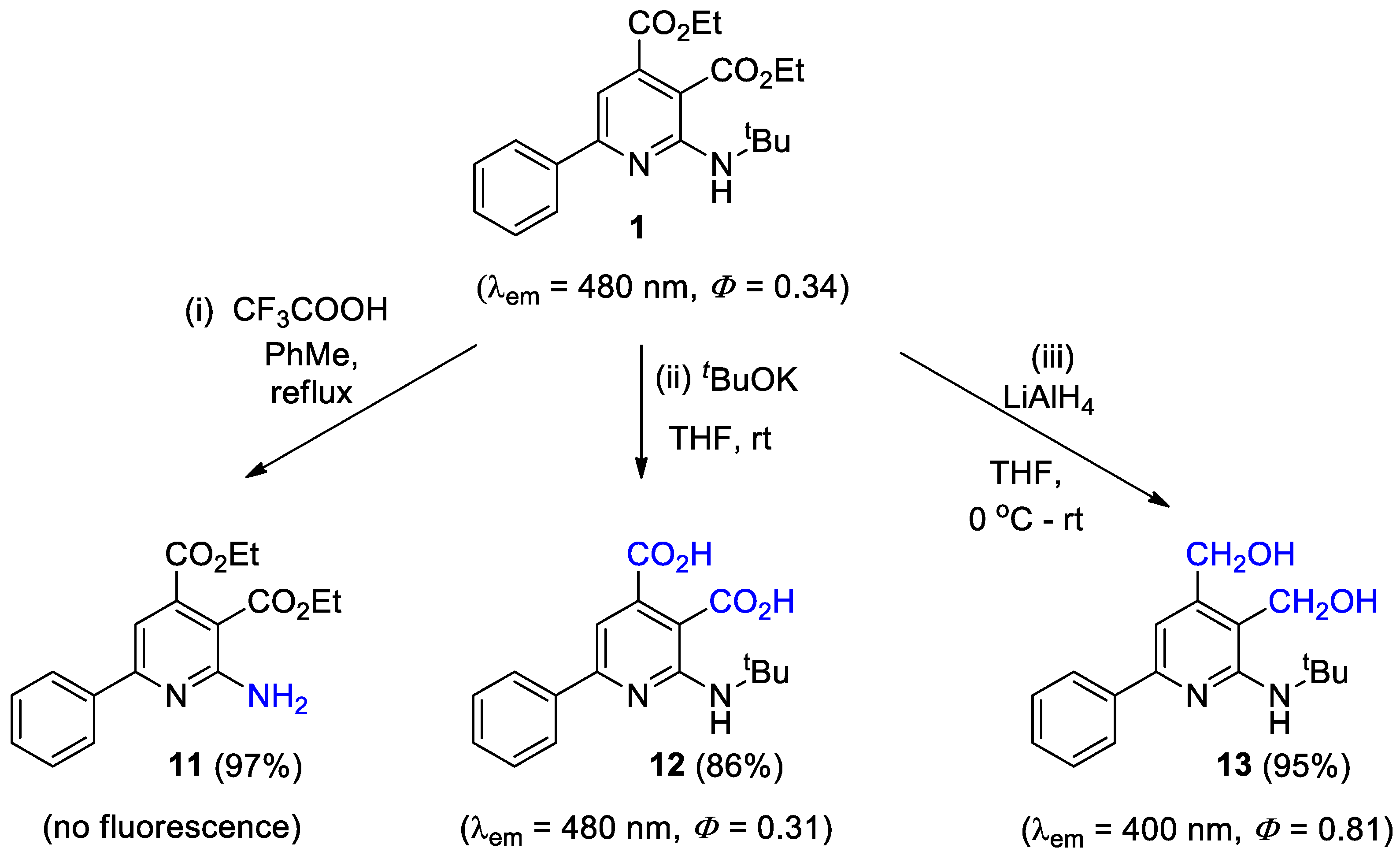
With diethyl 2-(tert-butylamino)-6-phenylpyridine-3,4-dicarboxylate (1) in hand, the functional groups of which were transformed as follows (Scheme 2): (1) The tertiary butyl group of 1 was cleaved using CF3COOH to furnish compound 11, which showed no fluorescence. The free amino group could be further transformed to other functional groups. (2) The ester groups were hydrolyzed to carboxylic acid to afford compound 12, which still had a good quantum yield (0.31). (3) Compound 1 was reduced to a high Φ (0.81) compound 13 using LiAlH4 in excellent yield, but the λem was reduced to 400 nm.
2.3. Synthesis and Fluorescent Properties of Pre-Fluorescence Aminopyridine
As shown in Scheme 3, the BocNH-substituted aminopyridine 14 was first synthesized under the standard conditions shown in Section 2.1. Then, the azido substituted aminopyridine 15 was synthesized via a Sandmeyer reaction, and it had no fluorescence because of the quenching effect from the electron-rich nitrogen of the azido group. With catalytic Cu(II), the azide aminopyridine 15 could reacted smoothly with phenylacetylene or propynol in a mixed solution of ethanol and water (1:1) to afford triazole products 16 or 17 at room temperature. Aminopyridines 16 and 17 both fluoresce at 480 nm because of the elimination of the quenching through the formation of the triazole ring from the azido group [11,13]. In comparison to 0.03 of the starting materials 15, the quantum yield of two products, 16 and 17, is 0.35 and 0.43, respectively. The cycloaddition of triazole-substituted aminopyridine 17 underwent significant fluorescence enhancement of 0.03 to 0.45 upon click reaction at a level that was 14 times as high as its starting azido aminopyridine 15.
2.4. Kinetic Experiments of the Click Reaction of Pre-Fluorescence Aminopyridine
The efficiency of the click reaction is a determining factor during the bio-application [28,29,30,31,32]. As the acceleration of rate of the cycloaddition may be achieved in various conditions, the rate of CuAAC between pre-fluorescence azido aminopyridine 15 and propargyl alcohol was measured, and the result showed in Scheme 4. Reactions were performed with 50 μM 15, 500 μM propargyl alcohol, 500 μM sodium ascorbate, 50 μM CuSO4, and 100 μM ligands. These reactions were carried out in a mixture of EtOH (DMF) and H2O solutions. The fluorescence of triazole product 17 were measured every minute during a total time course of 35 min.
Six conditions were evaluated. Condition A: no ligand, EtOH/H2O = 1:1; Condition B: TBTA (100 μM), EtOH/H2O = 1:1; Condition C: THPTA (100 μM), EtOH/H2O = 1:1; Condition D: TBTA (100 μM), DMF/H2O = 20:80; Condition E: THPTA (100 μM), DMF/H2O = 20:80; and Condition F: THPTA (100 μM), DMF/H2O = 5:95. (TBTA = tris[(1-benzyl-1H-1,2,3-triazol-4-yl)methyl]amine, THPTA = tris(3-hydroxypropyltriazolylmethyl)amine).
Among these conditions, TBTA in 50% H2O with 50% EtOH (condition B) showed the highest activity in accelerating the cycloaddition as the fluorescent intensity reached 1200 with 3 min, followed by THPTA in 80% H2O with 20% DMF (condition E). Notably, only after 10 min did the fluorescence intensity of the latter tend to go up comparing to other conditions. Besides, when the proportion of DMF and H2O changed to 5% and 95% (condition F), a slight acceleration could be seen, indicating that the mixture of DMF and H2O is a better solvent for THPTA as ligand in comparison with H2O and EtOH (condition C). Unfortunately, we could hardly see the difference between TBTA in the mixed solvent with 20% DMF (condition D) and the controlled condition with no ligand.
2.5. In Vitro “Clicking and Probing” on Conjugated BSA
Based on the above results, we further evaluated whether the newly designed fluorescent probe could be applied to a real biological environment for the purpose of labeling. A clicking-and-probing experiment was carried out between aminopyridines and BSA conjugated with an alkynyl group.
As shown in Scheme 5, BSA was conjugated with a terminal alkyne as follows: BSA (10 mg/mL) was incubated with 1 mM TCEP (TCEP = Tris(2-carboxyethyl)phosphine) in PBS (PBS = phosphate buffered saline) buffer at room temperature for 30 min. Then 1-(prop-2-yn-1-yl)-1H-pyrrole-2,5-dione was added to the solution at a final concentration of 1 mM, and the whole solution was incubated at room temperature for 4 h. Five volumes of methanol were added to the solution to precipitate the protein, and the mixture was then centrifuged at 10,000 rpm for 10 min. The supernatant was discarded, and the precipitate was washed three times with methanol, and then redissolved in PBS buffer containing 2% SDS.
Thus, we detected the fluorescence of the in vitro click reaction. Click reactions were performed in a 20 μL system with 40 μg above conjugated BSA, 50–500 mM probe 15, 10 mM sodium ascorbate, 200 μM CuSO4, and 200 μM indicated ligands (TBTA/THPTA/BTTES/BTTAA), respectively. Reaction mixtures were incubated and shaken gently at room temperature for 1 h. For in-gel fluorescence detection, the reaction mixture was mixed with 5X loading buffer and directly loaded on SDS gel for electrophoresis. Gels were analyzed with a ChemiDocTM MP system (Biorad) using a 530/28 filter. After fluorescence detection, the gel was stained with Coomassie Blue to indicate the protein loading amount (Scheme 6). In-gel fluorescence detection indicated that the labeling efficiency of compound 15 increased in a concentration-dependent manner, and 200 μM gave a best result (for more details, see Supplementary Materials Figure S18). Also, all of the four commonly used ligands, TBTA, THPTA, BTTES, and BTTAA, could facilitate the clicking-and-probing labeling process. Among them, BTTES showed the lowest activity in click reaction, followed by TBTA and THPTA, with BTTAA exhibiting the highest activity.
3. Materials and Methods
3.1. General
All reactions were performed in a Schlenk reaction flask. All solvents were purchased from Sinopharm Chemical Reagent (Beijing, China), and THF was redistilled by sodium. The boiling point of petroleum ether is between 60 and 90 °C. For chromatography, 200–300 mesh silica gel (Qingdao, China) was employed. All Rh catalysts and BSA (A1933) were purchased from Sigma-Aldrich (Shanghai, China). All ligands and substrates were purchased from Energy Chemical.
1H and 13C NMR spectra were recorded at 400 MHz and 100 MHz with Varian Mercury 400 spectrometer ((Agilent Technologies, Palo Alto, CA, USA) at ambient temperature. Chemical shifts are reported in ppm relative to chloroform (1H, δ 7.26; 13C, δ 77.00), DMSO (1H, δ 2.50; 13C, δ 39.52). IR spectra were recorded with a Nicolet AVATAR 330 FT-IR spectrometer (Thermo Fisher Scientific, Madison, WI, USA). UV absorbance and fluorescence were measured on a PerkinElmer Lambada 650S (PerkinElmer Inc., Waltham, MA, USA) and HITACHI F-4500 (Hitachi Ltd., Tokyo, Japan), respectively. Mass spectra were obtained on a Waters Auto Purification LC/MS system (Waters Corp., Milford, MA, USA). HMRS were obtained on a Bruker Apex IV FTMS spectrometer (Bruker Corp., Karlsruhe, Germany). Kinetic experiments were performed using a 96-well BioTek Synergy Hybrid Plate Reader and carried out by the EnspireTM 2300 Multilabel Reader (PerkinElmer Inc., Waltham, MA, USA). In-gel fluorescence was detected by a Bio-Rad ChemiDocTM MP system (Bio-Rad Laboratories, Hercules, CA, USA) using a 530/28 filter.
3.2. Experimental Procedures for the Synthesis of 1–17
In a 5 mL tube, catalyst [Rh(COD)Cl]2 (0.005 mmol) and ligand 2,2′-bpy (0.01 mmol) were dissolved in 1,4-dioxane (2 mL). Vinyl azide (0.2 mmol) and isonitrile (0.2 mmol) were added by syringe after the tube was sealed under N2 atmosphere and the reaction mixture was stirred at rt. After the spot of vinyl azide disappeared on TLC, NH4Cl (0.2 mmol), NaHCO3 (0.2 mmol), and alkyne (0.4 mmol) were added and the mixture was heated to 120 °C for 8 h. The reaction solution was concentrated in vacuum, and the residue was chromatographed with petroleum and ethyl acetate as eluent to afford aminopyridine products 1–10 and 14.
In a 5 mL tube, 1 (0.2 mmol) and CF3OOH (0.4 mmol) were dissolved in PhMe (2 mL). The mixture was stirred and refluxed until 1 disappeared, which was judged by TLC. The reaction solution was concentrated in vacuum, and the residue was chromatographed with petroleum and ethyl acetate as eluent (PE/EA = 3:1) to afford 11.
In a 5 mL tube under N2 atmosphere, 1 (0.20 mmol) and t-BuOK (0.80 mmol) were added in THF (2 mL). The mixture was stirred at rt until 1 disappeared, which was judged by TLC. The solution pH was adjusted to 3 by HCl (3 M). Then the mixture partitioned between EtOAc and H2O, and the organic phase was separated, dried (Na2SO4), and concentrated in vacuum. The residue was chromatographed with petroleum and ethyl acetate as eluent (PE/EA = 1:1) to afford 12.
In a 5 mL tube under N2 atmosphere, 1 (0.20 mmol) was dissolved in THF (2 mL), and then LiAlH4 (0.4 mmol) was added slowly in an ice bath. The mixture was stirred at rt until 1 disappeared, which was judged by TLC. The solution pH was adjusted to 7 by HCl (3 M). Then the mixture partitioned between EtOAc and H2O, and the organic phase was separated, dried (Na2SO4) and concentrated in vacuum. The residue was chromatographed with petroleum and ethyl acetate as eluent (PE/EA = 3:1) to afford 13.
In a 5 mL tube under N2 atmosphere, 14 (0.20 mmol) was dissolved in a mixed solution of THF (1 mL) and water (1 mL), and then conc. HCl (0.5 mL) was added slowly in an ice bath. The mixture was stirred at rt until 14 disappeared, which was judged by TLC. Then NaN3 (0.24 mmol) was added and the reaction mixture was stirred at rt for another 2h. Then the mixture partitioned between EtOAc and H2O, and the organic phase was separated, dried (Na2SO4) and concentrated in vacuum. The residue was chromatographed with petroleum and ethyl acetate as eluent (PE/EA = 20:1) to afford 15.
In a 5 mL tube, catalyst CuSO4 (0.005 mmol) and NaAsc (0.01 mmol) were dissolved in a mixed solution of EtOH (1 mL) and water (1 mL). Then, azido aminopyridine 15 (0.2 mmol) and alkyne (0.24 mmol) were added and the reaction mixture was stirred at rt until 15 disappeared, which was judged by TLC. Then the mixture was partitioned between EtOAc and H2O, and the organic phase was separated, dried (Na2SO4), and concentrated in vacuum. The residue was chromatographed with petroleum and ethyl acetate as eluent (PE/EA = 10:1) to afford 16 and 17.
3.3. Characterization Details
Diethyl 2-(tert-butylamino)-6-phenylpyridine-3,4-dicarboxylate (1). Light yellow solid. 1H NMR (400 MHz, CDCl3) δ 8.08–8.03 (m, 2H), 7.97 (s, 1H), 7.50–7.42 (m, 3H), 6.98 (s, 1H), 4.36 (q, J = 7.2 Hz, 2H), 4.30 (q, J = 7.1 Hz, 2H), 1.57 (s, 9H), 1.38 (t, J = 7.2 Hz, 3H), 1.34 (t, J = 7.2 Hz, 3H). 13C NMR (100 MHz, CDCl3) δ 169.05, 167.06, 159.24, 157.85, 145.95, 138.51, 130.00, 128.76, 127.36, 105.81, 101.13, 61.82, 61.42, 51.85, 29.31, 14.27, 14.14. HRMS (ESI+), m/z [M + H]+, calcd for C21H27N2O4: 371.1971, found: 371.1959. λA = 270 nm, λex = 390 nm, λem = 480 nm, Φ = 0.34.
Diethyl 2-(benzylamino)-6-phenylpyridine-3,4-dicarboxylate (2). Light yellow solid. 1H NMR (400 MHz, CDCl3) δ 8.27 (s, 1H), 8.02–7.97 (m, 2H), 7.46–7.37 (m, 5H), 7.36–7.30 (m, 2H), 7.28–7.23 (m, 1H), 7.02 (s, 1H), 4.87 (d, J = 5.6 Hz, 2H), 4.38 (q, J = 7.2 Hz, 2H), 4.31 (q, J = 7.2 Hz, 2H), 1.39 (t, J = 7.2 Hz, 3H), 1.33 (t, J = 7.1 Hz, 3H). 13C NMR (100 MHz, CDCl3) δ 168.88, 166.73, 159.86, 157.83, 145.99, 139.79, 138.15, 130.17, 128.71, 128.64, 127.65, 127.42, 127.12, 106.86, 101.20, 61.92, 61.51, 45.28, 14.26, 14.12. HRMS (ESI+), m/z [M + H]+, calcd for C24H25N2O4: 405.1814, found: 405.1797. λA = 270 nm, λex = 390 nm, λem = 480 nm, Φ = 0.31.
Diethyl 2-(cyclohexylamino)-6-phenylpyridine-3,4-dicarboxylate (3). Light yellow solid. 1H NMR (400 MHz, CDCl3) δ 8.08–8.00 (m, 2H), 7.89 (d, J = 7.3 Hz, 1H), 7.50–7.41 (m, 3H), 6.95 (s, 1H), 4.37 (q, J = 7.2 Hz, 2H), 4.31 (q, J = 7.2 Hz, 2H), 2.13–2.03 (m, 2H), 1.82–1.73 (m, 2H), 1.69–1.59 (m, 1H), 1.52–1.45 (m, 2H), 1.44–1.21 (m, 10H). 13C NMR (100 MHz, CDCl3) δ 169.07, 166.81, 159.84, 157.44, 146.05, 138.38, 130.08, 128.73, 127.33, 105.98, 100.48, 61.84, 61.35, 49.42, 32.99, 26.08, 24.94, 14.26, 14.16. HRMS (ESI+), m/z [M + H]+, calcd for C23H29N2O4: 397.2127, found: 397.2112. λA = 270 nm, λex = 390 nm, λem = 480 nm, Φ = 0.44.
Diethyl 2-(tert-butylamino)-6-(4-(trifluoromethyl)phenyl)pyridine-3,4-dicarboxylate (4). Light yellow solid. 1H NMR (400 MHz, CDCl3) δ 8.16 (d, J = 8.2 Hz, 2H), 7.90 (s, 1H), 7.71 (d, J = 8.1 Hz, 2H), 6.99 (s, 1H), 4.42–4.29 (m, 4H), 3.63 (q, J = 6.7 Hz, 2H), 1.72–1.63 (m, 2H), 1.52–1.43 (m, 2H), 1.43–1.32 (m, 6H), 0.98 (t, J = 7.3 Hz, 3H). 13C NMR (101 MHz, CDCl3) δ 168.71, 166.70, 158.17, 158.12, 146.12, 141.68, 131.67 (q, J = 32.5 Hz), 127.65, 125.68 (q, J = 3.8 Hz), 124.21 (q, J = 544.5, 272.2 Hz), 106.43, 101.87, 62.02, 61.61, 41.11, 31.64, 20.46, 14.27, 14.14, 14.07. HRMS (ESI+), m/z [M + H]+, calcd for C22H26F3N2O4: 489.1845, found: 489.1845. λA = 270 nm, λex = 390 nm, λem = 485 nm, Φ = 0.31.
Diethyl 2-(tert-butylamino)-6-(p-tolyl)pyridine-3,4-dicarboxylate (5). Light yellow solid. 1H NMR (400 MHz, CDCl3) δ 7.99–7.91 (m, 3H), 7.32–7.21 (m, 2H), 6.94 (s, 1H), 4.42–4.23 (m, 4H), 2.41 (s, 3H), 1.56 (s, 9H), 1.42–1.30 (m, 6H). 13C NMR (100 MHz, CDCl3) δ 169.17, 167.10, 159.30, 157.88, 145.90, 140.25, 135.79, 129.51, 127.32, 105.53, 100.73, 61.80, 61.36, 51.82, 29.32, 21.52, 14.27, 14.16. HRMS (ESI+), m/z [M + H]+, calcd for C22H29N2O4: 385.2127, found: 385.2108. λA = 270 nm, λex = 390 nm, λem = 485 nm, Φ = 0.27.
Diethyl 2-(tert-butylamino)-6-(o-tolyl)pyridine-3,4-dicarboxylate (6). Light yellow solid. 1H NMR (400 MHz, CDCl3) δ 7.94 (s, 1H), 7.43–7.40 (m, 1H), 7.31–7.28 (m, 1H), 7.27–7.24 (m, 2H), 6.58 (s, 1H), 4.37–4.27 (m, 4H), 2.41 (s, 3H), 1.47 (s, 9H), 1.39–1.31 (m, 6H). 13C NMR (100 MHz, CDCl3) δ 168.89, 167.20, 162.79, 157.71, 145.06, 140.33, 135.82, 130.79, 129.57, 128.63, 125.80, 109.94, 100.71, 61.81, 61.48, 51.87, 29.46, 20.72, 14.26, 14.14. HRMS (ESI+), m/z [M + H]+, calcd for C22H29N2O4: 385.2127, found: 385.2114. λA = 270 nm, λex = 390 nm, λem = 485 nm, Φ = 0.32.
Diethyl 2-(tert-butylamino)-6-(4-bromophenyl)pyridine-3,4-dicarboxylate (7). Light yellow solid. 1H NMR (400 MHz, CDCl3) δ 7.97 (s, 1H), 7.93–7.89 (m, 2H), 7.61–7.57 (m, 2H), 6.93 (s, 1H), 4.36 (q, J = 7.3 Hz, 2H), 4.30 (q, J = 7.3 Hz, 2H), 1.55 (s, 9H), 1.38 (t, J = 7.2 Hz, 3H), 1.34 (t, J = 7.1 Hz, 3H). 13C NMR (75 MHz, CDCl3) δ 168.82, 166.98, 158.09, 157.82, 146.11, 137.52, 131.98, 128.91, 124.56, 105.63, 101.79, 61.88, 61.51, 51.90, 29.29, 14.26, 14.12. HRMS (ESI+), m/z [M + H]+, calcd for C21H26BrN2O4: 449.1076, found: 449.1051. λA = 270 nm, λex = 390 nm, λem = 485 nm, Φ = 0.22.
Diethyl 2-(tert-butylamino)-6-octylpyridine-3,4-dicarboxylate (8). Light yellow solid. 1H NMR (400 MHz, CDCl3) δ 7.87 (s, 1H), 6.30 (s, 1H), 4.47–4.12 (m, 4H), 2.71–2.57 (m, 2H), 1.82–1.66 (m, 2H), 1.48 (s, 9H), 1.39–1.18 (m, 16H), 0.87 (t, J = 6.9 Hz, 3H). 13C NMR (100 MHz, CDCl3) δ 169.20, 167.18, 166.30, 157.94, 145.17, 108.27, 99.57, 61.65, 61.19, 51.75, 38.59, 32.00, 29.59, 29.41, 29.35, 29.33, 29.29, 28.62, 22.81, 14.24, 14.14. HRMS (ESI+), m/z [M + H]+, calcd for C23H39N2O4: 407.2904, found: 407.2900. λA = 258 nm, λex = 345 nm, λem = 455 nm, Φ = 0.02.
Dimethyl 2-(cyclohexylamino)-6-phenylpyridine-3,4-dicarboxylate (9). Light yellow solid. 1H NMR (400 MHz, CDCl3) δ 8.12–7.97 (m, 2H), 7.82 (d, J = 7.2 Hz, 1H), 7.57–7.37 (m, 3H), 6.97 (s, 1H), 4.33–4.15 (m, 1H), 3.91 (s, 3H), 3.84 (s, 3H), 2.13–2.02 (m, 2H), 1.88–1.70 (m, 2H), 1.54–1.30 (m, 6H). 13C NMR (100 MHz, CDCl3) δ 169.52, 167.16, 160.02, 157.38, 145.70, 138.28, 130.17, 128.76, 127.34, 105.95, 100.43, 52.79, 52.32, 49.48, 32.98, 26.07, 24.94. HRMS (ESI+), m/z [M + H]+, calcd for C21H25N2O4: 369.1814, found: 369.1799. λA = 270 nm, λex = 390 nm, λem = 480 nm, Φ = 0.35.
Bis(2-methoxyethyl) 2-(tert-butylamino)-6-phenylpyridine-3,4-dicarboxylate (10). Light yellow solid. 1H NMR (400 MHz, CDCl3) δ 8.04 (dd, J = 7.7, 1.9 Hz, 2H), 7.82 (s, 1H), 7.45 (m, 3H), 7.01 (s, 1H), 4.64–4.23 (m, 4H), 3.84–3.57 (m, 4H), 3.39 (d, J = 3.4 Hz, 6H), 1.56 (s, 9H). 13C NMR (100 MHz, CDCl3) δ 168.92, 166.93, 159.34, 157.71, 145.66, 138.48, 130.03, 128.76, 127.39, 106.10, 101.31, 70.31, 70.21, 65.03, 64.41, 59.20, 58.95, 51.91, 29.31. HRMS (ESI+), m/z [M + H]+, calcd for C23H31N2O6: 431.2177, found: 431.2164. λA = 270 nm, λex = 390 nm, λem = 480 nm, Φ = 0.30.
Diethyl 2-amino-6-phenylpyridine-3,4-dicarboxylate (11). Light yellow solid. 1H NMR (400 MHz, CDCl3) δ 8.35 (s, 2H), 7.98–7.77 (m, 2H), 7.67–7.42 (m, 3H), 6.96 (s, 1H), 4.52–4.25 (m, 4H), 1.50–1.31 (m, 6H). 13C NMR (100 MHz, CDCl3) δ 166.96, 164.79, 157.46, 157.19, 148.73, 133.59, 131.82, 129.26, 128.04, 108.88, 104.33, 62.64, 62.51, 14.17, 14.05. HRMS (ESI+), m/z [M + H]+, calcd for C17H19N2O4: 315.1345, found: 315.1332. λA = 270 nm, λex = 390 nm, λem = 480 nm, Φ = 0.01.
2-(tert-Butylamino)-6-phenylpyridine-3,4-dicarboxylic acid (12). Light yellow solid. 1H NMR (400 MHz, DMSO-d6) δ 13.39 (s, 2H), 8.27–8.00 (m, 3H), 7.65–7.36 (m, 3H), 7.17 (s, 1H), 1.53 (s, 9H). 13C NMR (100 MHz, DMSO-d6) δ 169.47, 168.42, 157.49, 157.25, 147.70, 137.80, 130.04, 128.89, 126.82, 105.18, 102.11, 51.03, 28.87. HRMS (ESI+), m/z [M + H]+ calcd for C17H19N2O4: 315.1345, found: 315.1333. λA = 270 nm, λex = 390 nm, λem = 480 nm, Φ = 0.30.
(2-(tert-Butylamino)-6-phenylpyridine-3,4-diyl)dimethanol (13). Light yellow solid. 1H NMR (400 MHz, CDCl3) δ 8.03–7.97 (m, 2H), 7.47–7.38 (m, 2H), 7.38–7.32 (m, 1H), 6.92 (s, 1H), 4.46 (d, J = 11.7 Hz, 4H), 1.54 (s, 9H). 13C NMR (100 MHz, CDCl3) δ 157.93, 154.40, 147.28, 139.66, 128.66, 128.61, 126.64, 115.72, 108.20, 63.11, 57.75, 51.58, 29.63. HRMS (ESI+), m/z [M + H]+, calcd for C17H23N2O2: 287.1760, found: 287.1748. λA = 330 nm, λex = 335 nm, λem = 400 nm, Φ = 0.81.
Diethyl 6-(4-((tert-butoxycarbonyl)amino)phenyl)-2-(cyclohexylamino)pyridine-3,4-dicarboxylate (14). Light yellow solid. 1H NMR (400 MHz, CDCl3) δ 7.99 (dd, J = 8.6, 1.4 Hz, 2H), 7.89 (d, J = 7.2 Hz, 1H), 7.45 (d, J = 7.8 Hz, 2H), 6.90 (s, 1H), 6.65 (s, 1H), 4.36 (q, J = 7.2 Hz, 2H), 4.30 (q, J = 7.1 Hz, 2H), 4.25–4.16 (m, 1H), 2.15–2.03 (m, 2H), 1.84–1.70 (m, 2H), 1.71–1.60 (m, 2H), 1.54 (s, 8H), 1.45–1.29 (m, 10H). 13C NMR (100 MHz, CDCl3) δ 169.93, 169.21, 166.83, 159.20, 157.42, 152.56, 146.02, 140.28, 132.87, 128.23, 118.19, 105.33, 61.83, 61.27, 49.48, 32.98, 28.45, 27.04, 26.10, 24.97, 14.26, 14.18. HRMS (ESI+), m/z [M + H]+, calcd for C28H38N3O6: 512.2755, found: 512.2750. λA = 270 nm, λex = 390 nm, λem = 480 nm, Φ = 0.67.
Diethyl 6-(4-azidophenyl)-2-(cyclohexylamino)pyridine-3,4-dicarboxylate (15). Light yellow solid. 1H NMR (400 MHz, CDCl3) δ 8.05 (d, J = 8.0 Hz, 2H), 7.90 (d, J = 7.4 Hz, 1H), 7.11 (d, J = 7.9 Hz, 2H), 6.90 (s, 1H), 4.41–4.27 (m, 4H), 4.24–4.18 (m, 1H), 2.15–2.03 (m, 2H), 1.82–1.73 (m, 2H), 1.69–1.61 (m, 2H), 1.50–1.42 (m, 2H), 1.42–1.32 (m, 8H). 13C NMR (100 MHz, CDCl3) δ 169.01, 166.75, 158.63, 157.41, 146.15, 141.84, 135.13, 128.86, 119.33, 105.53, 61.90, 61.40, 49.49, 32.96, 26.08, 24.95, 14.27, 14.17. HRMS (ESI+), m/z [M + H]+, calcd for C23H28N5O4: 438.2136, found: 438.2205. λA = 270 nm, λex = 390 nm, λem = 480 nm, Φ = 0.03.
Diethyl 2-(cyclohexylamino)-6-(4-(4-phenyl-1H-1,2,3-triazol-1-yl)phenyl)pyridine-3,4-dicarboxylate (16). Light yellow solid. 1H NMR (400 MHz, CDCl3) δ 8.26 (s, 1H), 8.22 (d, J = 8.8 Hz, 2H), 7.96–7.89 (m, 4H), 7.53–7.45 (m, 2H), 7.42–7.35 (m, 1H), 6.99 (s, 1H), 4.43–4.29 (m, 4H), 4.27–4.20 (m, 1H), 2.15–2.06 (m, 2H), 1.85–1.75 (m, 2H), 1.71–1.62 (m, 2H), 1.54–1.43 (m, 2H), 1.43–1.33 (m, 6H), 1.30–1.24 (m, 2H). 13C NMR (100 MHz, CDCl3) δ 168.85, 166.69, 158.05, 157.40, 148.70, 146.27, 138.71, 138.04, 130.25, 129.10, 128.77, 128.67, 126.03, 120.47, 117.47, 105.92, 101.28, 62.00, 61.54, 49.59, 32.94, 26.07, 24.95, 14.28, 14.16. HRMS (ESI+), m/z [M + H]+, calcd for C31H34N5O4: 540.2605, found: 540.2598. λA = 255 nm, λex = 390 nm, λem = 480 nm, Φ = 0.35.
Diethyl 2-(cyclohexylamino)-6-(4-(4-(hydroxymethyl)-1H-1,2,3-triazol-1-yl)phenyl)pyridine-3,4-dicarboxylate (17). Light yellow solid. 1H NMR (400 MHz, CDCl3) δ 8.20 (d, J = 7.2 Hz, 2H), 8.05 (s, 1H), 7.91 (d, J = 7.1 Hz, 1H), 7.84 (d, J = 7.2 Hz, 2H), 6.98 (s, 1H), 4.92 (s, 2H), 4.44–4.29 (m, 4H), 4.27–4.20 (m, 1H), 2.17–2.02 (m, 2H), 1.86–1.65 (m, 4H), 1.56–1.42 (m, 8H), 1.44–1.30 (m, 8H). 13C NMR (100 MHz, CDCl3) δ 168.84, 166.68, 158.01, 157.40, 146.27, 138.83, 137.97, 128.79, 120.57, 105.94, 101.32, 62.01, 61.55, 56.88, 49.57, 32.93, 26.06, 24.93, 14.28, 14.16. HRMS (ESI+), m/z [M + H]+, calcd for C26H32N5O5: 494.2398, found: 494.2392. λA = 280 nm, λex = 390 nm, λem = 480 nm, Φ = 0.45. (for details, see Supplementary Materials Figures S1–S17).
4. Conclusions
We synthesized a series of fluorescent molecules with an aminopyridine scaffold and investigated their fluorescent properties. The majority of obtained compounds exhibit good fluorescence characteristics, among which 2 with a cyclohexyl substituting the amine group has the highest quantum yield over 0.40, and its characteristic wavelength meets the requirements as a fluorescent molecule. By means of derivatizing, we designed a bioorthogonally activated smart probe that is nonfluorescent and generates a highly fluorescent fluorophore after a click-labeling reaction with alkynes. This represents a 14-fold change in fluorescence intensity. The efficiency of this “turn on” and “turn off” strategy is also investigated, indicating the reaction could be conducted rapidly with the optimized condition. Hence, the biochemical application of this probe was demonstrated by the BSA protein conjugated “clicking-and-probing” experiment. Therefore, the fluorescent molecules based on multisubstituted aminopyridines have a potential prospect in extensive application of biochemical detection and analysis.
Supplementary Materials
The following supporting information can be downloaded online: Figures S1–S17 (1H and 13C NMR spectra of compounds); Figure S18 (Fluorescence scan of SDS-PAGE gel and coomassie blue staining of SDS-PAGE gel).
Author Contributions
Conceptualization and supervision, Z.Z.; methodology, Z.Z., X.L. and L.D.; experiment and characterization, Z.L., Y.L., W.C. and S.P.; Writing-original draft, Z.L. and Y.L.; Writing-review and editing, Z.Z. All authors have read and agreed to the published version of the manuscript.
Funding
This research was funded by the National Key Research and Development program of China (2017YFD0201300) and the Fundamental Research Funds for the Central Universities (China Agricultural University 2020-00109015).
Institutional Review Board Statement
Not applicable.
Informed Consent Statement
Not applicable.
Data Availability Statement
The data presented in this study are available in supplementary material.
Conflicts of Interest
The authors declare no conflict of interest.
Sample Availability
Samples of the compounds in this article are available from the authors.
References
- Gonçalves, T.; Sameiro, M. Fluorescent labeling of biomolecules with organic probes. Chem. Rev. 2009, 109, 190–212. [Google Scholar] [CrossRef] [PubMed]
- Grammel, M.; Hang, H.C. Chemical reporters for biological discovery. Nat. Chem. Biol. 2013, 9, 475–484. [Google Scholar] [CrossRef] [PubMed] [Green Version]
- Lang, K.; Chin, J.W. Cellular incorporation of unnatural amino acids and bioorthogonal labeling of proteins. Chem. Rev. 2014, 114, 4764–4806. [Google Scholar] [CrossRef] [PubMed]
- Zhang, Y.; Li, S.; Zhang, H.; Xu, H. Design and application of receptor-targeted fluorescent probes based on small molecular fluorescent Dyes. Bioconjug. Chem. 2021, 32, 4–24. [Google Scholar] [CrossRef]
- Algar, W.R.; Massey, M.; Rees, K.; Higgins, R.; Krause, K.D.; Darwish, G.H.; Peveler, W.J.; Xiao, Z.; Tsai, H.-Y.; Gupta, R.; et al. Photoluminescent nanoparticles for chemical and biological analysis and imaging. Chem. Rev. 2021, 121, 9243–9358. [Google Scholar] [CrossRef]
- Patterson, D.M.; Nazarova, L.A.; Prescher, J.A. Finding the right (bioorthogonal) chemistry. ACS Chem. Biol. 2014, 9, 592–605. [Google Scholar] [CrossRef]
- Ji, X.; Pan, Z.; Yu, B.; Cruz, L.K.D.L.; Zheng, Y.; Ke, B.; Wang, B. Click and release: Bioorthogonal approaches to “on-demand” activation of prodrugs. Chem. Soc. Rev. 2019, 48, 1077–1094. [Google Scholar] [CrossRef]
- Zhang, F.-G.; Chen, Z.; Tang, X.; Ma, J.-A. Triazines: Syntheses and inverse electron-demand diels–alder reactions. Chem. Rev. 2021, 121, 14555–14593. [Google Scholar] [CrossRef]
- Jewett, J.C.; Bertozzi, C.R. Synthesis of a fluorogenic cyclooctyne activated by Cu-free click chemistry. Org. Lett. 2011, 13, 5937–5939. [Google Scholar] [CrossRef] [Green Version]
- Shieh, P.; Dien, V.T.; Beahm, B.J.; Castellano, J.M.; Wyss-Coray, T.; Bertozzi, C.R. Calfluors: A universal motif for fluorogenic azide probes across the visible spectrum. J. Am. Chem. Soc. 2015, 137, 7145–7151. [Google Scholar] [CrossRef] [Green Version]
- Ji, X.; Ji, K.; Chittavong, V.; Aghoghovbia, R.E.; Zhu, M.; Wang, B. Click and fluoresce: A bioorthogonally activated smart probe for wash-free fluorescent labeling of biomolecules. J. Org. Chem. 2017, 82, 1471–1476. [Google Scholar] [CrossRef] [Green Version]
- Liu, J.; Abdullah, M.A.A.; Yang, L.; Wang, J. Fast affinity induced reaction sensor based on a fluorogenic click reaction for quick detection of protein biomarkers. Anal. Chem. 2020, 92, 647–653. [Google Scholar] [CrossRef]
- Sivakumar, K.; Xie, F.; Cash, B.M.; Long, S.; Barnhill, H.N.; Wang, Q. A fluorogenic 1,3-dipolar cycloaddition reaction of 3-azidocoimarins and acetylences. Org. Lett. 2004, 6, 4603–4606. [Google Scholar] [CrossRef]
- Shieh, P.; Hangauer, M.J.; Bertozzi, C.R. Fluorogenic azidofluoresceins for biological imaging. J. Am. Chem. Soc. 2012, 134, 17428–17431. [Google Scholar] [CrossRef] [Green Version]
- Lee, Y.; Cho, W.; Sung, J.; Kim, E.; Park, S.B. Monochromophoric design strategy for tetrazine-based colorful bioorthogonal probes with a single fluorescent core skeleton. J. Am. Chem. Soc. 2018, 140, 974–983. [Google Scholar] [CrossRef] [Green Version]
- Eaton, D.F. Reference materials for fluorescence measurement. Pure Appl. Chem. 1988, 60, 1107–1114. [Google Scholar] [CrossRef]
- Zonouzi, A.; Izakian, Z.; Weng Ng, S. Novel synthesis of some new fluorescent 2-amino-3-cyanopyridines. Heterocycles 2012, 85, 2713. [Google Scholar] [CrossRef]
- Li, H.; Petersen, J.L.; Wang, K.K. Cascade cyclizations via N,4-didehydro-2-(phenylamino)pyridine biradicals/zwitterions generated from enyne-carbodiimides. J. Org. Chem. 2003, 68, 5512–5518. [Google Scholar] [CrossRef]
- Shi, F.; Tu, S.; Fang, F.; Li, T. One-pot synthesis of 2-amino-3-cyanopyridine derivatives under microwave irradiation without solvent. Arkivoc 2005, 2005, 137–142. [Google Scholar] [CrossRef] [Green Version]
- Londregan, A.T.; Jennings, S.; Wei, L. General and mild preparation of 2-aminopyridines. Org. Lett. 2010, 12, 5254–5257. [Google Scholar] [CrossRef]
- Vamos, M.; Cosford, N.D.P. 2-Aminopyridines via reaction of pyridine N-oxides and activated isocyanides. J. Org. Chem. 2014, 79, 2274–2280. [Google Scholar] [CrossRef] [Green Version]
- He, X.; Shang, Y.; Yu, Z.; Fang, M.; Zhou, Y.; Han, G.; Wu, F.J. FeCl3-catalyzed four-component nucleophilic addition/intermolecular cyclization yielding polysubstituted pyridine derivatives. J. Org. Chem. 2014, 79, 8882–8888. [Google Scholar] [CrossRef]
- Weng, Y.; Kuai, C.; Lv, W.; Cheng, G. Synthesis of 2-aminopyridines via a base-promoted cascade reaction of N-propargylic β-enaminones with formamides. J. Org. Chem. 2018, 83, 5002–5008. [Google Scholar] [CrossRef]
- Kumar, D.; Vemula, S.R.; Cook, G.R. Merging C–H bond functionalization with amide alcoholysis: An route to 2-aminopyridines. ACS Catal. 2016, 6, 3531–3536. [Google Scholar] [CrossRef]
- Li, Z.; Huo, T.; Li, L.; Feng, S.; Wang, Q.; Zhang, Z.; Pang, S.; Zhang, Z.; Wang, P.; Zhang, Z. Rh-catalyzed reaction of vinyl azides with isonitriles and alkynes/benzynes. Org. Lett. 2018, 20, 7762–7765. [Google Scholar] [CrossRef]
- Yang, M.; Meng, X.H.; Wang, Z.; Gong, Y.; Zhao, Y.L. Rhodium/copper-cocatalyzed coupling-cyclization of o-alkenyl arylisocyanides with vinyl azides: One-pot synthesis of α-carbolines. Org. Chem. Front. 2020, 7, 3493–3498. [Google Scholar] [CrossRef]
- Optically dilute measurements withrefractive-index corrections, maximal absorption of the solutions 0.04. Calibrated spectrometer (Perkin Elmer LS 50B); measurements referenced to 9,10-diphenylanthracene (Φ = 0.95). In Principles of Fluorescence Spectroscopy, 2nd ed.; Lakowicz, J.R. (Ed.) Kluwer Academic/Plenum Publishers: New York, NY, USA, 1999. [Google Scholar]
- Hong, V.; Presolski, S.I.; Ma, C.; Finn, M.G. Analysis and optimization of copper-catalyzed azide–alkyne cycloaddition for bioconjugation. Angew. Chem. Int. Ed. 2009, 48, 9879–9883. [Google Scholar] [CrossRef] [Green Version]
- del Amo, D.S.; Wang, W.; Jiang, H.; Besanceney, C.; Yan, A.C.; Levy, M.; Liu, Y.; Marlow, F.L.; Wu, P. Biocompatible Copper(I) Catalysts for in Vivo Imaging of Glycans. J. Am. Chem. Soc. 2010, 132, 16893–16899. [Google Scholar] [CrossRef] [Green Version]
- Besanceney, C.; Jiang, H.; Zheng, T.; Feng, L.; del Amo, D.S.; Wang, W.; Klivansky, L.M.; Marlow, F.L.; Liu, Y.; Wu, P. Increasing the efficacy of bioorthogonal click reactions for bioconjugation: A comparative study. Angew. Chem. Int. Ed. 2011, 50, 8051–8056. [Google Scholar] [CrossRef]
- Bevilacqua, V.; King, M.; Chaumontet, M.; Nothisen, M.; Gabillet, S.; Buisson, D.; Puente, C.; Wagner, A.; Taran, F. Copper-chelating azides for efficient click conjugation reactions in complex media. Angew. Chem. Int. Ed. 2014, 53, 5872–5876. [Google Scholar] [CrossRef]
- Su, Y.; Li, L.; Wang, H.; Wang, X.; Zhang, Z. All-in-One azides: Empowered click reaction for in vivo labeling and imaging of biomolecules. Chem. Commun. 2016, 52, 2185–2188. [Google Scholar] [CrossRef] [PubMed]
Scheme 1.
The application of Aminopyridine in “clicking and probing”.

Scheme 2.
Fluorescent Properties of 6-Phenyl Substituted Aminopyridine derivatives.

Scheme 3.
Synthesis and Fluorescent Properties of Pre-fluorescence Aminopyridine.


Scheme 4.
Kinetic Experiments of the Click Reaction of Pre-fluorescence Aminopyridine 15.


Scheme 5.
Conjugation of terminal alkynyl group to BSA.

Scheme 6.
Fluorescent labelling of BSA protein by compound 15 using ligand-assisted CuAAC reaction.


Table 1.
Fluorescent Properties of Aminopyridines.

| Entry | Structure (with Isolated Yield) | λA/nm 1 | λex/nm | λem/nm 2 | ϕ 3 |
|---|---|---|---|---|---|
| 1 |  1 (71%) | 270 | 390 | 480 | 0.34 |
| 2 |  2 (24%) | 270 | 390 | 480 | 0.31 |
| 3 |  3 (65%) | 270 | 390 | 480 | 0.44 |
| 4 |  4 (52%) | 270 | 390 | 485 | 0.31 |
| 5 |  5 (60%) | 270 | 390 | 485 | 0.27 |
| 6 |  6 (52%) | 270 | 390 | 485 | 0.32 |
| 7 |  7 (53%) | 270 | 390 | 485 | 0.22 |
| 8 |  8 (17%) | 258 | 345 | 455 | 0.02 |
| 9 |  9 (67%) | 270 | 390 | 480 | 0.35 |
| 10 |  10 (57%) | 270 | 390 | 480 | 0.30 |
1 Absorption maxima in ethanol (longest wavelength transition); 2 Maxima of the corrected emission spectra in ethanol; 3 External standard: 9,10-diphenylanthracene (ϕ = 0.95, in cyclohexane).
Publisher’s Note: MDPI stays neutral with regard to jurisdictional claims in published maps and institutional affiliations. |
© 2022 by the authors. Licensee MDPI, Basel, Switzerland. This article is an open access article distributed under the terms and conditions of the Creative Commons Attribution (CC BY) license (https://creativecommons.org/licenses/by/4.0/).
Share and Cite
MDPI and ACS Style
Li, Z.; Li, Y.; Chang, W.; Pang, S.; Li, X.; Duan, L.; Zhang, Z. Synthesis and Fluorescent Properties of Aminopyridines and the Application in “Click and Probing”. Molecules 2022, 27, 1596. https://doi.org/10.3390/molecules27051596
AMA Style
Li Z, Li Y, Chang W, Pang S, Li X, Duan L, Zhang Z. Synthesis and Fluorescent Properties of Aminopyridines and the Application in “Click and Probing”. Molecules. 2022; 27(5):1596. https://doi.org/10.3390/molecules27051596
Chicago/Turabian StyleLi, Zongyang, Yaxuan Li, Wenxu Chang, Sen Pang, Xuefeng Li, Liusheng Duan, and Zhenhua Zhang. 2022. "Synthesis and Fluorescent Properties of Aminopyridines and the Application in “Click and Probing”" Molecules 27, no. 5: 1596. https://doi.org/10.3390/molecules27051596