Article

Fortification of Fermented Camel Milk with Salvia officinalis L. or Mentha piperita Leaves Powder and Its Biological Effects on Diabetic Rats

by
Magdy Ramadan Shahein
1,
Mahmoud Ibrahim El-Sayed
2,
Enrique Raya-Álvarez
3,
Abdelmoneim Ahmed Elmeligy
4,
Mohamed A. Mohamady Hussein
5,
Murad A. Mubaraki
6,
Ahmad Agil
7 and
Ehab Kotb Elmahallawy
8,9,*
1
Department of Food Science and Technology, Faculty of Agriculture, Tanta University, Tanta 31527, Egypt
2
Department of Dairy Technology Research, Food Technology Research Institute, Agricultural Research Center, Giza 12622, Egypt
3
Rheumatology Department, Hospital Universitario San Cecilio, Av. de la Investigación, s/n, 18016 Granada, Spain
4
Department of Pathology, Faculty of Veterinary Medicine, Suez Canal University, Ismailia 41522, Egypt
5
Department of Pharmacology, Medical Research and Clinical Studies Institute, National Research Centre, 33 El Bohouth St., Dokki, Giza 12622, Egypt
6
Clinical Laboratory Sciences Department, College of Applied Medical Sciences, King Saud University, Riyadh 12372, Saudi Arabia
7
Department of Pharmacology, Biohealth Institute Granada (IBs Granada) and Neuroscience Institute, School of Medicine, University of Granada, 18016 Granada, Spain
8
Departamento de Sanidad Animal, Grupo de Investigación en Sanidad Animal y Zoonosis (GISAZ), Facultad de Veterinaria, Universidad de Córdoba, 14004 Córdoba, Spain
9
Department of Zoonoses, Faculty of Veterinary Medicine, Sohag University, Sohag 82524, Egypt
*
Author to whom correspondence should be addressed.
Molecules 2023, 28(15), 5749; https://doi.org/10.3390/molecules28155749
Submission received: 14 June 2023 / Revised: 17 July 2023 / Accepted: 24 July 2023 / Published: 29 July 2023

Abstract

:
The incorporation of fermented camel milk with natural additives possesses numerous benefits for the treatment of various pathological and metabolic conditions. The present study investigated the impact of fortification of fermented camel milk with sage or mint leaves powder (1 and 1.5%, respectively) on glucose and insulin levels, lipid profile, and liver and kidney functions in alloxan-induced diabetic rats. The gross chemical composition of sage and peppermint leaves powder was studied. The chemical composition of sage and mint extracts was performed using liquid chromatography–electrospray ionization–tandem mass spectrometry (LC-MS) of sage and mint extracts. Furthermore, a total of forty-two adult normal male albino rats were included in this study, whereas one group was kept as the healthy control group (n = 6 rats) and diabetes was induced in the remaining animals (n = 36 rats) using alloxan injection (150 mg/kg of body weight). Among diabetic rats groups, a control group (n = 6 rats) was kept as the diabetic control group whereas the other 5 groups (6 rats per group) of diabetic rats were fed fermented camel milk (FCM) or fermented camel milk fortified with 1 and 1.5% of sage or mint leaves powder. Interestingly, the oral administration of fermented camel milk fortified with sage or mint leaves powder, at both concentrations, caused a significant decrease in blood glucose level and lipid profile, and an increase in insulin level compared to the diabetic control and FCM groups. Among others, the best results were observed in the group of animals that received fermented camel milk fortified with 1.5% sage powder. In addition, the results revealed that the fermented camel milk fortified with sage or mint leaves powder improved the liver and kidney functions of diabetic rats. Our study concluded that the use of sage and mint leaves powder (at a ratio of 1.5%) with fermented camel milk produces functional food products with anti-diabetic activity.

1. Introduction

Diabetes mellitus (DM) is a chronic metabolic condition described by persistent hyperglycemia due to the incapability of the pancreas to produce enough insulin, or the organ cannot use the produced insulin (insulin resistance), or a combination of both [1,2]. The available data from IDF confirmed that, in 2021, the number of people (20 to 79-year-olds) suffering from diabetes was predestined to be near 537 million [3]. This number is foreseeable to reach 643 million in 2030 and 783 million by 2045. Moreover, diabetes increases the serum level of urea and creatinine [4]. According to the most recent statistics from the Food and Agriculture Organization (FAO), the camel world population is estimated to be around 32.6 million [5]. Camel’s milk is a vital part of the staple diet in several parts of the world, especially in the arid and semi-arid zones. Camel’s milk is rich in health-beneficial substances, such as lactoferrin, lysozyme, lactoperoxidase, bioactive peptides, mono and polyunsaturated fatty acids, minerals (calcium, magnesium, copper, iron, zinc, phosphorous, potassium, and sodium), immunoglobulins and vitamins including, B1, B2 and C [6,7,8,9]. Camel milk has been known as a source for the production of dairy products with excellent therapeutic properties such as fermented milk [10]. Raw and fermented camel milk is found to have many health benefits such as anticancer, antimicrobial, antioxidant, anti-inflammatory, anti-diabetic, anti-diarrhea, hypocholesterolemic, and angiotensin I-converting enzyme (ACE) inhibitory activities [11,12,13,14]. It was reported that camel milk is a unique source of nutrients and is considered a superfood with high medicinal values [1,9,15,16]. Camel milk has been shown to improve other pathophysiological aspects related to diabetes as a chronic disease such as obesity, insulin resistance, wound healing and inflammation [17,18]. Camel milk improves diabetes complications such as wounds, kidney and liver failures, and oxidative stress. Also, camel milk improves diabetes complications such as liver and kidney failures, wounds, and oxidative stress [19]. In a study performed by Meena et al. (2016) [17], camel milk intake could enhance the level of antioxidative enzymes (catalase, glutathione peroxidase, and superoxide dismutase) in the pancreas in diabetic rats and, thus, could ameliorate hyperglycemia and oxidative damage. Furthermore, a previous study [20] found that raw camel milk caused an increase in insulin secretion, and reduced about 30–35% of required insulin in type 1 diabetes patients.
Fortifications of dairy products with natural substances enhance the physicochemical characterizations and nutritive values of functional food, and therefore, have attracted many research projects over the last few decades [21,22,23,24,25,26,27,28,29]. Many studies reported that herbal therapy is considered an effective way to the management of diabetes. These plants contain phenolics, flavonoids, carotenoids, alkaloids, terpenoids, and glycosides and may exert anti-diabetic effects to some extent [30,31]. Despite its traditional applications in food flavoring, Mentha spp. are widely used for treating not only fever and cold but also cardiovascular and gastrointestinal disorders as folk medicines [32]. Rajeshwari et al. 2012 [33], reported that the administration of mint leaves powder (5 g/day) to type 2 diabetes patients for 60 days reduced oxidative stress by decreasing lipid peroxidation, protein oxidation, increased serum beta carotene, and vitamin A, E, and C levels. Among others, the anti-diabetic actions of natural plant products are expressed by their role in activation of the antioxidant enzymes such as catalase, glutathione peroxidase, and superoxide dismutase [34,35], where the level of antioxidant enzymes such as superoxide dismutase was elevated in the presence of mentha extract [36]. In addition, it improved the activity of some antioxidant enzymes, i.e., glutathione-S-transferase (GST), in addition to the content of reduced glutathione (GSH). Chandirasegaran et al., 2014 [37] detected a significant decrease in blood glucose and creatinine levels as well as an increase in insulin levels of diabetic rats after being treated with mint (300 mg/kg B.W) for 45 days. These findings determined that mint possesses anti-diabetic activity against streptozotocin-induced diabetic rats. Sage, Salvia officinalis L., is well-reputed to cure diabetes or restrain its complications [38]. A previous study [39] found that the treatment of alloxan-induced diabetic rats with aqueous and ethanol extracts of Salvia officinalis leaves at a concentration of 100 mg/kg B.W for 14 days decreased the levels of blood glucose, triglycerides, and total cholesterol. The suggested mechanisms for anti-diabetic actions of salvia species extracts are the increase in insulin sensitivity, activation of pancreatic b-cells, and peripheral use of glucose, and the inactivation of insulinase enzyme and the glycogenolysis reduction decreases the absorption of glucose from the intestine and increases the synthesis of glucose in the liver [40]. The protective effect of sage extract against the depletion of antioxidant enzymes may contribute to its anti-diabetic effects [39]. Limited information is available about the biological effects of fermented camel milk fortified with sage and mint (Mentha piperita) leaves powder on alloxan-induced diabetic rats. The present study aimed to evaluate the effects of camel milk fortified with sage and mint leaves powder on the biochemical markers of alloxan-induced diabetic rats.

2. Results

2.1. Proximate Chemical Composition of Peppermint and Sage Leaves Powder and Their Antioxidant Activity

The proximate chemical composition of peppermint and sage leaves powder is shown in Table 1. Many studies reported that the plant extracts were used effectively to treat diabetic animals, where their components could induce one or more of the following actions: antioxidant, antihyperglycemic, increasing release of insulin, and anti-glycation agents [41,42,43,44]. Data in Table 2 showed the antioxidant activity of mint and sage extracts. The results found that sage extract was higher in total phenolic contentment (7.35 mg GAE/g) than mint extract (6.60 mg GAE/g), while the mint extract was the highest in total flavonoids (184 µg/mL). Moreover, a higher DPPH scavenging activity (%) was found with sage extract, while a higher FRAP value was observed with mint extract.

2.2. Chemical Composition of Sage and Mint Extracts Using Liquid Chromatography–Electrospray Ionization–Tandem Mass Spectrometry of Sage and Mint Extracts (LC-MS)

As shown in Table 3, the potential bioactive compounds of both SE and ME were identified using LC-ESI-MS/MS. As depicted in Table 3, twenty compounds were identified by comparing the fragmentation peaks with those of the corresponding standards. Out of them, 16 identified compounds of SE and ME were found and comprise chlorogenic acid, gallic acid, caffeic acid, rutin coumaric acid, naringenin, quercetin, ellagic, 3.4-dihydroxybenzoic acid, acid hesperetin, methyl gallate, kaempferol, ferulic acid, syringic acid, apigenin, and luteolin.

2.3. Effect of Fermented Camel Milk on Alloxan-Induced Diabetic Rats

2.3.1. Serum Glucose and Insulin Determination

The anti-diabetic properties of camel milk are very complex, involving many cellular and molecular mechanisms and aspects of metabolism and transport of glucose as well as the synthesis and secretion of insulin [45,46]. Data presented in Figure 1 showed the effect of fermented camel milk fortified with sage and mint leaves powders by ratio 1 and 1.5% on plasma glucose and insulin levels of diabetic rats. Results indicated that a higher plasma glucose level (253 mg/dL) was observed in the diabetic control group. On the other hand, the oral intake of fermented camel milk with or without fortification by sage and mint leaves powders significantly (p < 0.05) decreased the plasma glucose level in diabetic rats, while the normal rats were not affected. The oral intake of fermented camel milk fortified with saga and mint powder (FCMM1, FCMM2, FCMS1, and FCMS2) caused a significant decrease in the plasma glucose level compared with the group of fermented camel milk (FCM), and a higher decrease was found with FCMM2 and FCMS2 groups (186.3 and 175.2 mg/dL, respectively). The induction with alloxan caused a significant (p < 0.05) decrease in the insulin level in rats’ plasma (Figure 1). On the other hand, the oral intake of fermented camel milk with or without sage and mint powder significantly (p < 0.05) increases the insulin level in the blood again. The results showed that higher insulin levels were observed in the healthy control group (35.9 µU/mL), followed by the animal groups’ intake of fermented camel milk fortified with 1 and 1.5% sage powder (29.11 and 30.2 µU/mL, respectively), while no significant (p > 0.05) differences were found between FCM, FCMM1, and FCMM2 groups (27.3, 27.4, and 28.6 µU/mL, respectively).

2.3.2. Lipid Profile

Data in Figure 2 showed that after eight weeks of animal induction using alloxan, the animal untreated with fermented camel milk (diabetic control) displayed an increase in plasma total triglyceride (TG) and total cholesterol (TC), compared with healthy control and animal groups treated with fermented camel milk (FCM with or without mint and sage powders, FCMS and FCMM). The results showed that the oral administration of FCM or FCMS and FCMM significantly decreased TG and TC in diabetic rats groups. A higher decrease in TG and TC levels was found in FCMS2 and FCMM2 groups compared to FCMS1, FCMM1, and FCM groups. The results determined that the oral administration of fermented camel milk (FCM) or fermented camel milk fortified with 1 and 1.5% of sage or mint powder (FCMS and FCMM) caused a significant decrease in low-density lipoprotein cholesterol (LDL-c) and very low-density lipoprotein cholesterol (VLDL-c) levels compared with diabetic control group, while the high-density lipoprotein cholesterol (HDL-c) was significantly increased. The higher decrease in LDL values was found with FCMS2 and FCMM2 groups and there were no significant differences between the two groups, while the lowest value of VLDL was observed with FCMM2. Also, higher values of HDL were found in FCMM2 and FCMS2 groups compared with all other groups. It seems that the oral administration of fermented camel milk fortified with sage and mint powder by a ratio of 1 and 1.5% improved the lipid profile of diabetic rats, and the best results were found with an additional ratio of 1.5% of each herpes powder.

2.4. Liver Functions

In the current study, treatment of diabetic rats with fermented camel milk’s for 8 weeks significantly improved liver functions as evidenced by the following observations. Induction of rats with alloxan alone (diabetic control group) caused a significant (p < 0.05) increase in ALP, AST, and ALT compared with the healthy control group (Figure 3). These increases in ALP and AST were significantly (p < 0.05) decreased after being treated with FCM (FCM group) and FCM fortified with 1 or 1.5% of sage and mint powder. Meanwhile, the values of ALT were significantly (p < 0.05) decreased in FCMS1, FCMS2, and FCMM2 groups, while FCM and FCMM1 were not affected, compared with the diabetic control group. The treatment with fermented camel fortified with 2% sage powder (FCMS2 group) reduced the increase in liver functions to be close to the normal range. No significant (p > 0.05) differences were observed between FCMS1 and FCMM2.

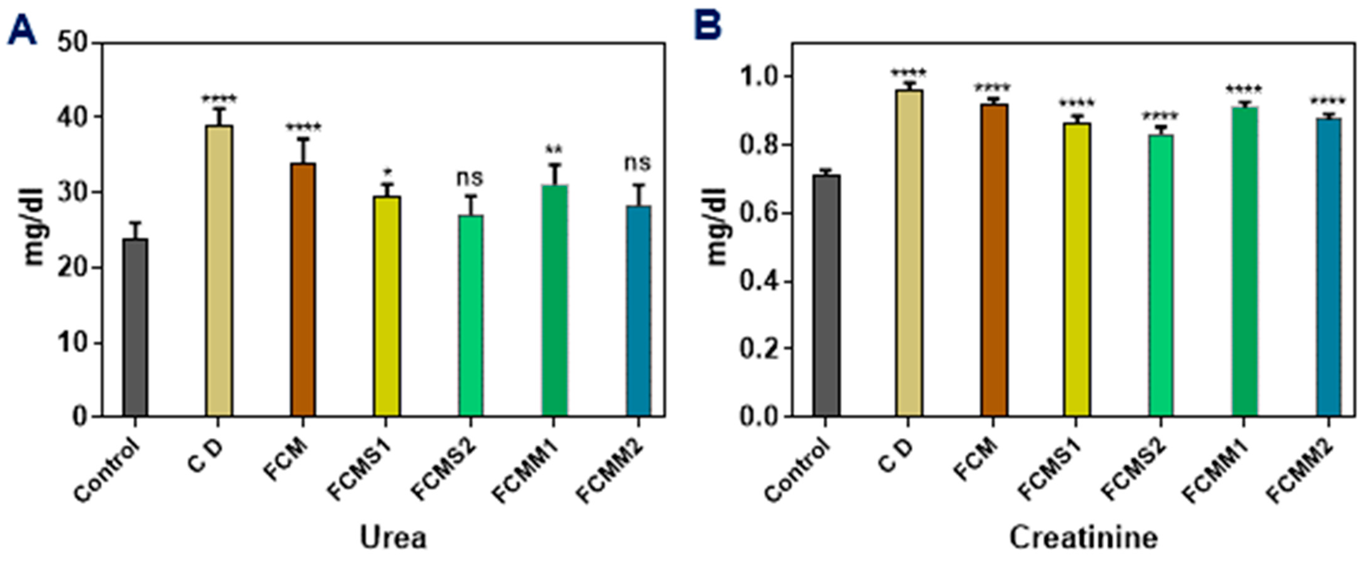
2.5. Kidney Functions

A serious complication of diabetes disease is diabetic nephropathy (DN), which is the most popular cause of chronic kidney disease, especially in Western countries, affecting 30–40% of patients with type 1 and type 2 diabetes [47]. As displayed in Figure 4, the induction of rats with alloxan significantly (p < 0.05) increased serum urea and creatinine levels and the high values were found in the positive control group as compared to the negative control group. Treatment with FCM or FCM fortified with sage and mint powder significantly decreased serum urea and creatinine levels. The higher decrease in urea level was observed with fermented camel milk samples containing 1.5% of sage or mint powder (FCS2 and FCMM2), followed by the samples containing 1% of sage and mint powder (FCS1 and FCMM1), and then samples of FCM alone. Concerning creatinine levels in diabetic rats groups, the results showed that the creatinine levels were significantly decreased administration of FCM, FCMS, and FCMM, and a higher decrease was found with FCMS1, FCMS2, and FCMM2, followed by FCMM1 and FCM groups.

3. Discussion

In the present study, we confirmed that the supplementation of fermented camel milk with sage and mint powder increased its anti-diabetic effects on alloxan-induced diabetic rats, whereas the oral administration of fermented camel milk fortified with sage and mint powder caused a significant decrease in blood glucose level and lipid profile and increase in insulin level compared to the control (+) and FCM groups. In relation to the chemical composition of sage and mint powder, which is shown in Table 2, the results of LC-MS revealed higher contents of phenolic and flavonoids in the study. Quantitative LC-MS/MS analysis revealed that the most abundant compounds in ME in order from high to low were naringenin, caffeic acid, luteolin, dihydroxybenzoic acid, hesperetin, chlorogenic acid, and apigenin. While the most abundant compounds in SE were naringenin, caffeic acid, luteolin, chlorogenic acid, apigenin, dihydroxybenzoic acid, quercetin, and coumaric acid. According to LC-MS analysis, phenolic and flavonoid compounds were identified and quantified (µg/g) in sage and mint extract. The polyphenols and flavonoid contents in the SE and ME were clearly superior according to LC-MS analysis. Importantly, the levels of naringenin, as a flavanone (RT 14.9.0 min), accounted for 2137 µg/g in SE and 4933 µg/g in ME. Caffeic acid (RT 8.04 min) accounted for 394.8 µg/g in SE and 339.9 µg/g in ME, whereas luteolin acid (RT 8.04 min) accounted for 372.4 µg/g in SE and 281.5 µg/g in ME. The obtained LC-MS results could be consistent with a previous study that declared that SE is rich in phenolic acids such as caffeic acid, and in flavonoids, in particular luteolin [48]. Also, previous studies [49,50] pointed out that ME is rich in naringenin, caffeic acid, and hydroxybenzoic acid, which is similar to the LC-MS of ME in this study. Other compounds were found at low levels but still in considered amounts such as apigenin, chlorogenic acid, hydroxybenzoic acid, quercetin, and coumaric acid in SE, and hesperetin, apigenin, rutin, and gallic acid in ME. The anti-diabetic activity of the dosage sage and mint powder could be attributed to these three compounds. Naringenin, a flavanon which is found in high amount in sage, was demonstrated for its anti-diabetic properties though inducing glucose uptake [51]. Additionally, Naringenin was reported for its tangible role to protect β cells from cytokine-induced cell death [52]. Caffeic acid is a phenolic acid, was found in a considered amount in SE according to LC-MS, and might play an important role in the anti-diabetic activity of the dosage form, due to its antioxidant activity and the capability to reduce the glucose level, enhance glucose-stimulated insulin secretion and glucose sensitivity [53,54]. Moreover, caffeic acid could be an effective agent in alleviating diabetes and associated complications [55]. Luteolin and apigenin as flavone class (flavonoids) showed anti-diabetic effects through activating expression levels of insulin receptor substrate-1, insulin receptor substrate-2, and phosphatidylinositol 3-kinase/protein kinase B pathway in IR-HepG2 cells [56]. High-glucose-simulated diabetes is associated with excessive production of reactive oxygen species (ROS) and advanced glycation end-products (AGEs). The increased levels of ROS increase oxidative stress with subsequent tissue damage (cellular inflammasomes) and are associated with insulin resistance [57]. Most of the identified compounds either in SE or ME such as caffeic acid, naringenin, chlorogenic acid, gallic acid, rutin, coumaric acid, quercetin, and ferulic acid are well known for their antioxidant activity, and therefore, such compounds can counteract the insulin resistance and can induce the glucose uptake. Moreover, these compounds such as chlorogenic acid were reported for their anti-diabetic effect through enhancing the insulin secretion and sensitivity.
It should be noted that experimental induction of diabetes by alloxan increased the level of plasma glucose significantly over the control level as reported elsewhere [58]. Also, in alloxan-induced diabetic rats, the levels of triglyceride, total cholesterol, LDL, VLDL, ALP, AST, and ALT were increased as declared in the results. The hyperglycemia induced by alloxan contributes to oxidative imbalance and increases oxidative stress. It is noteworthy to mention that alloxan induces diabetes in the diabetic model through the generation of reactive oxygen species (ROS) in excess levels, where ROS could be generated in this reaction involving alloxan, and then the alloxan is converted to a reduced product of dialuric acid combined with inhibition for the synthesis and secretion of insulin [59,60]. These findings cleared the relationship between the antioxidant compounds and the secretion of insulin in the body. In our study, the oral administration of FCM fortified with mint or sage leaves powder increased the secretion of insulin in diabetic rats and this may be due to the antioxidant components in mint and sage. Many studies reported the relationship between the antioxidant components in medicinal herbs such as sage and mint and potential anti-diabetic properties. Menthol and other volatile compounds in the leaves of M. piperita may be responsible for antioxidant and antioxidant activities [61]. Also, mint (M. piperita) leaf extract possesses a high amount of phenolic content, flavonoid content, and flavonols. Rosmarinic acid, caffeic acid, and its derivatives, and chlorogenic are the main phenolic compounds of the genus Mentha as well as the presence of some salvianolic acids [6,62]. In vitro assays have shown free radical (hydroxyls radicals, nitric oxide, hydrogen peroxide radicals, superoxide radicals, and DPPH radical) scavenging activities of extracts from different Mentha spp. [63,64,65]. The obtained results in this study agree with Agawane et al. [65], who found that the methanolic leaves extract of Mentha arvensis L. showed the ability to scavenge DPPH free radicals which was found to be 78% at a concentration of 1000 mg/mL. The effect of antioxidative components on the inhibition of DPPH radicals is considered to be due to their ability to donate hydrogen [66].
The blood glucose level in diabetic rats was significantly decreased after administration of FCM, FCMS, and FCMM; this finding is in agreement with that found by Hussain et al., 2021 [67], who observed that the mean blood glucose in diabetic mice decreased from 346 (mg/dL) to 140 (mg/dL) after being treated with camel milk (83 mL/kg body weight for 7 weeks), which is not significantly different from the diabetic mice receiving glibenclamide (anti-diabetic drug) in a dose of 600 µg/kg body weight (blood glucose of 125 mg/dL). Also, Shori and Baba, 2014 [68] reported that fermented plain camel milk had higher anti-diabetic activity than fermented plain cow milk. The oral intake of camel milk (at a dose of 250 mL/24 h/15 rats) reduced the blood glucose level from 462.3 ± 37.8 to 96.7 ± 11.1 mg/dL [69], while oral administration of camel milk for three weeks decreased the level of blood glucose of alloxan-induced diabetic rats from 10.88 ± 0.55 to 6.22 ± 0.5 mmol/L [70]. On the same side, Hamad et al., 2011 [71] noted that camel milk had higher anti-diabetic activity (49%) compared with buffalo and cow milk (11%) in diabetic Sprague Dawley rats. Previous studies [72,73] observed that camel milk had a significant hypoglycemic effect when administered to type 1 diabetic patients as an adjunct therapy for 3 months. Also, Agrawal et al. 2005 [74] reported that camel milk as an adjunct to insulin therapy appears to be safe and efficacious in improving long-term glycemic control and helps in reduction in the doses of insulin in patients with type 1 diabetes. One of the suggested mechanisms of the anti-diabetic effect of camel milk might be attributable to the inhibition of various metabolic enzymes such as dipeptidyl peptidase IV [DPP-IV, an enzyme that degrades the insulin-secreting incretin hormones gastric inhibitory polypeptide (GIP) and glucagon-like peptide (GLP), α-glucosidase and α-amylase [75]. The potential inhibition of DPP-IV is due to bioactive peptides resulting after hydrolysis of camel milk proteins throughout proteolysis or fermentation process [12], particularly bioactive peptides released from whey proteins [76,77]. Additionally, the presence of hydrophobic amino acids in the bioactive peptides is considered an additional factor for DPP-IV inhibition because these amino acids may further enhance interaction with the active site of DPP-IV [78,79]. Another study suggested that the anti-diabetic activity of camel milk due to its effect on the insulin receptors [80], while Mehaia et al., 1995 [81] reported that the content of insulin-like proteins in camel milk was three times more than in cow milk.
In the present study, it was observed that the corporation between sage or mint powder and fermented camel milk increased anti-diabetic activity (decreased glucose level and increased insulin level in blood plasma). This could be attributed to the additional biological activity of mint or sage extract as an antioxidant and anti-diabetic agent along with fermented camel milk. According to previous studies, the anti-diabetic activity of sage leaves powder due to its activity in reduced the blood glucose level and also inhibits the activity of the intestinal maltase and sucrase enzymes [39,82]. A previous study [83] found that the oral administration of peppermint juice for 21 days significantly (p < 0.0010) decreased the blood glucose level in alloxan-induced diabetic rats. Diabetes is associated with an increase in oxidative stress as shown by an increase in free radicals and decreased activities of catalase (CAT), superoxide dismutase (SOD), glutathione S-transferase (GST), glutathione peroxidase (GPX), and GSH [84]. Free radicals play an important role in the development of both type I and type II diabetes [85]. A previous work [86] reported that the elevation in plasma insulin levels in the sage extract-treated STZ diabetic rats could be due to substances present in the plant extract which stimulate insulin secretion or which protect the intact functional b-cells from further deterioration so that they keep active and continue to insulin production. The same study [86] showed that the methanol extract of S. officinalis causes a significant reduction in glucose concentrations on STZ -induced hyperglycemic rats. Also, a previous study [39] found that the alloxan-induced diabetic rats treated with aqueous and ethanol extracts (100 mg/kg) of sage (Salvia officinalis) leaves showed a significant reduction (p < 0.05) in fasting blood glucose. The effects of plants on diabetes disease were summarized as increasing insulin secretion, increasing glucose uptake by fat tissues and skeletal muscle, inhibiting the production of liver glucose, and inhibiting the absorption of glucose in the intestinal [40]. These results in agreement with previous work [69] noted that the oral administration of camel milk reduced the increase in TG, TC, LDL-C, and VLDL-C in diabetic rats compared with the diabetic control group. A previous study [87] evaluated the effects of camel milk on the TC, HDL, and TG levels in type 1D and type 2D, respectively, and their findings agreed with our results that camel milk normalized the alteration in TG and HDL-c, while reducing the increase in total cholesterol (TC) levels. Therefore, camel milk can give promising results when used as a dietary supplement for patients of type 1D. In the same line, Khattab et al., 2012 [88] found that treated diabetic rats with sage leaves induced significant improvement in lipid profile parameters as compared with the non-treated diabetic group and concluded that sage had a potent hypoglycemic activity and related this effect to its antioxidant activities. Regulation of the levels of cholesterol and triglyceride in the blood is an important way to protect humans from coronary heart disease. It was found that administration of sage infusion for 12 weeks reduced total cholesterol, triglycerides, and low-density lipoprotein (LDL-c) in rats, while HDL-c was increased [89]. Also, a previous study [39] indicated that ethanolic and water extracts of sage leaves significantly lowered cholesterol and TG levels. Moreover, many studies cleared the significant role of mint leaves on diabetic rats. This hypolipidemic effect of sage may be related to the inhibition of hepatic de novo synthesis or the activation of β-oxidation [89]. A previous study [90] reported that treatment of diabetic rats with M. piperita caused a reduction in the levels of cholesterol, LDL-c, and triglycerides and increase the levels of HDL-c. Also, a previous work [91] found that treatment of hyperlipidemic rats with aqueous extract of Mentha piperita leaves extract for 21 days significantly reduced serum total cholesterol, triglycerides, and LDL-c, and was associated with a significant increase in HDL-c levels and decrease in the atherogenic index in indicating its potent anti-hyperlipidemic and antiatherogenic activity.
It should be stressed that AST, ALT, and LDH are enzymes mainly found in hepatocyte cytosol and cell membrane. They are good markers considerably used to evaluate hepatotoxicity and integrity of the membrane [92]. The increase in activities of plasma ALT, AST, ACP, ALP, and LDH means that diabetes caused hepatic dysfunction. Therefore, the increment of the activities of ALT, AST, ACP, ALP, and LDH in plasma may be mainly due to the leakage of these enzymes from the liver cytosol into the bloodstream which gives an indication of the hepatotoxic effect of alloxan [93,94]. A previous report [92] noted that the increase in liver enzyme activities in diabetic rats was reduced after being treated with sage essential oil. Similarly, in alloxan diabetic rats, ALT, AST, and ALP activities were superior to those in normal rats, but recovered after oral administration of fermented camel milk fortified with sage and mint powder. These results are similar to that found in a previous study [86], which reported that the recovery of liver cell integrity was obtained after treatment by sage. Oral administration of ethanolic extract of sage leaves to diabetic rats, lowered serum glucose, triglycerides, total cholesterol, urea, creatinine, AST, ALT, and enhanced plasma insulin, depending on the increasing dose [42].
Induction of hyperglycemia caused an increase in serum creatinine and urea levels, excessive proteinuria, and marked deterioration of kidney function, and microscopic examination of sections of the kidneys of diabetic animals showed pathological features of glomerulosclerosis, with abnormal extracellular matrix (ECM) accumulation, glomerular matrix expansion, tubular alveolar degeneration, and fibrosis, and increased urinary excretion [1]. The observed increase in serum creatinine, urea, and uric acid of diabetic animals compared with the nondiabetic control group agrees with previous work [42], while the consumption of camel milk caused a significant decrease in creatinine and urea in diabetic rats and this could be attributed to the hypoglycemic and antioxidant effects of camel milk [1]. The reported powerful hypoglycemic action of camel milk in diabetic patients is hypothesized to abolish the glucose-driven metabolic pathways. Intensive glycemic control in type 1 and type 2 diabetes mellitus patients results in a decrease in microalbuminuria. So, the observed renal protective effects of camel milk treatment in diabetic rats could be assigned to the glucose homeostatic action of camel milk. Our results were in accord with the earlier findings [72], which found a significant reduction of microalbuminuria in type 1 diabetes mellitus patients receiving camel milk, along with their standard anti-diabetic therapy suggesting a direct protective effect of camel milk against diabetic nephropathy [67,95,96].
The increase in serum alanine aminotransferase (ALT) and aspartate aminotransferase (AST) activities may indicate liver tissue damage, probably by altered cell membrane permeability leading to the leak of the enzymes from the tissues to the serum. Alanine and aspartate aminotransaminases are considered to be sensitive indicators of hepatocellular damage and within limit can provide a quantitative evaluation of the degree of damage to the liver [97]. Diabetes has a strong relationship with renal and liver diseases [98]. Camel milk was reported to protect the liver and kidney function from failure. A previous study [1] found that the administration of camel milk to the control animals caused insignificant changes in the glomerulotubular morphology in comparison to the non-camel milk-treated control animals. Furthermore, the kidney slices obtained from the diabetic animals and stained with the hematoxylin and eosin showed glomerular expansion and tubular alveolar degeneration. A previous work [99] mentioned that the induction with streptozotocin caused damage to the kidney tissue of diabetic rats, and the untreated group showed severe glomerular necrosis with lymphocyte hyperplasia when compared with the normal. This result is similar to the work carried out by Trujillo et al. [100], who reported that abnormal levels of serum urea usually signify decreased renal function, so plasma urea is a recognized marker of glomerular filtration rate (GFR) and in nephropathy.

4. Material and Methods

4.1. Materials and Chemicals

Camel milk (total solids 11.84%, protein 3.22%, fat 3.43%, pH 6.60, and acidity 0.175%) was obtained from a private farm in El-Arish, North Sinai Governorate, Egypt. Commercially available lyophilized culture (Yo-fast 88, contains Streptococcus thermophillus and Lactobacillus delbrueckii ssp. bulgaricus) was purchased from Chr. Hansen Laboratories, Hoersholm, Denmark. Mint (Mentha piperita) and sage (Salvia officinalis) leaves were obtained from El-Arish local market, North Sinai Governorate, Egypt. Alloxan monohydrate, analytical reagent grade purchased from Sigma Chemical Co. (Sigma-Aldrich Company Ltd., Gillingham, UK). 1, 1-dipheny l-2-picryl-hydrazyl (DPPH) was purchased from Sigma-Aldrich (Munich, Germany). Potassium ferricyanide, ferric chloride, and gallic acid were purchased from Loba Chemie, Mumbai, India. Formic acid (LC grade) was provided by Fisher Chemical. Methanol and acetonitrile (LC grade) were supplied from Supelco, where Milli-Q water was used.

4.2. Methods

4.2.1. Preparation of Mint and Sage Leaves Powder

The leaves of mint and sage were dried at 30–40 °C by the hybrid solar convective drying system (C.C.P. Parma—Parma, Italy), and then the leaves were ground until they become a powder.

4.2.2. Preparation of Sage and Mint Extracts for Proximate Chemical Analysis

Five grams of mint and sage leaves powders were mixed with 100 mL ethanol solution 75%, stirring for 2 h at room temperature. Finally, the mixtures were filtered by Whatman No. 1 and the extracts were stored at 4 °C until analysis [101]. Furthermore, the gross chemical compositions of sage and peppermint leaves powder used for different treatments of fermented camel milk samples were determined and analyzed for moisture, protein, fat, and ash. Crude fiber contents were determined, while carbohydrate was calculated by difference according to the standard methods of the association of official analytical chemists [102].

4.2.3. Preparation of Standard Compounds and Extract Samples Solutions and Identification of Their Chemical Composition of Sage and Mint Extracts and Their Compounds Using LC-ESI-MS/MS

Stock standard solutions in 1 mg/mL of standard compounds were prepared in methanol. Then, the stock solutions of each standard were diluted to 10 µg/mL and kept at 4 °C. The working standards mixture solution (200 ppb) was prepared by mixing 20 µL of each stock solution and then completing the volume to 1 mL using methanol. Then, 3 mg of the dried extract (mint extract or sage extract) was dissolved in 1 mL 80% methanol, sonicated for 15 min, filtered through a 0.45 µm syringe filter, and finally injected into LC-MS/MS. A blank 80% methanol was injected before each sample. The analysis of the extracts of sage and mint extracts was performed using liquid chromatography–electrospray ionization–tandem mass spectrometry (LC-ESI-MS/MS) with an ExionLC AC system for separation and SCIEX Triple Quad 5500+ MS/MS system equipped with electrospray ionization (ESI) for detection [103]. MRM transitions and the optimized mass spectrometer parameters of each compound are listed in Supplementary Table S1 and LC-ESI-MS spectra of standards and extracts are shown in Supplementary Figures S1–S3. The chromatographic conditions were optimized where the separation was performed using ZORBAX Eclipse Plus C18 Column (4.6 × 100 mm, 1.8 µm). The mobile phases consisted of two eluents, A: 0.1% formic acid in water; B: acetonitrile (LC grade). The mobile phase elusion was programmed as follows: 2% B from 0–1 min, 2–60% B from 1–21 min, 60% B from 21–25 min, and 2% B from 25.01–28 min. The flow rate was optimized at 0.8 mL/min and the injection volume was 3 µL. Positive and negative ionization modes were applied in the same run for MRM analysis of the selected standard compounds, with the following parameters: curtain gas: 25 psi; ion spray voltage: 4500 and −4500 for positive and negative modes, respectively; source temperature: 400 °C; ion source gas 1 and 2 were 55 psi with a declustering potential: 50; collision energy: 25; collision energy spread: 10.

4.2.4. Antioxidant Activity of Sage and Mint Extracts

Determination of Total Phenolic Contents (TPC) of Sage and Mint Extracts

The total phenolic contents of sage and mint extracts were determined according to the method of Abirami et al., 2014 [104]. Total phenol content (TPC) was expressed as Gallic acid equivalent (mg GAE/g plant material or extract).

Determination of Total Flavonoids (TF)

The TF content of sage and mint extracts was determined based on the method of Barros et al., 2011 [105]. A calibration curve of Rutin was prepared and TF content was determined.

DPPH Scavenging Activity %

The scavenging activity of 1,1-diphenyl-2-picrylhydrazyl (DPPH) radical was determined according to the method of Lim and Quah, 2007 [106]. Triplicate tubes were prepared for each extract. The results were expressed as % radical scavenging activity.
R a d i c a l   s c a v e n g i n g   a c t i v i t y % = A c o n t r o l A s a m p l e A c o n t r o l × 100
IC50, which denotes the amount (mg) of the plant powder in 1 mL solution required to reduce the initial concentration of DPPH radicals by 50%, was also calculated. Ascorbic acid was used as a standard.

Ferric Reducing Antioxidant Power (FRAP)

The FRAP was determined according to the method of Oyaizu, 1986 [107]. Triplicate tubes were prepared for each extract. The FRAP values, expressed in mg GAE/g, were derived from a standard curve.

4.2.5. Physicochemical Analysis of Camel Milk

Total solids (%), protein (%), and fat (%) of camel milk were determined using the AOAC procedures [108]. The pH of camel milk was measured using a digital pH meter (Martini, Italy). Titratable acidity (lactic acid %) of raw camel milk was evaluated by titration with NaOH (0.1 N) in the presence of phenolphthalein as an indicator. All analyses were performed in triplicate.

4.2.6. Preparation of Fermented Camel Milk (FCMs)

Camel milk was divided into five portions. The first portion served as a control (FCM). Four portions of camel milk were supplemented with sage and mint leaves powder at levels of 1 and 1.5% (FCMS1 (1% sage), FCMS2 (1.5% sage), FCMM1 (1% mint), and FCMM2 (1.5% mint), and these percentages were chosen according to the sensory evaluation of fermented camel milk fortified with different percentages of these herbs (0.5, 1, 1.5, and 2%). Fermented milk was prepared according to Tamime and Robinson, 2007 [109]. Camel’s milk was heated at 72 °C/15 s, cooled to 40 °C, and then inoculated with 0.3% yogurt starter culture. Camel milk was incubated at 42 ± 1 °C until the pH value was decreased to approximately 4.6. The resultant fermented camel milk of all treatments was kept in a refrigerator (4 ± 1 °C) until use.

4.2.7. Animals and Treatments

The Induction of Experimental Diabetes

Alloxan was dissolved in a saline solution (0.9% sodium chloride, pH 7). Diabetes was induced in normal healthy male albino rats by receiving an intra-peritoneal injection dose of alloxan 150 mg/kg body weight, according to the method described elsewhere [110]. After three days of the injection with alloxan, fasting blood samples were obtained to estimate fasting serum glucose higher than 200 mg/dL in rats, which were considered diabetes by the National Diabetes Data Group [111].

Experimental Design

Forty-two adult normal male albino rats of Sprague Dawley strain (140 ± 10 g) were obtained from the Vaccine and Immunity Organization, Ministry of Health, Helwan, Egypt. Animals were housed 6 per cage and fed on a basal diet prepared based on the American Institute of Nutrition [112] and consisting of 12% casein, 10% sugars, 10% sunflower oil, 1% vitamin mixtures, 4% mineral mixtures, 4% fiber, 58.50% starch, 0.3% DL-methionin, and 0.2% choline chloride, and given free access to fresh water ad libitum. Rats were acclimated for 2 weeks at 25 ± 1 °C with a 12 h dark and light cycle. The experimental period was 8 weeks after stabilization of diabetes for 1 week and the animals were divided into 7 major groups (6 rats per group) and treated based on the experimental protocol:
(1) healthy control (C group); (2) diabetic control (DC group); (3) diabetic + 85 mL FCM/kg B.W/day (FCM group); (4) diabetic + 85 mL FCMS1/kg B.W/day (FCMS1 group); (5) diabetic + 85 mL FMCS2/kg B.W/day (FCMS2 group); (6) diabetic + 85 mL FCMM1/kg B.W/day (FCMM1 group); and (7) diabetic + 85 mL FCMM2/kg B.W/day (FCMM2). The oral dose of fermented camel milk was chosen based on the study of Althnaian et al., 2013 [113]. At the end of the experimental period, rats fasted for 12 h, were anesthetized with ether, and killed. Fasting blood samples were collected in heparinized tubes from the killed animals and then centrifuged at 7200× g at 4 °C for 20 min (Sigma centrifuge 113, VWR International) to obtain plasma. The obtained plasma was stored at −80 °C until used for analyses [114].

Blood Biochemical and Enzymes Activities

Stored plasma samples were analyzed for plasma glucose concentration according to the method of Trinder [110] and National Diabetes Data Group [115]. Urea was determined as described elsewhere [116,117,118]. Creatinine was determined according to the method of Bartels and Böhmer, 1971 [119] and Fabiny and Ertingshausen, 1971 [120]. Triglycerides were determined according to the method of Bucolo and David, 1973 [121] and Fossati and Prencipe, 1982 [122]. Cholesterol was determined according to the method of Meiattini et al. 1978 [123]. High-density lipoprotein (HDL) cholesterol was determined according to the method of Grove, 1979 [124] and Burstein et al. 1970 [125]. Low-density lipoprotein (LDL) was determined by the calculation (cholesterol-(TG/5+HDL). Very low-density lipoprotein (VLDL) was calculated by dividing the values of TG by a factor of 5. The activities of plasma aspartate transaminase (AST) and alanine transaminase (ALT) were assayed by the method of Reitman and Frankel, 1957 [126]. Alkaline phosphatase (AlP) activity was determined in plasma according to the method of Belfield and Goldberg, 1971 [127]. Commercial kitts of the previous assays were obtained from Biosystems S.A. (Barcelona, Spain) (for glucose, cholesterol, HDL, TG, urea, and creatinine); QUIMICA CLINICA APLICADA S.A (Amposta, Spain) (for AST, ALT); and Biodiagnostic (ARE) (for ALP).

Determination of Blood Insulin Level

Insulin levels were estimated according to Abraham et al., 1978 [128] and Wilson and Miles, 1977 [129] using an ELISA kit by Linco Research Inc., Missouri, USA.

4.2.8. Statistical Analysis

The results were analyzed using GraphPad Prism 8.0.2 software, GraphPad Software Inc, California, USA. The results were expressed as means ± SD, where the statistical significance was evaluated using one-way ANOVA. followed by Tukey’s correction (* p < 0.0332; ** p < 0.0021, *** p < 0.0002, **** p < 0.0001, versus control).

5. Conclusions

The current study concluded that supplementation of camel milk with sage and mint leaves powder ameliorated and normalized the changes in glucose, total cholesterol, and triglyceride levels in the blood of diabetic rats. The most marked findings were found with the fortification of fermented camel milk with sage leaves powder at a ratio of 1.5%. Given the above findings, it could be concluded that sage and mint leaves powder (at a ratio of 1.5%) can be used to produce healthy and functional fermented camel milk with high antioxidant activity and anti-diabetic activity. The presented promising data could be beneficial and encourage us to set up a further future study utilizing the major components in the fermented milk (e.g., bioactive peptides) with the major antioxidant components in sage or mint extracts to obtain the best dosage form with studying its effect on metabolites and key genes involved in the glucose metabolism pathway, as a potential promising anti-diabetic product in the future.

Supplementary Materials

The following supporting information can be downloaded from: https://www.mdpi.com/article/10.3390/molecules28155749/s1. Table S1: MRM transitions and the optimized mass spectrometer parameters; Figures S1–S3: the mass liquid chromatography–electrospray ionization–tandem mass spectra of standards; sage and mint extracts are provided in the Supplementary file.

Author Contributions

M.R.S., A.A.E. and M.I.E.-S. were involved in the conception of the research idea and methodology design, and supervision, performed data analysis and interpretation, and prepared the manuscript for publication and revision. E.R.-Á., A.A., M.A.M.H., M.A.M. and E.K.E. were involved in the methodology and drafted and prepared the manuscript for publication and revision. All authors have read and agreed to the published version of the manuscript. The funders had no role in data collection and analysis, the decision to publish, or the preparation of the manuscript.

Funding

Researchers Supporting Project number (RSPD2023R655), King Saud University, Riyadh, Saudi Arabia.

Institutional Review Board Statement

Ethics approval of studies using rats was obtained from Research Ethical Committee (REC), The Institutional Animal Care and Use Committee (ICUC), Tanta University, Egypt (Approval number: IACUC-SCI-TU-0246).

Informed Consent Statement

Not applicable.

Data Availability Statement

Data is contained within the article.

Acknowledgments

E.K.E was supported by a postdoctoral contract María Zambrano (University of Córdoba) from the Program of Requalification of the Spanish University System (Spanish Ministry of Universities) financed by the European Union-NextGenerationEU. The authors would like to thank Researchers Supporting Project number (RSPD2023R655), King Saud University, Riyadh, Saudi Arabia for funding this research work.

Conflicts of Interest

The authors declare no conflict of interest.

Sample Availability

Not applicable.

List of Abbreviations

CM: camel milk; FCM: fermented camel milk; DM: diabetes mellitus; IDF: International Diabetes Federation; DPPH: 1, 1-diphenyl-2-picryl-hydrazyl; TPC: total phenol content; FRAP: ferric reducing antioxidant power; HDL-c: high-density lipoprotein cholesterol; LDL-c: low-density lipoprotein cholesterol; VLDL-c: very low-density lipoprotein cholesterol; TG: triglycerides; TC: total cholesterol; AST: aspartate transaminase; ALT: alanine transaminase; AlP: alkaline phosphatase.

References

  1. Korish, A.A.; Abdel Gader, A.G.; Korashy, H.M.; Al-Drees, A.M.; Alhaider, A.A.; Arafah, M.M. Camel milk attenuates the biochemical and morphological features of diabetic nephropathy: Inhibition of Smad1 and collagen type IV synthesis. Chem.-Biol. Interact. 2015, 229, 100–108. [Google Scholar] [CrossRef] [PubMed]
  2. Al Kury, L.T.; Abdoh, A.; Ikbariah, K.; Sadek, B.; Mahgoub, M. In Vitro and In Vivo Antidiabetic Potential of Monoterpenoids: An Update. Molecules 2021, 27, 182. [Google Scholar] [CrossRef] [PubMed]
  3. Federation, I.D. IDF Diabetes Atlas, 10th ed.; International Diabetes Federation: Brussels, Belgium, 2021; Available online: https://diabetesatlas.org/atlas/tenth-edition/ (accessed on 31 December 2021).
  4. Sharma, S.; Kulkarni, S.K.; Chopra, K. Curcumin, the active principle of turmeric (Curcuma longa), ameliorates diabetic nephropathy in rats. Clin. Exp. Pharmacol. Physiol. 2006, 33, 940–945. [Google Scholar] [CrossRef]
  5. FAO Stat. 2019. Available online: http://www.fao.org/faostat/en/#home (accessed on 30 November 2006).
  6. Ereifej, K.; Aludatt, M.; Alkhalidy, H.; Alli, I.; Rababah, T. Comparison and characterisation of fat and protein composition for camel milk from eight Jordanian locations. Food Chem. Food Chem. 2011, 127, 282–289. [Google Scholar] [CrossRef]
  7. Mati, A.; Senoussi-Ghezali, C.; Si Ahmed Zennia, S.; Almi-Sebbane, D.; El-Hatmi, H.; Girardet, J.-M. Dromedary camel milk proteins, a source of peptides having biological activities—A review. Int. Dairy J. 2017, 73, 25–37. [Google Scholar] [CrossRef]
  8. Kamal, M.; Karoui, R. Monitoring of mild heat treatment of camel milk by front-face fluorescence spectroscopy. LWT Food Sci. Technol. 2017, 79, 586–593. [Google Scholar] [CrossRef]
  9. Swelum, A.A.; El-Saadony, M.T.; Abdo, M.; Ombarak, R.A.; Hussein, E.O.S.; Suliman, G.; Alhimaidi, A.R.; Ammari, A.A.; Ba-Awadh, H.; Taha, A.E.; et al. Nutritional, antimicrobial and medicinal properties of Camel’s milk: A review. Saudi J. Biol. Sci. 2021, 28, 3126–3136. [Google Scholar] [CrossRef]
  10. Quan, S.; Tsuda, H.; Miyamoto, T. Angiotensin I-converting enzyme inhibitory peptides in skim milk fermented with Lactobacillus helveticus 130B4 from camel milk in Inner Mongolia, China. J. Sci. Food Agric. 2008, 88, 2688–2692. [Google Scholar] [CrossRef]
  11. Agrawal, R.P.; Tantia, P.; Jain, S.; Agrawal, R.; Agrawal, V. Camel milk: A possible boon for type 1 diabetic patients. Cell. Mol. Biol. 2013, 59, 99–107. [Google Scholar] [PubMed]
  12. Kamal, H.; Jafar, S.; Mudgil, P.; Murali, C.; Amin, A.; Maqsood, S. Inhibitory properties of camel whey protein hydrolysates toward liver cancer cells, dipeptidyl peptidase-IV, and inflammation. J. Dairy Sci. 2018, 101, 8711–8720. [Google Scholar] [CrossRef] [Green Version]
  13. Solanki, D.; Hati, S. Fermented camel milk: A Review on its bio-functional properties. Emir. J. Food Agric. 2018, 30, 268–274. [Google Scholar]
  14. El-Sayed, M.; Awad, S.; Abou-Soliman, N. Improving the Antioxidant Properties of Fermented Camel Milk Using Some Strains of Lactobacillus. Food Nutr. Sci. 2021, 12, 352–371. [Google Scholar] [CrossRef]
  15. Mohammadabadi, T. The unique effects of camel milk as adjunctive super food on the health. World J. Pharm. Sci. 2021, 9, 97–106. [Google Scholar]
  16. Rasheed, Z. Medicinal values of bioactive constituents of camel milk: A concise report. Int. J. Health Sci. 2017, 11, 1–2. [Google Scholar]
  17. Meena, S.; Rajput, Y.S.; Pandey, A.K.; Sharma, R.; Singh, R. Camel milk ameliorates hyperglycaemia and oxidative damage in type-1 diabetic experimental rats. J. Dairy Res. 2016, 83, 412–419. [Google Scholar] [CrossRef]
  18. Badr, G.; Sayed, L.H.; Omar, H.E.M.; Abd El-Rahim, A.M.; Ahmed, E.A.; Mahmoud, M.H. Camel Whey Protein Protects B and T Cells from Apoptosis by Suppressing Activating Transcription Factor-3 (ATF-3)-Mediated Oxidative Stress and Enhancing Phosphorylation of AKT and IκB-α in Type I Diabetic Mice. Cell. Physiol. Biochem. Int. J. Exp. Cell. Physiol. Biochem. Pharmacol. 2017, 41, 41–54. [Google Scholar] [CrossRef]
  19. Agrawal, R.P.; Saran, S.; Sharma, P.; Gupta, R.P.; Kochar, D.K.; Sahani, M.S. Effect of camel milk on residual beta-cell function in recent onset type 1 diabetes. Diabetes Res. Clin. Pract. 2007, 77, 494–495. [Google Scholar] [CrossRef] [PubMed]
  20. Fallah, Z.; Feizi, A.; Hashemipour, M.; Kelishadi, R. Effect of fermented camel milk on glucose metabolism, insulin resistance, and inflammatory biomarkers of adolescents with metabolic syndrome: A double-blind, randomized, crossover trial. J. Res. Med. Sci. Off. J. Isfahan Univ. Med. Sci. 2018, 23, 32. [Google Scholar] [CrossRef]
  21. Atwaa, E.S.H.; Shahein, M.R.; El-Sattar, E.S.A.; Hijazy, H.H.A.; Albrakati, A.; Elmahallawy, E.K. Bioactivity, Physicochemical and Sensory Properties of Probiotic Yoghurt Made from Whole Milk Powder Reconstituted in Aqueous Fennel Extract. Fermentation 2022, 8, 52. [Google Scholar] [CrossRef]
  22. Atwaa, E.S.H.; Shahein, M.R.; Radwan, H.A.; Mohammed, N.S.; Aloraini, M.A.; Albezrah, N.K.A.; Alharbi, M.A.; Sayed, H.H.; Daoud, M.A.; Elmahallawy, E.K. Antimicrobial Activity of Some Plant Extracts and Their Applications in Homemade Tomato Paste and Pasteurized Cow Milk as Natural Preservatives. Fermentation 2022, 8, 428. [Google Scholar] [CrossRef]
  23. Shahein, M.R.; Atwaa, E.S.H.; Alrashdi, B.M.; Ramadan, M.F.; Abd El-Sattar, E.S.; Siam, A.A.H.; Alblihed, M.A.; Elmahallawy, E.K. Effect of Fermented Camel Milk Containing Pumpkin Seed Milk on the Oxidative Stress Induced by Carbon Tetrachloride in Experimental Rats. Fermentation 2022, 8, 223. [Google Scholar] [CrossRef]
  24. Shahein, M.R.; Atwaa, E.S.H.; Elkot, W.F.; Hijazy, H.H.A.; Kassab, R.B.; Alblihed, M.A.; Elmahallawy, E.K. The Impact of Date Syrup on the Physicochemical, Microbiological, and Sensory Properties, and Antioxidant Activity of Bio-Fermented Camel Milk. Fermentation 2022, 8, 192. [Google Scholar] [CrossRef]
  25. Shahein, M.R.; Atwaa, E.S.H.; El-Zahar, K.M.; Elmaadawy, A.A.; Hijazy, H.H.A.; Sitohy, M.Z.; Albrakati, A.; Elmahallawy, E.K. Remedial Action of Yoghurt Enriched with Watermelon Seed Milk on Renal Injured Hyperuricemic Rats. Fermentation 2022, 8, 41. [Google Scholar] [CrossRef]
  26. Shahein, M.R.; Atwaa, E.S.H.; Radwan, H.A.; Elmeligy, A.A.; Hafiz, A.A.; Albrakati, A.; Elmahallawy, E.K. Production of a Yogurt Drink Enriched with Golden Berry (Physalis pubescens L.) Juice and Its Therapeutic Effect on Hepatitis in Rats. Fermentation 2022, 8, 112. [Google Scholar] [CrossRef]
  27. Shahein, M.R.; Atwaa, E.-S.H.; Babalghith, A.O.; Alrashdi, B.M.; Radwan, H.A.; Umair, M.; Abdalmegeed, D.; Mahfouz, H.; Dahran, N.; Cacciotti, I.; et al. Impact of Incorporating the Aqueous Extract of Hawthorn (C. oxyanatha) Leaves on Yogurt Properties and Its Therapeutic Effects against Oxidative Stress Induced by Carbon Tetrachloride in Rats. Fermentation 2022, 8, 200. [Google Scholar] [CrossRef]
  28. Shahein, M.R.; Elkot, W.F.; Albezrah, N.K.A.; Abdel-Hafez, L.J.M.; Alharbi, M.A.; Massoud, D.; Elmahallawy, E.K. Insights into the Microbiological and Physicochemical Properties of Bio-Frozen Yoghurt Made with Probiotic Strains in Combination with Jerusalem Artichoke Tubers Powder. Fermentation 2022, 8, 390. [Google Scholar] [CrossRef]
  29. Swelam, S.; Zommara, M.A.; Abd El-Aziz, A.E.-A.M.; Elgammal, N.A.; Baty, R.S.; Elmahallawy, E.K. Insights into Chufa Milk Frozen Yoghurt as Cheap Functional Frozen Yoghurt with High Nutritional Value. Fermentation 2021, 7, 255. [Google Scholar] [CrossRef]
  30. Shokri, F.; Shokoohi, M.; Niazkar, H.R.; Abadi, A.; Kalarestaghi, H.; Ahin, M. Investigation the Spermatogenesis and Testis Structure in Diabetic Rats After Treatment With Galega officinalis Extract. Crescent J. Med. Biol. Sci. 2019, 6, 31–36. [Google Scholar]
  31. Abtahi-Eivari, S.H.; Shokoohi, M.; Ghorbani, M.; Halimi, M.; Hajizadeh, H.; Pourlak, T.; Bahrami, J.; Ghoreishi, Z. Effects of hydroalcoholic extracts of cloves (Syzygium aromaticum) on serum biomarkers, antioxidant status and histopathological changes of kidneys in diabetic rats. Crescent J. Med. Biol. Sci. 2021, 8, 269–275. [Google Scholar]
  32. Eftekhari, A.; Khusro, A.; Ahmadian, E.; Dizaj, S.M.; Hasanzadeh, A.; Cucchiarini, M. Phytochemical and nutra-pharmaceutical attributes of Mentha spp.: A comprehensive review. Arab. J. Chem. 2021, 14, 103106. [Google Scholar] [CrossRef]
  33. Rajeshwari, C.; Preeti, M.; Andallu, B. Efficacy of mint (Mentha spicata L.) leaves in combating oxidative stress in type 2 diabetes. Int. J. Life Sci. 2012, 1, 28–34. [Google Scholar]
  34. Mihailović, M.; Dinić, S.; Arambašić Jovanović, J.; Uskoković, A.; Grdović, N.; Vidaković, M. The influence of plant extracts and phytoconstituents on antioxidant enzymes activity and gene expression in the prevention and treatment of impaired glucose homeostasis and diabetes complications. Antioxidants 2021, 10, 480. [Google Scholar] [CrossRef] [PubMed]
  35. Nazir, N.; Zahoor, M.; Nisar, M.; Khan, I.; Ullah, R.; Alotaibi, A. Antioxidants isolated from elaeagnus umbellata (Thunb.) protect against bacterial infections and diabetes in streptozotocin-induced diabetic rat model. Molecules 2021, 26, 4464. [Google Scholar] [CrossRef]
  36. Hassan, H.A.; Hafez, H.S.; Goda, M.S. Mentha piperita as a pivotal neuro-protective agent against gamma irradiation induced DNA fragmentation and apoptosis: Mentha extract as a neuroprotective against gamma irradiation. Cytotechnology 2013, 65, 145–156. [Google Scholar] [CrossRef] [Green Version]
  37. Chandirasegaran, G.; Elanchezhiyan, C.; Suhasini, S.; Babby, A. Antihyperglycemic activity of Mentha piperita ethanol leaves extract on streptozotocin induced diabetic rats. Int. J. Pharm. Res. Sch 2014, 3, 2277–7873. [Google Scholar]
  38. Hamidpour, M.; Hamidpour, R.; Hamidpour, S.; Shahlari, M. Chemistry, Pharmacology, and Medicinal Property of Sage (Salvia) to Prevent and Cure Illnesses such as Obesity, Diabetes, Depression, Dementia, Lupus, Autism, Heart Disease, and Cancer. J. Tradit. Complement. Med. 2014, 4, 82–88. [Google Scholar] [CrossRef] [Green Version]
  39. Khashan, K.T.; Al-Khefaji, K.A. Effects of Salvia officinalis L. (sag) leaves extracts in normal and alloxan -induced diabetes in white rats. Int. J. Sci. Eng. Res. 2015, 2015, 20–28. [Google Scholar]
  40. Mahdizadeh, R.; Moein, S.; Soltani, N.; Malekzadeh, K.; Moein, M. Study the molecular mechanism of salvia species in prevention of diabetic. Int. J. Pharm. Sci. Res. 2018, 9, 4512–4521. [Google Scholar] [CrossRef]
  41. Agawane, S.B.; Gupta, V.S.; Kulkarni, M.J.; Bhattacharya, A.K.; Koratkar, S.S. Chemo-biological evaluation of antidiabetic activity of Mentha arvensis L. and its role in inhibition of advanced glycation end products. J. Ayurveda Integr. Med. 2019, 10, 166–170. [Google Scholar] [CrossRef]
  42. Eidi, A.; Eidi, M. Antidiabetic effects of sage (Salvia officinalis L.) leaves in normal and streptozotocin-induced diabetic rats. Diabetes Metab. Syndr. Clin. Res. Rev. 2009, 3, 40–44. [Google Scholar] [CrossRef]
  43. Figueroa-Pérez, M.G.; Gallegos-Corona, M.A.; Ramos-Gomez, M.; Reynoso-Camacho, R. Salicylic acid elicitation during cultivation of the peppermint plant improves anti-diabetic effects of its infusions. Food Funct. 2015, 6, 1865–1874. [Google Scholar] [CrossRef] [PubMed]
  44. Rahman, M.M.; Dhar, P.S.; Anika, F.; Ahmed, L.; Islam, M.R.; Sultana, N.A.; Cavalu, S.; Pop, O.; Rauf, A. Exploring the plant-derived bioactive substances as antidiabetic agent: An extensive review. Biomed. Pharmacother. 2022, 152, 113217. [Google Scholar] [CrossRef] [PubMed]
  45. Shori, A.B. Camel milk as a potential therapy for controlling diabetes and its complications: A review of in vivo studies. J. Food Drug Anal. 2015, 23, 609–618. [Google Scholar] [CrossRef] [PubMed]
  46. Ayoub, M.A.; Palakkott, A.R.; Ashraf, A.; Iratni, R. The molecular basis of the anti-diabetic properties of camel milk. Diabetes Res. Clin. Pract. 2018, 146, 305–312. [Google Scholar] [CrossRef]
  47. Molitch, M.E.; DeFronzo, R.A.; Franz, M.J.; Keane, W.F.; Mogensen, C.E.; Parving, H.H.; Steffes, M.W. Nephropathy in diabetes. Diabetes Care 2004, 27 (Suppl. S1), S79–S83. [Google Scholar] [CrossRef] [Green Version]
  48. Lima, C.F.; Valentao, P.C.; Andrade, P.B.; Seabra, R.M.; Fernandes-Ferreira, M.; Pereira-Wilson, C. Water and methanolic extracts of Salvia officinalis protect HepG2 cells from t-BHP induced oxidative damage. Chem.-Biol. Interact. 2007, 167, 107–115. [Google Scholar] [CrossRef] [Green Version]
  49. Farnad, N.; Heidari, R.; Aslanipour, B. Phenolic composition and comparison of antioxidant activity of alcoholic extracts of Peppermint (Mentha piperita). J. Food Meas. Charact. 2014, 8, 113–121. [Google Scholar] [CrossRef]
  50. Yu, M.; Gouvinhas, I.; Rocha, J.; Barros, A.I. Phytochemical and antioxidant analysis of medicinal and food plants towards bioactive food and pharmaceutical resources. Sci. Rep. 2021, 11, 10041. [Google Scholar] [CrossRef]
  51. Bhattacharya, S.; Christensen, K.B.; Olsen, L.C.; Christensen, L.P.; Grevsen, K.; Færgeman, N.J.; Kristiansen, K.; Young, J.F.; Oksbjerg, N. Bioactive components from flowers of Sambucus nigra L. increase glucose uptake in primary porcine myotube cultures and reduce fat accumulation in Caenorhabditis elegans. J. Agric. Food Chem. 2013, 61, 11033–11040. [Google Scholar] [CrossRef]
  52. Lin, C.-Y.; Ni, C.-C.; Yin, M.-C.; Lii, C.-K. Flavonoids protect pancreatic beta-cells from cytokines mediated apoptosis through the activation of PI3-kinase pathway. Cytokine 2012, 59, 65–71. [Google Scholar] [CrossRef]
  53. Bhattacharya, S.; Oksbjerg, N.; Young, J.; Jeppesen, P. Caffeic acid, naringenin and quercetin enhance glucose-stimulated insulin secretion and glucose sensitivity in INS-1E cells. Diabetes Obes. Metab. 2014, 16, 602–612. [Google Scholar] [CrossRef] [PubMed]
  54. Oršolić, N.; Sirovina, D.; Odeh, D.; Gajski, G.; Balta, V.; Šver, L.; Jazvinšćak Jembrek, M. Efficacy of caffeic acid on diabetes and its complications in the mouse. Molecules 2021, 26, 3262. [Google Scholar] [CrossRef] [PubMed]
  55. Ganguly, R.; Singh, S.V.; Jaiswal, K.; Kumar, R.; Pandey, A.K. Modulatory effect of caffeic acid in alleviating diabetes and associated complications. World J. Diabetes 2023, 14, 62. [Google Scholar] [CrossRef] [PubMed]
  56. Miao, L.; Zhang, H.; Cheong, M.S.; Zhong, R.; Garcia-Oliveira, P.; Prieto, M.A.; Cheng, K.-W.; Wang, M.; Cao, H.; Nie, S. Anti-diabetic potential of apigenin, luteolin, and baicalein via partially activating PI3K/Akt/Glut-4 signaling pathways in insulin-resistant HepG2 cells. Food Sci. Hum. Wellness 2023, 12, 1991–2000. [Google Scholar] [CrossRef]
  57. Bagul, P.K.; Deepthi, N.; Sultana, R.; Banerjee, S.K. Resveratrol ameliorates cardiac oxidative stress in diabetes through deacetylation of NFkB-p65 and histone 3. J. Nutr. Biochem. 2015, 26, 1298–1307. [Google Scholar] [CrossRef] [PubMed]
  58. Sheweita, S.A.; Newairy, A.A.; Mansour, H.A.; Yousef, M.I. Effect of some hypoglycemic herbs on the activity of phase I and II drug-metabolizing enzymes in alloxan-induced diabetic rats. Toxicology 2002, 174, 131–139. [Google Scholar] [CrossRef]
  59. Behr, G.A.; da Silva, E.G.; Ferreira, A.R.; Cerski, C.T.; Dal-Pizzol, F.; Moreira, J.C. Pancreas beta-cells morphology, liver antioxidant enzymes and liver oxidative parameters in alloxan-resistant and alloxan-susceptible Wistar rats: A viable model system for the study of concepts into reactive oxygen species. Fundam. Clin. Pharm. 2008, 22, 657–666. [Google Scholar] [CrossRef]
  60. Ceretta, L.B.; Réus, G.Z.; Abelaira, H.M.; Ribeiro, K.F.; Zappellini, G.; Felisbino, F.F.; Steckert, A.V.; Dal-Pizzol, F.; Quevedo, J. Increased oxidative stress and imbalance in antioxidant enzymes in the brains of alloxan-induced diabetic rats. Exp. Diabetes Res. 2012, 8, 302682. [Google Scholar] [CrossRef] [Green Version]
  61. Samarth, R.M. Protection against radiation induced hematopoietic damage in bone marrow of Swiss albino mice by Mentha piperita (Linn). J. Radiat. Res. 2007, 48, 523–528. [Google Scholar] [CrossRef] [Green Version]
  62. Samarth, R.M.; Samarth, M. Protection against radiation-induced testicular damage in Swiss albino mice by Mentha piperita (Linn.). Basic Clin. Pharmacol. Toxicol. 2009, 104, 329–334. [Google Scholar] [CrossRef]
  63. Sun, Z.; Wang, H.; Wang, J.; Zhou, L.; Yang, P. Chemical Composition and Anti-Inflammatory, Cytotoxic and Antioxidant Activities of Essential Oil from Leaves of Mentha piperita Grown in China. PLoS ONE 2014, 9, e114767. [Google Scholar] [CrossRef] [Green Version]
  64. Brahmi, F.; Boulekbache-Makhlouf, L.; Yalaoui-Guellal, D.; Mohamed, C.; Madani, K. Comparative study on the antioxidant effect of aqueous and ethanolic extracts of Mentha pulegium L. grown at two different locations. PhytoChem. BioSub. J. 2014, 8, 138–149. [Google Scholar]
  65. Fatiha, B.; Didier, H.; Naima, G.; Khodir, M.; Martin, K.; Léocadie, K.; Caroline, S.; Mohamed, C.; Pierre, D. Phenolic composition, in vitro antioxidant effects and tyrosinase inhibitory activity of three Algerian Mentha species: M. spicata (L.), M. pulegium (L.) and M. rotundifolia (L.) Huds (Lamiaceae). Ind. Crop. Prod. 2015, 74, 722–730. [Google Scholar] [CrossRef]
  66. El-Sayed, M.; Awad, S.; Ibrahim, A. Impact of Purslane (Portulaca oleracea L.) Extract as Antioxidant and Antimicrobial Agent on Overall Quality and Shelf Life of Greek-Style Yoghurt. Egypt. J. Food Sci. 2019, 47, 51–64. [Google Scholar] [CrossRef]
  67. Hussain, H.; Wattoo, F.H.; Wattoo, M.H.S.; Gulfraz, M.; Masud, T.; Shah, I.; Ali, S.; Alavi, S.E. Camel milk as an alternative treatment regimen for diabetes therapy. Food Sci. Nutr. 2021, 9, 1347–1356. [Google Scholar] [CrossRef]
  68. Shori, A.B.; Baba, A.S. Comparative antioxidant activity, proteolysis and in vitro α-amylase and α-glucosidase inhibition of Allium sativum-yogurts made from cow and camel milk. J. Saudi Chem. Soc. 2014, 18, 456–463. [Google Scholar] [CrossRef] [Green Version]
  69. Mansour, A.A.; Nassan, M.A.; Saleh, O.M.; Soliman, M.M. Protective Effect of Camel Milk As Anti-Diabetic Supplement: Biochemical, Molecular And Immunohistochemical Study. Afr. J. Tradit. Complement. Altern. Med. AJTCAM 2017, 14, 108–119. [Google Scholar] [CrossRef] [PubMed] [Green Version]
  70. Sboui, A.; Khorchani, T.; Djegham, M.; Agrebi, A.; Elhatmi, H.; Belhadj, O. Anti-diabetic effect of camel milk in alloxan-induced diabetic dogs: A dose-response experiment. J. Anim. Physiol. Anim. Nutr. 2010, 94, 540–546. [Google Scholar] [CrossRef] [PubMed]
  71. Hamad, E.M.; Abdel-Rahim, E.A.; Romeih, E.A. Beneficial effect of camel milk on liver and kidneys function in diabetic Sprague-Dawley rats. Int. J. Dairy Sci. 2011, 9, 190–197. [Google Scholar] [CrossRef] [Green Version]
  72. Agrawal, R.; Swami, S.C.; Beniwal, R.; Kochar, D.; Sahani, M.S.; Tuteja, F.; Ghorui, S. Effect of camel milk on glycemic control, lipid profile and diabetes quality of life in type 1 diabetes: A randomised prospective controlled cross over study. Indian J. Anim. Sci. 2003, 73, 1105–1110. [Google Scholar]
  73. Agrawal, R.P.; Dogra, R.; Mohta, N.; Tiwari, R.; Singhal, S.; Sultania, S. Beneficial effect of camel milk in diabetic nephropathy. Acta Bio-Med. Atenei Parm. 2009, 80, 131–134. [Google Scholar]
  74. Agrawal, R.P.; Beniwal, R.; Sharma, S.; Kochar, D.K.; Tuteja, F.C.; Ghorui, S.K.; Sahani, M.S. Effect of raw camel milk in type 1 diabetic patients: 1 year randomised study. J. Camel Pract. Res. 2005, 12, 27–35. [Google Scholar]
  75. Mudgil, P.; Kamal, H.; Yuen, G.C.; Maqsood, S. Characterization and identification of novel antidiabetic and anti-obesity peptides from camel milk protein hydrolysates. Food Chem. 2018, 259, 46–54. [Google Scholar] [CrossRef]
  76. Nongonierma, A.B.; Cadamuro, C.; Le Gouic, A.; Mudgil, P.; Maqsood, S.; FitzGerald, R.J. Dipeptidyl peptidase IV (DPP-IV) inhibitory properties of a camel whey protein enriched hydrolysate preparation. Food Chem. 2019, 279, 70–79. [Google Scholar] [CrossRef] [PubMed]
  77. Mudgil, P.; Baby, B.; Ngoh, Y.-Y.; Kamal, H.; Vijayan, R.; Gan, C.-Y.; Maqsood, S. Molecular binding mechanism and identification of novel anti-hypertensive and anti-inflammatory bioactive peptides from camel milk protein hydrolysates. LWT 2019, 112, 108193. [Google Scholar] [CrossRef]
  78. Nongonierma, A.B.; FitzGerald, R.J. Features of dipeptidyl peptidase IV (DPP-IV) inhibitory peptides from dietary proteins. J. Food Biochem. 2019, 43, e12451. [Google Scholar] [CrossRef] [Green Version]
  79. Ashraf, A.; Mudgil, P.; Palakkott, A.; Iratni, R.; Gan, C.Y.; Maqsood, S.; Ayoub, M.A. Molecular basis of the anti-diabetic properties of camel milk through profiling of its bioactive peptides on dipeptidyl peptidase IV (DPP-IV) and insulin receptor activity. J. Dairy Sci. 2021, 104, 61–77. [Google Scholar] [CrossRef]
  80. Abdulrahman, A.O.; Ismael, M.A.; Al-Hosaini, K.; Rame, C.; Al-Senaidy, A.M.; Dupont, J.; Ayoub, M.A. Differential Effects of Camel Milk on Insulin Receptor Signaling—Toward Understanding the Insulin-Like Properties of Camel Milk. Front. Endocrinol. 2016, 7, 4. [Google Scholar] [CrossRef] [Green Version]
  81. Mehaia, M.A.; Hablas, M.A.; Abdel-Rahman, K.M.; El-Mougy, S.A. Milk composition of Majaheim, Wadah and Hamra camels in Saudi Arabia. Food Chem. 1995, 52, 115–122. [Google Scholar] [CrossRef]
  82. Gayathri, G.A.; Mahalingam, G. Review on enhancement of glucose uptake and up-regulation of glucose transporters by antidiabetic medicinal plants. Asian J. Pharm. Clin. Res. 2016, 9, 34–39. [Google Scholar]
  83. Angel, J.; Sailesh, S.K.; Mukkadan, J.K. A study on Anti—Diabetic effect of peppermint in alloxan induced diabetic model of wistar rats. J. Clin. Biomed. Sci. 2013, 3, 177–181. [Google Scholar] [CrossRef]
  84. Awad, S.; El-Sayed, M.I.; Wahba, A.; El Attar, A.; Yousef, M.I.; Zedan, M. Antioxidant activity of milk protein hydrolysate in alloxan-induced diabetic rats. J. Dairy Sci. 2016, 99, 8499–8510. [Google Scholar] [CrossRef] [PubMed] [Green Version]
  85. Marjani, A. Lipid peroxidation alterations in type 2 diabetic patients. Pak. J. Biol. Sci. PJBS 2010, 13, 723–730. [Google Scholar] [CrossRef] [Green Version]
  86. Eidi, M.; Eidi, A.; Zamanizadeh, H. Effect of Salvia officinalis L. leaves on serum glucose and insulin in healthy and streptozotocin-induced diabetic rats. J. Ethnopharmacol. 2005, 100, 310–313. [Google Scholar] [CrossRef] [PubMed]
  87. Ejtahed, H.S.; Niasari Naslaji, A.; Mirmiran, P.; Zraif Yeganeh, M.; Hedayati, M.; Azizi, F.; Moosavi Movahedi, A. Effect of camel milk on blood sugar and lipid profile of patients with type 2 diabetes: A pilot clinical trial. Int. J. Endocrinol. Metab. 2015, 13, e21160. [Google Scholar] [CrossRef] [Green Version]
  88. Khattab, H.A.; Mohamed, R.A.; Hashemi, J.M. Evaluation of Hypoglycemic Activity of Salvia officinalis L. (Sage) Infusion on Streptozotocin-Induced Diabetic Rats. J. Am. Sci. 2012, 8, 411–416. [Google Scholar]
  89. Hernández-Saavedra, D.; Pérez Ramírez, I.; Ramos-Gómez, M.; Mendoza-Díaz, S.; Reynoso-Camacho, R. Phytochemical characterization and effect of Calendula officinalis, Hypericum perforatum, and Salvia officinalis infusions on obesity-associated cardiovascular risk. Med. Chem. Res. 2016, 25, 163–172. [Google Scholar] [CrossRef]
  90. Barbalho, S.M.; Damasceno, D.C.; Spada, A.P.; da Silva, V.S.; Martuchi, K.A.; Oshiiwa, M.; Machado, F.M.; Mendes, C.G. Metabolic Profile of Offspring from Diabetic Wistar Rats Treated with Mentha piperita (Peppermint). Evid.-Based Complement. Altern. Med. Ecam 2011, 2011, 430237. [Google Scholar] [CrossRef] [Green Version]
  91. Radovanović, B.C.; Radovanović, A.N.; Souquet, J.M. Phenolic profile and free radical-scavenging activity of Cabernet Sauvignon wines of different geographical origins from the Balkan region. J. Sci. Food Agric. 2010, 90, 2455–2461. [Google Scholar] [CrossRef] [PubMed]
  92. Belhadj, S.; Hentati, O.; Hammami, M.; Ben Hadj, A.; Boudawara, T.; Dammak, M.; Zouari, S.; El Feki, A. Metabolic impairments and tissue disorders in alloxan-induced diabetic rats are alleviated by Salvia officinalis L. essential oil. Biomed. Pharmacother. Biomed. Pharmacother. 2018, 108, 985–995. [Google Scholar] [CrossRef]
  93. Bouché, C.; Serdy, S.; Kahn, C.R.; Goldfine, A.B. The cellular fate of glucose and its relevance in type 2 diabetes. Endocr. Rev. 2004, 25, 807–830. [Google Scholar] [CrossRef]
  94. El-Sayed, M.I.; Awad, S.; Wahba, A.; El Attar, A.; Yousef, M.I.; Zedan, M. In Vivo Anti-diabetic and Biological Activities of Milk Protein and Milk Protein Hydrolyaste. Adv. Dairy Res. 2016, 4, 1–6. [Google Scholar] [CrossRef]
  95. Mohamad, R.H.; Zekry, Z.K.; Al-Mehdar, H.A.; Salama, O.; El-Shaieb, S.E.; El-Basmy, A.A.; Al-said, M.G.; Sharawy, S.M. Camel milk as an adjuvant therapy for the treatment of type 1 diabetes: Verification of a traditional ethnomedical practice. J. Med. Food 2009, 12, 461–465. [Google Scholar] [CrossRef] [PubMed]
  96. Agrawal, R.P.; Jain, S.; Shah, S.; Chopra, A.; Agarwal, V. Effect of camel milk on glycemic control and insulin requirement in patients with type 1 diabetes: 2-years randomized controlled trial. Eur. J. Clin. Nutr. 2011, 65, 1048–1052. [Google Scholar] [CrossRef] [PubMed] [Green Version]
  97. Al-Habori, M.; Al-Aghbari, A.; Al-Mamary, M.; Baker, M. Toxicological evaluation of Catha edulis leaves: A long term feeding experiment in animals. J. Ethnopharmacol. 2002, 83, 209–217. [Google Scholar] [CrossRef]
  98. Alsahli, M.; Gerich, J.E. Hypoglycemia in Patients with Diabetes and Renal Disease. J. Clin. Med. 2015, 4, 948–964. [Google Scholar] [CrossRef] [Green Version]
  99. Daniel, E.E.; Mohammed, A.; Tanko, Y.; Ahmed, A. Effect of Lycopene on Altered Kidney Antioxidant Enzymes Activity and Functions in Streptozotocin-Induced Diabetic Wistar Rats. Cell Biol. 2015, 3, 1–13. [Google Scholar] [CrossRef] [Green Version]
  100. Trujillo, J.; Chirino, Y.I.; Molina-Jijón, E.; Andérica-Romero, A.C.; Tapia, E.; Pedraza-Chaverrí, J. Renoprotective effect of the antioxidant curcumin: Recent findings. Redox Biol. 2013, 1, 448–456. [Google Scholar] [CrossRef] [Green Version]
  101. Öztürk, H.İ.; Aydın, S.; Sözeri, D.; Demirci, T.; Sert, D.; Akın, N. Fortification of set-type yoghurts with Elaeagnus angustifolia L. flours: Effects on physicochemical, textural, and microstructural characteristics. Lwt Food Sci. Technol. 2018, 90, 620–626. [Google Scholar] [CrossRef]
  102. Association of Official Analytical Chemists; Association of Official Agricultural Chemists (US). Official Method of Analysis: Association of Analytical Chemists; Association of Official Analytical Chemists: Rockville, MD, USA, 2012; pp. 121–130. [Google Scholar]
  103. Singh, P.; Bajpai, V.; Khandelwal, N.; Varshney, S.; Gaikwad, A.N.; Srivastava, M.; Singh, B.; Kumar, B. Determination of bioactive compounds of Artemisia Spp. plant extracts by LC–MS/MS technique and their in-vitro anti-adipogenic activity screening. J. Pharm. Biomed. Anal. 2021, 193, 113707. [Google Scholar] [CrossRef]
  104. Abirami, A.; Nagarani, G.; Siddhuraju, P. In vitro antioxidant, anti-diabetic, cholinesterase and tyrosinase inhibitory potential of fresh juice from Citrus hystrix and C. maxima fruits. Food Sci. Hum. Wellness 2014, 3, 16–25. [Google Scholar] [CrossRef] [Green Version]
  105. Barros, L.; Cabrita, L.; Boas, M.V.; Carvalho, A.M.; Ferreira, I.C.F.R. Chemical, biochemical and electrochemical assays to evaluate phytochemicals and antioxidant activity of wild plants. Food Chem. 2011, 127, 1600–1608. [Google Scholar] [CrossRef]
  106. Lim, Y.; Quah, E.P.L. Antioxidant Properties of Different Cultivars of Portulaca oleracea. Food Chem. 2007, 103, 734–740. [Google Scholar] [CrossRef]
  107. Oyaizu, M. Studies on Products of Browning Reaction. Antioxidative Activities of Products of Browning Reaction Prepared from Glucosamine. Jpn. J. Nutr. Diet. 1986, 44, 307–315. [Google Scholar] [CrossRef] [Green Version]
  108. Association of Official Analytical Chemist. Official Methods of Analysis of Association of Official Analytical Chemistry, 18th ed.; AOAC International: Gaithersburg, MD, USA, 2005. [Google Scholar]
  109. Tamime, A.Y.; Robinson, R.K. Tamime and Robinson’s Yoghurt: Science and Technology, 3rd ed.; Woodhead Publishing Limited: Cambridge, UK, 2007; pp. 11–118. [Google Scholar]
  110. Desai, A.C.; Bhide, M.B. Hypoglycaemic activity of Hamiltonia suaveolens. Indian J. Med. Res. 1985, 81, 86–91. [Google Scholar] [PubMed]
  111. National Diabetes Data Group. Classification and diagnosis of diabetes mellitus and other categories of glucose intolerance. Diabetes 1994, 28, 1039–1057. [Google Scholar]
  112. AIN. American Institute of Purified Diet for Laboratory Rodent, Final Report. J. Nutr. 1993, 123, 1939–1951. [Google Scholar] [CrossRef] [PubMed] [Green Version]
  113. Althnaian, T.; Albokhadaim, I.; El-Bahr, S.M. Biochemical and histopathological study in rats intoxicated with carbontetrachloride and treated with camel milk. SpringerPlus 2013, 2, 57. [Google Scholar] [CrossRef] [Green Version]
  114. El-Demerdash, F.M.; Yousef, M.I.; El-Naga, N.I. Biochemical study on the hypoglycemic effects of onion and garlic in alloxan-induced diabetic rats. Food Chem. Toxicol. Int. J. Publ. Br. Ind. Biol. Res. Assoc. 2005, 43, 57–63. [Google Scholar] [CrossRef]
  115. Trinder, P. Determination of Glucose in Blood Using Glucose Oxidase with an Alternative Oxygen Acceptor. Ann. Clin. Biochem. 1969, 6, 24–27. [Google Scholar] [CrossRef]
  116. Chaney, A.L.; Marbach, E.P. Modified reagents for determination of urea and ammonia. Clin. Chem. 1962, 8, 130–132. [Google Scholar] [CrossRef] [PubMed]
  117. Searcy, R.L.; Reardon, J.E.; Foreman, J.A. A new photometric method for serum urea nitrogen determination. Am. J. Med. Technol. 1967, 33, 15–20. [Google Scholar] [PubMed]
  118. Tabacco, A.; Meiattini, F.; Moda, E.; Tarli, P. Simplified enzymic/colorimetric serum urea nitrogen determination. Clin. Chem. 1979, 25, 336–337. [Google Scholar] [CrossRef] [PubMed]
  119. Bartels, H.; Böhmer, M. Micro-determination of creatinine. Clin. Chim. Acta Int. J. Clin. Chem. 1971, 32, 81–85. [Google Scholar] [CrossRef]
  120. Fabiny, D.L.; Ertingshausen, G. Automated reaction-rate method for determination of serum creatinine with the CentrifiChem. Clin. Chem. 1971, 17, 696–700. [Google Scholar] [CrossRef] [PubMed]
  121. Bucolo, G.; David, H. Quantitative determination of serum triglycerides by the use of enzymes. Clin. Chem. 1973, 19, 476–482. [Google Scholar] [CrossRef]
  122. Fossati, P.; Prencipe, L. Serum triglycerides determined colorimetrically with an enzyme that produces hydrogen peroxide. Clin. Chem. 1982, 28, 2077–2080. [Google Scholar] [CrossRef] [PubMed]
  123. Meiattini, F.; Prencipe, L.; Bardelli, F.; Giannini, G.; Tarli, P. The 4-hydroxybenzoate/4-aminophenazone chromogenic system used in the enzymic determination of serum cholesterol. Clin. Chem. 1978, 24, 2161–2165. [Google Scholar] [CrossRef]
  124. Grove, T.H. Effect of reagent pH on determination of high-density lipoprotein cholesterol by precipitation with sodium phosphotungstate-magnesium. Clin. Chem. 1979, 25, 560–564. [Google Scholar] [CrossRef]
  125. Burstein, M.; Scholnick, H.R.; Morfin, R. Rapid method for the isolation of lipoproteins from human serum by precipitation with polyanions. J. Lipid Res. 1970, 11, 583–595. [Google Scholar] [CrossRef]
  126. Reitman, S.; Frankel, S. A colorimetric method for the determination of serum glutamic oxalacetic and glutamic pyruvic transaminases. Am. J. Clin. Pathol. 1957, 28, 56–63. [Google Scholar] [CrossRef] [PubMed]
  127. Belfield, A.; Goldberg, D.M. Revised assay for serum phenyl phosphatase activity using 4-amino-antipyrine. Enzyme 1971, 12, 561–573. [Google Scholar] [CrossRef] [PubMed]
  128. Abraham, E.C.; Huff, T.A.; Cope, N.D.; Wilson, J.B., Jr.; Bransome, E.D., Jr.; Huisman, T.H. Determination of the glycosylated hemoglobins (HB AI) with a new microcolumn procedure. Suitability of the technique for assessing the clinical management of diabetes mellitus. Diabetes 1978, 27, 931–937. [Google Scholar] [CrossRef] [PubMed]
  129. Wilson, M.A.; Miles, L.E. Radioimmunoassay of Insulin in Handbook of Radio Immunoassay; Abraham, G.E., Ed.; Marcel Dekker Inc.: New York, NY, USA, 1977; p. 275. [Google Scholar]
Figure 1. Effect of fermented camel milk fortified with sage and mint leaves powders on (A) glucose level and (B) insulin level of normal and diabetic rats. Control: normal healthy rats; CD: control diabetic rats; FCM: fermented camel milk; FCMS1: fermented camel milk with 1.0% sage leaves powder; FCMS2: fermented camel milk with 1.5% sage leaves powder; RCMM1: fermented camel milk with 1.0% mint leaves powder; FCMM2: fermented camel milk with 1.5% mint leaves powder. Data are expressed as average ± SD (n = 6). *** p < 0.0002, **** p < 0.0001, versus control.
Molecules 28 05749 g001
Figure 2. Effect of fermented camel milk fortified with sage and mint leaves powders on lipid profile in plasma of normal and diabetic rats (A, B, C, D, and E, respectively). Control: normal healthy rats; CD: control diabetic rats; FCM: fermented camel milk; FCMS1: fermented camel milk with 1.0% sage leaves powder; FCMS2: fermented camel milk with 1.5% sage leaves powder; RCMM1: fermented camel milk with 1.0% mint leaves powder; FCMM2: fermented camel milk with 1.5% mint leaves powder. Data are expressed as average ± SD (n = 6). * p < 0.0332; ** p < 0.0021, *** p < 0.0002, **** p < 0.0001, versus control.
Molecules 28 05749 g002
Figure 3. Effect of fermented camel milk fortified with sage and mint leaves powders on liver enzymes in plasma of normal and diabetic rats (A, B, and C, respectively). Control: normal healthy rats; CD: control diabetic rats; FCM: fermented camel milk; FCMS1: fermented camel milk with 1.0% sage leaves powder; FCMS2: fermented camel milk with 1.5% sage leaves powder; RCMM1: fermented camel milk with 1.0% mint leaves powder; FCMM2: fermented camel milk with 1.5% mint leaves powder. Graphs show mean values ± SD (n = 6). Data are expressed as average ± SD (n = 6). ** p < 0.0021, *** p < 0.0002, **** p < 0.0001, versus control.
Molecules 28 05749 g003
Figure 4. Effect of fermented camel milk fortified with sage and mint leaves powders on urea and creatinine levels of normal and diabetic rats (A and B, respectively). Control: normal healthy rats; CD: control diabetic rats; FCM: fermented camel milk; FCMS1: fermented camel milk with 1.0% sage leaves powder; FCMS2: fermented camel milk with 1.5% sage leaves powder; RCMM1: fermented camel milk with 1.0% mint leaves powder; FCMM2: fermented camel milk with 1.5% mint leaves powder. Data are expressed as average ± SD (n = 6). * p < 0.0332; ** p < 0.0021, **** p < 0.0001, versus control.
Molecules 28 05749 g004
Table 1. Proximate chemical composition of peppermint and sage leaves powder.
Composition%Peppermint (Mentha peperita) Leaves Powder (%)Sage (Salvia officinalis) Leaves Powder (%)
Ash 217.12
Crude9.426.23
Lipids 5.687.91
Proteins 7.68.73
Carbohydrates 56.3250.01
Table 2. Antioxidant activity of saga and mint leaves extracts.
PropertySageMint
DPPH (%)71.64 ± 3.45 a45.32 ± 3.45 b
FRAP (mg GAE/g)0.236 ± 0.008 b0.466 ± 0.041 a
Total phenolic (mg GA/g)7.35 ± 0.026 a6.60 ± 0.137 b
Total flavonoids (µg/mL)170.87 ± 4.04 b184.92 ± 4.96 a
Mean values (±standard deviation), with different small letters, a and b, are significantly different at p < 0.05.
Table 3. LC-MS of mint extract and sage extract.
NRTCompoundMint
Extract µg/g
Sage
Extract µg/g
17.35Chlorogenic acid 40.07155.47
212.9Daidzein NDND
33.89Gallic acid 24.346.81
48.04Caffeic acid 339.87394.85
59.73Rutin 25.470.15
69.53Coumaric acid 15.4336.12
79.5Vanillin ND18.50
815.03Naringenin 4933.352137.01
913.56Quercetin 4.1645.62
109.94Ellagic acid 9.6117.80
115.733.4-Dihydroxybenzoic acid 123.5354.87
1215.57Hesperetin 81.012.61
1314.1Cinnamic acid NDND
147.42Methyl gallate 0.100.08
1515.33Kaempferol 0.299.99
1610.21Ferulic acid 10.9117.26
178.37Syringic acid 20.1333.89
1815.03Apigenin 38.16107.64
197.35Catechin NDND
2013.5Luteolin 281.54372.40
ND: not detected.
Disclaimer/Publisher’s Note: The statements, opinions and data contained in all publications are solely those of the individual author(s) and contributor(s) and not of MDPI and/or the editor(s). MDPI and/or the editor(s) disclaim responsibility for any injury to people or property resulting from any ideas, methods, instructions or products referred to in the content.

