Article

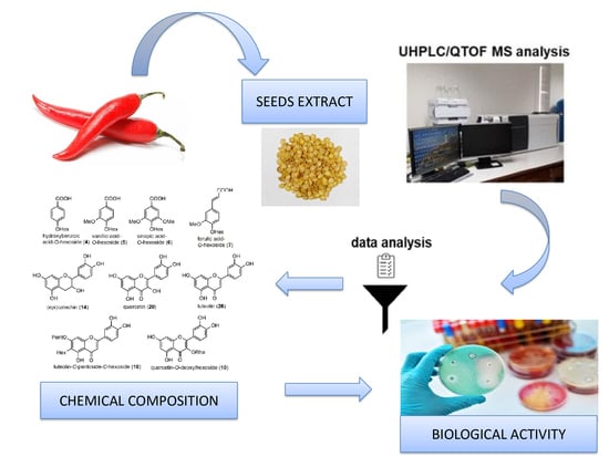
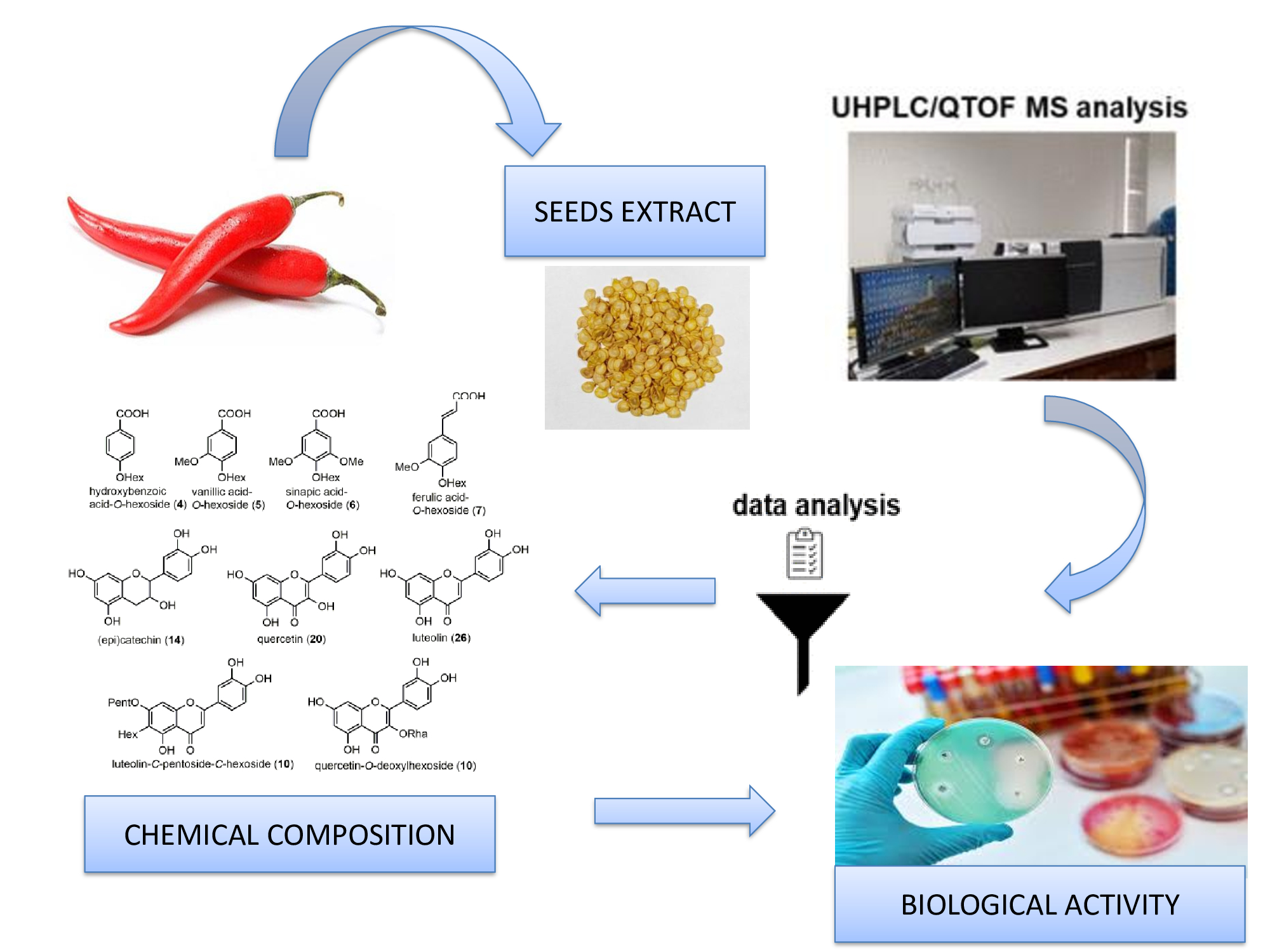
Untargeted Phytochemical Profiling, Antioxidant, and Antimicrobial Activities of a Tunisian Capsicum annuum Cultivar

1
Laboratory of Electrochemistry and Environment, National School of Engineers of Sfax, University of Sfax-Tunisia, B.P “1173”, Sfax 3038, Tunisia
2
Faculty of Sciences and Technology of SidiBouzid, University of Kairouan, Campus University Agricultural City, Sidi Bouzid 9100, Tunisia
3
Laboratory of Plant-Soil-Environment Interactions, LR21ES01, Faculty of Sciences of Tunis, University of Tunis EL Manar, Tunis 2092, Tunisia
4
Biothermodynamics, TUM School of Life Sciences, Technical University of Munich, 85354 Freising, Germany
5
Department of Pharmacognosy and Phytotherapy, Grigore T. Popa University of Medicine and Pharmacy, 700115 Iasi, Romania
6
Department of Chemistry of Natural Products, Medical University of Lublin, 20-093 Lublin, Poland
7
Laboratory of Medicinal and Environmental Chemistry, High Institute of Biotechnology of Sfax, University of Sfax, Sfax 3038, Tunisia
8
Higher Institute of Biotechnology of Sfax, University of Sfax, B.P “1175”, Sfax 3038, Tunisia
*
Authors to whom correspondence should be addressed.
These authors contributed equally to this work.
Molecules 2023, 28(17), 6346; https://doi.org/10.3390/molecules28176346
Submission received: 28 July 2023 / Revised: 20 August 2023 / Accepted: 25 August 2023 / Published: 30 August 2023
(This article belongs to the Special Issue Discovery, Isolation, and Mechanisms of Bioactive Natural Products)

Abstract

:
Peppers are among the spices possessing a wide plethora of biological properties due to their excellent supply of health-related metabolites. Capsicum annuum L. (Solanaceae) is cultivated throughout Tunisia, and there is a shortage of information on the identification of the secondary metabolites in the seeds of this species as well as on their biological activities. In the present work, we intended to undertake a chemical characterization of the bioactive compounds from the hydro-methanolic seed extract of C. annuum as well as an evaluation of its broad spectrum of antimicrobial and antioxidant activities. The chemical profile was evaluated by RP-HPLC-DAD-QTOF-MS/MS, whereas the total phenol and flavonoid content, antioxidant, and antimicrobial activities were determined in in vitro assays. In this work, 45 compounds belonging to various phytochemical classes, such as organic acids (2), phenolic compounds (4 phenolic acids and 5 flavonoids), capsaicinoids (3), capsianosides (5), fatty acids (13), amino acids (1), sphingolipids (10), and steroids (2) were identified in the hydro-methanolic seed extract of C. annuum. The phenolic and flavonoid content (193.7 mg GAE/g DW and 25.1 mg QE/g DW, respectively) of the C. annuum extract correlated with the high antiradical activity (IC50 = 45.0 µg/mL), reducing power (EC50 = 61.3 µg/mL) and chelating power (IC50 = 79.0 µg/mL) activities. The hydro-methanolic seed extract showed an important antimicrobial activity against seven bacterial and four fungal strains. In fact, the inhibition zones (IZs) for bacteria ranged from 9.00 ± 1.00 mm to 12.00 ± 0.00 mm; for fungi, the IZs ranged from 12.66 ± 0.57 mm to 13.66 ± 0.57 mm. The minimal inhibition concentration and minimal bactericidal concentration values showed that the extract was more effective against fungi than bacteria.

Graphical Abstract

1. Introduction

The genus Capsicum belongs to the Solanaceae family and consists of about 27 species [1]. It was considered a typical American genus, and several species of Capsicum were cultivated for thousands of years by pre-Columbian civilizations [2]. Capsicum species are popular vegetables and spices grown worldwide, especially in tropical and subtropical countries [3]. Based on the taxonomic identification of these plant species (peppers), four varieties are recognized, namely, Capsicum annuum var. abbreviatum, Capsicum annuum var. acuminatum, Capsicum annuum var. grossum, and Capsicum frutescens var. baccatum [4].
In Tunisia, pepper cultivars are mainly represented by C. annuum species, with a number of chili pepper landraces [5] cultivated throughout the country, in addition to a few C. frutescens cultivars [6,7]. Interest in peppers has increased not only because of their high nutritional value as a vegetable, food ingredient, and coloring agent in the food industry [8,9,10] but also for their cosmetic and medical uses [11,12]. Indeed, pepper is recognized as an excellent source of health-related metabolites, such as ascorbic acid (vitamin C), carotenoids (provitamin A), tocopherols (vitamin E), polyphenols, and capsaicinoids [13]. Polyphenols, including flavonoids, are secondary metabolites that are abundant in plants. It is known that these components play a protective role against pathogens and UVB light in the interaction between plants and the environment [14,15]. Therefore, polyphenolic compounds have attracted a great deal of attention, since they act as antioxidants and protect the human body from oxidative stress, which is the main cause of different degenerative processes. Thus, consuming fruits and vegetables is reversely correlated with the development of chronic diseases [16]. Due to their health benefits, polyphenols have gained a great deal of attention, especially in vegetables such as peppers that are consumed in large amounts worldwide. Thus, peppers are among the vegetables that provide a rich source of various bioactive compounds with potential biological properties. For example, the appreciable amount of phenols and flavonoids in the ethanolic extracts of C. annuum contributed to their antiradical activities [17]. In another study, methanolic extracts from C. annuum were reported to inhibit 4-hydroxy-2-nonenal- and H2O2-induced DNA damage [18].
On the other hand, only a few studies have shown that C. annuum has antimicrobial activities against different microorganisms [19,20,21]. Koffi-Nevry et al. [19] demonstrated that C. annuum exhibited inhibitory activity against Escherichia coli, Vibrio cholerae, Staphylococcus aureus, and Pseudomonas aeruginosa. Moreover, Sree Sandhya and Vijayakumar [20] reported that an ethanolic extract of C. annuum var. glabriusculum inhibited two microbial strains (Staphylococcus aureus and Streptococcus mutans). More recently, aqueous, ethanolic, and ethyl acetate extracts of C. annuum were tested against several pathogenic fungal strains (Alternria sp., Penicillium sp., Fusarium sp., Aspergillus flavus, and Aspergillus niger). It was demonstrated that these extracts inhibited all the strains used except for Aspergillus niger [21].
Furthermore, there is a shortage of information on the identification of secondary metabolites in the seeds of the Tunisian C. annuum cultivar as well as on their antioxidant activities. To the best of our knowledge, only one study was carried out by LC-MS/MS on the identification of phenolic compounds in the ethanolic seed extract of three types of Australian-grown bell peppers (green, red, and yellow) and the estimation of their antioxidant potential [22]. As a valorization of the by-products of C. annuum (seeds) discarded by a Tunisian harissa factory located in the northeast of Tunisia (Cap Bon), this study was geared towardsa chemical characterization of the bioactive compounds from a hydro-methanolic seed extract of C. annuum as well as an evaluation of its broad spectrum of antioxidant and antimicrobial activities.

2. Results and Discussion

2.1. Metabolite Characterization of C. annuum Seed Extract by RP-HPLC-DAD-QTOF-MS/MS

In the present work, the metabolite profiling of the hydro-methanolic extract of C. annuum seeds was established using RP-HPLC-DAD-QTOF-MS/MS. Figure 1A,B presents the base peak chromatogram (BPC) profiles of the analyzed extract in both negative (A) and positive (B) ionization modes. The peak numbers are given following the elution order (retention time, TR), as listed in Table 1. Table 1 also summarizes the experimental m/z of the precursor ion, molecular formula, and main MS/MS fragments in the negative or positive ionization modes. The identified compounds were classified into various groups, as follows: organic acids (2), phenolic compounds (4 phenolic acids and 5 flavonoids), capsaicinoids (3), capsianosides (5), fatty acids (13), amino acids (1), sphingolipids (10), and steroids (2). Using the abovementioned method, it was possible to identify 45 compounds belonging to various phytochemical classes in the C. annuum hydro-methanolic seed extract.

2.1.1. Organic Acids

In the polar region of the BPC (Figure 1A), two organic acids with low molecular masses were identified in the hydro-methanolic extract of C. annuum seeds (negative ionization mode). Galactonic/gluconic acid (peak 1, C6H12O7) was detected as the precursor ion [M−H] at m/z 195.0563. Its MS/MS spectra showed fragment ions at m/z 177.0444 [M–H–H2O], 159.0358 [M–H–2H2O], 129.0233 [M–H–2H2O–CH2O]. In addition, RP-HPLC-DAD-QTOF-MS was helpful in the identification of citric acid (peak 3) with the precursor ion [M−H] at m/z 191.0241 and the molecular formula C6H8O7. Moreover, the structure of this organic acid was confirmed by the presence of the diagnostic fragment ion at m/z 111.0409, corresponding to [M–H–CO2–2H2O] in its MS/MS spectra. Citric acid was previously described in the literature; its fragmentation patterns were in accordance with previous studies [23,24,25]. In fact, citric acid was identified in many Balkan pepper accessions [25] as well as in a population of wild Piquin Chili (C. annuum var. glabriusculum) [26].

2.1.2. Phenolic Compounds

Two sub-classes of phenolic compounds were characterized in the negative ionization mode, namely, phenolic acids and flavonoids (Figure 2). In the case of phenolic acids, four compounds were identified in the BPC (retention time range 10.2–16.5 min). Peak 4, with the pseudo-molecular ion [M−H] at m/z 299.0845 (C13H16O8), was assigned to hydroxybenzoic acid-O-hexoside. It is worth mentioning that this compound was previously reported in peppers such as C. chinense [27] and C. annuum [28]. A derivative of this compound, namely, vanillic acid-O-hexoside (peak 5), was shown to possess the precursor ion at m/z 329.0943 (C13H16O8), indicating the presence of an additional methoxy group. Furthermore, two hydroxycinnamic acid derivatives were assigned, sinapic acid-O-glucoside (peak 6, [M−H] at m/z 385.1938) and ferulic acid-O-hexoside (peak 7, [M−H] at m/z 355.1106). The MS/MS spectra of the latter compound showed two abundant fragment ions at m/z 193.0542 [M–H–hexosyl] and 175.0440 [M–H–hexosyl–H2O]; these fragments are in agreement with those reported by Leng et al. [22]. Both compounds were previously detected in Capsicum peppers [27,29].
The second class of phenolic compounds detected in the present study was flavonoids (Table 1). They are among the most important phenolic compounds distributed mainly in the Capsicum genus, such as C. annuum var. glabriusculum [26] and C. chinense [27]. Five compounds belonging to various flavonoid classes (flavones, flavanols, and flavonols) were identified in the seeds of C. annuum. In fact, two luteolin derivatives were detected, namely luteolin-O-pentoside-C-hexoside (peak 10, [M−H] at m/z 579.1437) and its free aglycon luteolin (peak 26, [M−H] at m/z 285.0407). The glycosylated derivative showed, in its MS/MS spectrum, the loss of the pentosyl group (−132 Da), indicating the O-glycosylation with pentose. However, the subsequent fragment ion at m/z 327.0554 was obtained from the previous ion by the diagnostic removal of a group with 120 Da, indicative of a C-glycosylation with a hexose unit. Luteolin and its derivative were previously reported in Capsicum peppers by several authors [27,29,30]. Three other flavonoid peaks were detected in the hydro-methanolic extract of C. annuum seeds. (Epi)catechin (peak 14), with the precursor ion [M−H] at m/z 289.1038 (C15H14O6), showed diagnostic fragments at m/z 245.0865 [M–H–CO2] and 205.0511 [M–H–C4H4O2], which were consistent with previous studies [22,31,32]. This compound was previously found in the ethanolic seed extracts of three types of Australian-grown bell peppers [27]. Quercetin-O-deoxyhexoside (peak 11) showed a precursor ion [M−H] at m/z 447.1011 (C21H20O11), which was fragmented to m/z 271.0443, 255.0404, and 179.0086, as mentioned by Jeong et al. [33]. Quercetin (peak 20) exhibited a precursor ion [M−H] at m/z 301.0383 (C15H10O7) which generated the specific fragments at m/z 273.0376 and 178.9983, as cited by Santos et al. [30] and Schelz et al. [34]. Previously, quercetin-3-O-rhamnoside was found in C. annuum fruits [27], whereas quercetin was reported in the ethanolic extract of C. chinense ripe fruits [30], as well as in the methanolic extract of the unripe fruit of C. annuum [33].

2.1.3. Capsaicinoids

All capsaicinoids (amides) were characterized in the positive ionization mode, which showed the protonated molecule ions [M+H]+ of norhydrocapsaicin (peak 32, m/z at 294.2072), capsaicin (peak 34, m/z at 306.2069), and dihydrocapsaicin (peak 37, m/z at 308.2229), as shown in Table 1. Capsaicin and dihydrocapsaicin, which differ only by a double bond on their lateral carbonic chain (Figure 3), showed identical fragments at m/z 137.0579. However, capsaicin and dihydrocapsaicin showed a characteristic fragment at m/z 182.1524 and 184.1668, respectively. The importance of these compounds is related to several factors. On the one hand, they are considered to be the main active ingredients in C. annuum seeds [28]; hence, they are valuable pharmaceutical ingredients. On the other hand, they are responsible for the hot taste of peppers [29,30].

2.1.4. Capsianosides

Capsianosides are a large group of diterpenic glycosides characteristic of Capsicum peppers [35]. In the current study, five capsianoside derivatives (Figure 3) were putatively labeled by RP-HPLC-DAD-QTOF-MS/MS (negative ionization mode) in the hydro-methanolic extract of C. annuum seeds (Table 1), namely, capsianoside III (peak 16, C50H84O26), capsianoside IX (peak 18, C44H74O21), capsianoside XV (peak 19, C50H84O26), capsianoside II (peak 22, C50H84O25), andcapsianoside VIII (peak 25, C50H84O25). In their MS/MS spectra, the removal of the corresponding sugar units was diagnostically observed. For instance, in the case of capsianosides III and XV with [M−H] at m/z 1099.5317 and 1099.5379, respectively, the following fragment ions were noticed at m/z: 937.4759 [M–hexosyl–H], 775.4192 [M–2hexosyl–H], 629.3666 [M–2hexosyl–deoxyhexosyl–H], and 467.2929 [M–3hexosyl–deoxyhexosyl–H].

2.1.5. Fatty Acids

Thirteen oxygenated fatty acids were detected in the negative ionization mode in the non-polar region of the chromatogram (retention times from 34.5 to 59.3 min). Practically, all compounds were derived from octadecanoic acid (C18), with the differences residing in the number of oxygenated functions and double bonds; their m/z values ([M−H]) ranged from 293.2203 and 331.2124. Thus, acids with one double bond (e.g., trihydroxyoctadecenoic acid, dihydroxyoctadecenoic acid), two double bonds (e.g., hydroperoxyoctadecadienoic acid, hydroxyoctadecadienoic acid) and three double bonds (e.g., hydroxyoctadecatrienoic acid) were putatively identified in the C. annuum seed extract (Table 1). Fatty acids reported in our study were also previously documented in C. chinense extracts [36].

2.1.6. Amino Acids and Amino Alcohols (Sphingolipids)

One amino acid and 10 amino alcohols (sphingolipids) were tentatively assigned by RP-HPLC-DAD-QTOF-MS/MS (positive ionization mode) in the hydro-methanolic extract of C. annuum seeds (Table 1). The precursor ion [M+H]+ at m/z 294.1539 (C12H23NO7) and MS/MS fragment ions at m/z 258.1321, 230.1388, 144.0990, and 114.0984 were in agreement with the spectrometric data reported by Menezes et al. [36] for N-fructosyl(iso)leucine (peak 2).
Sphingolipids are structurally derived from fatty alcohols with one vicinal amino group and several additional hydroxyl groups (Figure 4). In the C. annuum extract, compounds with C12 (2-aminododecane-1,3-diol), C14 (tetradecaphytosphingosine, tetradecasphinganine), C16 (hexadecaphytosphingosine, hexadecasphinganine), C18 (phytosphingosine, sphinganine), and C20 (N-hydroxy arachidonoyl amine) atoms were tentatively identified. Since each derivative differed from the previous one by two carbon atoms and/or an extra hydroxyl group, these structural differences were easily noticeable in the MS spectra, which allowed for the annotation of the molecular formulas. The MS/MS fragmentation patterns of these amino alcohols generally show the characteristic loss of a hydroxyl group [M–H–H2O]+. Similar sphingolipids, such as sphinganine 1-phosphate, phytosphingosine, sphinganine 1-phosphate, sphinganine, 2-aminoicosane-1,3-diol, hexadecasphinganine, and soyacerebroside, were previously reported in C. annuum by Guevara et al. [37] and Cervantes-Hernandez et al. [38].

2.1.7. Steroids

Lastly, two steroidal glycosidic saponins were putatively annotated in the analyzed extract. Capsicoside A (peak 12, [M−H] at m/z 1421.6593) showed in its MS/MS spectra the successive loss of the sugar units bound to its aglycon, as follows: m/z 1259.5962 [M–hexosyl–H], 1097.4411 [M–2hexosyl–H], 935.4233 [M–3hexosyl–H], 773.4179 [M–4hexosyl-H]. Similarly, protodegalactotigonin (peak 17, [M−H] at m/z 1213.6011) presented an MS/MS fragment ion at m/z 1081.5401, 919.5061, and 757.4474, derived by the neutral successive cleavage of one pentose and two hexose units. These two steroidal glycosides were previously reported in C. annuum by Yahara et al. [39].

2.2. Total Phenolic Content, Flavonoid Content, and Antioxidant Activities of C. annuum Seed Extract

Table 2 presents the results for the polyphenol and flavonoid content of the C. annuum hydro-methanolic extract, as well as its antioxidant activities. In our study, we used three complementary methods for antioxidant activity determination, such as DPPH, reducing power, and chelating power assay [40]. As compared to BHT (11.5 µg/mL), vitamin C (37.0 µg/mL), and EDTA (32.5 µg/mL), the moderate polyphenols and flavonoids content (193.7 mg GAE/g DW and 25.1 mg QE/g DW, respectively) of the C. annuum hydro-methanolic extract necessarily show relatively high antiradical activity (IC50 = 45.0 µg/mL), reducing power (EC50 = 61.3 µg/mL) and chelating power (IC50 = 79.0 µg/mL) activities. Compared to other varieties of C. annuum species [4], the hydro-methanolic seed extract of C.annuum is relatively low in its content of polyphenols and flavonoids. In fact, the polyphenols content of the ethanolic extract of mature fruits of C. annuum var. abbreviatum, C. annuum var. acuminatum, C. annuum var. grossum ranged from 200.70 to 272.74 mg GAE/g DW, whereas the flavonoid content of the same extracts varied between 1223.71 and 1630.53 mg QE/g DW [4]. Moreover, the DPPH radical scavenging activities of this extract changed in the order: C. annuum var. abbreviatum > C. annuum var. acuminatum > C. annuum var. grossum [4].

2.3. Antimicrobial Activity of C. annuum Extract

The data presented in Table 3 demonstrate the inhibitory diameter zones (IZs) for both bacterial and fungal strains. Specifically, for the hydro-methanolic seed extract, the IZs range from 9.00 ± 1.00 mm to 12.00 ± 0.00 mm for bacterial strains and from 12.66 ± 0.57 mm to 13.66 ± 0.57 mm for fungal strains. On the other hand, for the reference antibiotic gentamycin, the IZs span from 21.33 ± 0.58 mm to 27.67 ± 1.53 mm for bacterial strains, and for amphotericin B, the range is 16.00 ± 0.00 mm to 18.00 ± 0.00 mm for fungal strains.
Upon conducting the statistical analysis, it was observed that the hydro-methanolic seed extract of C. annuum exhibited lower efficacy than the reference antibiotics. This suggests that the extract may not be as potent in inhibiting the growth of bacterial and fungal strains. Furthermore, the statistical analysis (p < 0.05) of the inhibition diameters indicates significant differences in the resistance of the strains against the hydro-methanolic seed extract. Interestingly, the Gram-negative bacteria displayed the highest sensitivity to the extract, as evidenced by the larger inhibitory zone observed for this group. Overall, these findings suggest that the hydro-methanolic seed extract of C. annuum may have limited antimicrobial effectiveness, particularly against Gram-negative bacteria. Further investigations and studies may be warranted to better understand the potential applications and limitations of this extract as an antimicrobial agent.
The quantitative method (Table 3) reveals the MIC and MBC values for the hydro-methanolic seed extract. The MIC values range from 0.15 mg/mL (for E. faecalis and M. luteus) to 1.875 mg/mL (for S. epidermidis, B. cereus, L. monocytogenes, and S. typhimurium), while the MBC values vary from 1.875 mg/mL (for M. luteus) to 7.50 mg/mL (for E. coli). In contrast, for the fungal strains, the MIC and MFC values were comparatively lower. This outcome indicates a higher level of sensitivity to the C. annuum hydro-methanolic seed extract among the tested bacterial strains. However, when comparing these values, it becomes apparent that the antibiotics or antifungal agents seem to exhibit higher activity than the hydro-methanolic seed extract.
The MBC/MIC and MFC/MIC ratios were utilized to gain insights into the antimicrobial effect of the hydro-methanolic seed extract. Based on the classification provided by Schaechter et al. [41] and Soro et al. [42], a ratio greater than four indicates a bacteriostatic or fungistatic effect, whereas a ratio equal to or lower than four suggests a bactericidal or fungicidal effect. Upon examining the ratios presented in Table 3, it becomes evident that the hydro-methanolic seed extract displayed bactericidal and fungicidal properties against all the tested strains. This finding indicates that the extract is effective in not only inhibiting the growth (bacteriostatic/fungistatic) but also in killing the bacteria and fungi (bactericidal/fungicidal) at or below the concentrations tested.
The observed antimicrobial activity of the hydro-methanolic seed extract can be attributed to its chemical composition, particularly its high content of phenolic compounds (193.7 ± 3.1 mg GAE/g). The RP-HPLC-DAD-QTOF-MS/MS analysis of the Tunisian C. annuum hydro-methanolic seed extract revealed the presence of several compounds known for their biological activity, which likely contribute to the extract’s antimicrobial properties. Some of these bioactive compounds include citric acid, hydroxybenzoic acid-O-hexoside, ferulic acid-O-hexoside, quercetin-O-deoxyhexoside, (epi)catechin, quercetin, luteolin, and capsaicin. Each of these compounds was previously studied for its potential antimicrobial effects, and their presence in the extract may synergistically enhance its overall antimicrobial activity against the tested bacterial and fungal strains. Citric acid is known for its acidic and chelating properties, which can inhibit bacterial growth [19,43,44,45]. Hydroxybenzoic acid-O-hexoside and ferulic acid-O-hexoside are phenolic compounds with reported antimicrobial and antioxidant properties [46,47]. Quercetin-O-deoxyhexoside, (epi)catechin, quercetin, and luteolin are flavonoids known for their antibacterial and antifungal activities. Capsaicin, a compound found in chili peppers, has also demonstrated antimicrobial properties against various pathogens [48].
The combined presence of these bioactive compounds in the hydro-methanolic seed extract likely contributes to its broad-spectrum antimicrobial efficacy against both bacterial and fungal strains. It is important to note that the synergistic interactions among these compounds and their individual concentrations can significantly influence the overall antimicrobial effectiveness of the extract.

3. Materials and Methods

3.1. Plant Material and Extraction Procedure

Mature fruits of Capsicum annuum were harvested from a farmer’s field in the CHAFFAR region (MAHRES, Sfax, Tunisia) at the end of September 2020. The fruits were air-dried at room temperature (25 °C) for 15 days. Then, the corresponding seeds were extracted from dried fruit and crushed into fine powder. Regarding the extraction procedure, 10 g of powder seeds were put in an amber glass bottle and homogenized in 100 mL of a mixture of methanol/water 80:20 (v/v) using an ultrasonic bath for 30 min at a power of 2500 W. After filtration with Whatman filter paper No. 42 (125 mm), the extract was evaporated with a rotary evaporator under vacuum at 40 °C. Finally, the dry extract was obtained with a yield of 7.6% and was kept at −20 °C until future analysis.

3.2. RP-HPLC–DAD-QTOF-MS/MS Analysis

The Capsicum annuum seed extract was analyzed by RP-HPLC-DAD-QTOF-MS/MS using the procedures described by Ammar et al. [49]. The chromatographic analytical procedures were performed on an Agilent 1200 Series (Agilent Technologies, Palo Alto, CA, USA) LC system coupled to a DAD and an Agilent 6540 Ultra-High-Definition (UHD) Accurate-Mass QTOF with a Jet Stream dual ESI interface. The instrument was equipped with a vacuum degasser, a binary pump, an autosampler with a thermostat, and a column compartment. The separations were carried out on a Phenomenex Gemini RP-18 (100 mm × 2 mm; i.d. 3 μm; Phenomenex, Torrance, CA, USA) column maintained at 20 °C. The mobile phase consisted of 0.1% formic acid in water (solvent A) and 0.1% formic acid in acetonitrile (solvent B). The elution gradient was used as follows: 0 min, 1% B; 13 min, 25% B; 20 min, 25% B; 25 min, 40% B; 30 min, 60% B; 35 min, 100% B; 40 min, 100% B; post-time 12 min. The flow rate was 0.20 mL/min. The ultraviolet (UV) spectra were recorded from 190 to 600 nm. The sample was diluted with a methanol/water mix of 80:20 (v/v) prior to the injection of 10 μL sample volume. The MS analyses were carried out using the following operating conditions: drying nitrogen temperature at 350 °C with a flow of 12 L/min; nebulizer pressure 40 psi; sheath gas temperature 400 °C with a flow of 12 L/min; capillary voltage, skimmer, and radiofrequency voltages of 4000, 645 and 750 V, respectively. The spectra were acquired in negative and positive ionization modes over a mass-to-charge (m/z) range of 100 to 1000. The reference mass correction of the sample was performed with a continuous infusion of Agilent API TOF reference mixture (61969–85001). The data analysis was carried out with Mass Hunter Qualitative Analysis B.06.00 (Agilent Technologies) software that enabled the generation of the molecular formula with a mass accuracy limit of 5 ppm and an MS ≥ 80 (related to the contribution to mass accuracy, isotope abundance, and isotope). For the retrieval of the chemical structure information, some databases were consulted as follows: PubChem (http://pubchem.ncbi.nlm.nih.gov), ChemSpider (http://www.chemspider.com), SciFinderScholar (https://scifinder.cas.org), Reaxys (http://www.reaxys.com), Phenol-Explorer (www.phenol-explorer.eu) and KNApSAcK Core System (http://kanaya.naist.jp/knapsackjsp/top.html) (accessed on 5 August 2023).

3.3. Total Polyphenol Compounds Analysis

Total polyphenol compounds were determined colorimetrically with Folin–Ciocalteu’s reagent according to the method of Gargouri et al. [50], with some modifications. Briefly, 50 μL of the suitable sample dilution was added to 250 μL of the Folin–Ciocalteu reagent. The mixture was shaken before adding 500 μL of Na2CO3 (20%) solution, adjusting with distilled water to a final volume of 5 mL, and mixed thoroughly. After incubation of the mixture for 30 min at 25 °C in darkness, the absorbance versus a prepared blank was read at 727 nm. A standard curve of gallic acid was used. The total phenolic content of the extract was expressed as mg gallic acid equivalents per gram of dry weight (mg GAE/g DW) through a calibration curve with gallic acid. The calibration curve range was 0–160 μg/mL (R2 = 0.98). The sample was analyzed in three replicates.

3.4. Total Flavonoid Content Analysis

The total flavonoid content was determined with AlCl3 reagent, according to Mouhamadi et al. [51], with some modifications. An amount of 1 mL of the diluted sample (500 mg/L in methanol) was added to 4 mL of distilled water and 300 μL of the NaNO2 solution (50%) and mixed for 6 min before adding 300 μL of AlCl3 (10%). After 5 min, 2 mL of NaOH (1M) was added. The final volume was adjusted to 10 mL with distilled water and thoroughly mixed. The absorbance of the mixture was determined at 510 nm against the same mixture, without the sample, as a blank. The total flavonoid content was expressed as mg quercetin/g dry weight (mg QE/g DW) through a calibration curve of quercetin. The calibration curve range was 0–120 μg/mL (R2 = 0.99). The sample was analyzed in three replicates.

3.5. Antioxidant Activity

3.5.1. DPPH Radical Scavenging Ability Assay

DPPH quenching ability of the hydro-methanolic extract was measured according to Bouaziz et al. [52]. Briefly, a volume of 500 μL of each sample at different concentrations (10 to 100 µg/mL) was added to 375 μL of 99% ethanol and 125 μL of DPPH solution (0.02% in ethanol) as the free radical source. The obtained mixtures were shaken and then incubated for 60 min in the dark at room temperature. The measurement of the scavenging capacity was carried out spectrophotometrically by controlling the decrease of absorbance at 517 nm. The DPPH, in its radical form, has an absorption band at 517 nm, which vanishes upon reduction by an antiradical compound. A low absorbance of the reaction mixture reveals high DPPH free radicalscavenging activity. BHT was used as a positive control, and the calculation of DPPH radicalscavenging activity was performed as follows:
%   scavenging   effect = A D P P H A E A D P P H   ×   100
where AE denotes the absorbance of the solution when the sample extract is added at a specific level, and ADPPH is the absorbance of the DPPH solution.
The antiradical activity was expressed as IC50 (µg/mL), the extract dose required to cause a 50% inhibition.

3.5.2. Reducing Power

The ability of the extract to reduce Fe3+ was assayed using the method described by Yildirim et al. [53]. Briefly, 1 mL of the hydro-methanolic extract at different concentrations was mixed with 2.5 mL of phosphate buffer (0.2M, pH 6.6) and 2.5 mL of 1% K3Fe(CN)6. The resulting mixture was incubated for 20 min at 50 °C. After the addition of 2.5 mL of 10% (w/v) trichloroacetic acid, the solution was manually shaken. Lastly, 2.5 mL of the supernatant solution was mixed with 2.5 mL of distilled water and 500 µL of 0.1% (w/v) ferric chloride. After 10 min, the absorbance was measured at 700 nm. The EC50 value (µg/mL) is the effective concentration at which absorbance was 0.5 for reducing power. BHT and Vitamin C were used as the positive control.

3.5.3. Chelating Effect on Ferrous Ions

The iron chelating activity of the different samples was estimated according to the protocol described by Dhouibi et al. [54], with slight modifications. Indeed, 50 μL of 2mM FeCl2, 4H2O was added to 100 μL of the extract at different concentrations (10 to 100 µg/mL) diluted in 450 μL of water. The obtained mixtures were incubated at room temperature for 5 min. The reactions were started by adding 200 μL of 5 mM of 3-(2-pyridyl)-5,6-bis (4-phenyl-sulfonic acid)-1,2,4-triazine (ferrozine). The mixtures were then strongly shaken and were left to stand at room temperature for 10 min. Similarly, the control tube was prepared, replacing the sample with distilled water. EDTA was used as the positive control.
The solutions absorbance was measured at 562 nm, and the inhibition percentage of ferrozine-Fe2+ complex formation was calculated as follows:
Metal   chelating   activity   %   Ab = ( A C + A B A S AC )   ×   100
where AC, AB and AS are the control absorbance, the blank, and the sample reaction tubes, respectively.

3.6. Antimicrobial Activities

3.6.1. Microorganisms

The bacterial strains were divided into 5 Gram-positive (S. epidermidis CIP 106510, E. feacalis ATCC 29212, M. luteus NCIMB 8166, B. cereus ATCC 11778, L. monocytogenes ATCC19115) and 2 Gram-negative bacteria (E. coli ATCC 35218, S. typhimurium LT2 DT104). The fungal species belonged to 4 Candida strains (C. albicans ATCC 90028; C. glabrata ATCC 90030; C. parapsilosis ATCC 22019; C. krusei ATCC 6258). These strains were chosen for their ability to cause serious human infections.

3.6.2. Disc-Diffusion Assay

The antimicrobial activity testing was done according to the protocol described by Ben Bnina et al. [55]. For the experiments, a small amount of the microorganism’s working stocks was added to a tube containing 9 mL of Mueller–Hinton broth (for bacteria) and Sabouraudchloramphenicol broth (for yeast strains). The mixture was then incubated at 37 °C for 18 to 24 h. The resulting overnight cultures were utilized to assess the antimicrobial activity of the extract in this study. The optical density was adjusted to 0.5 McFarland turbidity standards using a DENSIMAT (Biomérieux®, Marcy-l’Étoile, France). The respective bacteria and fungi were streaked onto MH or SB agar plates using a sterile swab.
Sterile filter discs with a diameter of 6 mm, made from Whatman paper No. 3, were soaked in an extract solution with a concentration of 150 mg/mL. These impregnated discs were then placed onto the appropriate agar media, which included the Sabouraud chloramphenicol broth (SB) and the Mueller–Hinton broth (MH). Gentamycin (10 μg/disc) and Amphotericin B (20 μg/disc) were used as positive reference standards. They were employed to gauge the susceptibility of a particular strain or isolate to each of the tested microbial species.
Following an incubation period at 37 °C lasting 18 to 24 h, the diameter of the inhibition zone around each disc was measured using a 1 mm flat rule. These measurements were interpreted according to the guidelines provided by the Committee of the French Society of the Antibiogram [56]. The dishes were kept in an incubator at 37 °C for 18 to 24 h for the microbial strains to develop. The extent of the inhibition zones surrounding each disc served as a measure of the antimicrobial activity. Every experiment was conducted three times (triplicate), and the average diameter of the inhibition zones was recorded.

3.6.3. Micro-Well Determination of MIC, MBC and MFC

Minimal inhibition concentration (MIC), minimal bactericidal concentration (MBC), and minimal fungicidal concentration (MFC) values were determined, as described by Zanati et al. [57], for all bacterial and fungal strains used in this study. A 100 μL aliquot from stock solutions of the extract was added into the first wells. Then, 100 μL from the serial dilutions were transferred into eleven consecutive wells. The last well containing 195 μL of the nutrient broth without the extract and 5 μL of the inoculum on each strip was used as the negative control. The final volume in each well was 200 μL. The plates were incubated at 37 °C for 18–24 h. The extract tested in this study was screened two times against each organism. The MIC (Minimal Inhibition Concentration) value was defined as the lowest concentration of the compounds to inhibit the growth of the microorganisms. The MBC (Minimal Bactericidal Concentration) and MFC (Minimal Fungicidal Concentration) values were interpreted as the highest dilution (lowest concentration) of the sample, which showed clear fluid with no development of turbidity and without visible growth. Furthermore, we determined the MBC/MIC and MFC/MIC ratios to better understand the potential bacteriostatic or bactericidal effects of our extract. Each test was carried out on a single occasion. According to the categorization provided by Schaechter et al. [41] and Soro et al. [42], if the ratio surpasses 4, it signifies a bacteriostatic or fungistatic effect. Conversely, if the ratio is 4 or lower, it suggests a bactericidal or fungicidal effect.

3.7. Statistical Analysis

The experiments (antioxidant and antimicrobial activities) were conducted in triplicates, and the average values were calculated using the SPSS 25.0 statistical package for Windows. Differences in means were analyzed using Duncan’s multiple-range tests with a 95% confidence interval (p ≤ 0.05).

4. Conclusions

Overall, the Tunisian C. annuum seed cultivar was proven to be abundant in various secondary metabolites endowed with considerable antioxidant potential as well as an important antimicrobial activity against seven bacterial and four fungal strains. The identified compounds in the hydro-methanolic seed extract of C. annuum belonged to various phytochemical classes, such as organic acids, phenolic compounds, capsaicinoids, capsianosides, fatty acids, amino acids, sphingolipids, and steroids. The combined presence of these bioactive compounds in this extract likely contributes to its broad-spectrum antimicrobial efficacy against both bacterial and fungal strains. Further research and studies may be needed to determine the exact mechanisms of action and potential applications of this extract as a natural antimicrobial agent.

Author Contributions

Conceptualization, Y.G., H.H., S.V.L., K.S.-W. and M.B.; formal analysis, J.A. and S.V.L.; investigation, M.B.; methodology, Y.G. and M.B.; project administration, K.S.-W. and M.B.; supervision and validation, K.S.-W. and M.B.; visualization, S.Z.; writing—original draft, Y.G. and S.Z.; writing—review and editing, Y.G., H.H., S.V.L., K.S.-W., S.Z. and M.B. All authors have read and agreed to the published version of the manuscript.

Funding

This research was funded by the Ministry of Higher Education and Scientific Research (Laboratory LR14ES08): Tunisia.

Institutional Review Board Statement

Not applicable.

Informed Consent Statement

Not applicable.

Data Availability Statement

The data presented in this study are available on request from thecorresponding author.

Conflicts of Interest

The authors declare no conflict of interest.

Sample Availability

Samples of the compounds are available from the authors.

References

  1. Bosland, P.W.; Votava, E.J. Peppers: Vegetable and Spice Capsicums; CABI Publishing: Wallingford, UK, 2000. [Google Scholar]
  2. da Veiga, V.F., Jr.; Wiedemann, L.S.M.; de Araujo, C.P., Jr.; da Silva Antonio, A. Chapter 1: Origin and Evolution of Capsicum. In Chemistry and Nutritional Effects of Capsicum; Royal Society of Chemistry: London, UK, 2022; pp. 1–14. [Google Scholar]
  3. Dimitrios, B. Sources of natural phenolic antioxidants. Trends Food Sci. 2006, 17, 505–512. [Google Scholar] [CrossRef]
  4. Olatunji, T.L.; Afolayan, A.J. Comparative quantitative study on phytochemicalcontents and antioxidant activities of Capsicum annuum L. and Capsicum frutescens L. Sci. World J. 2019, 2019, 4705140. [Google Scholar] [CrossRef]
  5. Lahbib, K.; Bnejdi, F.; ElGazzah, M. Selection of pepper parent from a collection of Capsicum annuum landraces on genetic diversity. J. Plant Breed. Crop Sci. 2013, 5, 68–72. [Google Scholar] [CrossRef]
  6. Zhani, K.; Hamdi, W.; Sedraoui, S.; Fendri, R.; Lajim, O.; Hannachi, C. Agronomic evaluation of Tunisian accessions of chili pepper (Capsicum frutescens L.). Int. Res. J. Eng. Technol. 2015, 2, 28–34. [Google Scholar]
  7. Zhani, K.; Hamdi, W.; Sedraoui, S.; Fendri, R.; Lajim, O.; Hannachi, C. A comparative study of morphological characterization of Tunisian accessions of Chili pepper (Capsicum frutescens L.). Int. Res. J. Eng. Technol. 2015, 2, 87–94. [Google Scholar]
  8. Loizzo, M.R.; Pugliese, A.; Bones, M.; Menichini, F.; Tundis, R. Evaluation of chemical profile and antioxidant activity of twenty cultivars from Capsicum annuum, Capsicum baccaum, Capsicum chacoense and Capsicum chinense: A comparison between fresh and processed peppers. Food Sci. Technol. 2015, 64, 623–631. [Google Scholar]
  9. Materska, M. Bioactive phenolics of fresh and freeze-dried sweet and semi-spicy pepper fruits (Capsicum annuum L.). J. Funct. Foods 2014, 7, 269–277. [Google Scholar] [CrossRef]
  10. Pugliese, A.; Loizzo, M.R.; Tundis, R.; O’Callaghan, Y.; Menichini, F.; O’Brie, N.; Galvin, K. The effect of domestic processing on the content and bioaccessibility of carotenoids from chili peppers (Capsicum species). Food Chem. 2013, 141, 2606–2613. [Google Scholar] [CrossRef]
  11. Halikowski Smith, S. In the shadow of a pepper-centric historiography: Understanding the global diffusion of capsicums in the sixteenth and seventeenth centuries. J. Ethnopharmacol. 2015, 167, 64–77. [Google Scholar] [CrossRef]
  12. Materska, M.; Konopacka, M.; Rogolinsk, J.; Slosarek, K. Antioxidant activity and protective effects against oxidative damage of human cells induced by X-radiation of phenolic glycosides isolated from pepper fruits Capsicum annuum L. Food Chem. 2015, 168, 546–553. [Google Scholar] [CrossRef]
  13. Howard, L.R.; Wildman, R.E.C. Antioxidant vitamin and phytochemical content of fresh and processed pepper fruit (Capsicum annuum). In Handbook of Nutraceuticals and Functional Foods, 2nd ed.; Wildman, R.E.C., Ed.; CRC Press: Boca Raton, FL, USA, 2007; pp. 165–191. [Google Scholar]
  14. Naczk, M.; Shahidi, F. Extraction and Analysis of Phenolics in Food. J. Chromatogr. 2004, 1054, 95–111. [Google Scholar] [CrossRef]
  15. Crozier, A.; Jaganath, I.B.; Clifford, M.N. Plant Secondary Metabolites: Occurrence, Structure and Role in the Human Diet; Crozier, A., Clifford, M.N., Ashihara, H., Eds.; Blackwell Publishing: Oxford, UK, 2006; p. 1. [Google Scholar]
  16. Pietta, P.; Minoggio, M.; Bramati, L. Studies in Natural Products Chemistry; Rahman, A., Ed.; Elsevier: Amsterdam, The Netherlands, 2003; p. 257. [Google Scholar]
  17. Tundis, R.; Loizzo, M.R.; Menichini, F.; Bonesi, M.; Conforti, F.; Statti, G.; De Luca, D.; de Cindio, B.; Menichini, F. Comparative study on the chemical composition, antioxidant properties and hypoglycaemic activities of two Capsicum annuum L. cultivars (Acuminatum small and Cerasiferum). Plant Foods Hum. Nutr. 2011, 66, 261–269. [Google Scholar] [CrossRef]
  18. Park, J.-H.; Jeon, G.-I.; Kim, J.-M.; Park, E. Antioxidant activity and antiproliferative action of methanol extracts of 4 different colored bell peppers (Capsicum annuum L.). Food Sci.Biotechnol. 2012, 21, 543–550. [Google Scholar] [CrossRef]
  19. Koffi-Nevry, R.; Kouassi, K.; Nanga, Z.; Koussémon, M.; Loukou, G. Antibacterial activity of two bell pepper extracts: Capsicum annuum L. and Capsicum frutescens. Int. J. Food Prop. 2012, 15, 961–971. [Google Scholar] [CrossRef]
  20. Sree Sandhya, M.V.; Vijayakumar, N. Comparative Study on Antimicrobial Activity of Eight Capsicum Species—A novel Therapeutic compound. Indian J. Res. 2016, 5, 103–107. [Google Scholar]
  21. Koffi, A.C.; Koffi, A.R.; Kossonou, Y.K.; et Koffi-Nevry, R. Activitéantimicrobienne et composition phytochimiqued’extraits de piment “Capsicum sp.”. ” Pharm. Méd. Tradit. Afr. 2021, 20, 29–38. [Google Scholar]
  22. Leng, Z.; Zhong, B.; Wu, H.; Liu, Z.; Rauf, A.; Bawazeer, S.; Suleria, H.A.R. Identification of Phenolic Compounds in Australian-Grown Bell Peppers by Liquid Chromatography Coupled with Electrospray Ionization-Quadrupole-Time-of-Flight-Mass Spectrometry and Estimation of Their Antioxidant Potential. ACS Omega 2022, 7, 4563–4576. [Google Scholar] [CrossRef] [PubMed]
  23. Pereira, C.; Barros, L.; Carvalho, A.M.; Ferreira, I.C.F.R. Use of UFLC-PDA for the analysis of organic acids in thirty-five species of food and medicinal plants. Food Anal. Methods 2013, 6, 1337–1344. [Google Scholar] [CrossRef]
  24. Abidi, J.; Ammar, S.; Ben Brahim, S.; Skalicka-Woźniak, K.; GhrabiGammar, Z.; Bouaziz, M. Use of ultra-high-performance liquid chromatography coupled with quadrupole-time-of-flight mass spectrometry system as valuable tool for an untargeted metabolomic profiling of Rumextunetanus flowers and stems and contribution to the antioxidant activity. J. Pharm Biomed. Anal. 2019, 162, 66–81. [Google Scholar] [CrossRef]
  25. Denev, P.; Todorova, V.; Ognyanov, M.; Georgiev, Y.; Yanakieva, I.; Tringovska, I.; Grozeva, S.; Kostova, D. Phytochemical composition and antioxidant activity of 63 Balkan pepper (Capsicum annuum L.) accessions. J. Food Meas. Charact. 2019, 13, 2510–2520. [Google Scholar] [CrossRef]
  26. Moreno-Ramírez, Y.R.; Martínez-Ávila, G.C.G.; González-Hernández, V.A.; Castro-López, C.; Torres-Castillo, J.A. Free Radical-Scavenging Capacities, Phenolics and Capsaicinoids in Wild Piquin Chili (Capsicum annuum var. Glabriusculum). Molecules 2018, 23, 2655. [Google Scholar] [CrossRef]
  27. del Aguiar, A.C.; da Fonseca Machado, A.P.; FigueiredoAngolini, C.; de Morais, D.R.; Baseggio, A.M.; NogueiraEberlin, M.; Maróstica Junior, M.R.; Julian, M. Sequential high-pressure extraction to obtain capsinoids and phenolic compounds from biquinho pepper (Capsicum chinense). J. Supercrit. Fluids 2019, 150, 112–121. [Google Scholar] [CrossRef]
  28. Morales-Soto, A.; Gómez-Caravaca, A.M.; García-Salas, P.; Segura-Carretero, A.; Fernández-Gutiérrez, A. High-performance liquid chromatography coupled to diode array and electrospray time-of-flight mass spectrometry detectors for a comprehensive characterization of phenolic and other polar compounds in three pepper (Capsicum annuum L.) samples. Food Res. Int. 2013, 51, 977–984. [Google Scholar] [CrossRef]
  29. Kelebek, H.; Sevindik, O.; Uzlasir, T.; Selli, S. LC-DAD/ESI MS/MS characterization of fresh and cooked Capia and Aleppo red peppers (Capsicum annuum L.) phenolic profiles. Eur. Food Res. Technol. 2020, 246, 1971–1980. [Google Scholar] [CrossRef]
  30. Santos, L.S.; Fernandes, C.C.; Santos, L.S.; de Deus, I.P.B.; de Sousa, T.L.; Miranda, M.L.D. Ethanolic extract from Capsicum chinense Jacq. ripe fruits: Phenolic compounds, antioxidant activity and development of biodegradable films. Food Sci. Technol.Camp. 2021, 41, 497–504. [Google Scholar] [CrossRef]
  31. Stöggl, W.M.; Huck, C.W.; Bonn, G.K. Structural elucidation of catechin and epicatechin in sorrel leaf extracts using liquid-chromatography coupled to diode array-, fluorescence-, and mass spectrometric detection. J. Sep. Sci. 2004, 27, 524–528. [Google Scholar] [CrossRef]
  32. Chang, C.; Wu, R. Quantification of (+)-catechin and (−)-epicatechin in coconut water by LC–MS. Food Chem. 2011, 126, 710–717. [Google Scholar] [CrossRef]
  33. Jeong, W.Y.; Jin, J.S.; Cho, Y.A.; Lee, J.H.; Park, S.; Jeong, S.W.; Kim, Y.H.; Lim, C.S.; Abd El-Aty, A.M.; Kim, G.S.; et al. Determination of polyphenols in three Capsicum annuum L. (bell pepper) varieties using high-performance liquid chromatographytandem mass spectrometry: Their contribution to overall antioxidant and anticancer activity. J. Sep. Sci. 2011, 34, 2967–2974. [Google Scholar] [CrossRef] [PubMed]
  34. Schelz, Z.; Molnár, J.; Fogliano, V.; Ferracane, R.; Pernice, R.; Shirataki, Y.; Motohashi, N. Qualitative Analysis of MDR-reversing Anastasia Black (Russian Black Sweet Pepper, Capsicum annuum, Solanaceae) Extracts and Fractions by HPLC and LC-MS-MS Methods. In Vivo 2006, 20, 651–656. [Google Scholar]
  35. Chilczuk, B.; Marciniak, B.; Stochmal, A.; Pecio, Ł.; Kontek, R.; Jackowska, I.; Materska, M. Anticancer Potential and Capsianosides identification in Lipophilic Fraction of Sweet Pepper (Capsicum annuum L.). Molecules 2020, 25, 3097. [Google Scholar] [CrossRef]
  36. Menezes, R.d.P.; Bessa, M.A.d.S.; Siqueira, C.d.P.; Teixeira, S.C.; Ferro, E.A.V.; Martins, M.M.; Cunha, L.C.S.; Martins, C.H.G. Antimicrobial, Antivirulence, and Antiparasitic Potential of Capsicum chinense Jacq. Extracts and Their Isolated Compound Capsaicin. Antibiotics 2022, 11, 1154. [Google Scholar] [CrossRef]
  37. Guevara, L.; Domínguez-Anaya, M.Á.; Ortigosa, A.; González-Gordo, S.; Díaz, C.; Vicente, F.; Corpas, F.J.; Pérez del Palacio, J.; Palma, J.M. Identification of Compounds with Potential Therapeutic Uses from Sweet Pepper (Capsicum annuum L.) Fruits and Their Modulation by Nitric Oxide (NO). Int. J. Mol. Sci. 2021, 22, 4476. [Google Scholar] [CrossRef]
  38. Cervantes-Hernández, F.; Ochoa-Alejo, N.; Martínez, O.; Ordaz-Ortiz, J.J. Metabolomic Analysis Identifies Differences between Wild and Domesticated Chili Pepper Fruits During Development (Capsicum annuum L.). Front. Plant Sci. 2022, 13, 893055. [Google Scholar] [CrossRef] [PubMed]
  39. Yahara, S.; Ura, T.; Sakamoto, C.; Nohara, T. Steroidal glycosides from Capsicum annuum. Phytochemistry 1994, 37, 831–835. [Google Scholar] [CrossRef]
  40. Chouaib, H.; Ayadi, I.; Zouari, S.; Fakhfakh, N.; Zaidi, S.; Zouari, N. Effect of phenological stage and geographical location on antioxydant activities of Tunisian horehound: Marrubiumvulgare L. (Lamiaceae). J. Biol. Act. Prod. Nat. 2012, 2, 232–238. [Google Scholar]
  41. Schaechter, M.; Medoff, G.; Barry, I.; Eisenstein, B.I. Microbiologie et PathologieInfectieuse; De Boeck University: Paris, France, 1999. [Google Scholar]
  42. Soro, D.; Kone, M.W.; Kamanz, I.K. Evaluation des activitésantimicrobiennes et anti-radicauxlibres de quelquestaxonsbioactifs de Cote d’Ivoire. Eur. J. Sci. Res. 2010, 40, 307–317. [Google Scholar]
  43. Elif, A.; Erdoğan, E. Antimicrobial activity of citric acid. Eur. J. For. Sci. 2020, 8, 295–301. [Google Scholar]
  44. Munirah, F.A. The synergistic effect of capsicum aqueous extract (Capsicum annuum) and chitosan against multidrug-resistant bacteria. J. King Saud Univ. Sci. 2023, 35, 102438. [Google Scholar]
  45. Sidra, K.; Zaheer, H.S.; Saira, R.; Naveed, A.; Shumaila Islam, M.; Akram, R.; Shahzad, N. Antimicrobial activity of citric acid functionalized iron oxide nanoparticles–Superparamagnetic effect. Ceram Int. 2020, 6, 10942–10951. [Google Scholar]
  46. Pascal, N.M.; Serena, M.; Julienne, N.; Davide, T.; Emilio, S. Phenolic compounds profile of water and ethanol extracts of Euphorbia hirta L.leaves showing antioxidant and antifungal properties. S. Afr. J. Bot. 2019, 127, 319–332. [Google Scholar]
  47. Egle, V.; Ilona, J.; Michail, S.; Ernesta, A.; Paulius, M.; Andrius, S.; Naglis, M. Advances and Prospects of Phenolic Acids Production, Biorefinery and Analysis. Biomolecules 2020, 10, 874. [Google Scholar]
  48. Marini, E.; Magi, G.; Mingoia, M.; Pugnaloni, A.; Facinelli, B. Antimicrobial and Anti-Virulence Activity of Capsaicin against Erythromycin-Resistant, Cell-Invasive Group a Streptococci. Front. Microbiol. 2015, 6, 1281. [Google Scholar] [CrossRef]
  49. Ammar, S.; Abidi, J.; Vlad Luca, S.; Boumendjel, M.; Skalicka-Woźniak, K.; Bouaziz, M. Untargeted metabolite profiling and phytochemical analysis based on RP-HPLC-DAD-QTOF-MS and MS/MS for discovering new bioactive compounds in Rumexalgeriensis flowers and stems. Phytochem. Anal. 2020, 31, 616–635. [Google Scholar] [CrossRef]
  50. Gargouri, B.; Ammar, S.; Zribi, A.; Mansour, A.B.; Bouaziz, M. Effect of growing region on quality characteristics and phenolic compounds of Chemlali extra-virgin olive oils. Acta Physiol. Plant. 2013, 35, 2801–2812. [Google Scholar] [CrossRef]
  51. Mouhamadi, N.; Meraghni, M.; Necib, A.; Jelaiel, L.; El Arbi, M.; Bouaziz, M. Comparative Study on Chemical Composition of Green and Black Table Olives Brines of the Endemic “Sigoise” Cultivar: Recovery of high—Added Values Compounds. Chem. Biodivers. 2023, 20, e202200596. [Google Scholar] [CrossRef]
  52. Bouaziz, M.; Jemai, H.; Khabou, W.; Sayadi, S. Oil content, phenolic profiling and antioxidant potential of Tunisian olive drupes. J. Sci. Food Agric. 2010, 90, 1750–1758. [Google Scholar] [CrossRef]
  53. Yildirim, A.; Mavi, A.; Oktay, M.; Kara, A.A.; Algur, Ö.F.; Bilaloglu, V. Comparison of antioxidant and antimicrobial activities of tilia (Tiliaargentea Desf Ex DC), sage (Salvia triloba L.) and black tea (Camellia sinensis) extracts. J. Agric. Food Chem. 2000, 48, 5030–5034. [Google Scholar] [CrossRef]
  54. Dhouibi, I.; Flamini, G.; Bouaziz, M. Comparative Study on the Essential Oils Extracted from Tunisian Rosemary and Myrtle: Chemical Profiles, Quality, and Antimicrobial Activities. ACS Omega 2023, 8, 6431–6438. [Google Scholar] [CrossRef]
  55. Ben Bnina, E.; Hajlaoui, H.; Chaieb, I.; Daami-Remadi, M.; Ben Said, M.; Ben Jannet, H. Chemical composition, antimicrobial and insecticidal activities of the tunisian Citrus aurantium essential oils. Czech J. Food Sci. 2019, 37, 81–92. [Google Scholar] [CrossRef]
  56. Cavallo, J.D.; Chardon, H.; Chidiac, C.; Choutet, P.; Courvalin, P.; Dabernat, H.; Drugeon, H.; Dubreuil, L.; Goldstein, F.; Jarlier, V.; et al. Comité de l’antibiogramme de la sociétéFrançaise de Microbiologie. Communiqué. 2006. Available online: https://www.sfm-microbiologie.org/wp-content/uploads/2020/07/Casfm_2005.pdf (accessed on 5 August 2023).
  57. Znatia, M.; Jabrane, A.; Hajlaoui, H.; Harzallah-Skhiri, F.; Bouajila, J.; Casanova, J.; Ben Jannet, H. Chemical Composition and in vitro Evaluation of Antimicrobial and Anti-acetylcholinesterase Properties of the Flower Oil of Ferula lutea. Nat. Prod. Commun. 2012, 7, 947–950. [Google Scholar]
Figure 1. Base peak chromatogram in the negative ionization mode (A); and positive ionization mode (B) of the RP-HPLC-DAD-QTOF-MS/MS analysis of the Tunisian C. annuum hydro-methanolic seed extract; with the red color as the peak number, according to Table 1.
Molecules 28 06346 g001
Figure 2. Potential structures of phenolic compounds identified in the Tunisian C. annuum hydro-methanolic seed extract.
Molecules 28 06346 g002
Figure 3. Potential structures of capsaicinoids and capsianosides identified in the Tunisian C. annuum hydro-methanolic seed extract.
Molecules 28 06346 g003
Figure 4. Potential structures of sphingolipids identified in the Tunisian C. annuum hydro-methanolic seed extract.
Molecules 28 06346 g004
Table 1. RP-HPLC-DAD-QTOF-MS/MS analysis of the Tunisian C. annuum hydro-methanolic seed extract.
NoProposed IdentityClassTR
(min)
Ion TypeHRMS
(m/z)
MFHRMS/MS (m/z)
1Galactonic/gluconic acidOrganic acid1.2[M−H]195.0563C6H12O7177.0444, 159.0358, 129.0233
2N-Fructosyl(iso)leucineAmino acid1.8[M+H]+294.1539C12H23NO7258.1321, 230.1388, 211.0604, 144.0990, 114.0984
3Citric acidOrganic acid2.0[M−H]191.0241C6H8O7173.0106, 154.9924, 129.0135, 111.089
4Hydroxybenzoic acid-O-hexosidePhenolic acid10.2[M−H]299.0845C13H16O8239.0589, 179.0409, 137.0301
5Vanillic acid-O-hexosidePhenolic acid11.6[M−H]329.0943C14H18O9209.0571, 167.0382, 125.0279
6Sinapic acid-O-hexosidePhenolic acid15.8[M−H]385.1938C17H22O10223.1360, 168.0088, 153.0899
7Ferulic acid-O-hexosidePhenolic acid16.5[M−H]355.1106C16H20O9235.0688, 217.0575, 193.0542, 175.0440
82-Aminododecane-1,3-diolSphingolipid17.7[M+H]+218.2116C12H27NO2200.2046, 156.1870
9TetradecaphytosphingosineSphingolipid19.3[M+H]+262.2377C14H31NO3200.1996, 109.0575
10Luteolin-O-pentoside-C-hexosideFlavonoid23.2[M−H]579.1437C26H28O16447.0987, 327.0554, 297.0437, 285.0445, 151.0065
11Quercetin-O-deoxyhexosideFlavonoid25.4[M−H]447.1011C21H20O11300.0480, 271.0443, 255.0404, 179.0086, 163.0230, 151.0156
12Capsicoside ASaponin26.2[M−H]1421.6593C63H106O351259.5962, 1097.4411, 935.4233, 773.4179, 663.2902
13TetradecasphinganineSphingolipid26.5[M+H]+246.2417C14H31NO2228.2353, 163.0689, 106.0814
14(Epi)catechinFlavonoid27.0[M−H]289.1038C15H14O6245.0865, 205.0511, 179.0332
15Hexadecaphytosphingosine ISphingolipid27.4[M+H]+290.2678C16H35NO3228.2282, 102.0922
16Capsianoside IIIDiterpene28.1[M−H]1099.5317C50H84O26937.4708, 793.4166, 775.4363, 629.3666, 479.3652
17ProtodegalactotigoninSaponin29.9[M−H]1213.6011C56H94O281081.5401, 919.5061, 757.4474
18Capsianoside IXCapsianoside30.1[M−H]937.4775C44H74O21791.4181, 629.3639, 483.2961, 467.2926
19Capsianoside XVCapsianoside31.5[M−H]1099.5310C50H84O26937.4766, 775.4302, 629.3895, 467.2933
20QuercetinFlavonoid31.9[M−H]301.0388C15H10O7273.0376, 178.9983, 107.0112
21HexadecasphinganineSphingolipid32.8[M+H]+274.2736C16H35NO2212.2237, 106.0752, 102.0930
22Capsianoside IICapsianoside33.1[M−H]1083.5379C50H84O25937.4759, 921.4850, 775.4192, 757.4132, 611.3466
23Phytosphingosine ISphingolipid33.3[M+H]+318.3009C18H39NO3256.2627, 102.0818
24Hexadecaphytosphingosine IISphingolipid34.0[M+H]+290.2701C16H35NO3242.2430, 171.0999, 122.0756
25Capsianoside VIIICapsianoside34.5[M−H]1083.5348C50H84O25937.4877, 921.4761, 775.4152, 757.4161, 629.3696, 467.3276
26LuteolinFlavonoid35.8[M−H]285.0407C15H14O6267.0298, 258.0453, 151.0030
27Trihydroxyoctadecenoic acid IFatty acid36.8[M−H]329.2410C18H34O5312.2237, 293.2175, 201.1169, 171.1047
28SphinganineSphingolipid37.9[M+H]+302.3064C18H39NO2106.0868
29Trihydroxyoctadecenoic acid IIFatty acid38.7[M−H]329.2411C18H34O5227.1443, 211.15406, 171.1143
30Phytosphingosine IISphingolipid39.0[M+H]+318.3018C18H39NO3300.2913, 122.0825
31Hydroxyoctadecatrienoic acid IFatty acid40.3[M−H]293.1838C18H30O3236.1074, 221.1527
32NordihydrocapsaicinCapsaicinoid42.4[M+H]+294.2072C17H27NO3170.1505, 137.0589, 123.1110
33Hydroperoxyoctadecadienoic acid IFatty acid42.4[M−H]311.2306C18H32O4293.1986, 275.2096, 256.1854, 223.1707, 207.1317
34Capsaicin Capsaicinoid43.2[M+H]+306.2069C18H27NO3182.1524, 170.1515, 153.1255, 137.0579
35Dihydroxyoctadecenoic acid IFatty acid45.1[M−H]313.2456C18H34O4250.5000, 183.1416, 129.0978
36Dihydroxyoctadecenoic acid IIFatty acid45.9[M−H]313.2466C18H34O4297.2344, 278.2035, 241.1174, 201.1164
37DihydrocapsaicinCapsaicinoid46.5[M+H]+308.2229C18H29NO3184.1668, 137.0587, 122.0350
38N-Hydroxy arachidonoyl amineSphingolipid48.3[M+H]+320.2581C20H33NO2262.1713, 123.0397
39Hydroxyoctadecadienoic acid IFatty acid52.6[M−H]295.2360C18H32O3277.2189, 195.1437
40Hydroxyoctadecatrienoic acid IFatty acid53.4[M−H]293.2200C18H30O3275.1981, 235.1642
41Hydroxyoctadecatrienoic acid IIFatty acid54.2[M−H]293.2192C18H30O3195.1651, 171.1170
42Hydroxyoctadecatrienoic acid IIIFatty acid54.9[M−H]293.2203C18H30O3236.1141, 185.1146
43Trihydroxyoctadecanoic acidFatty acid56.1[M−H]331.2124C18H36O5295.2367, 226.5369
44Hydroxyoctadecadienoic acid IIFatty acid57.5[M−H]295.2357C18H32O3277.2233, 195.1459, 171.1072, 123.1208
45Hydroxyoctadecadienoic acid IIIFatty acid59.3[M−H]295.2354C18H32O3249.2159, 141.1321
Table 2. Polyphenol content (mg GAE/g DW), flavonoid content (mg QE/g DW) and antioxidant activities (DPPH test, IC50), reducing power (FRAP, EC50), chelating power (CP, IC50) of the Tunisian C. annuum hydro-methanolic seed extract.
DPPHFRAPCPPolyphenol ContentFlavonoid Content
Extract45.0 ± 2.0 a61.3±0.6 a79.0 ± 1.0 a193.7 ± 3.125.1 ± 1.1
BHT11.5 ± 0.6 b23.0 ±1.0 c---
Vitamin C-37.0 ±2.0 b---
EDTA--32.5 ± 1.3 b--
Means (three replicates) followed by at least one same letter are not significantly different at p < 0.05.
Table 3. Zones of growth inhibition (IZ mm±SD), minimal inhibition concentration (MIC mg/mL), minimal bactericidal concentration (MBC mg/mL) and ratios (MBC/MIC and MFC/MIC) showing quantitative antimicrobial activity for the Tunisian C. annuum hydro-methanolic seed extract against human pathogenic bacteria and candida compared to that of the positive standard antibiotic/antifungal (gentamycin/amphotericin B).
MicroorganismsExtractAntibiotic/Antifungal
Gentamycin
Bacteria StrainsIZ aMICMBCMBC/MICIZ bMICMBCMBC/MIC
S. epidermidis CIP 106510 10.83 ± 0.76 b1.8753.7502 (Bactericidal)21.33 ± 0.58 efg0.0090.0394 (Bactericidal)
M. luteus NCIMB 816610.33 ± 0.57 bc0.9381.8752 (Bactericidal)27.67 ± 1.53 a0.0040.0194 (Bactericidal)
E. feacalis ATCC 292129.33 ± 0.57 c0.9383.7504 (Bactericidal)26.00 ± 1.00 b0.0040.0194 (Bactericidal)
B. cereus ATCC 117789.00 ± 1.00 c1.8753.7502 (Bactericidal)26.00 ± 1.00 b0.0040.0398 (Bacteriostatic)
E.coli ATCC 3521811.66 ± 0.57 a1.8757.5002 (Bactericidal)22.00 ±1.00 def0.0090.0394 (Bactericidal)
L. monocytogenes ATCC19115 11.00 ± 0.0 b1.8753.7504 (Bactericidal)23.00 ± 0.0 cd0.0190.0784 (Bactericidal)
S. typhimurium LT2 DT10412.00 ± 0.0 a1.8753.7502 (Bactericidal)20.33 ± 0.57 g0.0190.0392 (Bactericidal)
Yeast strains Amphotericin B
C. albicans ATCC 9002813.66 ± 0.57 a0.2340.9384 (Fungicidal)18 ± 0.0 a0.0780.3104 (Fungicidal)
C. glabrata ATCC 9003013.00 ± 1.00 a0.2340.9384 (Fungicidal)16.33 ± 0.57 b0.0090.0788 (Fungistatic)
C. parapsilosis ATCC 2201913.00 ± 0.0 a0.9381.8752 (Fungicidal)17.33 ± 0.57 a0.0390.0782 (Fungicidal)
C. krusei ATCC 625812.66 ± 0.57 ab0.2340.9384 (Fungicidal)16 ± 0.0 b0.0090.0194 (Fungicidal)
SD: Standard deviation; IZ a: Inhibition zone in diameter (mm) around the discs (6 mm) impregnated with 150 mg/mL of hydro-methanolicextract; IZ b: Inhibition zone in diameter (mm) of gentamycin (20 μg/disc) and amphotericin B (20 μg/disc) were used as positive reference standards antibiotic discs. Means (three replicates) followed by at least one same letter are not significantly different at p < 0.05.
Disclaimer/Publisher’s Note: The statements, opinions and data contained in all publications are solely those of the individual author(s) and contributor(s) and not of MDPI and/or the editor(s). MDPI and/or the editor(s) disclaim responsibility for any injury to people or property resulting from any ideas, methods, instructions or products referred to in the content.

