Article

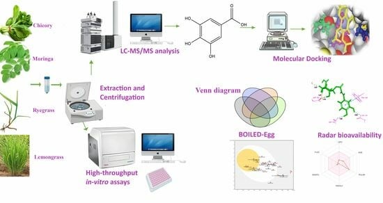
Optimized Extraction of Polyphenols from Unconventional Edible Plants: LC-MS/MS Profiling of Polyphenols, Biological Functions, Molecular Docking, and Pharmacokinetics Study

1
Department of Microbiology, Quaid-i-Azam University, Islamabad 45320, Pakistan
2
State Key Laboratory of Bioreactor Engineering, East China University of Science and Technology, Shanghai 200237, China
3
Department of Nutritional Sciences, Government College University Faisalabad, Faisalabad 38000, Pakistan
4
Department of Zoology, College of Science, King Saud University, Riyadh 11451, Saudi Arabia
5
School of Agriculture, Food and Ecosystem Sciences, Faculty of Science, The University of Melbourne, Parkville 3010, Australia
*
Author to whom correspondence should be addressed.
Molecules 2023, 28(18), 6703; https://doi.org/10.3390/molecules28186703
Submission received: 19 August 2023 / Revised: 12 September 2023 / Accepted: 13 September 2023 / Published: 19 September 2023
(This article belongs to the Special Issue Advances in Natural Products and Their Biological Activities)

Abstract

:
Plant bioactive phenolic metabolites have recently attracted the attention of researchers due to their numerous health advantages. Therefore, this study aimed to investigate with advanced techniques the bioactive metabolites and antioxidant and antidiabetic capacity of four unconventional edible plant leaves: lemongrass (Cymbopogon citratus (DC.) Stapf), chicory (Cichorium intybus L.), moringa (Moringa oleifera Lam.), and ryegrass (Lolium perenne L.). The extraction process was optimized using different solvents. These plants’ phenolic composition, identification, and characterization have been determined herein using LCESI-QTOF-MS/MS. This research identified 85 phenolic compounds, including 24 phenolic acids, 31 flavonoids, 7 stilbenes and lignans, and 17 other metabolites. Moreover, the study determined that moringa has the highest total phenolic content (TPC; 18.5 ± 1.01 mg GAE/g), whereas ryegrass has the lowest (3.54 ± 0.08 mg GAE/g) among the selected plants. It seems that, compared to other plants, moringa was found to have the highest antioxidant potential and antidiabetic potential. In addition, twenty-two phenolic compounds were quantified in these chosen edible plants. Rosmarinic acid, chlorogenic acid, chicoric acid, ferulic acid, protocatechuic acid, and caffeic acid were the most abundant phenolic acids. In silico molecular docking was also conducted to investigate the structure–function relationship of phenolic compounds to inhibit the alpha-glucosidase. Finally, the simulated pharmacokinetic characteristics of the most common substances were also predicted. In short, this investigation opens the way for further study into these plants’ pharmaceutical and dietary potential.

Graphical Abstract

1. Introduction

For thousands of years, people have utilized plants to improve their health. Until the 19th century, traditional plants were used for therapeutic purposes [1]. For example, herbal tea from plants treats digestive issues [2]. Traditional phytotherapy uses plants for medicinal purposes, and it has long been a choice for historical and modern treatment [3]. More than 80% of the population, particularly in underdeveloped nations, benefits from herbal products for medicinal purposes. Nowadays, it is a significant field of study to develop plant medications in addition to the traditional use of plants for treatments.
The phytochemicals found in medicinal plants enhance the control and prevention of metabolic diseases. These compounds have an additive and synergistic therapeutic impact [4,5]. Medicinal plants provide a variety of biologically active substances that have positive health effects. Phytochemicals from medicinal plants involve numerous complex biochemical, metabolic, and physiological mechanisms and have been studied for their impact on human health and wellness. Moringa and lemongrass are well-known plants due to their therapeutic potential. Moringa (Moringa oleifera) is used in herbal medicine and, as fresh leaves, a flavoring agent in many foods and beverages, and possesses cytotoxic and antibacterial properties [6]. Moringa is also a good source of antioxidant metabolites [7], used for gastric acidity treatment [8,9] aerophobia [10,11], nausea [12,13] vomiting, pharyngitis, liver issues, anorexia, stomach aches, etc. [14,15]. In vitro and animal studies indicate that moringa leaves may have more medical applications than are known at present. Studies have revealed that moringa has antibacterial, stress-relieving, and anticancer properties. The most well-known benefit of moringa is as a digestive aid. Irritable bowel syndrome-related stomach pain may be relieved by moringa oil, based on a brief study. There is sufficient evidence from pharmacological studies to support the biological effects of moringa and the phytochemicals found in the plant [16]. Additionally, mouth, throat, and sore throat irritation are also treated with it. Moringa (Moringa oleifera) is a nutrient-rich plant with various health benefits. It contains bioactive compounds like flavonoids, polyphenols, and alkaloids that exhibit antidiabetic properties. Moringa may help lower blood sugar levels by improving insulin secretion and reducing insulin resistance [17]. Additionally, its antioxidant and anti-inflammatory properties may play a role in mitigating diabetic complications.
Lemongrass (Cymbopogon citratus) contains compounds such as citral and polyphenols that have shown potential antidiabetic effects. Research suggests that lemongrass may help lower blood sugar levels by enhancing insulin sensitivity and improving glucose metabolism. Its antioxidant properties can also contribute to reducing oxidative stress, a factor linked to diabetes complications [18]. Further, lemongrass leaves have a strong, enticing, lemony scent and have been used for both culinary and medicinal purposes for many years. Traditionally, several illnesses and medical disorders have been treated with infusions produced from the plant’s fresh or dried leaves. These can include stomach issues, nervous system disorders, cough, rheumatism, nausea, and vomiting, as well as muscle and joint pain. In addition to having mild natural analgesic and urinary stimulant effects, research has revealed that the plant also contains anticancer, antioxidant, and anti-inflammatory properties [19,20]. As for protecting the stomach lining, lemongrass tea has been known to relieve gastrointestinal irritations, indigestion, and ulcers in the gastric area [21].
Unconventional edible plants may exhibit potential antioxidant and antidiabetic activities. Chicory (Cichorium intybus) is a plant whose roots are often roasted and used as a coffee substitute. It contains inulin, a type of soluble fiber that acts as a prebiotic, supporting gut health. Inulin can help regulate blood sugar levels by slowing the absorption of glucose in the digestive tract. Studies have indicated that chicory may have hypoglycemic effects and could potentially be beneficial for managing diabetes [22]. Chicory has several vital substances that are significant from a medicinal perspective. These substances include vitamins, chlorophyll, flavonoids, inulin, tannins, alkaloids, unsaturated sterols, saponins, and coumarins [23,24,25]. One of the most efficient ways to manage and lessen the consequences of chronic diseases like diabetes mellitus is to consume chicory, which is a rich source of terpenoids and phenolic compounds. Chicory plants also have therapeutic qualities such as antioxidant activity, usage for wound healing, assistance with diabetes, and the ability to fight harmful pathogens [26,27].
Ryegrass (Lolium perenne) is known for its high fiber content and slow digestion rate. The soluble and insoluble fibers in ryegrass contribute to improved glycemic control by slowing the release of glucose into the bloodstream. This can help prevent rapid spikes in blood sugar levels after meals. Ryegrass consumption has been associated with reduced risk of type 2 diabetes and improved insulin sensitivity. The ryegrass plant is an unconventional food plant that has a significant quantity of bioactive substances, including protein, fatty acids, alkylresorcinols, folates, tocotrienols, tocopherol, lignan, phytosterols, and phenolic acid [28,29,30]. In addition to helping with weight loss and appetite control, ryegrass consumption also lowers the risk of cancer, type 2 diabetes, cardiovascular disease (CVD), and other chronic diseases, along with mitigating allergic and inflammatory reactions [31,32].
Moreover, unconventional edible plants are excellent sources of antioxidants for preserving food and beverages. Numerous phenolic compounds that act as antioxidants, anticarcinogens, antimutagens, or anti-inflammatory agents are being studied for their potential to prevent cancer. Green edible plants are used for enhancing the organoleptic qualities (aroma, flavor, and taste) of many food products, while their possible health-promoting effects receive less attention [33]. The primary phytochemicals in these plants are polyphenols. Functional and therapeutic foods are in higher demand due to consumer demand for goods with possible health advantages. The most popular unconventional edible plants, moringa and lemongrass, have a variety of uses because they contain bioactive compounds that are employed in pharmacy, skincare products, and medications, in addition to their use as supplementary food [34]. To increase the effectiveness of medicinal effects, the synergistic effects of nontraditional edible plants were examined. It is true that integrating them into a single product can be done by mixing their complementary phenolic compositions, which may bring up synergic effects for edible plants in boosting quality and potential health impacts of products.
A few studies have been conducted previously using single LC-MS [35,36] and without mass spectrometry [37,38,39] or MS/MS with limited profiling of complex mixture of plant extracts [40]. Still, complex profiling using LC-MS/MS is lacking due to the complex structure of phytochemicals and unavailability of pure standards. LC-ESI-QTOF-MS/MS is equipment that provides a more reliable, sensitive, and authentic untargeted profile of phytochemicals from a complex mixture of plant extracts. To achieve this, samples were extracted using different solvents to analyze the profile of phenolic components and antioxidant activity. Phenolic compounds were assessed using LC-ESI-QTOF-MS/MS (electrospray ionization–quadrupole time-of-flight mass spectrometry). To estimate the total phenolic compounds and antioxidant potential, other methods include the total phenolic contents (TPC), total flavonoid contents (TFC), total condensed tannins (TCT), 2,2-azinobis-3-ethylbenzothiazoline-6-sulfonic acid (ABTS), hydroxyl radical scavenging assay (OH-RSA), DPPH, and FICA were used. The thorough profile of phenolics and their antioxidant and antidiabetic activities are examined for the first time in this group of edible plants. Additionally, using the pkCSM and SwissADME platforms, their predicted absorption, distribution, metabolism, excretion, and toxicity models were investigated. In silico molecular docking was also conducted to understand the structure–function relationship of phenolic compounds in these edible plants. This study demonstrates the potential and significance of medicinal and edible plants as a potential source of bioactive metabolites in various business sectors, including functional foods, skincare products, pharmaceutical and therapeutic products, and even the livestock sector.

2. Results and Discussions

2.1. Extraction Process Optimization for Total Phenolic Content in Selected Unconventional Edible Plants

The extraction process to extract total phenols from selected unconventional edible plants was optimized using different solvents. The results of different solvents extraction are given in Table 1.
Six different solvent systems such as methanol (80%), ethanol (80%), acidified methanol (80%), acidified ethanol (80%), acetone (80%), acidified acetone (80%), and water were used for the extraction process. In terms of 80% methanol, maximum extraction of total phenolic content in moringa was observed to be 18.0 ± 1.51 mg GAE/g, whereas chicory exhibited its minimum extractions of total phenolic content, i.e., 4.76 ± 0.11 mg GAE/g. Previously, an analysis of the methanolic extract revealed that the phenolic content in the moringa leaf extract was substantially (p ≤ 0.001) higher than that in the floral extract. The amount of phenols in moringa leaf extract was 111% higher than that in its flower extract. A leaf of M. oleifera Lam had 2.28 mg/mL total phenols, whereas a floral extract of the same plants contained 1.08 mg/mL total phenols [41]. Utilizing 80% ethanol as the solvent, the greatest yield of total phenolic content from moringa was measured at 16.2 ± 1.33 mg GAE/g, whereas the lowest total phenolic content extraction was recorded at 3.12 ± 0.02 mg GAE/g. The highest yield of total phenolic content from moringa, recorded at 18.5 ± 1.01, was achieved using 80% acidified methanol as the solvent. In contrast, the lowest recovery of total phenolic content, measuring 3.54 ± 0.03, was obtained in ryegrass when 80% acidified methanol was employed. The peak extraction of total phenolic content from moringa, reaching 17.1 ± 1.32 mg GAE/g, was obtained when utilizing 80% acidified ethanol, while the least total phenolic extraction was observed with acidified ethanol, at 3.43 ± 0.09 mg GAE/g. Vania Urías-Orona et al. also proposed the effect of the extraction solvent on the phenolic content in a commercial dietary supplement made from moringa oleifera leaves. According to their study, methanol (MeOH) and ethanol (EtOH) at concentrations of 100%, 80%, and 50% aqueous were used as extraction solvents. The amounts of total phenols collected varied between 55.98 and 226.20 mg ChlAE/g, with EtOH 50% and EtOH 100% having the highest and lowest contents, respectively. There were significant variations in the phenolics analyses (p < 0.05) [42]. Moringa’s total phenolic content was best extracted using 80% acetone and 80% acidified acetone, yielding 16.1 ± 1.09 mg GAE/g and 17.2 ± 1.47 mg GAE/g, respectively. On the other end, the least efficient extraction of total phenolic content in ryegrass, using 80% acetone and 80% acidified acetone, resulted in 1.84 ± 0.01 and 1.63 ± 0.02, respectively (Table 1). The phenolic activity in ryegrass following solvent extraction was discovered by Darius Povilaitis et al. in their work. The TPC values for ryegrass ranged widely (12.50.19–530.1 mg GAE/g), with acetone extracts having the highest TPC (up to 530.11 mg GAE/g), followed by hexane and then methanol extracts. As a result, the most effective solvent for extracting phenolic compounds from ryegrass was acetone [43]. Overall, 80% acidified methanol provided better extraction than other solvent mixtures.

2.2. Quantification of Total Polyphenols in Selected Plant Extracts

Based on the optimization results, we proceeded with 80% acidified methanol extraction for biological activities and LC-MS/MS analysis. Researchers have been very interested in phenolic compounds since a range of phenolic compounds are examined that have chemoprotective abilities, which function as antioxidant, anticarcinogenic, antimutagenic, or anti-inflammatory chemicals. Table 2 lists the phenolic concentrations in selected plants.
The Folin–Ciocalteu technique was used to determine the total phenolic content (TPC). The findings revealed that the phenolic content in moringa, measured at 18.5 ± 1.01 mg GAE/g, was higher than that of lemongrass, measured at 12.9 ± 0.12 mg GAE/g, chicory measured at 4.85 ± 0.06 mg GAE/g. The lowest measured was in the ryegrass sample, at 3.54 ± 0.08 mg GAE/g. Previously, a higher amount of TPC was measured in moringa at 103.28 ± 8.08 mg GAE/g than that of the other investigated samples [44]. The TPC (12.9 ± 0.12 mg GAE/g) in our study is lower than the TPC of Australian lemongrass (15.09 ± 0.88 mg QE/g) [20]. The TPC results might vary because different samples were used in the current study and other studies to assess the phenolic levels.
According to an analysis of total flavonoid content, moringas had a much higher quantity of flavonoid content (10.1 ± 0.83 mg QE/g) than lemongrass (6.19 ± 0.09 mg QE/g) and chicory (1.06 ± 0.01 mg QE/g). Findings show that ryegrass (1.52 ± 0.13 mg QE/g) has the lowest concentration of flavonoids among the selected group of plants. Flavonoids are the most common class of secondary plant metabolites and are employed in pharmaceutical, medical, and cosmetic applications because of their antioxidative, anti-inflammatory, anticarcinogenic, and antimutagenic properties [45]. In a similar study, the flavonoid content in ryegrass was investigated, and the same results of TFC were reported for ryegrass, revealing 0.01 ± 0.01 mg QE/g [36].
Chicory showed significantly higher levels of total tannin content (1.31 ± 0.15 mg CE/g) compared to other selected edible plants. Previous investigations on the measurement of total tannin contents found similar results in Australian lemongrass (1.36 ± 0.08 mg CE/g) [20], moringa (8.31 ± 1.58 mg CE/g) [44], ryegrass (2.11 ± 0.06 mg CE/g) [36], and chicory (0.84 ± 0.03 mg CE/g) [35]. Moringa is a suitable natural antioxidant or a useful ingredient in food, cosmetics, and pharmaceuticals due to its overall much greater phenolic chemical profile. Owing to their potential health advantages, flavonoids, the most abundant class of phytochemicals found in plants, fruits, and medicinal plants, have attracted much attention. These medicinal plants were never combined in previous research. These plants’ flavonoids and total phenolic contents vary significantly from each other. Overall, moringa contains the highest amount of total phenolic and total flavonoids than other studied unconventional edible plants.

2.3. Antioxidant and Antidiabetic Activities of Selected Plant Extracts

For the various kinds of activities that were analyzed, such as radical oxygen scavengers, metal chelators, reducing agents, and H+ ion donators, antioxidants are recognized as essential components. Several assays, including the DPPH, ABTS, ferrous ion chelating activity (FICA), and OH-RSA, were conducted to evaluate the antioxidant capacity of herbal plants. Table 3 shows the results of antioxidant and antidiabetic potential for selected samples (moringa, lemongrass, chicory, and ryegrass).
The cost-effective DPPH test is usually performed to estimate the samples’ capacity to neutralize free radicals in biological systems, which is based on their capacity to release electrons or hydrogen ions. DPPH, a free radical with a stable nitrogen center, loses part of its bluish-purple pigments when mixed with sample extracts [46]. When investigated for its ability to scavenge DPPH radicals, moringa (34.16 ± 2.32 mg AAE/g) showed considerably (p ≤ 0.05) greater antioxidant capacity than lemongrass (25.73 ± 0.18 mg AAE/g), chicory (16.61 ± 0.23 mg AAE/g), or ryegrass (11.18 ± 0.23 mg AAE/g). Similar radical-scavenging capacities in ryegrass (0.05 ± 0.01 mg AAE/g) were found in previous studies [36] on the antioxidant activity and total polyphenol content in a variety of unconventional dietary plants.
The findings of the ABTS test revealed that the activities in ryegrass (18.4 ± 0.56 mg AAE/g) were significantly (p ≤ 0.05) lower than those in other green plants (moringa 54.9 ± 4.24, lemongrass 44.81 ± 0.93, and chicory 35.4 ± 0.68 mg AAE/g). Additionally, a ferrous ion chelating assay was carried out to determine the antioxidant capacity of the sample extracts, and the results showed that moringa had a substantially greater potential than other green plants (7.49 ± 0.39 mg EDTA/g) (p ≤ 0.05). Previous research has also shown [44] the same results. Furthermore, hydroxyl radical scavenging activity was carried out to estimate the antioxidant profile, and the findings showed that moringa had greater activities. Overall, the findings in this study for all antioxidant assays indicated that moringa had the highest antioxidant activity and that ryegrass plants had the lowest antioxidant activity.
The alpha-glucosidase IC50 (μg/mL) value is the inhibitory concentration needed to block 50% of the enzyme’s activity of the α-glucosidase enzyme. The enzyme activity is more effectively inhibited by the inhibitor when the IC50 value is lower. In this current study, comparing the inhibitory activity in the four edible plants selected (lemongrass, chicory, ryegrass, and moringa) against α-glucosidase, our results revealed that compared among all selected plant extracts, ryegrass showed lower enzyme inhibition activity, with IC50 = 29.02 ± 2.17 µg/mL. Ryegrass was followed by chicory, which also exhibited moderate inhibitory activity against αglucosidase, with IC50 values of 16.41 ± 1.21 µg/mL. The IC50 for α-glucosidase inhibitory activities of lemongrass and moringa were 2.15 ± 0.13 and 1.89 ± 0.01 µg/mL, respectively, which are considered as strong. In terms of α-glucosidase inhibitory effects, Shiva Nouri et al. identified Moringa peregrina (MP) and Ferulago carduchorum (FC) as two promising plants [47]. The regression equation representing the percentage of enzyme inhibition at various doses was used to estimate the IC50 values for MP and FC extracts. An increased enzyme inhibitory activity is indicated by a lower IC50 value. Their findings showed that the IC50 for MP was 4.96 μg/mL and the IC50 for FC was 7.41 μg/mL. The MP extract found to be more effective at inhibiting α-glucosidase [47].

2.4. Correlation Analysis

Correlation analysis was performed between the phenolic content (TPC, TCT, and TFC) of the chosen samples and the antioxidant activity produced by the four separate assays (Table 4).
The TPC had a significant positive correlation with TCT (r2 = 0.99), DPPH (r2 = 0.99), FICA (r2 = 0.93), OH-RSA (r2 = 0.99), and ABTS (r2 = 0.93); the TFC had a positive correlation (p ≤ 0.1) with the all four antioxidant assays DPPH, OH-RSA, ABTS, and FICA. This seems to demonstrate a direct connection between the samples’ phenolic components and the antioxidant processes of ferric chelation, peroxyl inhibition, and free radical scavenging.
Flavonoids showed significant hydroxyl inhibition, free radical scavenging, and ferric ion chelation activity correlations. This further demonstrates the variety of phenolic and nonphenolic metabolites found in plant extracts. This may be related to the fact that a flavonoid’s capacity to act as an antioxidant is affected by the location of the hydroxyl group on the B-ring and whether it can deliver a free radical, either a hydrogen atom or an electron, or both. A biplot analysis (Figure 1) was conducted to investigate the correlation between active variables (TPC, TFC, TCT, DPPH, ABTS, FICA, OH-RSA, and alpha-glucosidase inhibition activity) and active observations (lemongrass, chicory, ryegrass, and moringa).
The interactions between antioxidant activity and phenolic compounds might also be affected by the conditions of the study, the mechanism behind the antioxidant reactions, and the collaborative or antagonistic effects of other compounds present in the reaction mixture.

2.5. Characterization of Phenolic Compounds Using LC-MS

Green herbaceous plants have currently gained the attention of scientists due to the discovery that they contain significant amounts of bioactive components, specifically phenolic metabolites, which have demonstrated potential health benefits in numerous studies [44,48,49]. Using the latest method of LC-ESI-QTOF-MS2, phenolic compounds from the targeted samples of moringa, lemongrass, chicory, and ryegrass plants were screened and characterized. We can identify all the bioactive components of plant samples with an advanced analytical approach. The compounds were qualitatively characterized using Agilent Mass Hunter software, version B.06.00, and Personal Compound Database Library (PCDA). Table 5 lists the results associated with the phenolic chemical profile of plants. A total of 85 phenolic compounds have been found by LC/MS analysis, including 24 phenolic acids, 31 flavonoids, 6 isoflavonoids, 7 stilbenes and lignans, and 17 other polyphenols. The base peak chromatograms (BPC) of the selected plants and MS/MS spectra of some selected compounds are given in Figures S1 and S2.

2.5.1. Phenolic Acids

Secondary plant metabolites called phenolic acids are well known for their potential health advantages. Owing to their potential for antiaging, antioxidant, antibacterial, anticancer, cardioprotective, anticancer, and anti-inflammatory activities, they are widely used in many products in the food, cosmetic, and pharmaceutical industries. The samples of green plants included a total of 24 phenolic acids, primarily hydroxycinnamic acid molecules. When CO2 and the hexosyl moiety from the parent ions are removed, these phenolic acids show a fragmentation pattern. Gallic acid (m/z 171.0298), protocatechuic acid (m/z 155.0339), and p-hydroxybenzoic acid (m/z 137.0244) were recognized as the compounds 1, 2, and 4 are benzoic acid derivatives, and they each displayed the product ions at m/z 109, 153, and 93 that existed in all samples, respectively. Another study [35] that used the LC-MS/MS approach to identify hydroxybenzoic acids and hydroxycinnamic acids in various Australian plants also found similar metabolites. Other hydroxybenzoic acids included protocatechuic acid 4-O glucoside (m/z 315.0721), which was only identified in two plants (lemongrass and moringa).
Twenty hydroxycinnamic acids were found. Only compound 7, a cinnamic acid derivative identified as 3-caffeoylquinic acid (m/z 353.0878) with product ions at m/z 253, 190, and 144, was found in all four samples. Compounds 5, 11, 12, 15, 20, and 21, all found in moringa, lemongrass, and chicory samples, were identified as 3-sinapoylquinic acid (m/z 399.1286), caffeic acid (m/z 179.035), 3-feruloylquinic acid (m/z 367.1034), p-coumaroyl glycolic acid (m/z 221.0455), cinnamic acid (m/z 147.0451) and p-coumaric acid (m/z 163.04). Compounds 11 and 12 produced ions at m/z 143, 135, 133, 298, and 288 together with 192 and 191. Compounds 20 and 21 have product ions at m/z 129, 103, and 119, respectively. Only lemongrass and ryegrass samples were analyzed for the product ions of compounds 10 and 17, which were identified as 1-O-sinapoyl-beta-D-glucose (m/z 385.114) and 1,2-diferuloylgentiobiose (m/z 693.2036), and showed ions at m/z 223, m/z 193, and m/z 134 after removing glucoside moiety (162 Da), water (18 Da), and CO2 (44 Da) from the precursor ion (m/z 385.1140), and then from product ions. p-Coumaroylquinic acid (m/z 337.0929), sinapic acid (m/z 223.0612), p and 3,5-diferuloylquinic acid (m/z 543.1508) were found to produce ions at m/z 193, m/z 191, m/z 179, m/z 149, and m/z 134, in contrast to compounds 13, 14, and 23, which were only found in lemongrass and moringa samples. Similar findings were reported during previous research [20,35] that identified polyphenols in several Australian plants and plants. Their findings reveal many hydroxycinnamic acids, some of which resembled the compounds found in the current research.

2.5.2. Flavonoids

Flavonoids are the most prevalent category of plant metabolites that are used in medical, beauty, and pharmacological industries for their antioxidative, anti-inflammatory, anticarcinogenic, and antimutagenic qualities [45]. In the current study, a total of 45 flavonoids were identified. Flavonoids were observed higher in amount than all the other phenolic compounds from the targeted samples. Observed categories of flavonoids included 4 flavanols, 4 flavanones, 7 flavones, 11 flavonols, 8 Isoflavonoids and others.
Theaflavin 3-O-gallate (m/z 715.1304) was the first observed flavanol, as compound 25, in negative mode and produced product ions at m/z 565 and 139, only identified in the lemongrass and ryegrass samples. No flavanol compounds were found in all four samples. In addition, two compounds were only identified in chicory and moringa: procyanidin trimer C1 and epigallocatechin 3-O-gallate-forming product ions at m/z 739, 695, 577, and 451 for compound 30, and 305 and 169 for compound 32. A similar (−)-epicatechin compound was observed in the lemongrass, moringa, and chicory samples in both modes, and previous results revealed the presence of similar compounds in lemongrass and moringa samples.

Flavonols

Compound 47 was observed as quercetin 3-O-xylosyl-glucuronide, forming product ions at m/z 301 in negative mode only, identified in the ryegrass plant source, while three flavanol compounds that were only identified in lemongrass were kaempferol 3-O-xylosyl-glucoside, kaempferide, and isorhamnetin 3-O-rutinoside. Compounds 49, 52, and 54 were revealed by their product ions at m/z 285, m/z 283, m/z 151, and m/z 315, respectively. Myricetin 3-O-rhamnoside and 3,7-dimethylquercetin were identified only in lemongrass and moringa.

2.5.3. Isoflavonoids

In this study, six Isoflavonoids were observed in the given samples. Compounds 56, 59, and 60, identified as 6″-O-malonylglycitin, dihydroformononetin, and glycitein 7-O-glucuronide, were only present in lemongrass and moringa samples, as revealed their product ions at m/z 533, m/z 253, m/z 137, m/z 441, m/z 283, and m/z 267. Daidzein 7-O-glucuronide was only identified in lemongrass and chicory, while 6″-O-malonylgenistin was only identified in moringa and chicory samples. An isoflavonoid was only identified from samples of moringa, referred to as equol 7-O-glucuronide (m/z 417.1191), and forming its ions on m/z 241.

2.5.4. Stilbenes and Lignans

The current study observed two stilbene and five lignan compounds from four studied samples. Compound 62 was identified as piceatannol (m/z 243.0663) and only found in moringa and chicory, while compound 63 was identified as 3′-hydroxy-3,4,5,4′-tetramethoxystilbene (m/z 303.1227), only observed in lemongrass and moringa samples. Numerous studies have been conducted on piceatannol, a stilbene with two phenol rings that was found in moringa and chicory, and has considerable health benefits including antioxidant, anticancer, antimutagenic, antiatherosclerotic, and anti-inflammatory properties [50,51]. LC-MS characterized bioactive substances, including lignans, which exhibit exceptional antioxidant and anticarcinogenic activities. Human ovarian, breast, and prostate cancers are protected or prevented from spreading by consuming plant lignans [52]. LC-MS helped to identify five lignans from the selected variety of samples. Compound 64, medioresinol (m/z 389.1595), was only identified in lemongrass and moringa when observed in positive mode, while compound 65, lariciresinol-sesquilignan (m/z 555.2235), was observed in both modes and found in only the lemongrass, chicory, and moringa samples. Other three lignans that were identified in negative mode were secoisolariciresinol-sesquilignan (m/z 557.2392), lariciresinol (m/z 359.15), and matairesinol (m/z 357.1343), and found only in the moringa sample. Another study [53] conducted on phytochemicals with cancer-fighting properties revealed some compounds observed from the lignan profile that were similar to the compounds identified in the current research.

2.5.5. Other Compounds

A few coumarin derivatives were identified from all samples, four of which were only observed in lemongrass and moringa samples. These compounds identify as coumarin (m/z 145.0295), esculin (m/z 339.0721), umbelliferone (m/z 161.0244), and mellein (m/z 177.0557) as revealed by their product ions at m/z 101, m/z 177, and m/z 133, respectively. Three phenolic compounds characterized from the moringa sample were only identified as rosmanol, rosmadial, and hydroxytyrosol 4-O-glucoside, forming product ions at m/z 301, m/z 299, m/z 153, and m/z 123, respectively. Other terpene molecules found only in lemongrass and moringa were carvacrol (compound 70) and carnosol (compound 72), observed to be fragmenting at m/z 107 and m/z 285, respectively.
Using LC-ESI-QTOF-MS/MS, we were able to screen and characterize 85 polyphenolic compounds with their product ions in samples of plants. All of these plant’s phenolic constituents have not yet been the subject of a single study. Screening for these bioactive components in these plants can provide a new field of study toward revealing their significant health advantages and new applications. The use of this sophisticated analytical method has significant promise for the discovery of novel, undiscovered bioactive chemicals. Low collision energies have the drawback of inability to locate the position of the native phenolic ring in the analysis of LC-MS/MS that underwent alteration.

2.6. Venn Distribution of Polyphenols in Plant Extracts

The Venn diagram is a practical, potent, and adaptive tool that can rapidly examine a large number of facts and transform it into information that is easy to understand. The Venn diagram illustrates the distribution of phenolic metabolites in lemongrass, moringa, ryegrass and chicory (Figure 2).
According to the Venn diagram (Figure 2A), three distinct phenolic compounds were found in the lemongrass sample, whereas moringa, chicory, and ryegrass, respectively, contained 8, 2, and 1 distinctive phenolic compounds. This shows that moringa possesses a wider variety of phenolic metabolites than other herbaceous extracts, which may help explain its increased TPC, TFC, and antioxidant potential (Table 1). It is interesting to see that moringa and lemongrass have the highest number of overlapping phenolic compounds. The total amount of flavonoid compounds in these edible plants is shown in a Venn diagram (Figure 2B). Compared to ryegrass and chicory, which have fewer distinct flavonoids, it was found that lemongrass and moringa have a wider diversity of unique flavonoids.
This figure also shows that there are eight flavonoid components that are identical in lemongrass and moringa, but no single flavonoid touches all four sources. The total phenolic acids in the chosen plants are shown in Figure 2C. It demonstrates that moringa contains the most phenolic acids. Chicory also contains sixteen phenolic acids. Moringa contains a total of sixteen phenolic compounds (Figure 2D). None of the other phenolic metabolites were found to overlap in ryegrass, moringa, or chicory.

2.7. Quantification/Semiquantification of Selected Phenolic Compounds

Table S1 lists the twenty-two quantified compounds that were measured in the lemongrass, moringa, ryegrass, and chicory plants. The most common type of compound in these plants is flavonoids. Moringa had the highest concentration of hydroxycinnamic acids, with rosmarinic acid having the highest concentration (748.32 ± 25.82 μg/g) 3-caffeoylquinic acid (103.93 ± 12.51 μg/g), 3-sinapoylquinic acid (125.23 ± 19.42 μg/g), and caffeine (264.05 ± 11.54 μg/g). Cinnamic acid was present in only three samples, lemongrass, chicory, and moringa (91.38 ± 8.87, 17.71 ± 1.18, and 31.32 ± 5.06, respectively). The lowest chlorogenic acid content was measured in moringa, whereas chlorogenic acid (3-caffeoylquinic acid) is the most prevalent phenolic acid in ryegrass (445.62± 31.52 μg/g). It was previously measured [36] to determine the amount of chlorogenic acid in nonconventional plants. Only chicory (286.12 ± 31.27 μg/g) and ryegrass (15.82 ± 1.59 μg/g) contain chicoric acid; p-hydroxybenzoic acid was measured in lemongrass (36.13 ± 0.73 g/g) and ryegrass (75.92 ± 5.67 μg/g). Lemongrass had the highest content of caffeic acid (366.78 ± 14.77 μg/g), and chicory plant extract had the lowest concentration (245.67 ± 21.55 g/g). Ryegrass plants also contained protocatechuic acid (63.14 ± 3.58 μg/g) and sinapic acid (51.05 ± 5.12 μg/g). Chicory contained gallic acid (23.59 ± 2.04 μg/g) and p-coumaric acid (154.90 ± 12.62 μg/g). Additionally, procyanidin B2 was detected to be highest in moringa (68.76 ± 4.12 μg/g) and lowest in lemongrass (44.62 ± 6.32 μg/g). Ryegrass had the highest content of diosmin (132.63 ± 11.06 μg/g), whereas moringa had the lowest value (9.51 ± 1.43 μg/g). Additionally, MetaboAnalyst 5.0 (www.metaboanalyst.ca accessed on 22 March 2023) was used to perform hierarchical heatmap clustering (Figure 3).
The heatmap figure shows that chicory included higher quantities of chicoric acid, ferulic acid, and caffeic acid than other measured phenolic components; moringa had higher quantities of rosmarinic acid, 3-sinapoylquinic acid, umbelliferone, carnosic acid, coumaric, procyanidin B2, and epicatechin. Ryegrass was found to have the highest levels of chlorogenic acid (3-caffeoylquinic acid), sinapic acid, protocatechuic acid, p-hydroxybenzoic acid, and diosmin; p-coumaric acid, caffeic acid, and pyrogallol were all present in higher concentrations in lemongrass.

2.8. In Silico Molecular Docking of Phenolic Compounds

By using in silico molecular docking, the roles of several phenolic compounds in α-glucosidase inhibitory activities were anticipated. By employing computational methods, it is feasible to predict with high accuracy the affinities and modalities of attachment of a target molecule (or ligand) to a particular protein (or receptor), for instance, molecular docking. The projected two-dimensional binding geometries of chlorogenic acid (A), diosmin (B), naringin (C), and procyanidin B2 (D) in the α-glucosidase protein (5NN8), in addition to the computed binding energies, are shown in Figure 4 and Table S2. Chlorogenic acid (Figure 4A) formed one hydrogen bond to each of the ASPs 404 and 518, an additional bond with a molecule of water, two with ASP 282, and one with the hydrophobic PHE 649, a process known as “π–π staking” with the same substance. Diosmin (Figure 4B) established five hydrogen bonds with the negatively charged ASP 282 and one with the analogously charged ASP 616. As part of its hydrogen bonding interactions, naringin (Figure 4C) formed interactions with PHE 525, EDO 1024, ASP 282, ARG 281, and LEU 678, along with one π–π stacking contact with TRP 481. Procyanidin B2 (Figure 4D) in 7E3I interacted with PHE 295 and TRP 86, and formed hydrogen bonds with THR 83, ASP 74, TYR 124, HID 447, and ASN 87.
The protein structure of 5NN8 was docked to punicafolin. Punicafolin was positioned in the binding region of 5NN8 throughout the molecular docking procedure and enabled interaction with the nearby amino acid residues. PHE 649 is reported to be a hydrophobic amino acid. Additionally, punicafolin formed hydrophobic connections with residues of the amino acids ALA 284, PHE 525 along with TRP 481. Additionally, quercitrin and myricitrin formed two hydrogen bonds with the negatively charged ASP 282 along with one each to the negatively charged ASP 616, the hydrophobic ALA 284, and the electronegative EDO 1024. They possessed a single π–π binding with TRP 481 and a single π–π stacking hydrophobically with PHE 525. Acarbose formed twelve hydrogen bonds, two with ASPs 518, 404, 282, and 523, and three OH groups from water molecules, which also formed hydrogen bonds with ASPs 645 and 281. Rutin specifically created four hydrogen bonds, including ones with ASP 404, 616, and 518.
The binding energies of punicafolin, rutin, acarbose, procyanidin B2, myricitrin, 3-feruloylquinic acid, taxifolin, diosmin, quercitrin, chlorogenic acid, quercetin-3-O-arabinoside, naringin, 3-p-coumaroylquinic acid, myricetin, quercetin, Isorhamnetin, quinic acid, luteolin, (-)-epicatechin, hesperetin, gallic acid, 3-O-sinapoylquinic acid, diosmetin, naringenin, p-hydroxybenzoic acid, salicylic acid, caffeic acid, p-coumaric acid, pyrogallol, chrysin, protocatechuic acid, 3-4-5-trimethoxyflavone, cinnamic acid, and coumarin in 5NN8 were calculated as −12.16, −11.14, −11.05, −10.95, −10.59, −10.32, −10.13, −9.84, −9.72, −9.62, −9.49, −9.40, −9.35, −9.28, −6.95, −6.68, −6.65, −6.52, −5.36, −6.28, −6.14, −5.91, −5.36, −5.33, −5.30, −5.20, −5.08, −4.80, −4.74, −4.19, −4.12, −4.09, −4.04, and −3.93, respectively. Punicafolin is expected from the given table to have a higher binding affinity than the other chosen phenolic compounds. Punicafolin, rutin, and acarbose (standard) have stronger ability of α-glucosidase inhibition than other compounds. It is interesting to note that myricitrin has a greater affinity for binding than 3-feruloylquinic acid, taxifolin, diosmin, quercitrin, chlorogenic acid, quercetin-3-O-arabinoside, naringin, 3-p-coumaroylquinic acid, or myricetin. Comparatively, Isorhamnetin, quinic acid, luteolin, (-)-epicatechin, hesperetin, and gallic acid have lower binding affinities than myricetin. In silico molecular docking is a prediction of possible interactions between target proteins (5NN8) and potential inhibitors. Therefore, it is critical to assess the inhibitory activities of individual purified phenolic compounds to establish the precise roles of individual bioactive compounds in the inhibition of α-glucosidase. Moreover, the insights into inhibitory mechanisms of bioactive polyphenolic compounds against α-glucosidase and other proteins involved in diabetic conditions can be revealed through advanced molecular dynamics techniques and free-energy calculations, and through inverse molecular docking.

2.9. ADMET Properties of Abundant Phenolic Compounds

Bioinformatics facilitates the effective continuation of pharmacokinetic studies by reducing the amount of experimental effort required to evaluate various metabolites [54]. This study examined the metabolites for potential bioavailability and therapeutic effects using pharmacokinetics characteristics such as absorption, distribution, metabolism, excretion, and toxicity (ADMET) screening. The major cause of drug molecules failing in clinical tests is that they take too long to produce results are unfavorable properties of ADMET in the biological system [55]. This study examined the ADMET characteristics of the most prominent phenolic compounds found in given plant extracts.

2.9.1. Absorption and Distribution

With the help of the pkCSM platform and the BOILED-Egg approach (Figure 5), the absorption of the phenolic compounds had been expected. Tables S3 and S4 and Figure 5 display the absorption data. It was expected that fourteen compounds, including carnosic acid, nobiletin, sinapic acid, caffeic acid, and others, would be absorbed in the digestive system, whereas ten compounds would be expected to pass through brain barriers. Cinnamic acid, which is present in moringa, lemongrass, and chicory, was also predicted to penetrate the blood–brain barrier more rapidly than other phenolic substances (Table S4).
Nineteen substances have an absorption rate of more than 80% in the human digestive tract, based on absorption prediction including p-Hydroxybenzoic acid (83.9%), p-coumaric acid (93.5%), cinnamic acid (94.8%), coumarin (97.3%), ellagic acid (86.7%), and a few others. Finding shown in Table S4 indicate that only the coumarin molecule is projected to have high skin permeability. In addition, the highest Caco-2 cell permeability is predicted for p-Hydroxybenzoic acid (1.151), p-coumaric acid (1.21), resveratrol (1.17), cinnamic acid (1.717), coumarin (1.649), benzoic acid (1.707), nobiletin (1.306), dihydrobiochanin A (0.981), 4-hydroxycoumarin (1.206), scopoletin (1.184), and dihydroquercetin (0.924). A chemical is said to have high Caco-2 permeability if the value is more than 0.90 [54]. Furthermore, compounds with Caco-2 permeability, intestinal uptake, a high bioavailability score, Lipinski’s rule of compliance, failure to cross the blood–brain barrier, inability to serve as P-gp substrates, and poor skin penetration should be effective medicines [56]. Most of the compounds that are not absorbed in the digestive tract can be converted into other derivatives by microbes in the gut and absorbed in the colon [57].
Flavonoids are linked to albumin after absorption and transported to the liver via the portal vein. However, flavonoids have a low bioavailability due to their restricted absorption, significant metabolism, and rapid excretion [58]. By using Diana’s approach [59], oral bioavailability of a few compounds are shown with radars (Figure 6). No substance exhibited anticipated oral bioavailability, as shown in Figure 5 and Table S5. Six physiochemical characteristics (size, polarity, lipophilicity, flexibility, saturation, and solubility) were analyzed by using the bioavailability radar to forecast the oral bioavailability of given metabolites [60].

2.9.2. Metabolism, Excretion, and Toxicity

A key function of the metabolism of bioactive chemicals (drugs) is played by cytochrome P450 (CYP) [56]. Table S7 provides information on the phenolic compounds’ anticipated metabolism and excretion. The CYP model (CYP1A2, CYP2D6, CYP3A4, CYP2C9, and CYP2C19) was used to estimate metabolism for substrate or inhibitor antioxidants. Higher amounts of other bioactive compounds may come from bioactive chemicals that inhibit the CYP process, increasing their toxicity, and vice versa. It is hypothesized that bioactive substances with higher overall clearance have increased bioavailability and hepatic metabolism (Table S6). Table S7 provides a simulated toxicity screening for the bioactive chemicals. The predicted results show that none of the bioactive substances inhibits the hERG 1 channel.

3. Materials and Methods

3.1. Materials

In all experiments, only analytical-grade chemicals were used. Sigma Aldrich (Darmstadt, Germany) provided the chemicals for the characterization and identification of compounds. Folin–Ciocalteu’s reagent, hydrated sodium acetate, vanillin, sodium phosphate dibasic heptahydrate, trichloroacetic acid, ethylenediaminetetraacetic acid (EDTA), ammonium molybdate, catechin, 2,2-diphenyl-1-picryl-hydrazyl-hydrate (DPPH), potassium ferrocyanide (III), 2,4,6 tripyridyl-s-triazine (TPTZ), 2,2’-azino-bis 3-ethylbenzothiazoline-6-sulfonic acid (ABTS), 3-hydrobenzoic acid, and quercetin were purchased from the Sigma Aldrich (Castle Hill, NSW, Australia) for the estimation of polyphenols and antioxidant capability.

3.2. Sample Preparation and Method Optimization for Extraction of Phenolic Compounds

Chicory, lemongrass, moringa, and ryegrass plants were all collected from fields of Faisalabad division, Pakistan. Samples were reconfirmed and identified from the Department of Plant Sciences, Quaid-i-Azam University, Islamabad, Pakistan. The plant samples were dried at room temperature for two days and oven-dried at 45 °C for three days. A fine powder was prepared by grinding using a laboratory grinder. The method of extraction was optimized using different solvents: 80% ethanol, 80% methanol, 80% acetone, 80% chloroform, and Milli-Q water. The following procedure was employed to extract phenolic compounds: Using a 2 g sample and 30 mL of 80% solvent (methanol, ethanol, acetone, chloroform) in Milli-Q water, extracts from the four selected edible plants were prepared in triplicate. Samples were extracted after shaking in an orbital shaker (ZWYR-240) for 16 h at 150 rpm and 4 °C. After centrifuging the samples at 8000 g for 20 min, the supernatant was separated from the solution and filtered using a 0.45 μm syringe filter. Afterward, samples were kept at −20 °C for a maximum of seven days before LC-MS/MS and spectrophotometric analysis.

3.3. Total Phenolic Content and Antioxidant Potential

3.3.1. Determination of Total Phenolic Content

The phenolic compound profile of samples was analyzed using the previously described method by Ali et al. [61]. First, 25 µL of Folin–Ciocalteu reagent (25% v/v) was taken with 200 µL of distilled water. Then, 25 µL of sample extract was added and incubated at 27 °C for 5 min. At last, 25 µL of sodium carbonate (10% w/w) was added to the reaction mixture and placed in incubation again in the dark at 27 °C for 1 h. Absorbance of the samples was recorded at 760 nm. Quantification of the total phenolic content was carried out by making a standard curve against gallic acid that ranged from 0 to 200 µg/mL in methanol. Results were recorded using units of GAE (milligram gallic acid equivalents) per gram of sample.

3.3.2. Total Flavonoid Content

Flavonoid content in the samples was analyzed using the method described by Ali et al. [62], with some modifications. The aluminum chloride colorimetric method was used to determine the TFC. An 80 µL sample extract was taken in 96-well plates and mixed with 80 µL AlCl3 solution and 120 µL sodium acetate aqueous solution (50%). After preparation of the reaction mixture, the sample was incubated in the dark at 27 °C for 2.5 h and absorbance was recorded on the spectrophotometer at 440 nm. For the quantification of flavonoid content, a standard curve (R2= 0.999) was constructed against 0–50 µg/mL of quercetin in methanol. The milligram quercetin equivalents per gram of the sample unit was used to express the results.

3.3.3. Total Tannin Content

TTC was carried out using a modified version of the Ali et al. [62] technique. First, 150 µL vanillin solution (4%) and 25 µL of the sample solution was added. Then, 25 µL of 32% H2SO4 was then poured into the mixture. The final sample was incubated at 25 °C for 15 min. The absorbance was determined at 500 nm and the standard catechin curve (0–1000 µg/mL) was constructed. The data are given as mg CE/g.

3.4. Antioxidant Activities of Edible Plants

3.4.1. ABTS Radical Scavenging Assay

ABTS assay was performed by following the methods described by Ali et al. [61] and Severo et al. [63]. In order to produce ABTS+ solution, a mixture of 140 mM potassium persulfate solution and 7 mM ABTS solution was incubated for 16 h in the dark. The solution was then diluted with ethanol to obtain an absorbance reading of 0.70 ± 0.02 at 734 nm. In a 96-well plate, 290 µL of ABTS+ solution and 10 µL of sample extract were added and incubated for 6 min at ambient temperature. The absorbance was measured at 734 nm. The measurement (reported as mg AAE/g) was performed by generating the standard curve against ascorbic acid values of 0–150 μg/mL in water.

3.4.2. DPPH Radical Scavenging Assay

The DPPH assay for all samples was performed in triplicate by following the method described by Fia et al. [64] with modifications. First, 25 µL sample extracts were mixed with 275 µL of DPPH dye (0.1M) in methanol using the 96-well plate method. The prepared reaction mixture was then incubated for 30 min at 25 °C in a dark place and recorded at 517 nm on the spectrophotometer. The radical scavenging capability was analyzed by constructing a standard curve against ascorbic acid (0–50 µg/mL) in water. The results observed were recorded as milligrams of ascorbic acid equivalents per gram of sample (mg AAE/g).

3.4.3. Hydroxyl Radical Scavenging Assay

The Fenton-type reaction method was used to determine the OH-RSA (hydroxyl radical scavenging activity) of the samples by following the method of Ali et al. [61]. In this method, 50 µL of anhydrous ferrous sulfate, FeSO47H2O (6 mM) and 50 µL of 6 mM hydrogen peroxide (30%) were mixed with 50 µL sample extract and incubated for 10 min at 25 °C. Then, 50 µL of 6 mM 3-hydroxybenzoic acid was added to the solution and the absorbance was assessed at 510 nm wavelength. Ascorbic acid concentrations between 0 and 300 μg/mL were used to construct a standard curve, and results are expressed in mg AAE/g.

3.4.4. Fe2+ Chelating Activity (FICA)

Ferrous ion chelating activity was determined by slightly modifying the methodology described by Patel [65] and Ali et al. [62]. In this method, 85 µL of water, 50 µL of ferrous chloride (2 mM), and 50 µL of ferrozine (5 mM) were mixed with 15 µL of extract and incubated at 25 °C for 10 min. The absorbance was determined at 562 nm wavelength. A standard curve was developed using EDTA at 0 to 50 μg/mL for quantification, and the data are expressed as mg EDTA/g.

3.5. Alpha-Glucosidase Inhibition Activity

By using the technique described by Xiong et al., alpha-glucosidase enzyme solution can be generated from intestinal acetone powder. Briefly, 25 mL of potassium phosphate buffer (0.12 M with 1% NaCl, pH 6.8) was supplemented with one gram of intestinal acetone powder. A Q55 sonicator (QSonica, CT, USA) was used to sonicate the mixture for 3 min at 50 Hz. The mixture was then centrifuged at 4000× g for 30 min at 4 °C. After collecting the supernatant, it was centrifuged once more for 20 min at 14,000× g. The experiment was finished in two days after the supernatant was kept in a freezer at −20 °C. The Bradford assay was used to determine the protein concentration of the α-glucosidase (4.99 ± 0.27 mg/mL), employing bovine serum albumin as the protein standard. The alpha-glucosidase inhibition assay was performed according to the modified approach of Ali et al. [62]. This was accomplished by combining 20 microliters of phenolic extract with 90 microliters of potassium phosphate buffering solution (0.12 M, pH 6.8) in a 96-well plate; 20 μL of alpha glucosidase solution was then added, and the mixture was incubated for 25 min at 37 °C. Following the incubation period, 20 μL of a 25 mM pNPG solution was incubated at 37 °C for 30 min. After dissolving the precipitates with 70 μL of dimethyl sulfoxide (DMSO), the absorbance was determined at 405 nm. The same formula used to calculate AChE inhibition activity was utilized to determine the inhibition of α-glucosidase in triplicate. Acarbose was employed as a reference substance.

3.6. LC-MS/MS Characterization of Phenolic Compounds

The separation and identification of phenolic metabolites from plant samples were performed using the method previously described by Ali et al. [60]. Phytochemicals were extracted and identified using the MassHunter Workstation Software (version B.06.00) from Agilent, Santa Clara, California, USA. An LC-ESI-Q-TOF-MS/MS (Accurate-Mass Q-TOF LC/MS Agilent 6520) equipped with Agilent HPLC 1200 series was used to analyze untargeted phenolic metabolites of native Australian plums, Davidson plums, quandong peaches, and muntries. The screening of the phenolic extracts was performed on a Synergi 4 μm Hydro-RP 80 LC column (250 × 4.6 mm) protected with a C18 ODS (4.0 × 2.0 mm) guard column (Phenomenex, Torrance, CA, USA). During the injection of an aliquot of 10 μL from each phenolic extract, the flow rates of mobile phase A (0.1% formic acid in Milli-Q water) along with mobile phase B (0.1% formic acid in acetonitrile) were 600 μL/min with the gradient as follows: 0–10 min (10–20% B), 10–20 min (20–25% B), 20–30 min, (25–30% B), 30–40 min (30–45% B), 40–50 min (45–60% B), 50–65 min (60–90% B), 65–67 min (90–100% B), 67–68 min (100–10% B), and 68–70 min (10% B). The auto MS/MS mode was utilized with the following LC parameters: scan mode 50–1300 amu, nitrogen gas flow rate (9 L/min) at 325 °C, capillary voltage (3500 V), nebulization 45 pressure, and collision energies (10, 20, and 40 eV). The identification and characterization of phenolic metabolites were carried out using the Agilent MassHunter Workstation Software Quality Analysis (version B.06.00) and the Personal Compounds Database Library (PCDL) for metabolites. Forty-one phenolic chemicals were semiquantified in this experiment, and each sample was run twice. In this experiment, 40 commercial standards MS/MS spectra were also obtained. Equations were produced using LC-MS/MS and a combination of 26 commercial standards [60].

3.7. In Silico Molecular Docking and Simulated Pharmacokinetics Study of the Most Abundant Phenolic Compounds

The pharmacokinetic properties were predicted using the pkCSM and SwissADME platforms, as described by Ali et al. [60,62].

3.8. Statistical Analysis

Minitab (version 18.0, Minitab, LLC, State College, PA, USA) and XLSTAT-2019.1.3 were used for analysis of variance (ANOVA), Pearson correlation, and biplot analysis. The biological activity and phenolic content data are presented as mean plus standard deviation.

4. Conclusions

LC-ESI-QTOF-MS/MS was used to characterize phenolic chemicals in unconventional edible plants in detail, including their identification and quantification. Our findings demonstrate that these plants are a rich source of phenolic compounds from various classes, and they further demonstrate the important part that flavonoids contribute to phenolic compounds’ aggregate. Moringa contains a higher amount of total phenolic and total flavonoids compared to other selected plants. Moringa measurements also revealed higher antioxidant and antidiabetic activity than other selected plants. Rosmarinic acid, chlorogenic acid, caffeic acid, pyrogallol, chicoric acid, and 3-sinapoylquinic acid are the most abundant phenolic compounds in the selected plants. These findings emphasize the significance of conducting thorough analyses of phenolic component profiles in natural sources, including occasionally neglected plants. Total phenolics and flavonoids in these samples showed considerable differences. Since they contain a wide variety of phenolic compounds, the complementary profile of phenolic compounds in plants makes it particularly attractive to use them as dietary supplements that enhance sensory and medicinal characteristics. Finally, these results demonstrated the antioxidant and antidiabetic activities of these plants, which can be used in many products for a variety of health-improving purposes. In silico molecular docking further helped to understand the structure–function relationship of phenolic compounds toward alpha-glucosidase inhibition activity. Researching simulated pharmacokinetics could help to understand the role of phenolic compounds in drug discovery. Additionally, this study could be helpful for researchers to screen and characterize phytochemicals in other plants.

Supplementary Materials

The following supporting information can be downloaded at: https://www.mdpi.com/article/10.3390/molecules28186703/s1, Figure S1: Base Peak chromatograms (BPC) of ryegrass, lemongrass, chicory and moringa in positive and negative modes; Figure S2: MS/MS spectra of some elected compounds in some selected plants; Table S1: Quantification of abundant phenolic compounds (μg/g); Table S2: The calculated binding energies of phenolic compounds; Table S3: Predicted absorption and distribution of selected compounds; Table S4: Pharmacokinetics properties of selected compounds; Table S5: Radar bioavailability properties of selected compounds; Table S6: Metabolism and excretion of selected compounds; Table S7: Predicted toxicity of abundant phenolic compounds.

Author Contributions

Conceptualization, methodology, formal analysis, investigation, validation, software, data curation, and visualization, H.S.K., W.A., S.N. and A.A.; writing—original draft preparation, H.S.K., A.A. and W.A.; writing—review and editing, M.A.F. and A.A.; supervision, A.A., project administration, resources, and funding source, A.A. and M.A.F. All authors have read and agreed to the published version of the manuscript.

Funding

Researchers Supporting Project number (RSPD2023R694), King Saud University, Riyadh, Saudi Arabia.

Institutional Review Board Statement

Not applicable.

Informed Consent Statement

Not applicable.

Data Availability Statement

The supporting data are available in the Supplementary Materials.

Acknowledgments

The authors would like to extend their sincere appreciation to the Researchers Supporting Project Number (RSPD2023R694), King Saud University, Riyadh, Saudi Arabia.

Conflicts of Interest

The authors declare no conflict of interest.

References

  1. Heinrich, M. Quality and safety of herbal medical products: Regulation and the need for quality assurance along the value chains. Br. J. Clin. Pharmacol. 2015, 80, 62–66. [Google Scholar] [CrossRef] [PubMed]
  2. Poswal, F.S.; Russell, G.; Mackonochie, M.; MacLennan, E.; Adukwu, E.C.; Rolfe, V. Herbal Teas and their Health Benefits: A Scoping Review. Plant Foods Hum. Nutr. 2019, 74, 266–276. [Google Scholar] [CrossRef] [PubMed]
  3. Ali, A.; Kiloni, S.M.; Cáceres-Vélez, P.R.; Jusuf, P.R.; Cottrell, J.J.; Dunshea, F.R. Phytochemicals, Antioxidant Activities, and Toxicological Screening of Native Australian Fruits Using Zebrafish Embryonic Model. Foods 2022, 11, 4038. [Google Scholar] [CrossRef]
  4. Rodriguez-Casado, A. The Health Potential of Fruits and Vegetables Phytochemicals: Notable Examples. Crit. Rev. Food Sci. Nutr. 2016, 56, 1097–1107. [Google Scholar] [CrossRef] [PubMed]
  5. Altemimi, A.; Lakhssassi, N.; Baharlouei, A.; Watson, D.G.; Lightfoot, D.A. Phytochemicals: Extraction, Isolation, and Identification of Bioactive Compounds from Plant Extracts. Plants 2017, 6, 42. [Google Scholar] [CrossRef]
  6. Tahira, R.; Naeemullah, M.; Akbar, F.; Masood, M.S. Major phenolic acids of local and exotic mint germplasm grown in Islamabad. Pak. J. Bot. 2011, 43, 151–154. [Google Scholar]
  7. Sharopov, F.S.; Sulaimonova, V.A.; Setzer, W.N. Essential oil composition of Mentha longifolia from wild populations growing in Tajikistan. J. Med. Act. Plants 2012, 1, 76–84. [Google Scholar]
  8. Londonkar, R.L.; Poddar, P.V. Studies on activity of various extracts of Mentha arvensis Linn against drug induced gastric ulcer in mammals. World J. Gastrointest. Oncol. 2009, 1, 82. [Google Scholar] [CrossRef]
  9. Langella, C.; Naviglio, D.; Marino, M.; Calogero, A.; Gallo, M. New food approaches to reduce and/or eliminate increased gastric acidity related to gastroesophageal pathologies. Nutrition 2018, 54, 26–32. [Google Scholar] [CrossRef]
  10. Cengher, M.; Strohmeier, C.W. Behavioral Assessment and Treatment of Aerophagia. Clin. Case Stud. 2021, 21, 15346501211064584. [Google Scholar] [CrossRef]
  11. Gaddamwar, A.G.; Rajput, P.; Parsodkar, V. Extraction of basil, padina, ajwain and development of oxygen garden in the school yard as a preventive measure for covid-19. Int. J. Res. Appl. Sci. Eng. Technol. 2020, 8, 1408–1411. [Google Scholar] [CrossRef]
  12. Amzajerdi, A.; Keshavarz, M.; Montazeri, A.; Bekhradi, R. Effect of mint aroma on nausea, vomiting and anxiety in pregnant women. J. Fam. Med. Prim. Care 2019, 8, 2597. [Google Scholar]
  13. Agus, Y.; Hidayat, R.A. The Effect of Ginger Water and Mint Leaves for Reduce Nausea and Vomiting in the 1st Trimester of Pregnancy. In Proceedings of the 1st International Conference on Health Science, ICHS 2020, Jakarta, Indonesia, 26–27 October 2020. [Google Scholar]
  14. Haider, M.; Zhong, L. Ethno-medicinal uses of plants from district Bahawalpur, Pakistan. Curr. Res. J. Biol. Sci. 2014, 6, 183–190. [Google Scholar]
  15. Srivastava, J.K.; Shankar, E.; Gupta, S. Chamomile: A herbal medicine of the past with a bright future. Mol. Med. Rep. 2010, 3, 895–901. [Google Scholar] [PubMed]
  16. Mikaili, P.; Mojaverrostami, S.; Moloudizargari, M.; Aghajanshakeri, S. Pharmacological and therapeutic effects of Mentha longifolia L. and its main constituent, menthol. Anc. Sci. Life 2013, 33, 131–138. [Google Scholar] [CrossRef]
  17. Mthiyane, F.T.; Dludla, P.V.; Ziqubu, K.; Mthembu, S.X.H.; Muvhulawa, N.; Hlengwa, N.; Nkambule, B.B.; Mazibuko-Mbeje, S.E. A Review on the Antidiabetic Properties of Moringa oleifera Extracts: Focusing on Oxidative Stress and Inflammation as Main Therapeutic Targets. Front. Pharmacol. 2022, 13, 940572. [Google Scholar] [CrossRef]
  18. Pereira, A.S.P.; Banegas-Luna, A.J.; Peña-García, J.; Pérez-Sánchez, H.; Apostolides, Z. Evaluation of the Anti-Diabetic Activity of Some Common Herbs and Spices: Providing New Insights with Inverse Virtual Screening. Molecules 2019, 24, 4030. [Google Scholar] [CrossRef]
  19. Balakrishnan, B.; Paramasivam, S.; Arulkumar, A. Evaluation of the lemongrass plant (Cymbopogon citratus) extracted in different solvents for antioxidant and antibacterial activity against human pathogens. Asian Pac. J. Trop. Dis. 2014, 4, S134–S139. [Google Scholar] [CrossRef]
  20. Ali, A.; Cottrell, J.J.; Dunshea, F.R. LC-MS/MS Characterization of Phenolic Metabolites and Their Antioxidant Activities from Australian Native Plants. Metabolites 2022, 12, 1016. [Google Scholar] [CrossRef]
  21. Moore-Neibel, K.; Gerber, C.; Patel, J.; Friedman, M.; Ravishankar, S. Antimicrobial activity of lemongrass oil against Salmonella enterica on organic leafy greens. J. Appl. Microbiol. 2012, 112, 485–492. [Google Scholar] [CrossRef]
  22. Ferrare, K.; Bidel, L.P.R.; Awwad, A.; Poucheret, P.; Cazals, G.; Lazennec, F.; Azay-Milhau, J.; Tournier, M.; Lajoix, A.D.; Tousch, D. Increase in insulin sensitivity by the association of chicoric acid and chlorogenic acid contained in a natural chicoric acid extract (NCRAE) of chicory (Cichorium intybus L.) for an antidiabetic effect. J. Ethnopharmacol. 2018, 215, 241–248. [Google Scholar] [CrossRef] [PubMed]
  23. Abbas, Z.K.; Saggu, S.; Sakeran, M.I.; Zidan, N.; Rehman, H.; Ansari, A.A. Phytochemical, antioxidant and mineral composition of hydroalcoholic extract of chicory (Cichorium intybus L.) leaves. Saudi J. Biol. Sci. 2015, 22, 322–326. [Google Scholar] [CrossRef] [PubMed]
  24. Shad, M.; Nawaz, H.; Rehman, T.; Ikram, N. Determination of some biochemicals, phytochemicals and antioxidant properties of different parts of Cichorium intybus L.: A comparative study. J. Anim. Plant Sci. 2013, 23, 1060–1066. [Google Scholar]
  25. Shad, M.A.; Nawaz, H.; Rehman, T.; Ahmad, H.B.; Hussain, M. Optimization of extraction efficiency of tannins from Cichorium intybus L.: Application of response surface methodology. J. Med. Plants Res. 2012, 6, 4467–4474. [Google Scholar]
  26. Zhang, Y.-J.; Li, S.; Gan, R.-Y.; Zhou, T.; Xu, D.-P.; Li, H.-B. Impacts of gut bacteria on human health and diseases. Int. J. Mol. Sci. 2015, 16, 7493–7519. [Google Scholar] [CrossRef]
  27. Bahmani, M.; Shahinfard, N.; Rafieian-Kopaei, M.; Saki, K.; Shahsavari, S.; Taherikalani, M.; Ghafourian, S.; Baharvand-Ahmadi, B. Chicory: A review on ethnobotanical effects of Cichorium intybus L. J. Chem. Pharm. Sci. 2015, 8, 672–682. [Google Scholar]
  28. Pinseel, E.; Kulichová, J.; Scharfen, V.; Urbánková, P.; Van de Vijver, B.; Vyverman, W. Extensive cryptic diversity in the terrestrial diatom Pinnularia borealis (Bacillariophyceae). Protist 2019, 170, 121–140. [Google Scholar] [CrossRef]
  29. Siedliska, A.; Baranowski, P.; Pastuszka-Woźniak, J.; Zubik, M.; Krzyszczak, J. Identification of plant leaf phosphorus content at different growth stages based on hyperspectral reflectance. BMC Plant Biol. 2021, 21, 28. [Google Scholar] [CrossRef]
  30. Sinkovič, L.; Demšar, L.; Žnidarčič, D.; Vidrih, R.; Hribar, J.; Treutter, D. Phenolic profiles in leaves of chicory cultivars (Cichorium intybus L.) as influenced by organic and mineral fertilizers. Food Chem. 2015, 166, 507–513. [Google Scholar] [CrossRef]
  31. Afzal, S.; Shehzad, A.; Randhawa, M.A.; Asghar, A.; Shoaib, M.; Jahangir, M.A. Health benefits and importance of utilizing wheat and rye. Pak. J. Food Sci. 2013, 23, 212–222. [Google Scholar]
  32. Hefni, M.; Witthöft, C.M. Effect of germination and subsequent oven-drying on folate content in different wheat and rye cultivars. J. Cereal Sci. 2012, 56, 374–378. [Google Scholar] [CrossRef]
  33. Aziz, M.A.; Adnan, M.; Khan, A.H.; Shahat, A.A.; Al-Said, M.S.; Ullah, R. Traditional uses of medicinal plants practiced by the indigenous communities at Mohmand Agency, FATA, Pakistan. J. Ethnobiol. Ethnomed. 2018, 14, 2. [Google Scholar] [CrossRef] [PubMed]
  34. Fatiha, B.; Madani, K.; Chibane, M.; Duez, P. Chemical Composition and Biological Activities of Mentha Species. In Aromatic and Medicinal Plants; Hany, A.E.-S., Ed.; IntechOpen: Rijeka, Croatia, 2017; pp. 47–79. [Google Scholar] [CrossRef]
  35. Iqbal, Y.; Ponnampalam, E.N.; Suleria, H.A.R.; Cottrell, J.J.; Dunshea, F.R. LC-ESI/QTOF-MS Profiling of Chicory and Lucerne Polyphenols and Their Antioxidant Activities. Antioxidants 2021, 10, 932. [Google Scholar] [CrossRef] [PubMed]
  36. Iqbal, Y.; Ponnampalam, E.N.; Cottrell, J.J.; Suleria, H.A.R.; Dunshea, F.R. Extraction and characterization of polyphenols from non-conventional edible plants and their antioxidant activities. Food Res. Int. 2022, 157, 111205. [Google Scholar] [CrossRef] [PubMed]
  37. Moyo, B.; Oyedemi, S.; Masika, P.J.; Muchenje, V. Polyphenolic content and antioxidant properties of Moringa oleifera leaf extracts and enzymatic activity of liver from goats supplemented with Moringa oleifera leaves/sunflower seed cake. Meat Sci. 2012, 91, 441–447. [Google Scholar] [CrossRef] [PubMed]
  38. Bayazid, A.B.; Park, S.H.; Kim, J.G.; Lim, B.O. Green chicory leaf extract exerts anti-inflammatory effects through suppressing LPS-induced MAPK/NF-κB activation and hepatoprotective activity in vitro. Food Agric. Immunol. 2020, 31, 513–532. [Google Scholar] [CrossRef]
  39. Baiano, A.; Romaniello, R.; Giametta, F.; Fiore, A. Optimization of Process Variables for the Sustainable Extraction of Phenolic Compounds from Chicory and Fennel By-Products. Appl. Sci. 2023, 13, 4191. [Google Scholar] [CrossRef]
  40. Zhu, Y.; Yin, Q.; Yang, Y. Comprehensive Investigation of Moringa oleifera from Different Regions by Simultaneous Determination of 11 Polyphenols Using UPLC-ESI-MS/MS. Molecules 2020, 25, 676. [Google Scholar] [CrossRef]
  41. Sankhalkar, S.; Vernekar, V. Quantitative and Qualitative Analysis of Phenolic and Flavonoid Content in Moringa oleifera Lam and Ocimum tenuiflorum L. Pharmacogn. Res. 2016, 8, 16–21. [Google Scholar] [CrossRef]
  42. Urías-Orona, V.; Gutiérrez-Soto, G.; Ruiz-Bautista, J.; Flores-Alonso, R.; Montiel-Ramos, I.; Martínez-Ávila, G.C.G.; Aranda-Ruiz, J.; Niño-Medina, G. Influence of extraction solvent on phenolic content and antioxidant capacity level of a commercial food supplement from Moringa oleifera leaves. Arch. Latinoam. Nutr. 2017, 67, 211–217. [Google Scholar]
  43. Povilaitis, D.; Šulniūtė, V.; Venskutonis, P.R.; Kraujalienė, V. Antioxidant properties of wheat and rye bran extracts obtained by pressurized liquid extraction with different solvents. J. Cereal Sci. 2015, 62, 117–123. [Google Scholar] [CrossRef]
  44. Ali, A.; Bashmil, Y.M.; Cottrell, J.J.; Suleria, H.A.R.; Dunshea, F.R. LC-MS/MS-QTOF Screening and Identification of Phenolic Compounds from Australian Grown Herbs and Their Antioxidant Potential. Antioxidants 2021, 10, 1770. [Google Scholar] [CrossRef] [PubMed]
  45. Panche, A.N.; Diwan, A.D.; Chandra, S.R. Flavonoids: An overview. J. Nutr. Sci. 2016, 5, e47. [Google Scholar] [CrossRef] [PubMed]
  46. Ivanišová, E.; Kántor, A.; Kačániová, M. Antioxidant activity and total polyphenol content of different varieties of grape from the small Carpathians wine region of Slovakia. Anim. Sci. Biotechnol. 2019, 52, 78–82. [Google Scholar]
  47. Nouri, S.; Moslehishad, M.; Hosseini, S.M.; Etemadi, M. Comparison of Antioxidant and Alpha-Glucosidase Inhibitory Properties of Moringa peregrina and Ferulago carduchorum Leaf Extracts and Microencapsulation of Superior Plant. J. Food Qual. 2022, 2022, 5887180. [Google Scholar] [CrossRef]
  48. Salah Eddine, N.; Tlais, S.; Alkhatib, A.; Hamdan, R. Effect of Four Grape Varieties on the Physicochemical and Sensory Properties of Unripe Grape Verjuice. Int. J. Food Sci. 2020, 2020, 6457982. [Google Scholar] [CrossRef]
  49. Salih Mahdi, H.; Parveen, A. Biosynthesis, Characterization and Antibacterial Activity of Gold Nanoparticles (Au-NPs) using Black Lemon Extract. Mater. Today Proc. 2019, 18, 5164–5169. [Google Scholar] [CrossRef]
  50. Piotrowska, H.; Kucinska, M.; Murias, M. Biological activity of piceatannol: Leaving the shadow of resveratrol. Mutat. Res. 2012, 750, 60–82. [Google Scholar] [CrossRef]
  51. Tang, Y.-L.; Chan, S.-W. A Review of the Pharmacological Effects of Piceatannol on Cardiovascular Diseases. Phytother. Res. 2014, 28, 1581–1588. [Google Scholar] [CrossRef]
  52. Cui, Q.; Du, R.; Liu, M.; Rong, L. Lignans and Their Derivatives from Plants as Antivirals. Molecules 2020, 25, 183. [Google Scholar] [CrossRef]
  53. Jeya Shree, T.; Poompavai, S.; Mahaboob Begum, S.M.F.; Gowrisree, V.; Hemalatha, S.; Sieni, E.; Sundararajan, R. Cancer-Fighting Phytochemicals: Another Look. J. Nanomed. Biother. Discov. 2019, 9, 162. [Google Scholar]
  54. Ali, A.; Cottrell, J.J.; Dunshea, F.R. Antioxidant, Alpha-Glucosidase Inhibition Activities, In Silico Molecular Docking and Pharmacokinetics Study of Phenolic Compounds from Native Australian Fruits and Spices. Antioxidants 2023, 12, 254. [Google Scholar] [CrossRef]
  55. Attique, S.A.; Hassan, M.; Usman, M.; Atif, R.M.; Mahboob, S.; Al-Ghanim, K.A.; Bilal, M.; Nawaz, M.Z. A Molecular Docking Approach to Evaluate the Pharmacological Properties of Natural and Synthetic Treatment Candidates for Use against Hypertension. Int. J. Environ. Res. Public. Health 2019, 16, 923. [Google Scholar] [CrossRef] [PubMed]
  56. Khalfaoui, A.; Noumi, E.; Belaabed, S.; Aouadi, K.; Lamjed, B.; Adnan, M.; Defant, A.; Kadri, A.; Snoussi, M.; Khan, M.A.; et al. LC-ESI/MS-Phytochemical Profiling with Antioxidant, Antibacterial, Antifungal, Antiviral and In Silico Pharmacological Properties of Algerian Asphodelus tenuifolius (Cav.) Organic Extracts. Antioxidants 2021, 10, 628. [Google Scholar] [CrossRef]
  57. Aherne, S.A.; O’Brien, N.M. Dietary flavonols: Chemistry, food content, and metabolism. Nutrition 2002, 18, 75–81. [Google Scholar] [CrossRef]
  58. Khan, J.; Deb, P.K.; Priya, S.; Medina, K.D.; Devi, R.; Walode, S.G.; Rudrapal, M. Dietary Flavonoids: Cardioprotective Potential with Antioxidant Effects and Their Pharmacokinetic, Toxicological and Therapeutic Concerns. Molecules 2021, 26, 4021. [Google Scholar] [CrossRef]
  59. Daina, A.; Michielin, O.; Zoete, V. SwissADME: A free web tool to evaluate pharmacokinetics, drug-likeness and medicinal chemistry friendliness of small molecules. Sci. Rep. 2017, 7, 42717. [Google Scholar] [CrossRef]
  60. Ali, A.; Cottrell, J.J.; Dunshea, F.R. Characterization, Antioxidant Potential, and Pharmacokinetics Properties of Phenolic Compounds from Native Australian Herbs and Fruits. Plants 2023, 12, 993. [Google Scholar] [CrossRef]
  61. Ali, A.; Wu, H.; Ponnampalam, E.N.; Cottrell, J.J.; Dunshea, F.R.; Suleria, H.A.R. Comprehensive Profiling of Most Widely Used Spices for Their Phenolic Compounds through LC-ESI-QTOF-MS2 and Their Antioxidant Potential. Antioxidants 2021, 10, 721. [Google Scholar] [CrossRef] [PubMed]
  62. Ali, A.; Cottrell, J.J.; Dunshea, F.R. Identification and characterization of anthocyanins and non-anthocyanin phenolics from Australian native fruits and their antioxidant, antidiabetic, and anti-Alzheimer potential. Food Res. Int. 2022, 162, 111951. [Google Scholar] [CrossRef] [PubMed]
  63. Severo, J.; Tiecher, A.; Chaves, F.C.; Silva, J.A.; Rombaldi, C.V. Gene transcript accumulation associated with physiological and chemical changes during developmental stages of strawberry cv. Camarosa. Food Chem. 2011, 126, 995–1000. [Google Scholar] [CrossRef]
  64. Fia, G.; Gori, C.; Bucalossi, G.; Borghini, F.; Zanoni, B. A Naturally Occurring Antioxidant Complex from Unripe Grapes: The Case of Sangiovese (v. Vitis vinifera). Antioxidants 2018, 7, 27. [Google Scholar] [CrossRef] [PubMed]
  65. Patel, R.M. Ferrous Ion Chelating Activity (FICA)—A Comparative Antioxidant Activity Evaluation of Extracts of Eleven Naturally Growing Plants of Gujarat, India. Int. J. Sci. Res. 2012, 2, 426–428. [Google Scholar] [CrossRef]
Figure 1. A biplot analysis of phenolic contents and biological activities of selected edible plants.
Molecules 28 06703 g001
Figure 2. Distribution of phenolic compounds (Venn diagram) in lemongrass (L), moringa (M), ryegrass (R), and chicory (C): total phenolic (A), flavonoids (B), phenolic acids (C), and other compounds (D).
Molecules 28 06703 g002
Figure 3. Clustered hierarchical heatmap showing quantified compounds from lemongrass (L), moringa (M), ryegrass (R), and chicory (C).
Molecules 28 06703 g003
Figure 4. Two-dimensional binding geometry of chlorogenic acid (A), diosmin (B), naringin (C), and procyanidin B2 (D).
Molecules 28 06703 g004
Figure 5. The BOILED-Egg method for monitoring phenolic chemicals, which are widely present. The red dots denote molecules that are not expected to be expelled by P-glycoprotein from the CNS, whereas the blue dots denote compounds expected to be expelled from the CNS.
Molecules 28 06703 g005
Figure 6. Radars to assess the oral bioavailability of Carnosol (A), Cinnamic acid (B), Gallic acid (C), Matairesinol (D), Medioresinol (E), and Mellein (F).
Molecules 28 06703 g006
Table 1. Determination of TPC (mg GAE/g) using different solvent extractions in lemongrass, moringa chicory, and ryegrass.
VariablesLemongrassChicoryRyegrassMoringa
80% methanol11.91 ± 0.56 b4.76 ± 0.11 a3.43 ± 0.09 c18.0 ± 1.51 a
80% ethanol11.31 ± 0.19 b4.57 ± 0.07 ab3.12 ± 0.02 cd16.2 ± 1.33 b
80% acidified methanol12.86 ± 0.12 a4.85 ± 0.08 a3.54 ± 0.03 cd18.5 ± 1.01 a
80% acidified ethanol11.01 ± 0.41 b4.45 ± 0.14 ab3.17 ± 0.01 cd17.1 ± 1.32 ab
80% acetone8.95 ± 0.13 c3.91 ± 0.06 b1.84 ± 0.01 c16.1 ± 1.09 b
80% acidified acetone7.42 ± 0.35 cd3.76 ± 0.12 b1.63 ± 0.02 c17.2 ± 1.47 ab
Water2.91 ± 0.05 e0.45 ± 0.02 c0.24 ± 0.02 b4.01 ± 0.09 c
The values (n = 3) are mean ± standard deviation (SD). Results with superscript letters (a–e) in each column are significantly different from each other.
Table 2. Determination of phenolic composition in plant extracts.
SamplesTPC (mg GAE/g)TFC (mg QE/g)TCT (mg CE/g)
Lemongrass12.9 ± 0.12 b6.19 ± 0.09 b0.90 ± 0.76 a
Chicory4.85 ± 0.06 c1.06 ± 0.01 c2.31 ± 0.15 c
Ryegrass3.54 ± 0.08 c1.52 ± 0.13 c0.58 ± 0.05 c
Moringa18.5 ± 1.01 a10.1 ± 0.83 a1.45 ± 0.01 b
TPC (total phenolic content), TFC (total flavonoid content), TCT (total condensed tannins). The values are shown as the mean standard deviation (n = 3) per gram of dry matter. Results in each column with superscript letters (a–d) are significantly different from each other (p ≤ 0.05). GAE (gallic acid equivalents); QE (quercetin equivalents); Catechin equivalent (CE).
Table 3. Antioxidant and antidiabetic potential of selected edible plants.
VariablesDPPH
(mg AAE/g)
ABTS
(mg AAE/g)
FICA
(μg EDTA/g)
OH-RSA
(mg AAE/g)
α-Glucosidase Inhibition Activity (μg/mL)
Lemongrass25.73 ± 0.18 a44.8 ± 0.93 a3.42 ± 0.11 a29.73 ± 0.48 a2.15 ± 0.13 a
Chicory16.61 ± 0.23 b35.4 ± 0.68 b2.52 ± 0.15 b17.69 ± 0.27 b16.41 ± 1.21 b
Ryegrass11.18 ± 0.21 c18.4 ± 0.56 c1.81 ± 0.05 c16.33 ± 0.39 b29.02 ± 2.17 c
Moringa34.16 ± 2.32 a54.9 ± 4.24 a7.49 ± 0.39 a41.6 ± 3.52 a1.89 ± 0.01 a
The values are mean ± SD per gram dried sample in triplicate (n = 3). AAE (ascorbic acid equivalents); EDTA (ethylenediaminetetraacetic acid); 2,2-azio-bis-3-ethylbenzothiazoline-6-sulfonic acid assay (ABTS); (2,2-diphenyl-1-picrylhydrazyl assay (DPPH); ferrous ion chelating activity (FICA); and hydroxyl radical scavenging activity (OH-RSA). Results with superscript letters (a–c) in each column are significantly different from each other
Table 4. Pearson’s correlation of biological activity and phenolic content.
VariablesTPCTFCTCTDPPHABTSFICAOH-RSA
TFC0.99
TCT−0.03−0.12
DPPH0.990.970.18
ABTS0.930.870.320.97
FICA0.930.930.150.930.86
OH-RSA0.990.99−0.020.980.910.96
α-glu0.940.890.110.960.970.790.91
Values in bold are different from 0 with a significance level of alpha = 0.1.
Table 5. LC-QTOF-MS/MS characterization of polyphenols from plant extracts.
No.Retention TimeESI +/−Theoretical m/zPrecursor m/zMass ErrorProduct IonsFormulaCompound NameSamples
Phenolic acids
Hydroxybenzoic acids
16.216[M − H]171.0288171.02985.9125C7H6O5Gallic acidL, M, C, R
210.718[M + H]+155.0339155.03411.3109C7H6O4Protocatechuic acidL, M, R, C
313.549[M − H]−315.0721315.07251.3153C13H16O9Protocatechuic acid 4-O-glucosideL, M
415.982[M − H]−137.0244137.02524.893C7H6O3p-Hydroxybenzoic acidR, L, M, R
Hydroxycinnamic acids
56.302* [M + H]+399.1286399.1279−1.8223, 191C18H22O103-Sinapoylquinic acidL, C, M
66.302[M + H]+311.1125311.11332.6147, 131, 103C15H18O7Cinnamoyl glucoseL, M
713.038* [M − H]−353.0878353.08790.3253, 190, 144C16H18O93-Caffeoylquinic acidL, M, C, R
813.170[M − H]−359.0772359.0769−0.8197, 179, 161, 135C18H16O8Rosmarinic acidC, M
913.720[M − H]−355.0671355.06843.7179, 161C15H16O10Caffeic acid 4-O-glucuronideC, R, M
1014.041[M − H]−385.114385.11400.0223, 193C17H22O101-O-Sinapoyl-beta-D-glucoseR, L
1118.203* [M − H]−179.035179.0349−0.6143, 135, 133C9H8O4Caffeic acidL, M, C, R
1218.341[M − H]−367.1034367.10350.3298, 288, 192, 191C17H20O93-Feruloylquinic acidL, C, M, R
1318.697* [M − H]−337.0929337.0926−0.9265, 173, 162C16H18O83-p-Coumaroylquinic acidL, M
1421.546* [M − H]−223.0612223.0607−2.2193, 179, 149, 134C11H12O5Sinapic acidL, M, R
1522.719[M − H]−221.0455221.0453−0.9163C11H10O5p-Coumaroyl glycolic acidC, M
1623.085[M − H]−193.0506193.0502−2.1178, 149, 134C10H10O4Ferulic acidL, M, C, R
1724.020[M − H]−693.2036693.2016−2.9193, 134C32H38O171,2-DiferuloylgentiobioseL, R
1824.095[M − H]−325.0929325.09362.2163, 119C15H18O8p-Coumaric acid 4-O-glucosideL, C
1924.681[M − H]−295.0459295.04714.1115C13H12O8p-Coumaroyl tartaric acidC, M
2026.759* [M − H]−147.0451147.0450−0.7129, 103C9H8O2Cinnamic acidL, C, M, R
2129.106* [M − H]−163.04163.04021.2119C9H8O3p-Coumaric acidL, C, M
2229.184[M − H]−529.1351529.13540.6193, 191, 179C26H26O121-Caffeoyl-5-feruloylquinic acidC, L
2333.081* [M − H]−543.1508543.1492−2.9193, 191, 134C27H28O123,5-Diferuloylquinic acidL, M
2436.425* [M + H]+871.2655871.2618−4.2676, 195, 177C42H46O201,2,2′-TriferuloylgentiobioseL, R, M
Flavonoids
Flavanols
254.873[M − H]−715.1304715.13100.8565, 139C36H28O16Theaflavin 3-O-gallateL, R
2617.002[M − H]−305.0667305.06732.0269, 219C15H14O7(+)-GallocatechinC, M
2721.174* [M − H]−577.1351577.13542.3451, 425, 289C30H26O12Procyanidin B2L, M, C, R
2821.246* [M + H]+291.0863291.0850−4.5291C15H14O6(-)-EpicatechinL, M, C, R
2922.814[M + H]+483.1133483.11330.0483C21H22O13(−)-Epigallocatechin 7-O-glucuronideL, C
3023.438[M − H]−865.1985865.20094.6739, 695, 577, 451C45H38O18Procyanidin trimer C1C, M
3123.590[M − H]−563.1195563.12062.0 C29H24O12TheaflavinC, R
3224.681[M − H]−457.0776457.07800.9305, 169C22H18O11(−)-Epigallocatechin 3-O-gallateC, M
Flavanones
334.136* [M − H]−609.1825609.18554.9301C28H34O15HesperidinL, C, M
3414.151[M − H]−595.1668595.1659−1.5459, 287, 151C27H32O15NeoeriocitrinC, R, M
3520.214 * [M − H]−579.1719579.17393.5271C27H32O14NaringinL, M
3626.216[M + H]+435.1286435.12900.9273C21H22O10Naringenin 7-O-glucosideL, C, M
Flavones
3711.827* [M + H]+403.1388403.13941.5237, 188, 145, 59C21H22O8NobiletinL, R
3815.587[M − H]−577.1563577.15834.2431, 269C27H30O14RhoifolinC, M
3919.524[M − H]−621.1097621.1073−3.9271C27H26O17Apigenin 7-O-diglucuronideR
4020.550[M − H]−343.0823343.0819−1.2327, 255, 241C18H16O7CirsilineolL, C
4121.076* [M − H]−593.1512593.1504−1.3449, 283C27H30O15Apigenin 6,8-di-C-glucosideL, C
4221.174* [M − H]−431.0983431.10024.4269C21H20O10Apigenin 6-C-glucosideM
4324.378* [M − H]−607.1668607.1651−2.8301, 300C28H32O15DiosminL, M
4424.681[M − H]−637.1046637.10754.6285C27H26O18Luteolin 7-O-diglucuronideC, L
4526.169[M − H]−461.1089461.10941.1299C22H22O11Chrysoeriol 7-O-glucosideC
4628.604* [M + H]+287.055287.05551.7287C15H10O63,4′,7-TetrahydroxyflavoneL, M
Flavonols
474.706[M − H]−609.1097609.11000.5301C26H26O17Quercetin 3-O-xylosyl-glucuronideR
4813.680[M − H]−627.1567627.15700.5303C27H32O17Taxifolin 4′,7-diglucosideC, R
4917.320* [M − H]−579.1355579.1350−0.9285C26H28O15Kaempferol 3-O-xylosyl-glucosideL
5021.683[M − H]−463.0882463.0872−2.2317C21H20O12Myricetin 3-O-rhamnosideL, M
5121.694* [M − H]−461.0725461.07464.6285, 113, 85C21H18O12Kaempferol 3-O-glucuronideR, L
5222.899[M − H]−298.0483298.0475−2.7283, 151C16H11O6KaempferideL
5323.391[M − H]−535.1093535.11174.5359C24H24O14Jaceidin 4′-O-glucuronideR, C
5423.701[M − H]−461.1089461.1076−2.8315C22H22O11Isorhamnetin 3-O-rutinosideL
5530.894* [M − H]−329.0667329.06804.0314, 299, 271C17H14O73,7-DimethylquercetinL, M
Isoflavonoids
564.501[M + H]+533.129533.12971.3533C25H24O136″-O-MalonylglycitinL, M
5722.837* [M − H]−429.0827429.08280.2253C21H18O10Daidzein 7-O-glucuronideL, C
5823.010[M − H]−517.0987517.10135.0271C24H22O136″-O-MalonylgenistinM, C
5926.707[M + H]+271.0965271.09681.1253, 137C16H14O4DihydroformononetinL, M
6054.991* [M − H]−459.0933459.0916−3.7441, 283, 267C22H20O11Glycitein 7-O-glucuronideM, L
6155.134[M − H]−417.1191417.1184−1.7241C21H22O9Equol 7-O-glucuronideM
Stilbenes
626.905[M − H]−243.0663243.06713.3225, 201, 174, 159C14H12O4PiceatannolM, C
639.737[M + H]+303.1227303.1225−0.7285C17H18O53′-Hydroxy-3,4,5,4′-tetramethoxystilbeneL, M
Lignans
6414.203[M + H]+389.1595389.15950.0389C21H24O7MedioresinolL, M
6518.156* [M − H]−555.2235555.2220−2.7359C30H36O10Lariciresinol-sesquilignanL, C, M
6619.885[M − H]−557.2392557.2390−0.4539, 521, 509, 361C30H38O10Secoisolariciresinol-sesquilignanM
6743.624[M − H]−359.15359.15041.1329, 192, 178, 175, 160C20H24O6LariciresinolM
6845.726[M − H]−357.1343357.13450.6 C20H22O6MatairesinolM
Other compounds
694.551[M − H]−345.1707345.1693−3.9301C20H26O5RosmanolM
7038.744* [M + H]+151.1118151.11212.0107C10H14OCarvacrolL, M
7145.817[M − H]−331.1915331.19201.5287C20H28O4Carnosic acidL, R, M
7250.044[M − H]−329.1758329.17662.4285C20H26O4CarnosolL, M
7362.904[M − H]−343.1551343.15561.5299C20H24O5RosmadialM
7449.606[M − H]−315.1085315.10973.8153, 123C14H20O8Hydroxytyrosol 4-O-glucosideM
754.009* [M + H]+127.039127.0387−2.4127C6H6O3PyrogallolL, C, M
7616.729[M − H]−717.1461717.14792.5520, 357, 179, 161C36H30O16Salvianolic acid BR, C
7717.201[M − H]−491.0983491.0978−1.0311, 267, 249C26H20O10Salvianolic acid CC, L
784.331* [M − H]−245.0455245.0444−4.5215, 201C13H10O5IsopimpinellinR, C, L
7918.203* [M − H]−135.0451135.0450−0.7107, 93, 79C8H8O2p-AnisaldehydeL, M
8014.188[M − H]−191.035191.0343−3.7175, 147C10H8O4ScopoletinM, C
8120.611* [M − H]−145.0295145.0293−1.4101C9H6O2CoumarinM, L
8226.806[M − H]−339.0721339.0705−4.7177C15H16O9EsculinM, L
8326.949* [M − H]−161.0244161.02471.9133C9H6O3UmbelliferoneL, M
8442.621[M − H]−177.0557177.0553−2.3133C10H10O3MelleinM, L
8530.115[M − H]−473.0725473.07362.3293, 311C22H18O12Chicoric acidR, C
Notes: chicory (C), ryegrass (R), moringa (M), lemongrass (L); * asterisk indicates that compound identified in both modes.
Disclaimer/Publisher’s Note: The statements, opinions and data contained in all publications are solely those of the individual author(s) and contributor(s) and not of MDPI and/or the editor(s). MDPI and/or the editor(s) disclaim responsibility for any injury to people or property resulting from any ideas, methods, instructions or products referred to in the content.

