
Extraction of Polysaccharides from Root of Pseudostellaria heterophylla (Miq.) Pax. and the Effects of Ultrasound Treatment on Its Properties and Antioxidant and Immune Activities
 , ,
, ,
Abstract
: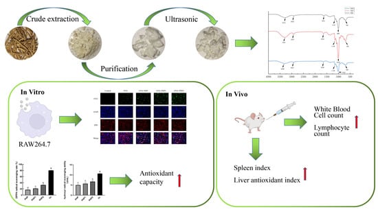
1. Introduction
2. Results and Discussion
2.1. Purification of PS
2.2. Characterization of PHP
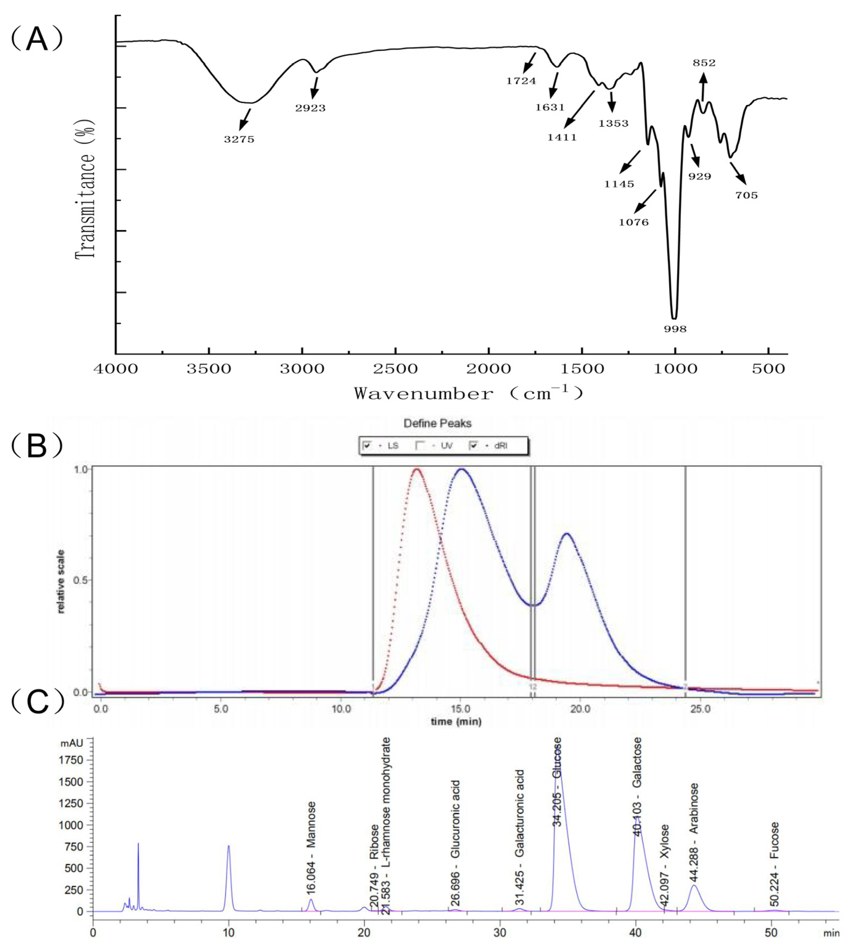
2.2.1. FTIR Analysis
2.2.2. Gel Permeation Chromatography Spectrum
2.2.3. Monosaccharide Composition of P. radix PHP
2.3. Characterization of Ultrasound PHP
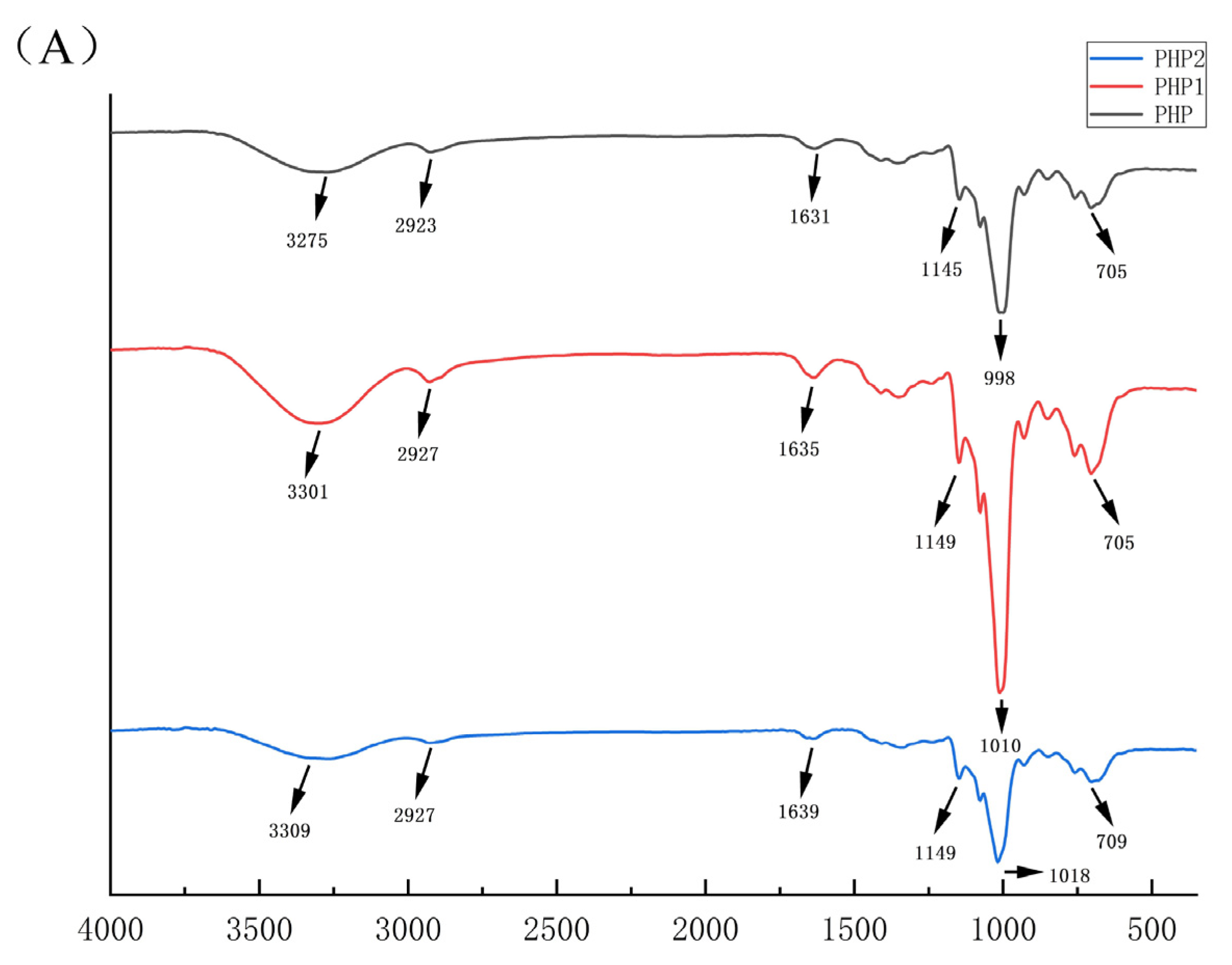
2.3.1. IR Analysis
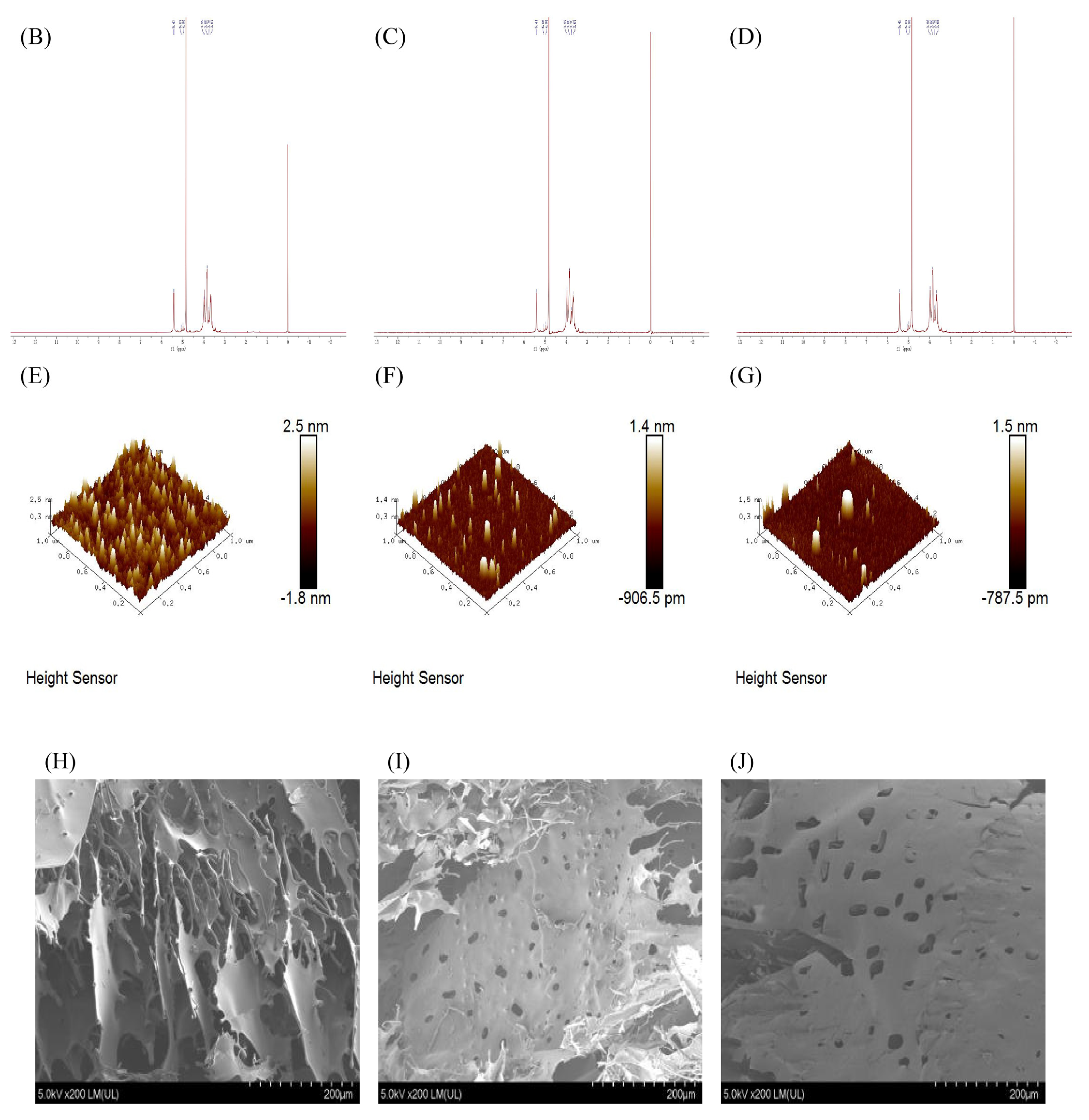
2.3.2. Nuclear Magnetic Resonance (NMR)
2.3.3. AFM and SEM Results
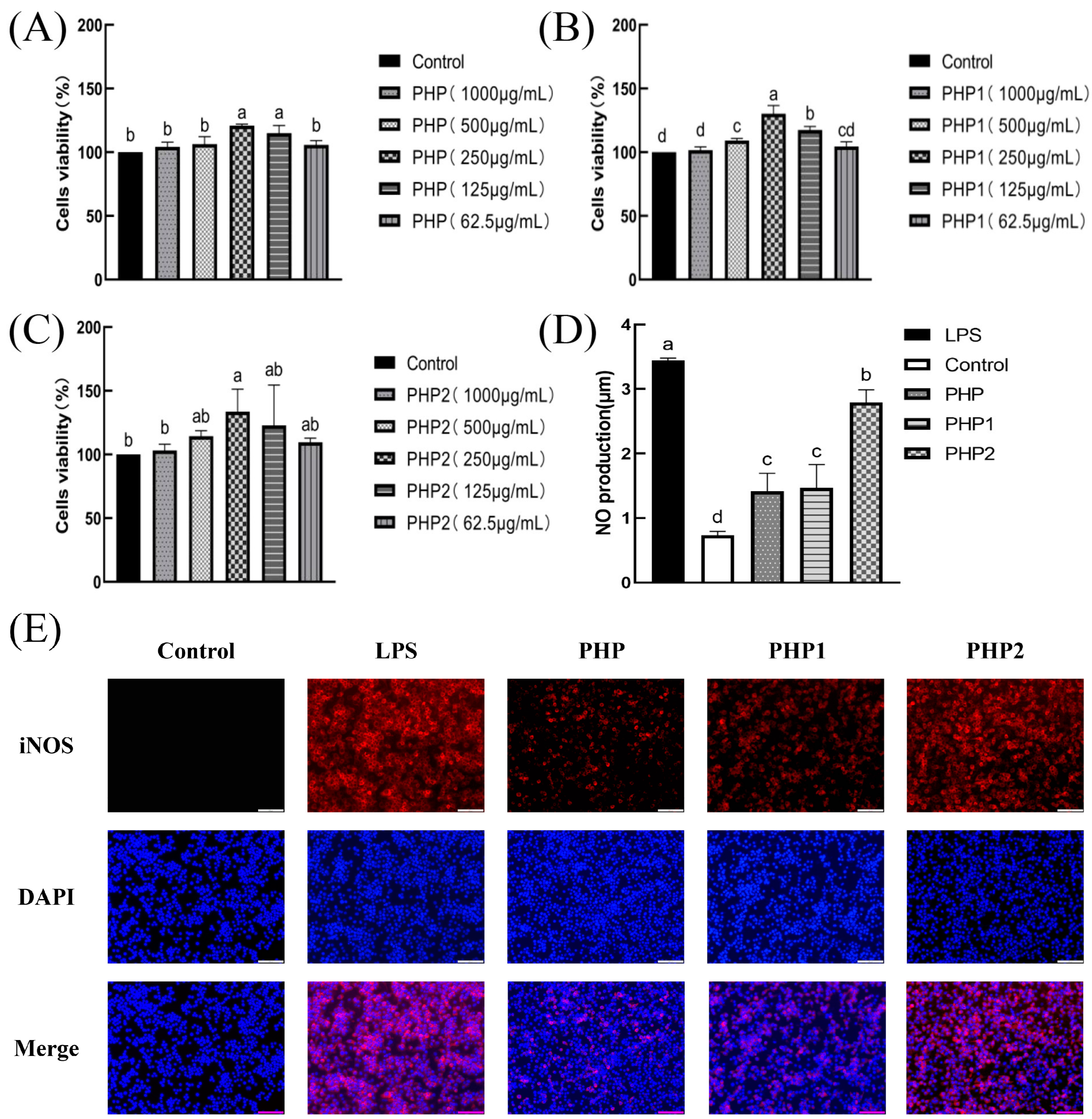
2.4. Activity of Ultrasonic PHP in RAW264.7
2.4.1. Analysis of Viability Test of RAW264.7
2.4.2. Analysis of NO Secretion Test and iNOS Expression of RAW264.7
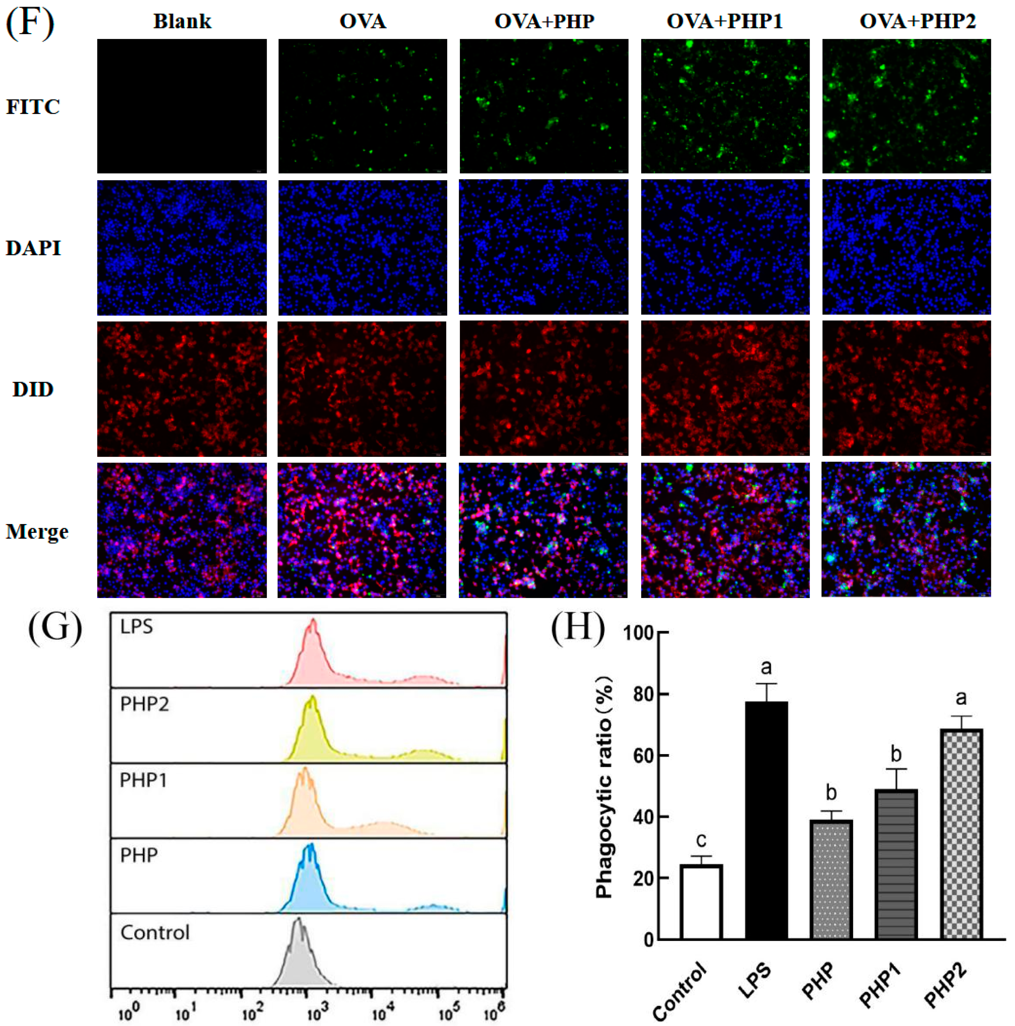
2.4.3. Analysis of Phagocytosis Test of RAW264.7
2.4.4. Analysis of the Polarization of RAW264.7
2.4.5. DPPH Radical Scavenging Ability
2.4.6. Hydroxyl Radical Scavenging Ability
2.5. Activity of Ultrasonic PHP In Vivo
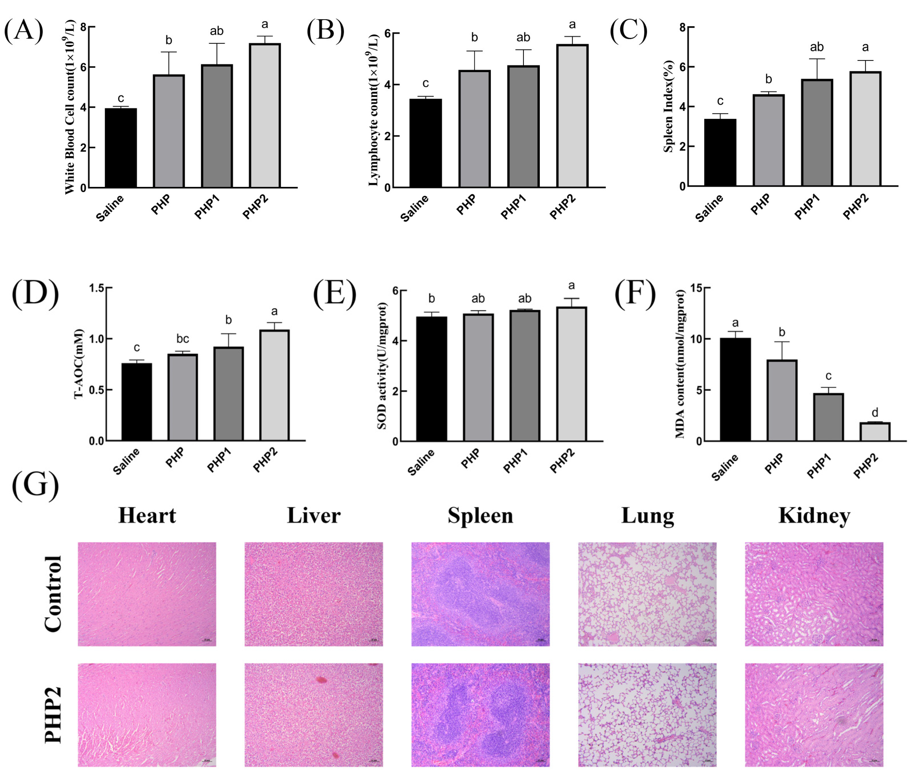
2.5.1. Blood Cell Analysis
2.5.2. Spleen Index
2.5.3. Antioxidant Activity
2.5.4. Histological Evaluation
3. Materials and Methods
3.1. Plant Materials and Chemicals
3.2. Extraction Method of PS
3.2.1. Quantification of PS via Standard Curve Method
3.2.2. Measurement of the PS Content
3.2.3. Single Factor Experiment and Orthogonal Experimental Design
3.2.4. Purification of PS
3.3. Characterization of PHP
3.3.1. Infrared Spectroscopy (IR) Analysis
3.3.2. Gel Permeation Chromatography (GPC) Analysis
3.3.3. Liquid Chromatography (LC) Sample Preparation
3.3.4. Liquid Chromatography (LC) Analysis
3.4. Preparation and Characterization of Ultrasound PHP
3.4.1. Preparation of Ultrasonic PHP
3.4.2. Comparison of IR Analysis
3.4.3. Nuclear Magnetic Resonance (NMR)
3.4.4. AFM and SEM Tests
3.5. In Vivo and In Vitro Activity of Ultrasonic–Treated PHP
3.5.1. Cells and Treatment
3.5.2. Viability Test
3.5.3. Nitric Oxide (NO) Secretion Test and iNOS Expression
3.5.4. Antigen Uptake Capacity Test
3.5.5. Effects of the Phenotypic of RAW264.7 Macrophages
3.5.6. DPPH Radical Scavenging Test
3.5.7. Hydroxyl Radical Scavenging Test
3.5.8. Animals and Treatment
3.5.9. Sample Collection and Preparation
3.5.10. Histological Evaluation
3.5.11. Data Analysis
4. Conclusions
Supplementary Materials
Author Contributions
Funding
Institutional Review Board Statement
Informed Consent Statement
Data Availability Statement
Conflicts of Interest
References
- Chen, J.; Pang, W.; Shi, W.; Yang, B.; Kan, Y.; He, Z.; Hu, J. Structural Elucidation of a Novel Polysaccharide from Pseudostellaria heterophylla and Stimulating Glucose Uptake in Cells and Distributing in Rats by Oral. Molecules 2016, 21, 1233. [Google Scholar] [CrossRef] [PubMed]
- Chen, J.; Pang, W.; Kan, Y.; Zhao, L.; He, Z.; Shi, W.; Yan, B.; Chen, H.; Hu, J. Structure of a pectic polysaccharide from Pseudostellaria heterophylla and stimulating insulin secretion of INS–1 cell and distributing in rats by oral. Int. J. Biol. Macromol. 2018, 106, 456–463. [Google Scholar] [CrossRef] [PubMed]
- Hua, Y.; Hou, Y.; Wang, S.; Ma, Y.; Liu, Z.; Zou, L.; Liu, X.; Luo, Y.; Liu, J. Comparison of Chemical Compositions in Pseudostellariae radix from Different Cultivated Fields and Germplasms by NMR–Based Metabolomics. Molecules 2016, 21, 1538. [Google Scholar] [CrossRef] [PubMed]
- Ng, C.; Wang, Y.C.; Ni, J.J.; So, P.S. Effects of phosphorus–modified biochar as a soil amendment on the growth and quality of Pseudostellaria heterophylla. Sci. Rep. 2022, 12, 7268. [Google Scholar] [CrossRef] [PubMed]
- Xu, G.B.; Zhu, Q.F.; Wang, Z.; Zhang, C.L.; Yang, X.; Zhang, J.J.; Wang, F.R.; Liu, J.; Zhou, M.; Wang, Y.L.; et al. Pseudosterins A–C, Three 1–Ethyl–3–formyl–beta–carbolines from Pseudostellaria heterophylla and Their Cardioprotective Effects. Molecules 2021, 26, 5045. [Google Scholar] [CrossRef] [PubMed]
- Hou, C.; Chen, L.; Yang, L.; Ji, X. An insight into anti–inflammatory effects of natural polysaccharides. Int. J. Biol. Macromol. 2020, 153, 248–255. [Google Scholar] [CrossRef] [PubMed]
- Yu, Y.; Shen, M.; Song, Q.; Xie, J. Biological activities and pharmaceutical applications of polysaccharide from natural resources: A review. Carbohydr. Polym. 2018, 183, 91–101. [Google Scholar] [CrossRef]
- Zhang, B.; Liu, N.; Hao, M.; Zhou, J.; Xie, Y.; He, Z. Plant–Derived Polysaccharides Regulated Immune Status, Gut Health and Microbiota of Broilers: A Review. Front. Vet. Sci. 2021, 8, 791371. [Google Scholar] [CrossRef]
- Ren, L.; Zhang, J.; Zhang, T. Immunomodulatory activities of polysaccharides from Ganoderma on immune effector cells. Food Chem. 2021, 340, 127933. [Google Scholar] [CrossRef]
- Zong, A.; Cao, H.; Wang, F. Anticancer polysaccharides from natural resources: A review of recent research. Carbohydr. Polym. 2012, 90, 1395–1410. [Google Scholar] [CrossRef]
- Shu, X.; Zhang, Y.; Jia, J.; Ren, X.; Wang, Y. Extraction, purification and properties of water–soluble polysaccharides from mushroom Lepista nuda. Int. J. Biol. Macromol. 2019, 128, 858–869. [Google Scholar] [CrossRef] [PubMed]
- Jung–Bum, L.; Azumi, T.; Kyoko, H.; Toshimitsu, H. Structures and antiviral activities of polysaccharides from Sargassum trichophyllum. Carbohydr. Polym. 2011, 86, 995–999. [Google Scholar]
- Saha, S.; Navid, M.H.; Bandyopadhyay, S.S.; Schnitzler, P.; Ray, B. Sulfated polysaccharides from Laminaria angustata: Structural features and in vitro antiviral activities. Carbohydr. Polym. 2012, 87, 123–130. [Google Scholar] [CrossRef] [PubMed]
- Wang, P.C.; Zhao, S.; Yang, B.Y.; Wang, Q.H.; Kuang, H.X. Anti–diabetic polysaccharides from natural sources: A review. Carbohydr. Polym. 2016, 148, 86–97. [Google Scholar] [CrossRef] [PubMed]
- Feng, H.; Du, X.; Tang, J.; Cao, X.; Han, X.; Chen, Z.; Chen, Y.; Zeng, X. Enhancement of the immune responses to foot–and–mouth disease vaccination in mice by oral administration of a novel polysaccharide from the roots of Radix Cyathulae officinalis Kuan (RC). Cell. Immunol. 2013, 281, 111–121. [Google Scholar] [CrossRef] [PubMed]
- Feng, H.; Fan, J.; Qiu, H.; Wang, Z.; Yan, Z.; Yuan, L.; Guan, L.; Du, X.; Song, Z.; Han, X.; et al. Chuanminshen violaceum polysaccharides improve the immune responses of foot–and–mouth disease vaccine in mice. Int. J. Biol. Macromol. 2015, 78, 405–416. [Google Scholar] [CrossRef]
- Li, X.; Zhu, Z.; Ye, L.; Kang, Z.; Zhang, X.; Huang, Y.; Zhang, B.; Zou, Y. Comparison of the Partial Structure and Antioxidant Activity of Polysaccharides from Two Species of Chinese Truffles. Molecules 2020, 25, 4345. [Google Scholar] [CrossRef] [PubMed]
- Pattanayak, M.; Samanta, S.; Maity, P.; Manna, D.K.; Sen, I.K.; Nandi, A.K.; Panda, B.C.; Chattopadhyay, S.; Roy, S.; Sahoo, A.K.; et al. Polysaccharide of an edible truffle Tuber rufum: Structural studies and effects on human lymphocytes. Int. J. Biol. Macromol. 2017, 95, 1037–1048. [Google Scholar] [CrossRef]
- Zhang, Z.; Wang, X.; Yu, S.; Zhao, M. Isolation and antioxidant activities of polysaccharides extracted from the shoots of Phyllostachys edulis (Carr.). Int. J. Biol. Macromol. 2011, 49, 454–457. [Google Scholar] [CrossRef]
- Fan, J.; Feng, H.; Yu, Y.; Sun, M.; Liu, Y.; Li, T.; Sun, X.; Liu, S.; Sun, M. Antioxidant activities of the polysaccharides of Chuanminshen violaceum. Carbohydr. Polym. 2017, 157, 629–636. [Google Scholar] [CrossRef]
- Jiang, J.; Kong, F.; Li, N.; Zhang, D.; Yan, C.; Lv, H. Purification, structural characterization and in vitro antioxidant activity of a novel polysaccharide from Boshuzhi. Carbohydr. Polym. 2016, 147, 365–371. [Google Scholar] [CrossRef] [PubMed]
- Wu, J.; Chen, R.; Tan, L.; Bai, H.; Tian, L.; Lu, J.; Gao, M.; Bai, C.; Sun, H.; Chi, Y. Ultrasonic disruption effects on the extraction efficiency, characterization, and bioactivities of polysaccharides from Panax notoginseng flower. Carbohydr. Polym. 2022, 291, 119535. [Google Scholar] [CrossRef] [PubMed]
- Liu, Y.; Liu, C.; Jiang, H.; Zhou, H.; Li, P.; Wang, F. Isolation, structural characterization and neurotrophic activity of a polysaccharide from Phellinus ribis. Carbohydr. Polym. 2015, 127, 145–151. [Google Scholar] [CrossRef] [PubMed]
- Sanandiya, N.D.; Siddhanta, A.K. Chemical studies on the polysaccharides of Salicornia brachiata. Carbohydr. Polym. 2014, 112, 300–307. [Google Scholar] [CrossRef] [PubMed]
- Chai, Z.; Huang, W.; Zhao, X.; Wu, H.; Zeng, X.; Li, C. Preparation, characterization, antioxidant activity and protective effect against cellular oxidative stress of polysaccharide from Cynanchum auriculatum Royle ex Wight. Int. J. Biol. Macromol. 2018, 119, 1068–1076. [Google Scholar] [CrossRef] [PubMed]
- Luo, B.; Dong, L.M.; Xu, Q.L.; Zhang, Q.; Liu, W.B.; Wei, X.Y.; Zhang, X.; Tan, J.W. Characterization and immunological activity of polysaccharides from Ixeris polycephala. Int. J. Biol. Macromol. 2018, 113, 804–812. [Google Scholar] [CrossRef]
- Wang, H.; Chen, J.; Ren, P.; Zhang, Y.; Omondi, O.S. Ultrasound irradiation alters the spatial structure and improves the antioxidant activity of the yellow tea polysaccharide. Ultrason. Sonochem. 2021, 70, 105355. [Google Scholar] [CrossRef]
- Song, Y.R.; Sung, S.K.; Shin, E.J.; Cho, C.W.; Han, C.J.; Hong, H.D. The Effect of Pectinase–Assisted Extraction on the Physicochemical and Biological Properties of Polysaccharides from Aster scaber. Int. J. Biol. Macromol. 2018, 19, 2839. [Google Scholar] [CrossRef]
- Liu, Y.; Huang, G. Extraction and derivatisation of active polysaccharides. J. Enzym. Inhib. Med. Chem. 2019, 34, 1690–1696. [Google Scholar] [CrossRef]
- Liu, Y.; Huang, W.; Dai, K.; Liu, N.; Wang, J.; Lu, X.; Ma, J.; Zhang, M.; Xu, M.; Long, X.; et al. Inflammatory response of gut, spleen, and liver in mice induced by orally administered Porphyromonas gingivalis. J. Oral Microbiol. 2022, 14, 2088936. [Google Scholar] [CrossRef]
- Carillon, J.; Rouanet, J.M.; Cristol, J.P.; Brion, R. Superoxide dismutase administration, a potential therapy against oxidative stress related diseases: Several routes of supplementation and proposal of an original mechanism of action. Pharm. Res. 2013, 30, 2718–2728. [Google Scholar] [CrossRef] [PubMed]
- Shang, H.; Zhou, H.; Duan, M.; Li, R.; Wu, H.; Lou, Y. Extraction condition optimization and effects of drying methods on physicochemical properties and antioxidant activities of polysaccharides from comfrey (Symphytum officinale L.) root. Int. J. Biol. Macromol. 2018, 112, 889–899. [Google Scholar] [CrossRef] [PubMed]
- Gu, Y.; Qiu, Y.; Wei, X.; Li, Z.; Hu, Z.; Gu, Y.; Zhao, Y.; Wang, Y.; Yue, T.; Yuan, Y. Characterization of selenium–containing polysaccharides isolated from selenium–enriched tea and its bioactivities. Food Chem. 2020, 316, 126371. [Google Scholar] [CrossRef] [PubMed]
- Wang, D.; Shao, S.; Zhang, Y.; Zhao, D.; Wang, M. Insight into Polysaccharides from Panax ginseng C. A. Meyer in Improving Intestinal Inflammation: Modulating Intestinal Microbiota and Autophagy. Front. Immunol. 2021, 12, 683911. [Google Scholar] [CrossRef]
- Chai, Y.; Zhao, M. Purification, characterization and anti–proliferation activities of polysaccharides extracted from Viscum coloratum (Kom.) Nakai. Carbohydr. Polym 2016, 149, 121–130. [Google Scholar] [CrossRef]
- Zhou, S.; Rahman, A.; Li, J.; Wei, C.; Chen, J.; Linhardt, R.J.; Ye, X.; Chen, S. Extraction Methods Affect the Structure of Goji (Lycium barbarum) Polysaccharides. Molecules 2020, 25, 936. [Google Scholar] [CrossRef]


| Analysis Item | Index | Peak 1 | Peak 2 |
|---|---|---|---|
| Molecular Weight | Distribution Index PD (Mw/Mn) | 2.501 | 1.342 |
| Number average molecular weight Mn (g/mol) | 463,500 | 125,900 | |
| Weight average molecular weight Mw (g/mol) | 1,159,000 | 168,900 |
| Levels | Bath Temperature | Extraction Time | Material to Water Ratio |
|---|---|---|---|
| 1 | 40 | 60 | 1:10 |
| 2 | 50 | 80 | 1:15 |
| 3 | 60 | 100 | 1:20 |
| Groups | Treatment |
|---|---|
| Saline | 0.5 mL Saline |
| PHP | 0.5 mL PHP (300 mg/kg B.W.) |
| PHP1 | 0.5 mL PHP1 (300 mg/kg B.W.) |
| PHP2 | 0.5 mL PHP2 (300 mg/kg B.W.) |
Disclaimer/Publisher’s Note: The statements, opinions and data contained in all publications are solely those of the individual author(s) and contributor(s) and not of MDPI and/or the editor(s). MDPI and/or the editor(s) disclaim responsibility for any injury to people or property resulting from any ideas, methods, instructions or products referred to in the content. |
© 2023 by the authors. Licensee MDPI, Basel, Switzerland. This article is an open access article distributed under the terms and conditions of the Creative Commons Attribution (CC BY) license (https://creativecommons.org/licenses/by/4.0/).
Share and Cite
Li, H.; Liu, Z.; Liu, Q.; Zhang, X.; Li, S.; Tang, F.; Zhang, L.; Yang, Q.; Wang, Q.; Yang, S.; et al. Extraction of Polysaccharides from Root of Pseudostellaria heterophylla (Miq.) Pax. and the Effects of Ultrasound Treatment on Its Properties and Antioxidant and Immune Activities. Molecules 2024, 29, 142. https://doi.org/10.3390/molecules29010142
Li H, Liu Z, Liu Q, Zhang X, Li S, Tang F, Zhang L, Yang Q, Wang Q, Yang S, et al. Extraction of Polysaccharides from Root of Pseudostellaria heterophylla (Miq.) Pax. and the Effects of Ultrasound Treatment on Its Properties and Antioxidant and Immune Activities. Molecules. 2024; 29(1):142. https://doi.org/10.3390/molecules29010142
Chicago/Turabian StyleLi, Hangyu, Ziwei Liu, Qianqian Liu, Xinnan Zhang, Sheng Li, Feng Tang, Linzi Zhang, Qian Yang, Qiran Wang, Shuyao Yang, and et al. 2024. "Extraction of Polysaccharides from Root of Pseudostellaria heterophylla (Miq.) Pax. and the Effects of Ultrasound Treatment on Its Properties and Antioxidant and Immune Activities" Molecules 29, no. 1: 142. https://doi.org/10.3390/molecules29010142