
Core-Hole Excitation Spectra of the Oxides and Hydrates of Fullerene C60 and Azafullerene C59N
by
 , , ,
, , ,
Xiong Li
1,2 ,
,
Shuyi Wang
1,3,
Jingdong Guo
3,
Ziye Wu
4,
Changrui Guo
4,
Shaohong Cai
4,5,* and
Mingsen Deng
4,* 1
College of Big Data and Information Engineering, Guizhou University, Guiyang 550025, China
2
School of Science, East China University of Technology, Nanchang 330013, China
3
Guizhou Provincial Key Laboratory of Computational Nano-Material Science, Guizhou Education University, Guiyang 550018, China
4
School of Information, Guizhou University of Finance and Economics, Guiyang 550025, China
5
Department of Resources and Environment, Moutai Institute, Renhuai 564507, China
*
Authors to whom correspondence should be addressed.
Molecules 2024, 29(3), 609; https://doi.org/10.3390/molecules29030609
Submission received: 3 December 2023
/
Revised: 18 January 2024
/
Accepted: 24 January 2024
/
Published: 26 January 2024
(This article belongs to the Special Issue Computational Chemistry Insights into Molecular Interactions)
Abstract
:The interaction of fullerenes and their derivatives with environmental molecules such as oxygen or water was crucial for the rational design of low-dimensional materials and devices. In this paper, the near-edge X-ray absorption fine structure (NEXAFS), X-ray emission spectroscopy (XES) and X-ray photoelectron spectroscopy (XPS) shake-up satellites were employed to distinguish the oxides and hydrates of the fullerene C60 and azafullerene C59N families. The study includes various isomers, such as the open [5,6] and closed [6,6] isomers of C60O, C60H(OH), C60-O-C60, C60H-O-C60H, C59N(OH) and C59N-O-C59N, based on density functional theory. These soft X-ray spectra offered comprehensive insights into the molecular orbitals of these azafullerene molecular groups. The oxygen K-edge NEXAFS, carbon and oxygen K-edge XPS shake-up satellite spectra provided valuable tools for distinguishing oxides or hydrates of fullerene C60 and azafullerene C59N. Our findings could significantly benefit the development of fullerene functional molecular materials and expand the application scope of soft X-ray spectroscopy as a molecular fingerprinting tool for the fullerene family.
1. Introduction
The carbon allotrope fullerene C60 and its molecular crystals are considered fundamental units in probing the mechanisms of transport and photoelectric processes. Their fast carrier mobility and strong electron affinity position fullerenes are at the forefront of electronic device applications, including field-effect transistors [1] or photovoltaics [2], nanomedicine [3], nanoelectronics [4], organic and inorganic nanocomposites [5,6]. The azafullerene C59N monomer, notable for its unpaired electrons and resultant free radical behavior [7], remains a vibrant research area despite its instability. For example, the report of Martín-Gomis et al. showed promising implications in the development of organic solar cells built with covalent donor-acceptor systems using C59N as the acceptor [8]. Further, the C59N system has been shown to achieve a more persistent charge-separated state [9], underscoring its potential in advanced applications.
However, the insertion of extrinsic species, such as oxygen and water, could substantially alter the physical properties of these electronic materials [10]. Numerous studies have reported evidence for the sensitivity of fullerene or azafullerene to the incorporation of such impurities [11,12,13,14]. To further understand these impacts, Lin et al. conducted a detailed study on the oxidation process of azafullerene C59N [15]. Research has established that oxides and hydrates of fullerene and azafullerene are key materials in constructing a variety of fullerene-based nanostructures [16,17]. As a straightforward derivative of fullerene, C60O has emerged as a foundational material for producing more complex fullerene oxides, playing a crucial role in the synthesis of diverse compounds [18,19,20]. Fedurco et al. and Jones et al. demonstrated that N@C60O represented a valuable intermediary for researchers engaged in quantum information processing at the molecular level [21]. The work of Mondal et al. has also highlighted the complex interplay between thermal and mechanical transformations within the C60O cluster, which could influence its thermoelectric properties through the modulation of intermolecular interactions [22]. Moreover, the C59N oxides have been identified as robust recyclable catalysts, featuring a rate-determining energy barrier of 0.80 eV [23].
The presence of oxides and water-related reactions in fullerene and azafullerene compounds often leads to substantial modifications in the physical properties of organic semiconductors. These modifications are instrumental in the fabrication of numerous fullerene nanostructures. However, the properties of the oxides and hydrates exhibit significant variations [18]. Consequently, understanding their electronic and chemical characteristics is essential for further exploration and utilization. In this regard, Palotás et al. have made a noteworthy contribution by providing the initial experimental infrared multiple-photon dissociation vibrational spectra of gaseous C60O+ and C60OH+. However, challenges persist, as the experimental band centered at 1333 cm−1 was not accurately reproduced by the theoretical [6,6]C60OH+ spectrum [24]. This discrepancy demands considerable efforts dedicated to acquiring a deeper understanding of these materials, as evident in the studies mentioned earlier [25,26,27,28], where X-ray spectra have proven to be valuable experimental methods for investigating the electronic structures of aza[60]fullerene [29,30,31].
Near-edge X-ray absorption fine structure (NEXAFS) is a well-established spectroscopic approach employed for examining the local molecular structure and acquiring insights into unoccupied molecular orbitals. X-ray emission spectroscopy (XES) was frequently utilized to investigate the occupancy density of molecular electronic states. Both NEXAFS and XES offered element and site-specific capabilities, making them potent tools for elucidating intricate electronic properties of materials. Additionally, X-ray photoelectron spectroscopy (XPS) shake-up satellites, engendered by excitations of valence electrons concomitant with the ionization of core electrons, provided information regarding the valence band of materials in the vicinity of the core-hole screening [32,33,34,35,36,37,38,39]. For instance, Klein et al. utilized the delta-self-consistent-field (ΔSCF) and transition potential (TP) methods for calculating XPS and NEXAFS transitions, allowing the prediction of K-shell information [40]. Meanwhile, Armillotta et al. investigated the mechanisms of metal intermolecular coordination using NEXAFS technology [41].
The open [5,6] and closed [6,6] isomers of C60O, as well as C60-O-C60, are examples of molecular crystals that had been synthesized [42,43,44]. Creegan et al. identified the epoxide isomer of C60O through 13C NMR and FTIR examinations, revealing that the oxygen atom bridges to carbon atoms of two hexagons with C2v symmetry [42]. Weisman et al. further demonstrated that C60O can be prepared in both closed [6,6] epoxide and open [5,6] annulene isomers, depending on the synthesis method [44]. The C60-O-C60 molecular crystal consists of a C60 dimer, and an oxygen atom bridged in a furan-like ring (Figure 1e) [43], while the two isomers of C60O are composed of a C60 monomer and an oxygen atom in the form of epoxide and annulene, respectively (Figure 1b,c) [42]. Erbahar et al. discovered that the most stable structure for a single oxygen atom within (C59N)2 involves the oxygen atom in an ether configuration, forming a cross-linking bond between two (C59N) units [45], namely C59N-O-C59N (Figure 1j). In the reaction between H2O and C60, the configuration of the cross-linking bond could also be considered, specifically C60H-O-C60H (Figure 1f). In this study, the NEXAFS, XES and XPS shake-up satellites for seven exemplary members of the hydrates and oxides of fullerene C60 and aza[60]fullerene C59N, namely open [5,6] and closed [6,6] isomers of C60O, C60H(OH), C60-O-C60, C60H-O-C60H, C59N(OH) and C59N-O-C59N, were studied by employing the full core-hole (FCH) potential method as well as equivalent core-hole Kohn–Sham (ECH-KS) approach. The study provides valuable insights that can support and guide further experimental investigations related to these oxide materials and water-involved reactions.
2. Results and Discussion
The optimized molecular models of the fullerene C60, aza[60]fullerene C59N, their oxides and hydrates are shown in Figure 1. The carbon atoms C1 and C1′, C2 and C2′, C3 and C3′, C4 and C4′ and C5 and C5′ are symmetrically equivalent. The binding energies (BE) of the H atom, O atom, OH, C59N, and C60 to the C59N and C60 monomers were calculated to obtain the stability of oxides and hydrates of the fullerenes and azafullerenes by
where is the electronic energy of C60, open [5,6] and closed [6,6] isomers of C60O, C60H(OH), C60-O-C60, C60H-O-C60H, C59N, C59N(OH), (C59N)2 and C59N-O-C59N molecules, Ei is the electronic energy of the isolated H atom, O atom, OH, C59N and C60 molecule, which are the monomers that constitute the ten molecules mentioned above. HOMO-LUMO energy gap (Egap) is calculated by
where are LUMO energies and are HOMO energies. The binding energies, HOMO and LUMO energies and Egap are collected in Table 1, which are verified well with the theoretical investigations based on the B3LYP [46] and LDA-PW91 [45] level. The symmetry point groups and the number of symmetrically inequivalent carbon atoms for the ten molecule groups are also shown in Table 1. The energy gaps decrease upon oxidation and hydration of fullerene C60, whereas the gaps increase for azafullerene C59N. The energy gap change trends indicated that the fullerene C60 becomes more reactive after oxidation and hydration, while azafullerene C59N becomes less reactive after oxidation and hydration. The binding energies confirm that these oxides and hydrates are more stable than the (C59N)2 molecule, with the C60H-O-C60H molecule identified as the most stable oxidation configuration.
The HOMOs and LUMOs of the ten molecular groups, calculated with B3LYP/6-311+G*, are presented in Figure S1, which are plotted with VESTA [47]. The isovalues for C60-O-C60, C60H-O-C60H, (C59N)2 and C59N-O-C59N molecules were set to 0.02, while that of the other six molecular groups was set to 0.04. The calculated energy of HOMO and LUMO of C60, C59N and (C59N)2 are in good agreement with the works of Mohammadi and colleagues [48,49,50]. It is notable that the two isomers of C60O had nearly the same energy gap and only a 2.11 kcal/mol difference in binding energy. Yet, they exhibit distinct electronic features, such as in O3 adsorption [18].
2.1. Carbon K-edge NEXAFS and XES
The NEXAFS and XES spectra of carbon K-edges of the selected molecules, including the open [5,6] and closed [6,6] isomers of C60O, C60H(OH), C60-O-C60, C60H-O-C60H, C59N(OH) and C59N-O-C59N, were plotted in Figure 2. The spectra of the C60, C59N, and (C59N)2 molecules were also plotted for comparison. Also, the experimental NEXAFS spectra of C60, [6,6]C60O and (C59N)2 molecules we could find are reproduced from the work of Käämbre et al. [51] and Schulte et al. [52] for comparison, which are plotted in dashed lines. It is clear that simulated NEXAFS spectra of C60 and [6,6]C60O molecules are in good agreement with the experimental results. Our simulated spectra reproduced all the important characteristics of the experiment results.
Carbon K-edge NEXAFS. The NEXAFS spectra in Figure 2a exhibit five distinct peaks (labeled A–E) at nearly identical energy positions for each molecule, as illustrated in Figure 1. In addition, the orbital transitions from the 1s to the LUMO (shown as red bars), LUMO + 1 (shown as green bars) and LUMO + 2 (shown as blue bars) in the NEXAFS spectra of the nine molecules, along with the discrete intensities (shown as red bars) of the NEXAFS spectrum of the fullerene C60 molecule were plotted below each convoluted spectrum. For the C59N molecule, the contributions from β-spin electrons are represented as negative numbers. Although the NEXAFS spectra of these ten molecules exhibit similar peak positions, some notable differences can be observed. Specifically, depending on their lower symmetry—C2h for (C59N)2, C2v for closed [6,6] isomer of C60O, C60-O-C60, C60H-O-C60H, C59N-O-C59N and Cs for open [5,6] isomer of C60O, C60H(OH) and C59N(OH), the NEXAFS spectra of these molecules exhibit a larger number of molecular orbital (MO) transitions from C 1s orbitals to unoccupied orbitals within each peak in comparison to that of C60. From the feature peak E to their low-energy region, which corresponds to the C K-shell 1s absorption edges, signals that the C K-shell 1s absorption edges of all ten molecules are nearly identical. This indicated that both fullerene C60 and aza[60]fullerene C59N maintain the same valence state of carbon even after oxidation or exposure to water.
In the oxides and hydrates of fullerene C60, the NEXAFS spectrum of the closed [6,6] isomer of C60O resembles that of the C60H(OH) and C60H-O-C60H, as they all have a relatively weak and broad peak C and a strong peak B, and a very weak feature peak A’ between the peaks A and B can be observed. While peak B of the NEXAFS spectra of the other two molecules open the [5,6] isomer of C60O, and C60-O-C60 exhibit a slightly higher intensity than peak C, the feature peak A’ only traced in the C60-O-C60 molecule. The feature peak A’ primarily arose from the carbon 1s orbital to the LUMO + 2. Notably, the transition 1s → LUMO + 2 is notably weaker in the open [5,6] isomer of C60O. The feature peak A’ is linked to the change of electronic structures of the C60 molecule upon its interaction with another atom or molecule.
It is evident that the NEXAFS spectra for C59N(OH), (C59N)2 and C59N-O-C59N molecules all have a weak feature peak A′, which is the marker of the oxides or reactions involving H2O of aza[60]fullerene C59N. However, they exhibit significant differences in comparison to that of the C59N molecule, which lacks peak A′. Remarkably, nearly all carbon atoms exhibit similar NEXAFS spectral feature peaks A, B, C, D and E, regardless of any specific molecule. However, there is one notable exception, where feature peak A lacks contribution from the nearest carbon atoms (C1, C2, C3, C4, C5 in Figure 1) neighboring the nitrogen and oxygen atoms. This distinction in feature peak A among the spectra of the neighbor of the nitrogen, oxygen and other carbon atoms could be attributed to their distinct local (chemical) environments.
Carbon K-edge XES. The XES spectra at the carbon K-edge of the ten molecules were plotted in Figure 2b. The discrete XES intensities of [5,6]C60O and C59N are shown in red bars (α-spin) and green bars (β-spin). These bars are superimposed on their convoluted XES spectra. The XES spectra of these ten molecules exhibit limited discernible differences in contrast to the NEXAFS spectra, except for specific peaks. The peak A in the C59N molecule can be attributed to the MO transition from the HOMO to the 1s orbital of carbon atom C3 [46], and the peak B can be attributed to the transitions from the HOMO to the 1s orbitals of other carbon atoms. Similarly, peak B in the [5,6]C60O isomer primarily arises from the MO transitions between the HOMO and the 1s orbital of the carbon atoms. Consequently, it might be beneficial to utilize the feature peak B in the XES spectrum of the [5,6]C60O isomer to examine its presence within C60. Although the experimental aspects might pose certain challenges, employing these distinctive features can provide valuable insights into their existence within the C60 crystalline and provide effective evidence for distinguishing the open [5,6] isomers of C60O from oxides and hydrates of C60.
In summary, the carbon K-edge NEXAFS and XES spectra provide electronic structure information about the oxides and hydrates of C60 and C59N, which can be used to distinguish the open [5,6] isomer of C60O. However, distinguishing other oxides and hydrates using these spectra remains challenging.
2.2. Nitrogen K-edge NEXAFS and XES
To detect the presence of C59N(OH) and C59N-O-C59N, which are the oxides or hydrates of aza[60]fullerene C59N, the nitrogen K-edge NEXAFS and XES spectra were simulated, as shown in Figure 3. The spectra of the C59N and (C59N)2 were also plotted for comparison. The experimental NEXAFS spectrum of (C59N)2 molecule is shown in dashed lines reproduced from the work of Schulte and colleagues [52]. It can be found that the simulated and experimental spectra are in good agreement.
Nitrogen K-edge NEXAFS. Both the C59N(OH) and C59N-O-C59N exhibit a faint shoulder A in the NEXAFS spectra (Figure 3a), which is attributed to the nitrogen 1s to LUMO transition. In contrast, the C59N molecule presents a relatively more pronounced first peak (peak A) at lower excitation energy, resulting from the nitrogen 1s → LUMO transition of β-spin. The MO transitions and corresponding energies of peak or shoulder A are available in Table S1. Despite the significance of the core-hole effect in comprehending NEXAFS spectra, the disparity observed in the nitrogen 1s → LUMO MO transition among the four molecules could be elucidated by considering the distribution of the LUMOs in the Ground state (GS). The LUMOs of C59N(β-spin), C59N(OH), (C59N)2 and C59N-O-C59N were plotted in Figure S1g–j, respectively. The LUMO of C59N exhibits a noticeable distribution around the nitrogen atom, whereas the LUMO of C59N(OH) exhibits a pronounced carbon character, similar to the (C59N)2, which fits well with prior results [53,54]. The LUMO in the C59N-O-C59N molecule is predominantly concentrated on the intermolecular bond of carbon, which contributes to the existence of a weak shoulder A in their NEXAFS spectra. Notably, the weak shoulder A observed in C59N(OH) and C59N-O-C59N are valuable for distinguishing them from C59N.
Nitrogen K-edge XES. The XES spectra at the nitrogen K-edge provide valuable insights, particularly when combining information obtained from GS calculations. In the case of the C59N(OH), (C59N)2 and C59N-O-C59N, their HOMOs exhibit some nitrogen characteristics, as illustrated in Figure S1h–j. For those compounds, the transitions from the HOMO to the nitrogen 1s orbital occur at relatively lower energies compared with the energy corresponding to the distinct peak A in the C59N molecule. The peak A in C59N originates from the transition from the HOMO (α-spin) to nitrogen 1s orbital transition [46]. Interestingly, the XES spectra of both C59N(OH) and C59N-O-C59N exhibit strong and sharp feature peaks B and D, resembling those observed in (C59N)2. This similarity is in stark contrast to the sharp and pronounced feature, peak C, observed in the C59N. These peaks are a result of transitions originating from deeper valence orbitals to the nitrogen 1s orbital(s), allowing for a clear distinction between C59N(OH) and C59N-O-C59N when compared with C59N.
2.3. Oxygen K-edge NEXAFS and XES
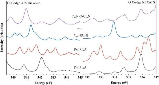
Oxygen K-edge NEXAFS. The simulated NEXAFS spectra at the oxygen K-edge of the seven oxygen-containing molecules are shown in Figure 4a. The experimental NEXAFS spectrum of [6,6]C60O molecule is shown in the dashed line, which is reproduced from the work of Wohlers et al. at 370K [55]. The energy positions of peak (shoulder) A (corresponds to 1s → LUMO molecular orbital transition) and peaks B and C are listed in Table 2. Both the [6,6]C60O and the C60H-O-C60H produce the first shoulder A or peak A with moderate intensity, which arises from the transition of oxygen 1s to the LUMO. The peak A of the NEXAFS spectra of the closed [6,6] isomer of C60O is observed at a higher excitation energy (532.35 eV), whereas it appears at a lower excitation energy in the C60H-O-C60H (531.97 eV). Conversely, the oxygen 1s → LUMO transition only produces a weak peak A in the C60H(OH) and a weak shoulder A in the [5,6]C60O, C60-O-C60, C59N(OH) and C59N-O-C59N molecules. The disparity observed in the oxygen 1s → LUMO transition among these seven molecules could also be elucidated by examining the distribution of their LUMOs in the GS. Unlike the [5,6]C60O, C60H(OH), C60-O-C60, C59N(OH) and C59N-O-C59N, whose LUMOs have strong carbon character (Figure S1b,d,e,h,j), the LUMOs of the [6,6]C60O and the C60H-O-C60H molecules present here also exhibit some distribution around the oxygen atom (Figure S1c,f). Therefore, their NEXAFS spectra exhibit a moderate shoulder A or peak A. In particular, the moderately strong shoulder A with its relatively high excitation energy in the [6,6]C60O and the moderately strong peak A with its relatively low excitation energy in the C60H-O-C60H can be considered as the fingerprint of their existence.
Furthermore, in the case of the [5,6]C60O, C60H(OH) and C59N(OH), the oxygen 1s → LUMO transition is shown in the initial peak A or shoulder A with a weak intensity at nearly identical higher excitation energies (532.80 eV, 532.64 eV and 532.78 eV), among the five molecules with pronounced carbon characteristics in their LUMOs. The molecular groups, including the open [5,6] isomer of C60O, C60H(OH) and C59N(OH), show that the absence of a strong peak B in comparison to the other two molecules, which can serve as the fingerprint of [5,6]C60O. Additionally, C60H(OH) can be distinguished by the presence of a distinct peak B, while the presence of C59N(OH) can be recognized by the strong peak B and weak peak C. The spectra of the other two molecules, C60-O-C60 and C59N-O-C59N, fall into another category, in which the oxygen 1s → LUMO transition generates the initial shoulder A with a weak intensity at nearly identical lower excitation energies (532.03 eV and 532.04 eV). These could be attributed to the influence of another carbon cage. At the same time, the energy positions of their peak B are also quite similar (532.64 eV and 532.51 eV). Nevertheless, the distinct feature peak C at 533.45 eV in the C59N-O-C59N molecule serves as its distinguishing characteristic compared with C60-O-C60.
Oxygen K-edge XES. The XES spectra at the oxygen K-edge are shown in Figure 4b. The MO transitions and corresponding energies of peak A are compared in Table S2. The XES spectrum of the [5,6] and [6,6] isomers of C60O, as well as the C59N-O-C59N, exhibit a discernible weak peak A at high energy (515.88 eV, 515.82 eV and 515.74 eV). These peaks arise from the HOMO to oxygen 1s orbital transition. The HOMOs of the [5,6] and [6,6] isomers of C60O and C59N-O-C59N, as depicted in Figure S1b,c,j, exhibit some characteristics of oxygen. This results in the manifestation of the moderate peak A in their XES spectra. Conversely, the HOMOs of C60H(OH), C60-O-C60 and C60H-O-C60H possess strong carbon attributes, as shown in Figure S1d–f. The transition from the HOMO to the oxygen 1s orbital does not result in the observation of any discernible peaks in their XES spectra. For the C59N(OH), although its HOMOs also exhibit little oxygen characteristics, the transition from the HOMO to the oxygen 1s orbital occurs at relatively lower energy (515.33 eV) compared with the [5,6] and [6,6] isomers of C60O, as well as C59N-O-C59N. Significantly, the XES spectra reveal distinct characteristics, including the moderately strong peak B in the C60-O-C60, as well as the uniformly strong and sharp peak C in all seven molecules. These peaks arise from transitions originating in the deeper valence orbitals to the oxygen 1s orbital, allowing for the distinction of oxides or hydrates of different fullerene and azafullerene.
2.4. XPS Shake-Up Satellites
Carbon K-edge XPS shake-up satellites. The calculated XPS shake-up satellites at the carbon K-edge for the fullerene C60, aza[60]fullerene C59N, their oxides and reactions involving H2O and (C59N)2 were plotted in Figure 5a. Additionally, spectra of C60, C59N and (C59N)2 were also provided for comparison. The XPS main lines of all symmetrically inequivalent carbon atoms are represented as red bars underneath each XPS shake-up spectrum of the other nine members. Contributions from the β-spin electrons in the C59N molecule were plotted as green bars. The experimental XPS shake-up satellite spectrum of (C59N)2 molecule is reproduced from the work of Schulte et al. [52], which is in agreement very well with our simulated spectrum. It is evident that the XPS shake-up satellites of the oxides or hydrates of fullerene C60 show significantly different profiles compared with those of C60, except for that of [6,6] C60O. This observation signifies an alteration in the electronic structures of the valence orbitals following the oxidation or reaction with water of the C60 molecule. Similarly, the spectra of the C59N(OH) and C59N-O-C59N exhibit distinct profiles compared with those of the C59N and aza[60]fullerene dimer (C59N)2.
The spectra of these ten molecules exhibit very similar profiles, featuring prominent peaks A and C located proximate to the XPS main line, except for C60 and [6,6]C60O. The presence of shoulder B can be attributed to contributions from several carbon atoms in C60H(OH). The primary contributions to the peaks A and C in the nine molecules have been elucidated and are collected in Table 3. It is interesting that except for the peak C in the open [5,6] isomer of C60O, C59N, C59N(OH) and C59N-O-C59N, resulting from the shake-up mechanisms, the primary contributions to the peaks A and C in the other five molecules are actually the XPS main lines of the atoms C1, C2, C3, C4 and C5. It is evident that the carbon atom C1 (sp2-like) is connected to the oxygen atom, C3 (in the C59N) and C5 (in the C59N, C59N(OH), (C59N)2 and C59N-O-C59N) are bonded to the nitrogen atom with sp2-like, while the carbon atoms C2 (connected to the oxygen atom), C3 (in the C60H(OH), C60-O-C60, C60H-O-C60H, C59N(OH), (C59N)2, and C59N-O-C59N) and C4 (in the C60H(OH), C60-O-C60, and C60H-O-C60H) are sp3-like atoms in the closed [6,6] isomer of C60O, C60H(OH), C60-O-C60, C60H-O-C60H, C59N(OH), (C59N)2 and C59N-O-C59N (Figure 1b–j). Hence, their binding energies exhibit a slight increase compared with those of other carbon atoms. The hybridization of carbon atoms and the blue-shifted binding energy of these hybridized carbon atoms relative to the XPS main line for the oxides or hydrates of fullerene C60 and aza[60]fullerene C59N are collected in Table 4.
The local electronic structures of the C3 and C4 atoms in the oxides or hydrates of fullerene C60 show minimal changes after bonding with other atoms (like H atom), such as (C4 in the C60H(OH) and C4 in the C60H-O-C60H), or a large molecule (like C3 in the C60-O-C60), although they become an sp3-like carbon, and consequently lead to blue-shifted binding energy—around 1 eV in the C60H(OH) (C4), C60-O-C60 (C3) and C60H-O-C60H (C4) in comparison to their XPS main line. In contrast, sp3-like carbon atoms bonded to oxygen atoms are shown a pronounced blue-shifted in their binding energy—approximately 2 eV in the closed [6,6] isomer of C60O(C2), C60H(OH) (C3), C60-O-C60 (C4) and C60H-O-C60H (C3) when compared with their XPS main line. The larger blue shifts demonstrated the local electronic structures of the sp3-like C2, C3 and C4 atoms changing a lot after being connected to an oxygen atom (such as C2 in the [6,6]C60O, C3 in the C60H(OH) and C60H-O-C60H, C4 in the C60-O-C60). The C K-edge XPS shake-up satellite spectra reveal the distinctive features of the [6,6]C60O molecule, characterized by a strong peak C and the absence of peak A, while C60H(OH) exhibits a unique shoulder B in its spectrum. Therefore, C K-edge XPS shake-up satellites serve as a valuable technique for detecting the presence of [6,6]C60O and C60H(OH) molecules, enabling their differentiation from other fullerene C60 oxides and hydrates.
In the oxides or hydrates of azafullerene C59N, the binding energy of carbon atom C5 is blue-shifted by approximately 1 eV relative to the XPS main line, while the binding energy of carbon atom C3 remains unchanged in the C59N molecule [46]. However, upon oxidation (e.g., C59N-O-C59N) or reaction with water (e.g., C59N(OH)), the local electronic structure of the carbon atom C3 undergoes significant alteration, transforming it into a sp3-like carbon atom in the C59N(OH) and C59N-O-C59N. Consequently, a substantial blue-shift binding energy of around 3 eV is observed in the C59N(OH) and C59N-O-C59N compared to their XPS main line, while the blue-shift is approximately 2 eV in the (C59N)2[46]. The unique peak D in the spectra of C59N(OH) and C59N-O-C59N molecules distinguish them from C59N and (C59N)2 molecules. However, the C K-edge XPS shake-up satellite spectra could not differentiate between C59N(OH) and C59N-O-C59N molecules.
It is noteworthy that only C1 exhibits a sp2-like hybridization, leading to a subtle blue shift in binding energy relative to the XPS main line among all the carbon atoms bonded to an oxygen atom. The uniqueness of the C1 atom demonstrates a remarkable change in the localized electronic structure of C1 in the [5,6]C60O isomer after breaking the five- and six-membered ring structures of the fullerene cage.
Nitrogen K-edge XPS shake-up satellites. The alterations in electronic structures can be elucidated through the analysis of nitrogen K-edge XPS shake-up satellites (Figure 5b). The energy of peak B in the spectra for the C59N, C59N(OH), (C59N)2 and C59N-O-C59N are shown in Table 5. In both C59N(OH) and C59N-O-C59N, peak A is relatively weaker compared with that of the C59N. Furthermore, peak B in C59N-O-C59N exhibits a blue shift of approximately 0.15 eV compared with peak B of the C59N(OH). An extensive assignment of the principal contributions to peaks A and B, which are observed in the nitrogen K-edge XPS shake-up satellites of these four members within the C59N family, is provided in Table S3. The information can serve as a valuable reference for future experimental investigations. It is evident that, apart from C59N and C59N(OH), feature B in the other two molecules is attributed to excitations originating from deeper valence orbitals.
Oxygen K-edge XPS shake-up satellites. The alterations in electronic structures can also be discerned from the oxygen K-edge XPS shake-up satellites, as depicted in Figure 5c. The energies of peaks A, B and C in the oxygen K-edge XPS shake-up satellites are shown in Table 6 for the seven oxygen-containing molecules. The [6,6]C60O, C60H(OH), C60-O-C60 and C60H-O-C60H exhibit three feature peaks A, B and C. The other three molecules, open [5,6] isomer of C60O, C59N(OH) and C59N-O-C59N, do not show a discernible peak B, highlighting a noticeable difference between the [6,6]C60O, C60H(OH), C60-O-C60 and C60H-O-C60H. In comparison to the [5,6]C60O molecule, the featured peak A of C59N(OH) demonstrates a red shift of 0.52 eV, whereas the characteristic peak A of the C59N-O-C59N exhibits a red shift of 1.00 eV. In contrast, in comparison with [6,6]C60O, the characteristic peak A of C60H(OH) shows a red shift of 0.42 eV, the feature peak A of the C60-O-C60 exhibits a red shift of 0.66 eV, and the characteristic peak A of the C60H-O-C60H shows a red shift of 1.00 eV. Hence, the oxygen K-edge XPS shake-up satellites offer a valuable technique to detect the presence of these seven molecules, which can also be used to distinguish them from other oxides or hydrates family members of fullerene C60 and aza[60]fullerene C59N.
The detailed assignments of the primary contributions to peaks A, B and C within the oxygen K-edge XPS shake-up satellites for these seven oxygen-containing molecules are collected in Table S4, which can provide a valuable reference for future experiments. It is notable that peak B includes excitations from deeper valence orbitals in C60-O-C60. With the exception of C60H(OH) and C59N(OH) molecules, peak C also encompasses excitations from deeper valence orbitals in the remaining five molecules.
3. Computational Details
The ten groups of molecular models are presented in Figure 1, including fullerene(C60), C60O with oxygen in the [5,6] open annulene and [6,6] closed epoxide isomers, C60H(OH), ether-oxygen-bridged fullerene dimer C60-O-C60, C60H-O-C60H, aza[60]fullerene C59N, C59N(OH), (C59N)2 and C59N-O-C59N, in which C60, C59N and (C59N)2 molecules are used for comparison. All the molecular configurations were optimized, and the single point energy calculations were performed with the Gaussian16 program at the B3LYP/6-31G(d,p) level [56], and the optimized configurations were subsequently validated as energy minima through frequency calculations. All structures were optimized without initial symmetry constraints. The geometries with Cartesian coordinates of all optimized structures are listed in Table S7 The lowest frequencies of these structures are collected in Table S5, while the energies of the ten molecules with different spin multiplicities are collected in Table S6. The electronic state with the lowest energy was applied for the soft X-ray spectra simulations. Nitrogen, oxygen and their attached carbon atoms are shown in Figure 1b–j, where atoms C1 and C1′, C2 and C2′, C3 and C3′, C4 and C4′ and C5 and C5′ are symmetrically equivalent atoms.
The Z + 1 approximation and TP method have been employed for the efficient computation of large systems [57,58,59], which are characterized by a high number of MOs in the NEXAFS and XES spectroscopy calculations. To ensure computing precision, the FCH potential method was adopted for the accurate characterization of NEXAFS spectra [60,61,62,63,64]. The triple- quality individual gauge for localized orbital (IGLO-Ⅲ) basis set was selected for the excited carbon, nitrogen or oxygen atoms, while other non-excited atoms were calculated by employing model core potentials (MCP), which was defined as the Huzinaga’s procedure [65] implemented in the StoBe program [66,67,68]. This computing strategy ensured the accuracy and speed for large-scale computing in our soft X-ray spectroscopy calculations [60,61]. Additionally, to enhance the accuracy, an augmented diffuse basis set (19s, 19p and 19d) for the excited carbon, nitrogen or oxygen atoms was utilized in the NEXAFS spectra calculations.
The NEXAFS spectra and ∆Kohn–Sham (∆KS) [60] calculations were carried out using StoBe [66,67,68]. The relativistic effects were accounted for by introducing energy shifts of +0.2 eV for the carbon K-edge, +0.3 eV for the nitrogen K-edge and +0.5 eV for the oxygen K-edge [69]. The NEXAFS spectra were generated by convoluting discrete intensities with a Lorentzian function, which incorporated spectral broadening. For energies below the ionization potential (IP), the full width at half maximum (FWHM) was set to 0.3 eV. In the range from the IP to 5 eV above, the FWHM increased linearly up to 1.0 eV. For energies beyond that range, a constant FWHM of 1.0 eV was employed. The XES spectra were simulated employing the BioNano-LEGO developed by Yi Luo’s group [70]. GS wave functions were derived from the Gaussian16 program [56] at the B3LYP/6-311+G* level. The computed XES spectra underwent additional convolution with a Lorentzian function, employing an FWHM of 0.3 eV.
The ECH-KS method was employed for XPS shake-up satellite spectra calculations [71,72,73], which has been introduced for the calculation of the numerous excited states necessary for the XPS shake-up satellites in large systems [74]. The 6-31G basis set was applied to the non-ionized atoms, while the IGLO-III basis set was utilized for the core-ionized carbon, nitrogen or oxygen atom [75]. The shake-up satellites were calculated by the ECH-KS method, which was consistent with ECH-TDDFT calculations and experimental results, as previously validated [75,76,77]. In our calculations, the XPS shake-up satellites were also performed by BioNano-LEGO [70]. The shake-up intensities were convolved with a Lorentzian function with an FWHM of 0.3 eV.
4. Conclusions
The core-hole excitation spectroscopic techniques, specifically NEXAFS, XES and XPS shake-up satellites, were employed to differentiate between various members of the oxides or hydrates family members of fullerene C60 and aza[60]fullerene C59N. These members include the open [5,6] and closed [6,6] isomers of C60O, C60H(OH), C60-O-C60, C60H-O-C60H, C59N(OH) and C59N-O-C59N. These spectra provided us with comprehensive insights into both the unoccupied and occupied molecular orbitals of the molecular groups under study. Meanwhile, the oxygen K-edge NEXAFS, carbon and oxygen K-edge XPS shake-up satellite spectra were employed to distinguish oxides or hydrates of fullerene C60 and azafullerene C59N. The approach allowed us to delve deeply insights into the relationships between individual spectral peaks and the electronic and geometric structures of the molecules within the fullerene C60 and aza[60]fullerene C59N families. These findings not only enhance our understanding of these complex molecular systems but also pave the way for future research and applications in the field of nanomaterial science.
Supplementary Materials
The following supporting information can be downloaded at: https://www.mdpi.com/article/10.3390/molecules29030609/s1, Figure S1: The HOMOs and LUMOs of the fullerene C60, aza[60]fullerene, their oxides and reactions involving H2O calculated at B3LYP/6-311+G* level. The carbon atoms C1 and C1′, C2 and C2′, C3 and C3′ and C4 and C4′, which were connected to the oxygen atom, were symmetrically equivalent; Table S1: MO transitions and the corresponding energies for peak or shoulder A in the nitrogen K-edge NEXAFS of the C59N, C59N(OH), (C59N)2 and C59N-O-C59N molecules; Table S2: MO transitions and the corresponding energies for peak A in the oxygen K-edge XES of the open [5,6] and closed [6,6] isomers of C60O, C59N(OH) and C59N-O-C59N molecules; Table S3: Assignments of the nitrogen K-edge XPS shake-up satellites of the C59N, C59N(OH), (C59N)2 and C59N-O-C59N molecules; Table S4: Assignments of the oxygen K-edge XPS shake-up satellites of the open [5,6] and closed [6,6] isomers of C60O, C60H(OH), C60-O-C60, C60H-O-C60H, C59N(OH) and C59N-O-C59N molecules; Table S5: The lowest frequencies of all optimized structures; Table S6: The energies of the fullerene C60, aza[60]fullerene C59N, their oxides and hydrates molecules at different spin multiplicities; Table S7: Molecule coordinate of the open [5,6] and closed [6,6] isomers of C60O, C60H(OH), C60-O-C60, C60H-O-C60H, C59N(OH) and C59N-O-C59N molecules.
Author Contributions
Conceptualization, X.L.; methodology, M.D.; software, X.L., M.D. and J.G.; validation, S.W., Z.W. and C.G.; writing—original draft preparation, X.L.; writing—review and editing, M.D.; visualization, X.L.; project administration, S.C. and M.D. All authors have read and agreed to the published version of the manuscript.
Funding
This research was funded by the National Natural Science Foundation of China (No. 11964004, 22103018), the Program for Innovative Research Team of Guizhou Province (Grant QKHPTRC(2020)5023) and the Natural Science and Technology Foundation of Guizhou Province (grant QKHBasic[2019]1044).
Institutional Review Board Statement
Not applicable.
Informed Consent Statement
Not applicable.
Data Availability Statement
Data are contained within the article and Supplementary Materials.
Acknowledgments
The authors acknowledge Hujun Shen in GZEU for useful discuss.
Conflicts of Interest
The authors declare no conflicts of interest.
References
- Nam, S.; Khim, D.; Martinez, G.T.; Varambhia, A.; Nellist, P.D.; Kim, Y.; Anthopoulos, T.D.; Bradley, D.D. Significant Performance Improvement in n-Channel Organic Field-Effect Transistors with C60:C70 Co-Crystals Induced by Poly (2-ethyl-2-oxazoline) Nanodots. Adv. Mater. 2021, 33, 2100421. [Google Scholar] [CrossRef] [PubMed]
- Benatto, L.; Marchiori, C.; Talka, T.; Aramini, M.; Yamamoto, N.; Huotari, S.; Roman, L.; Koehler, M. Comparing C60 and C70 as acceptor in organic solar cells: Influence of the electronic structure and aggregation size on the photovoltaic characteristics. Thin Solid Films 2020, 697, 137827. [Google Scholar] [CrossRef]
- Alipour, E.; Alimohammady, F.; Yumashev, A.; Maseleno, A. Fullerene C60 containing porphyrin-like metal center as drug delivery system for ibuprofen drug. J. Mol. Model. 2020, 26, 7. [Google Scholar] [CrossRef]
- Hong, I.-H.; Gao, C.-J.; Lin, K.-B.; Kaun, C.-C. Self-organized C70/C60 heterojunction nanowire arrays on Si(110) for Si-based molecular negative differential resistance nanodevices. Appl. Surf. Sci. 2020, 531, 147338. [Google Scholar] [CrossRef]
- Chen, L.; Wang, X.; Kumar, S. Thermal transport in fullerene derivatives using molecular dynamics simulations. Sci. Rep. 2015, 5, 12763. [Google Scholar] [CrossRef] [PubMed]
- Socol, M.; Preda, N.; Costas, A.; Borca, B.; Popescu-Pelin, G.; Mihailescu, A.; Socol, G.; Stanculescu, A. Thin films based on cobalt phthalocyanine: C60 fullerene: ZnO hybrid nanocomposite obtained by laser evaporation. Nanomaterials 2020, 10, 468. [Google Scholar] [CrossRef]
- Tanuma, Y.; Knaflič, T.; Anézo, B.; Stangel, C.; Volkmann, J.; Tagmatarchis, N.; Wegner, H.A.; Arčon, D.; Ewels, C.P. Long Spin Coherence Times on C59N-C60 Heterodimer Radicals Entrapped in Cycloparaphenylene Rings. J. Phys. Chem. C 2023, 127, 6552–6561. [Google Scholar] [CrossRef]
- Martín-Gomis, L.; Rotas, G.; Ohkubo, K.; Fernández-Lázaro, F.; Fukuzumi, S.; Tagmatarchis, N.; Sastre-Santos, Á. Does a nitrogen matter? Synthesis and photoinduced electron transfer of perylenediimide donors covalently linked to C59N and C60 acceptors. Nanoscale 2015, 7, 7437–7444. [Google Scholar] [CrossRef]
- Rotas, G.; Stranius, K.; Tkachenko, N.; Tagmatarchis, N. Ultralong 20 Milliseconds Charge Separation Lifetime for Photoilluminated Oligophenylenevinylene-Azafullerene Systems. Adv. Funct. Mater. 2018, 28, 1702278. [Google Scholar] [CrossRef]
- Yin, H.; Lin, H.; Zong, Y.; Wang, X.-D. The recent advances in C60 micro/nanostructures and their optoelectronic applications. Org. Electron. 2021, 93, 106142. [Google Scholar] [CrossRef]
- Kianezhad, M.; Youzi, M.; Vaezi, M.; Nejat Pishkenari, H. Unidirectional motion of C60-based nanovehicles using hybrid substrates with temperature gradient. Sci. Rep. 2023, 13, 1100. [Google Scholar] [CrossRef]
- Vinnikov, N.; Dolbin, A.; Basnukaeva, R.; Gavrilko, V.; Eselson, V.; Buravtseva, L. Quantum effects in the low-temperature thermal expansion of fullerite C60 doped with a 4He impurity. Low Temp. Phys. 2022, 48, 791–797. [Google Scholar] [CrossRef]
- Elistratova, M.; Zakharova, I. Temperature-dependent photoluminescence of thin tetraphenylporphyrin-based thin films and their composites with C60 fullerene. J. Mater. Sci. Mater. Electron. 2022, 33, 15554–15562. [Google Scholar] [CrossRef]
- Sivkov, D.V.; Petrova, O.V.; Nekipelov, S.V.; Vinogradov, A.S.; Skandakov, R.N.; Bakina, K.A.; Isaenko, S.I.; Ob’edkov, A.M.; Kaverin, B.S.; Vilkov, I.V.; et al. Quantitative Characterization of Oxygen-Containing Groups on the Surface of Carbon Materials: XPS and NEXAFS Study. Appl. Sci. 2022, 12, 7744. [Google Scholar] [CrossRef]
- Lin, I.H.; Lu, Y.H.; Chen, H.T. Nitrogen-doped C60 as a robust catalyst for CO oxidation. J. Comput. Chem. 2017, 38, 2041–2046. [Google Scholar] [CrossRef]
- Tang, K.; Chen, X.; Ding, X.; Yu, X.; Yu, X. MoS2/graphene oxide/C60-OH nanostructures deposited on a quartz crystal microbalance transducer for humidity sensing. ACS Appl. Nano Mater. 2021, 4, 10810–10818. [Google Scholar] [CrossRef]
- Esrafili, M.D.; Janebi, H. B-, N-doped and BN codoped C60 heterofullerenes for environmental monitoring of NO and NO2: A DFT study. Mol. Phys. 2020, 118, e1631495. [Google Scholar] [CrossRef]
- Tabari, L.; Farmanzadeh, D. Interesting adsorption behavior of C60O fullerene oxide isomers toward O3 and CO molecules: A DFT study. Appl. Surf. Sci. 2019, 479, 569–575. [Google Scholar] [CrossRef]
- Dattani, R.; Gibson, K.F.; Few, S.; Borg, A.J.; DiMaggio, P.A.; Nelson, J.; Kazarian, S.G.; Cabral, J.T. Fullerene oxidation and clustering in solution induced by light. J. Colloid Interface Sci. 2015, 446, 24–30. [Google Scholar] [CrossRef] [PubMed]
- Sanchís, J.; Freixa, A.; López-Doval, J.C.; Santos, L.H.; Sabater, S.; Barceló, D.; Abad, E.; Farré, M. Bioconcentration and bioaccumulation of C60 fullerene and C60 epoxide in biofilms and freshwater snails (Radix sp.). Environ. Res. 2020, 180, 108715. [Google Scholar] [CrossRef] [PubMed]
- Jones, M.A.; Britz, D.A.; Morton, J.J.; Khlobystov, A.N.; Porfyrakis, K.; Ardavan, A.; Briggs, G.A.D. Synthesis and reactivity of N@C60O. Phys. Chem. Chem. Phys. 2006, 8, 2083–2088. [Google Scholar] [CrossRef]
- Mondal, T.; Tripathi, A.; Tiwari, A.; Zhang, J.; Shripathi, T.; Shinohara, H.J.J.o.A.P. Temperature and pressure induced Raman studies of C60 oxide. J. Appl. Phys. 2018, 124, 195105. [Google Scholar] [CrossRef]
- Gao, B.; Chen, G. CO oxidization catalyzed by B, N, and their co-doped fullerenes: A first-principles investigation. RSC. Adv. 2019, 9, 21626–21636. [Google Scholar] [CrossRef]
- Palotás, J.; Martens, J.; Berden, G.; Oomens, J. Laboratory IR Spectra of the Ionic Oxidized Fullerenes C60O+ and C60OH+. J. Phys. Chem. A 2022, 126, 2928–2935. [Google Scholar] [CrossRef] [PubMed]
- Schulz, P.; Kelly, L.L.; Winget, P.; Li, H.; Kim, H.; Ndione, P.F.; Sigdel, A.K.; Berry, J.J.; Graham, S.; Brédas, J.L.; et al. Tailoring Electron-Transfer Barriers for Zinc Oxide/C60 Fullerene Interfaces. Adv. Funct. Mater. 2014, 24, 7381–7389. [Google Scholar] [CrossRef]
- Tanaka, Y.; Kanai, K.; Ouchi, Y.; Seki, K. Oxygen effect on the interfacial electronic structure of C60 film studied by ultraviolet photoelectron spectroscopy. Chem. Phys. Lett. 2007, 441, 63–67. [Google Scholar] [CrossRef]
- Wang, X.-B.; Woo, H.-K.; Kiran, B.; Wang, L.-S. Photoelectron Spectroscopy and Electronic Structures of Fullerene Oxides: C60Ox− (x = 1–3). J. Phys. Chem. A 2005, 109, 11089–11092. [Google Scholar] [CrossRef] [PubMed]
- Lim, J.; Park, J.; Yeom, G.Y. Interfacial electronic structure of molybdenum oxide on the fullerene layer, a potential hole-injecting layer in inverted top-emitting organic light-emitting diodes. Curr. Appl. Phys. 2013, 13, 1037–1041. [Google Scholar] [CrossRef]
- Pan, F.; Ni, K.; Xu, T.; Chen, H.; Wang, Y.; Gong, K.; Liu, C.; Li, X.; Lin, M.-L.; Li, S.; et al. Long-range ordered porous carbons produced from C60. Nature 2023, 614, 95–101. [Google Scholar] [CrossRef]
- Tropin, T.V.; Karpets, M.L.; Kosiachkin, Y.; Gapon, I.V.; Gorshkova, Y.E.; Aksenov, V.L. Evaluation of fullerenes C60/C70 layers in polystyrene thin films by neutron and X-ray reflectometry. Fuller. Nanotub. Carbon Nanostructures 2021, 29, 819–824. [Google Scholar] [CrossRef]
- Asad, K.; Stergiou, A.; Kourtellaris, A.; Tagmatarchis, N.; Chronakis, N. First Synthesis of the Inherently Chiral Trans-4′ Bisadduct of C59N Azafullerene by Using Cyclo-[2]-dodecylmalonate as a Tether. Chem. Eur. J. 2021, 27, 13879–13886. [Google Scholar] [CrossRef]
- Couto, R.C.; Kjellsson, L.; Ågren, H.; Carravetta, V.; Sorensen, S.L.; Kubin, M.; Bülow, C.; Timm, M.; Zamudio-Bayer, V.; von Issendorff, B.; et al. The carbon and oxygen K-edge NEXAFS spectra of CO+. Phys. Chem. Chem. Phys. 2020, 22, 16215–16223. [Google Scholar] [CrossRef]
- de Kock, S.; Skudler, K.; Matsidik, R.; Sommer, M.; Müller, M.; Walter, M. NEXAFS spectra of model sulfide chains: Implications for sulfur networks obtained from inverse vulcanization. Phys. Chem. Chem. Phys. 2023, 25, 20395–20404. [Google Scholar] [CrossRef] [PubMed]
- Wasowicz, T.; Ljubić, I.; Kivimäki, A.; Richter, R. Core–shell excitation of isoxazole at the C, N, and O K-edges–an experimental NEXAFS and theoretical TD-DFT study. Phys. Chem. Chem. Phys. 2022, 24, 19302–19313. [Google Scholar] [CrossRef] [PubMed]
- Cole, J.; Henderson, Z.; Thomas, A.G.; Compeán-González, C.L.; Greer, A.J.; Hardacre, C.; Venturini, F.; Garzon, W.Q.; Ferrer, P.; Grinter, D.C.; et al. Near-ambient pressure XPS and NEXAFS study of a superbasic ionic liquid with CO2. J. Phys. Chem. C 2021, 125, 22778–22785. [Google Scholar] [CrossRef]
- Braglia, L.; Tavani, F.; Mauri, S.; Edla, R.; Krizmancic, D.; Tofoni, A.; Colombo, V.; D’angelo, P.; Torelli, P. Catching the Reversible Formation and Reactivity of Surface Defective Sites in Metal–Organic Frameworks: An Operando Ambient Pressure-NEXAFS Investigation. J. Phys. Chem. Lett. 2021, 12, 9182–9187. [Google Scholar] [CrossRef] [PubMed]
- Johnson, M.; Hawly, T.; Zhao, B.; Halik, M.; Nefedov, A.; Fink, R. Field-induced modification of the electronic structure in BTBT-based organic thin films observed by NEXAFS spectroscopy. Appl. Phys. Lett. 2022, 121, 183503. [Google Scholar] [CrossRef]
- Couto, R.C.; Hua, W.; Lindblad, R.; Kjellsson, L.; Sorensen, S.L.; Kubin, M.; Bülow, C.; Timm, M.; Zamudio-Bayer, V.; von Issendorff, B.; et al. Breaking inversion symmetry by protonation: Experimental and theoretical NEXAFS study of the diazynium ion, N2H+. Phys. Chem. Chem. Phys. 2021, 23, 17166–17176. [Google Scholar] [CrossRef] [PubMed]
- Lever, F.; Mayer, D.; Metje, J.; Alisauskas, S.; Calegari, F.; Düsterer, S.; Feifel, R.; Niebuhr, M.; Manschwetus, B.; Kuhlmann, M.; et al. Core-level spectroscopy of 2-thiouracil at the sulfur L1-and L2,3-edges utilizing a SASE free-electron laser. Molecules 2021, 26, 6469. [Google Scholar] [CrossRef] [PubMed]
- Klein, B.P.; Hall, S.J.; Maurer, R.J. The nuts and bolts of core-hole constrained ab initio simulation for K-shell x-ray photoemission and absorption spectra. J. Phys. Condens. Matter 2021, 33, 154005. [Google Scholar] [CrossRef]
- Armillotta, F.; Bidoggia, D.; Biasin, P.; Annese, A.; Cossaro, A.; Verdini, A.; Floreano, L.; Peressi, M.; Vesselli, E. Spectroscopic fingerprints of iron-coordinated cobalt and iron porphyrin layers on graphene. Cell Rep. Phys. Sci. 2023, 4, 101378. [Google Scholar] [CrossRef]
- Creegan, K.M.; Robbins, J.L.; Robbins, W.K.; Millar, J.M.; Sherwood, R.D.; Tindall, P.J.; Cox, D.M.; McCauley, J.P.; Jones, D.R. Synthesis and characterization of C60O, the first fullerene epoxide. J. Am. Chem. Soc. 1992, 114, 1103–1105. [Google Scholar] [CrossRef]
- Lebedkin, S.; Ballenweg, S.; Gross, J.; Taylor, R.; Krätschmer, W. Synthesis of C120O: A new dimeric [60] fullerene derivative. Tetrahedron Lett. 1995, 36, 4971–4974. [Google Scholar] [CrossRef]
- Weisman, R.B.; Heymann, D.; Bachilo, S.M. Synthesis and characterization of the “missing” oxide of C60:[5,6]-open C60O. J. Am. Chem. Soc. 2001, 123, 9720–9721. [Google Scholar] [CrossRef] [PubMed]
- Erbahar, D.; Susi, T.; Rocquefelte, X.; Bittencourt, C.; Scardamaglia, M.; Blaha, P.; Guttmann, P.; Rotas, G.; Tagmatarchis, N.; Zhu, X.; et al. Spectromicroscopy of C60 and azafullerene C59N: Identifying surface adsorbed water. Sci. Rep. 2016, 6, 35605. [Google Scholar] [CrossRef] [PubMed]
- Deng, Y.; Gao, B.; Deng, M.; Luo, Y. A comparative theoretical study on core-hole excitation spectra of azafullerene and its derivatives. J. Chem. Phys. 2014, 140, 124304. [Google Scholar] [CrossRef] [PubMed]
- Momma, K.; Izumi, F. VESTA: A three-dimensional visualization system for electronic and structural analysis. J. Appl. Crystallogr. 2008, 41, 653–658. [Google Scholar] [CrossRef]
- Mohammadi, M.D.; Abdullah, H.Y.; Louis, H.; Etim, E.E.; Edet, H.O. Evaluating the detection potential of C59X fullerenes (X= C, Si, Ge, B, Al, Ga, N, P, and As) for H2SiCl2 molecule. J. Mol. Liq. 2023, 387, 122621. [Google Scholar] [CrossRef]
- Sood, P.; Kim, K.C.; Jang, S.S. Electrochemical and electronic properties of nitrogen doped fullerene and its derivatives for lithium-ion battery applications. J. Energy Chem. 2018, 27, 528–534. [Google Scholar] [CrossRef]
- Nattagh, F.; Hosseini, S.; Esrafili, M.D. Effects of B and N doping/codoping on the adsorption behavior of C60 fullerene towards aspirin: A DFT investigation. J. Mol. Liq. 2021, 342, 117459. [Google Scholar] [CrossRef]
- Käämbre, T.; Qian, L.; Rubensson, J.-E.; Guo, J.-H.; Såthe, C.; Nordgren, J.; Palmqvist, J.-P.; Jansson, U. Study of oxygen-C60 compound formation by NEXAFS and RIXS. Eur. Phys. J. D 2001, 16, 357–360. [Google Scholar] [CrossRef]
- Schulte, K.; Wang, L.; Moriarty, P.; Prassides, K.; Tagmatarchis, N. Resonant processes and Coulomb interactions in (C59N)2. J. Chem. Phys. 2007, 126, 184707. [Google Scholar] [CrossRef] [PubMed]
- Tan, Z.; Zhang, D.; Tian, H.-R.; Wu, Q.; Hou, S.; Pi, J.; Sadeghi, H.; Tang, Z.; Yang, Y.; Liu, J.; et al. Atomically defined angstrom-scale all-carbon junctions. Nat. Commun. 2019, 10, 1748. [Google Scholar] [CrossRef] [PubMed]
- Wang, S.; Zhao, R.; Zheng, T.; Lu, Z.; Fang, Y.; Xie, H.; Wang, W.; Xue, W. Rational Design of a Low-Dimensional and Metal-free Heterostructure for Efficient Water Oxidation: DFT and Experimental Studies. Langmuir 2022, 38, 12562–12569. [Google Scholar] [CrossRef]
- Wohlers, M.; Werner, H.; Herein, D.; Schedel-Niedrig, T.; Bauer, A.; Schlögl, R. Reaction of C60 and C70 with molecular oxygen. Synth. Met. 1996, 77, 299–302. [Google Scholar] [CrossRef]
- Frisch, M.J.; Schlegel, H.B.; Scuseria, G.E.; Robb, M.A.; Scalmani, G.; Barone, V.; Petersson, G.A.; Li, X.; Caricato, M.; Marenich, A.V.; et al. Gaussian 16, Revision A.03; Gaussian Inc.: Wallingford, CT, USA, 2016. [Google Scholar]
- Triguero, L.; Pettersson, L.; Ågren, H. Calculations of near-edge X-ray-absorption spectra of gas-phase and chemisorbed molecules by means of density-functional and transition-potential theory. Phys. Rev. B 1998, 58, 8097. [Google Scholar] [CrossRef]
- Delesma, F.A.; Van den Bossche, M.; Grönbeck, H.; Calaminici, P.; Köster, A.M.; Pettersson, L.G. A chemical view on X-ray photoelectron spectroscopy: The ESCA molecule and surface-to-bulk XPS shifts. ChemPhysChem 2018, 19, 169–174. [Google Scholar] [CrossRef]
- Hua, W.; Gao, B.; Luo, Y. First-principle simulation of soft X-ray spectroscopy. Prog. Chem. 2012, 24, 964. [Google Scholar]
- Nyberg, M.; Luo, Y.; Triguero, L.; Pettersson, L.G.; Ågren, H. Core-hole effects in x-ray-absorption spectra of fullerenes. Phys. Rev. B 1999, 60, 7956. [Google Scholar] [CrossRef]
- Bassan, A.; Nyberg, M.; Luo, Y. Identifying isomers of C78 by means of x-ray spectroscopy. Phys. Rev. B 2002, 65, 165402. [Google Scholar] [CrossRef]
- Gao, B.; Liu, L.; Wang, C.; Wu, Z.; Luo, Y. Spectral identification of fullerene C82 isomers. J. Chem. Phys. 2007, 127, 164314. [Google Scholar] [CrossRef] [PubMed]
- Brena, B.; Luo, Y. Electronic structures of azafullerene C48N12. J. Chem. Phys. 2003, 119, 7139–7144. [Google Scholar] [CrossRef]
- Qi, J.; Hua, W.; Gao, B. Theoretical study of two Ih-symmetry-breaking C60 isomers and their chlorinated species in core-excited and ground states. Chem. Phys. Lett. 2012, 539, 222–228. [Google Scholar] [CrossRef]
- Pettersson, L.G.; Wahlgren, U.; Gropen, O. Effective core potential parameters for first-and second-row atoms. J. Chem. Phys. 1987, 86, 2176–2184. [Google Scholar] [CrossRef]
- Becke, A.D. Density-functional exchange-energy approximation with correct asymptotic behavior. Phys. Rev. A 1988, 38, 3098. [Google Scholar] [CrossRef] [PubMed]
- Perdew, J.P. Density-functional approximation for the correlation energy of the inhomogeneous electron gas. Phys. Rev. B 1986, 33, 8822. [Google Scholar] [CrossRef] [PubMed]
- Hermann, K.; Pettersson, L.; Casida, M.; Daul, C.; Goursot, A.; Koester, A.; Proynov, E.; St-Amant, A.; Salahub, D.R.; Carravetta, V.; et al. StoBe-deMon v.3.3; Royal Institute of Technology: Stockholm, Sweden; Berlin, Germany, 2014. [Google Scholar]
- Vall-Llosera, G.; Gao, B.; Kivimäki, A.; Coreno, M.; Ruiz, J.; de Simone, M.; Ågren, H.; Rachlew, E. The C 1s and N 1s near edge x-ray absorption fine structure spectra of five azabenzenes in the gas phase. J. Chem. Phys. 2008, 128, 044316. [Google Scholar] [CrossRef]
- Gao, B.; Jiang, J.; Liu, K.; Luo, Y. Bionano-Lego, Version 2.0; Royal Institute of Technology: Stockholm, Sweden, 2008. [Google Scholar]
- Brena, B.; Carniato, S.; Luo, Y. Functional and basis set dependence of K-edge shake-up spectra of molecules. J. Chem. Phys. 2005, 122, 184316. [Google Scholar] [CrossRef]
- Brena, B.; Luo, Y.; Nyberg, M.; Carniato, S.; Nilson, K.; Alfredsson, Y.; Åhlund, J.; Mårtensson, N.; Siegbahn, H.; Puglia, C. Equivalent core-hole time-dependent density functional theory calculations of carbon 1s shake-up states of phthalocyanine. Phys. Rev. B 2004, 70, 195214. [Google Scholar] [CrossRef]
- Brena, B.; Luo, Y. Time-dependent DFT calculations of core electron shake-up states of metal-(free)-phthalocyanines. Radiat. Phys. Chem. 2006, 75, 1578–1581. [Google Scholar] [CrossRef]
- Gao, B.; Wu, Z.; Luo, Y. A density functional theory study of shake-up satellites in photoemission of carbon fullerenes and nanotubes. J. Chem. Phys. 2008, 128, 234704. [Google Scholar] [CrossRef]
- Kutzelnigg, W.; Fleischer, U.; Schindler, M. The IGLO-method: Ab initio calculation and interpretation of NMR chemical shifts and magnetic susceptibilities. Deuterium Shift Calc. 1990, 23, 165–262. [Google Scholar]
- Zhang, B.-B.; Lin, J.; Song, X.-N.; Wang, C.-K.; Hua, W.; Ma, Y. Identification of oxidation states in γ-graphyne by computational XPS and NEXAFS spectra. Appl. Surf. Sci. 2023, 609, 155134. [Google Scholar] [CrossRef]
- Ming, J.; Zhang, J.-R.; Song, X.-N.; Li, X.; Hua, W.; Ma, Y. First-principles simulation of X-ray spectra of graphdiyne and graphdiyne oxides at the carbon K-edge. Phys. Chem. Chem. Phys. 2023, 25, 32421–32429. [Google Scholar] [CrossRef] [PubMed]
Figure 1.
The optimized molecular models of the fullerene C60, aza[60]fullerene C59N, their oxides, and hydrates, the carbon atoms C1 and C1′, C2 and C2′, C3 and C3′, C4 and C4′ and C5 and C5′ are symmetrically equivalent.
Figure 1.
The optimized molecular models of the fullerene C60, aza[60]fullerene C59N, their oxides, and hydrates, the carbon atoms C1 and C1′, C2 and C2′, C3 and C3′, C4 and C4′ and C5 and C5′ are symmetrically equivalent.

Figure 2.
(a) NEXAFS spectra at the carbon K-edge of the fullerene C60, aza[60]fullerene C59N, their oxides and hydrates and (C59N)2. The orbital transitions from the 1s to the LUMO (shown as red bars), LUMO + 1 (shown as green bars) and LUMO + 2 (shown as blue bars) in the NEXAFS spectra of the nine molecules, along with the discrete intensities (shown as red bars) of the NEXAFS spectrum of the fullerene C60 molecule were plotted below each convoluted spectrum. For the C59N molecule, the contributions from β-spin electrons were marked as negative numbers. Experimental NEXAFS spectra of C60, [6,6]C60O [51] and (C59N)2 molecules [52] have been shifted −0.5 eV (shown as dashed lines). (b) Simulated XES spectra at the carbon K-edge of the fullerene C60, aza[60]fullerene C59N, their oxides and hydrates and (C59N)2. The discrete XES intensities of all molecules were depicted in the form of red bars (α-spin) and green bars (β-spin, represented as negative numbers) within the convoluted XES spectra.

Figure 3.
(a) Simulated NEXAFS spectra at the nitrogen K-edge of C59N, C59N(OH), (C59N)2 and C59N-O-C59N molecules, discrete intensities before convolution were represented by red bars for α-spin and green bars for β-spin. The experimental NEXAFS spectra of (C59N)2 molecule [52] are shown in dashed lines. (b) Simulated XES spectra at the nitrogen K-edge of these four molecules, discrete intensities of XES of all molecules were in red (α-spin) and green bars (β-spin, represented as negative numbers) within the convoluted XES spectra.

Figure 4.
(a) Simulated NEXAFS spectra at the oxygen K-edge of the open [5,6] and closed [6,6] isomers of C60O, C60H(OH), C60-O-C60, C60H-O-C60H, C59N(OH) and C59N-O-C59N, discrete intensities before convolution were in red bars. The experimental NEXAFS spectrum of [6,6]C60O molecule [55] is shown in dashed lines. (b) Simulated XES spectra at the oxygen K-edge of these seven oxygen-containing molecules, discrete intensities of XES of all molecules were in red bars within the convoluted XES spectra.

Figure 5.
(a) XPS shake-up satellites at the carbon K-edge for the fullerene C60, aza[60]fullerene C59N, their oxides and hydrates and (C59N)2, along with the discrete intensities of the C60 molecule before convolution were depicted as red bars. The XPS main lines of all symmetrically inequivalent carbon atoms are represented as red bars underneath each XPS shake-up spectrum of the other nine members. Contributions from the β-spin electrons in the C59N molecule are shown as green bars. The experimental XPS shake-up satellite spectrum of (C59N)2 molecule [52] is shown as dashed lines. (b) XPS shake-up satellites at the nitrogen K-edge for C59N, C59N(OH), (C59N)2 and C59N-O-C59N molecules, along with the discrete intensities before convolution, are depicted as red bars. Contributions from the β-spin electrons are also shown as green bars under the spectrum in the C59N molecule. (c) XPS shake-up satellites at the oxygen K-edge for the open [5,6] and closed [6,6] isomers of C60O, C60H(OH), C60-O-C60, C60H-O-C60H, C59N(OH) and C59N-O-C59N, along with the discrete intensities before convolution are shown as red bars.


Table 1.
Binding energies calculated using the B3LYP/6-31G(d,p) level, HOMO and LUMO energies, Egap, symmetry point groups and the number of symmetrically inequivalent carbon atoms for the fullerene C60, aza[60]fullerene C59N, their oxides and hydrates.
Table 1.
Binding energies calculated using the B3LYP/6-31G(d,p) level, HOMO and LUMO energies, Egap, symmetry point groups and the number of symmetrically inequivalent carbon atoms for the fullerene C60, aza[60]fullerene C59N, their oxides and hydrates.
| Molecule | HOMO Energy (eV) | LUMO Energy (eV) | Egap (eV) | BE (kcal/mol) | Sym. | Number of Symmetrically Inequivalent Carbon |
|---|---|---|---|---|---|---|
| C60 | −5.99 | −3.23 | 2.76 | – | Ih | 1 |
| [5,6]C60O | −5.90 | −3.30 | 2.60 | −78.92 | Cs | 32 |
| [6,6]C60O | −5.94 | −3.33 | 2.61 | −76.81 | C2v | 16 |
| C60H(OH) | −5.75 | −3.19 | 2.56 | −122.22 | Cs | 32 |
| C60-O-C60 | −5.77 | −3.31 | 2.46 | −94.02 | C2v | 32 |
| C60H-O-C60H | −5.71 | −3.27 | 2.44 | −220.17 | C2v | 32 |
| C59N | −4.58 (α spin) −5.94 (β spin) | −3.29 (α spin) −3.46 (β spin) | 1.29 (α spin) 2.48 (β spin) | – | Cs | 31 |
| C59N(OH) | −5.62 | −3.24 | 2.38 | −72.53 | Cs | 31 |
| (C59N)2 | −5.52 | −3.29 | 2.23 | −31.81 | C2h | 31 |
| C59N-O-C59N | −5.55 | −3.33 | 2.22 | −123.60 | C2v | 31 |
Table 2.
Energy of peak (shoulder) A, peaks B and C in the O K-edge NEXAFS for the [5,6] and [6,6] isomers of C60O, C60H(OH), C60-O-C60, C60H-O-C60H, C59N(OH) and C59N-O-C59N.
Table 2.
Energy of peak (shoulder) A, peaks B and C in the O K-edge NEXAFS for the [5,6] and [6,6] isomers of C60O, C60H(OH), C60-O-C60, C60H-O-C60H, C59N(OH) and C59N-O-C59N.
| Molecule | Peak (Shoulder) A (eV) (1s → LUMO) | Peak B (eV) | Peak C (eV) |
|---|---|---|---|
| [5,6]C60O | 532.80 | − | − |
| [6,6]C60O | 532.35 | 532.98 | − |
| C60H(OH) | 532.64 | 533.93 | − |
| C60-O-C60 | 532.03 | 532.64 | − |
| C60H-O-C60H | 531.97 | 533.00 | − |
| C59N(OH) | 532.78 | 533.82 | 532.97 |
| C59N-O-C59N | 532.04 | 532.51 | 533.45 |
Table 3.
The C K-edge XPS shake-up satellites of the fullerene C60, aza[60]fullerene C59N, their oxides and hydrates.
Table 3.
The C K-edge XPS shake-up satellites of the fullerene C60, aza[60]fullerene C59N, their oxides and hydrates.
| Molecule | A | C |
|---|---|---|
| [5,6]C60O | C1: 291.284 eV | … a |
| [6,6]C60O | - | C2: 292.035 eV |
| C60H(OH) | C4: 290.888 eV | C3: 292.257 eV |
| C60-O-C60 | C3: 291.055 eV | C4: 292.064 eV |
| C60H-O-C60H | C4: 290.735 eV | C3: 292.114 eV |
| C59N | C5: 291.245 eV (α spin) | … a |
| C5: 291.228 eV (β spin) | ||
| C59N(OH) | C5: 291.096 eV | … a |
| (C59N)2 | C5: 290.989 eV | C3: 292.003 eV |
| C59N-O-C59N | C5: 290.952 eV | … a |
a XPS shake-up satellites originating from multiple carbon atoms.
Table 4.
The hybridization of carbon atoms and the blue-shifted binding energy of these hybridized carbon atoms relative to the XPS main line in the oxides or hydrates of fullerene C60 and aza[60]fullerene C59N.
Table 4.
The hybridization of carbon atoms and the blue-shifted binding energy of these hybridized carbon atoms relative to the XPS main line in the oxides or hydrates of fullerene C60 and aza[60]fullerene C59N.
| Molecule | sp2-like Carbon Atom (Blue-Shift) | sp3-like Carbon Atom (Blue-Shift) |
|---|---|---|
| [5,6]C60O | C1 (∼1 eV) | - |
| [6,6]C60O | - | C2 (∼2 eV) |
| C60H(OH) | - | C3 (∼2 eV) |
| - | C4 (∼1 eV) | |
| C60-O-C60 | - | C3 (∼1 eV) |
| - | C4 (∼2 eV) | |
| C60H-O-C60H | - | C3 (∼2 eV) |
| - | C4 (∼1 eV) | |
| C59N | C3 (–) | - |
| C5 (∼1 eV) | - | |
| C59N(OH) | C5 (∼1 eV) | C3 (∼3 eV) |
| (C59N)2 | C5 (∼1 eV) | C3 (∼2 eV) |
| C59N-O-C59N | C5 (∼1 eV) | C3 (∼3 eV) |
Table 5.
Energy of peak B in the N K-edge XPS shake-up satellites for the C59N, C59N(OH), (C59N)2 and C59N-O-C59N molecules.
Table 5.
Energy of peak B in the N K-edge XPS shake-up satellites for the C59N, C59N(OH), (C59N)2 and C59N-O-C59N molecules.
| Molecule | Energy of Peak B (eV) |
|---|---|
| C59N | 409.19 |
| C59N(OH) | 409.31 |
| (C59N)2 | 409.34 |
| C59N-O-C59N | 409.46 |
Table 6.
Energy of peaks A, B and C in the O K-edge XPS shake-up satellites for the [5,6] and [6,6] isomers of C60O, C60H(OH), C60-O-C60, C60H-O-C60H, C59N(OH) and C59N-O-C59N.
Table 6.
Energy of peaks A, B and C in the O K-edge XPS shake-up satellites for the [5,6] and [6,6] isomers of C60O, C60H(OH), C60-O-C60, C60H-O-C60H, C59N(OH) and C59N-O-C59N.
| Molecule | Energy of Peak A (eV) | Energy of Peak (Shoulder) B (eV) | Energy of Peak C (eV) |
|---|---|---|---|
| [5,6]C60O | 540.81 | - | 542.11 |
| [6,6]C60O | 540.53 | 541.14 | 541.75 |
| C60H(OH) | 540.11 | 540.54 | 541.33 |
| C60-O-C60 | 539.87 | 540.89 | 541.10 |
| C60H-O-C60H | 539.53 | 539.94 | 540.76 |
| C59N(OH) | 540.29 | - | 541.44 |
| C59N-O-C59N | 539.81 | - | 541.00 |
Disclaimer/Publisher’s Note: The statements, opinions and data contained in all publications are solely those of the individual author(s) and contributor(s) and not of MDPI and/or the editor(s). MDPI and/or the editor(s) disclaim responsibility for any injury to people or property resulting from any ideas, methods, instructions or products referred to in the content. |
© 2024 by the authors. Licensee MDPI, Basel, Switzerland. This article is an open access article distributed under the terms and conditions of the Creative Commons Attribution (CC BY) license (https://creativecommons.org/licenses/by/4.0/).
Share and Cite
MDPI and ACS Style
Li, X.; Wang, S.; Guo, J.; Wu, Z.; Guo, C.; Cai, S.; Deng, M. Core-Hole Excitation Spectra of the Oxides and Hydrates of Fullerene C60 and Azafullerene C59N. Molecules 2024, 29, 609. https://doi.org/10.3390/molecules29030609
AMA Style
Li X, Wang S, Guo J, Wu Z, Guo C, Cai S, Deng M. Core-Hole Excitation Spectra of the Oxides and Hydrates of Fullerene C60 and Azafullerene C59N. Molecules. 2024; 29(3):609. https://doi.org/10.3390/molecules29030609
Chicago/Turabian StyleLi, Xiong, Shuyi Wang, Jingdong Guo, Ziye Wu, Changrui Guo, Shaohong Cai, and Mingsen Deng. 2024. "Core-Hole Excitation Spectra of the Oxides and Hydrates of Fullerene C60 and Azafullerene C59N" Molecules 29, no. 3: 609. https://doi.org/10.3390/molecules29030609