Article

Structure-Activity Relationships for the 9-(Pyridin-2’-yl)- aminoacridines

by
Michael D. Mosher
*,
Kristi L. Holmes
and
Katherine S. Frost
Department of Chemistry, University of Nebraska at Kearney, 905 W. 25th St., Kearney, Nebraska 68849-1150, USA
*
Author to whom correspondence should be addressed.
Molecules 2004, 9(3), 102-108; https://doi.org/10.3390/90300102
Submission received: 19 January 2004 / Accepted: 25 January 2004 / Published: 28 February 2004
(This article belongs to the Special Issue Biologically Relevant Heterocyclic Compounds)

Abstract

:
A series of 9-(pyridin-2’-yl)-aminoacridines was prepared and analyzed for their ability to change the thermal denaturation temperature of genomic calf thymus DNA. Development of a QSAR equation indicated that electron withdrawing goups on the pyridine ring promoted the interaction with double stranrded DNA.
Keywords:
Acridine; QSAR; pyridine; DNA

Introduction

This year approximately 1.3 million new cancer cases will be diagnosed in the United States. This estimate does not include diagnoses of in situ non-invasive cancer or the approximately 1 million cases of non-melanoma skin cancer. According to the American Cancer Society, more than half a million Americans will die of cancer in 2004 – more than 1500 people each day [1].
The Morbidity and Mortality Weekly Report produced by the Centers for Disease Control reports that death rates in the United States have generally declined for some cancers, although many cancer death rates have risen [2]. Chemotherapeutic treatment of cancerous tissues has made little headway in the fight to rid the world of cancer.
One area of advance has been in the use of DNA intercalating drugs such as m-amsacrine (m-AMSA, 1), a successful antiproliferative agent used in the fight against childhood leukemia (Scheme 1) [3]. The aminoacridines as a class, however, suffer from rapid metabolic decomposition via hydrolysis or thiolysis of the 9-amino group to give acridone and the corresponding aniline derivative [4]. Thus, m-AMSA, for example, exhibits a half-life of approximately 30 minutes in fresh mouse blood. [5]
Scheme 1.
Scheme 1.
Molecules 09 00102 g002
Recent experimental evidence suggests that therapeutic 9-aminoacridines disrupt the activity of topoisomerase-II by binding to the DNA topoisomerase-II complex. [6] The ternary complex leads to an interruption of the normal topoisomerase-II activity and resulting in numerous lethal strand scissions in the host DNA. The ability of the acridine nucleus to intercalate the DNA base pair stack is vital to the formation of the stable ternary complex. This implies that potential acridine therapeutics must exist as planar, aromatic systems, with large substituents located only at C9. Moreover, the potential drug must be able to form a cation at physiological pH.

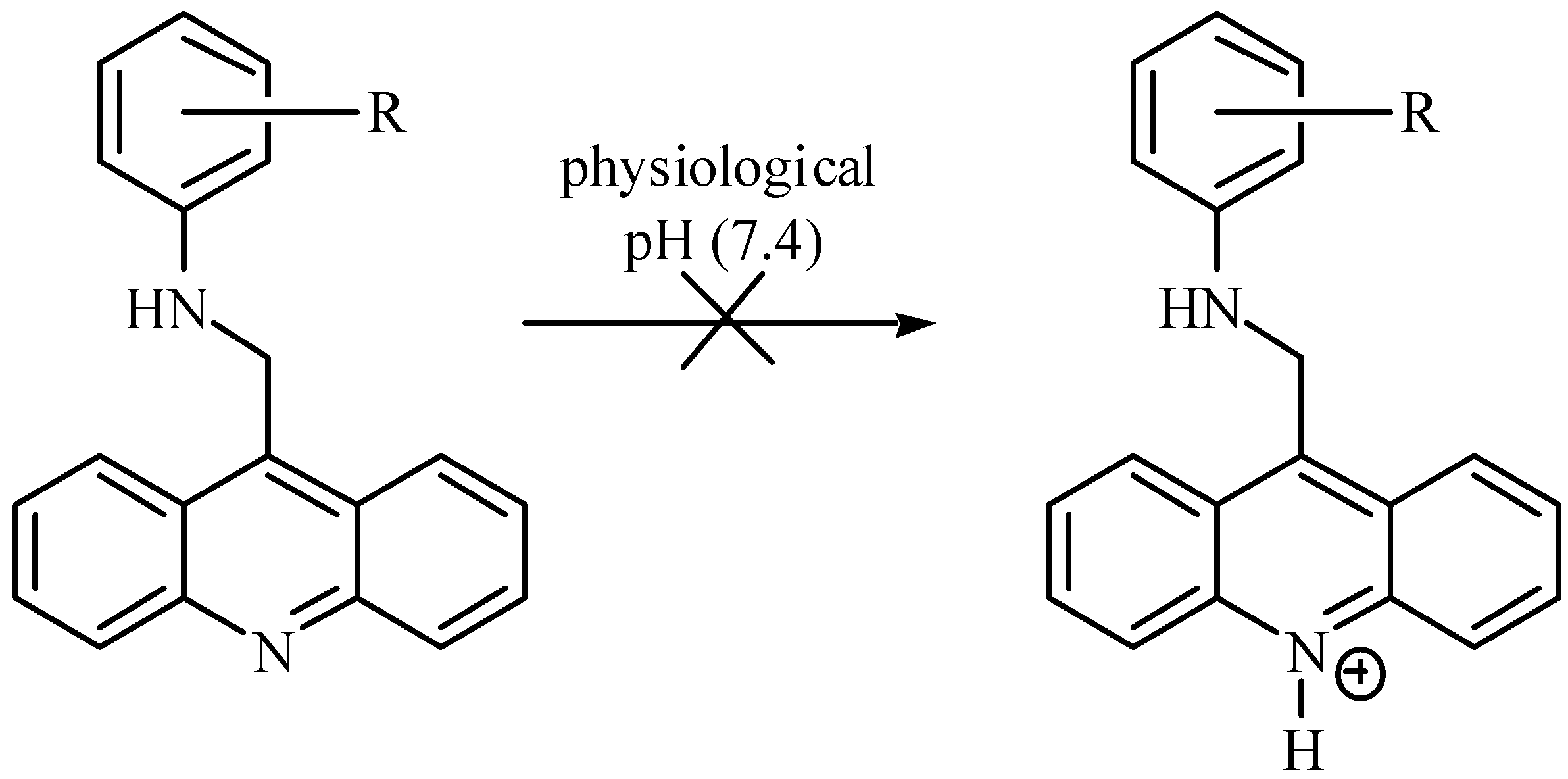
Results and Discussion
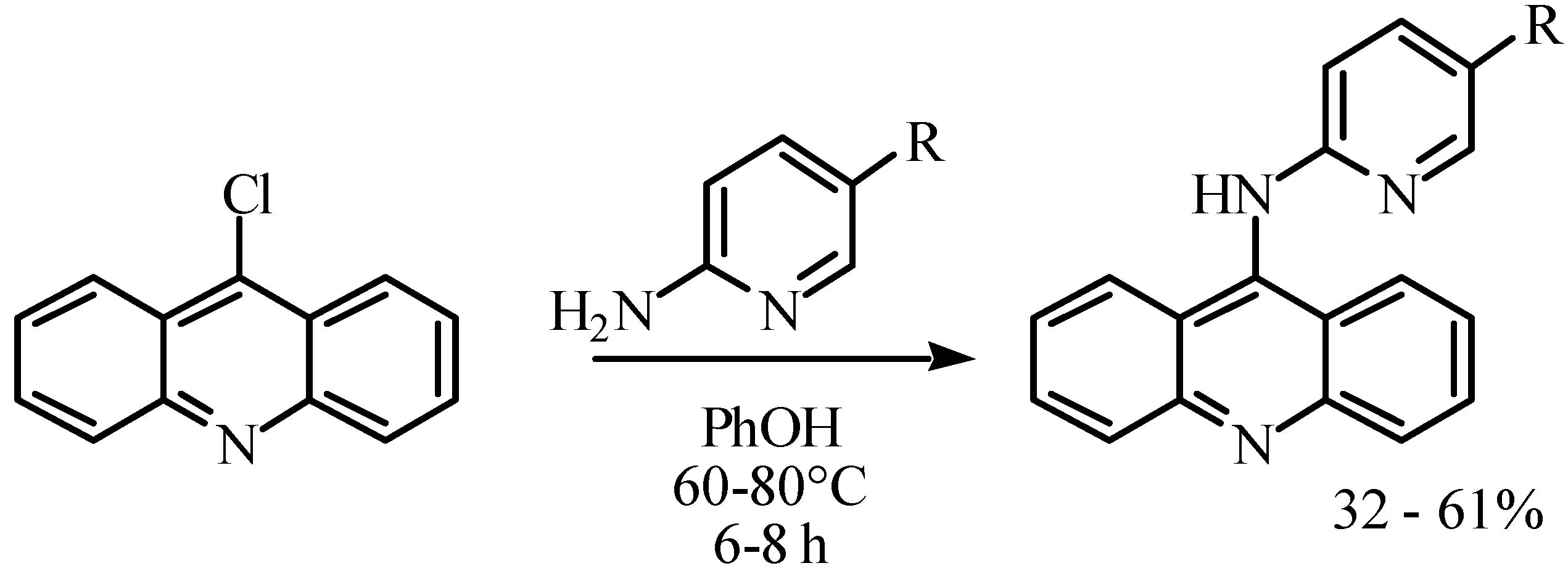
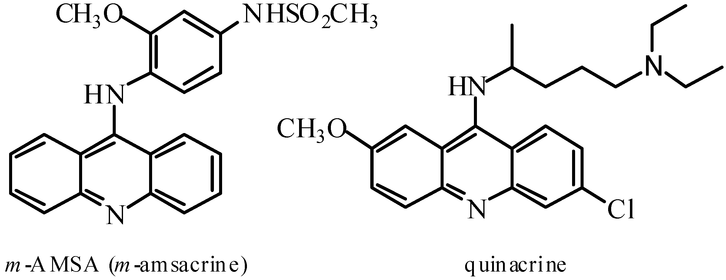
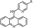
Problems with the use of 9-aminoacridines as therapeutic agents focus on the relative ease of hydrolysis and/or thiolysis of the C9-N bond, resulting in reduction or loss of biological activity [7]. In an effort to eliminate cleavage of this bond as a viable metabolic pathway, we prepared a series of 9-aminomethylacridines. However, no change in the thermal denaturation temperature of calf-thymus DNA due to these compounds was observed. Moreover, the limited antibiotic effect observed in this series most likely arose from unspecific toxicity. Subsequent analysis of the basicity of these compounds indicated that protonation of these compounds does not occur at physiological pH, thus they lack the requirements for strong association with DNA via intercalation [8].
Scheme 2.
Scheme 2.
Molecules 09 00102 g003
We next directed our efforts toward the preparation of a compound with a greater affinity for DNA. Five mono-substituted 9-(pyridin-2'-yl)-aminoacridines were designed, synthesized and evaluated for their ability to change the thermal denaturation temperature of calf-thymus DNA. These compounds were prepared in acceptable overall synthetic yield by condensation (Scheme 3). Modifications to the condensation reaction conditions (solvent, time, temperature) failed to improve the yields of the isolated products. In addition, while 9-chloroacridine can be purchased, we found that it was more convenient to prepare this compound by the Ullmann condensation [9]. The product of this reaction, N-phenylanthranilic acid (60% yield), underwent an intramolecular Friedel-Crafts acylation in the presence of phosphorus oxychloride to give a nearly quantitative yield of 9-chloroacridine. [10]
Scheme 3.
Scheme 3.
Molecules 09 00102 g004
A Hantzch congener set was developed for the 9-(pyridin-2’-yl)-aminoacridines in order to explore any electronic effects associated with DNA intercalation. The resulting compounds were analyzed for their ability to bind DNA through the measurement of the change in thermal denaturation temperature of genomic calf thymus DNA (Table 1) [11].
Table 1.
Table 1.
Molecules 09 00102 i001CompoundR∆Tm (90% confidence limits)
2aH15.3 ± 0.6 °C
2bCH36.79 ± 0.8 °C
2cNO2Not detnd.
2dCl18.3 ± 1.1°C
2eBr19.4 ± 1.4°C
Correlation of the ∆Tm and Hammett sigma values [12] for each compound illustrated a trend (Figure 1). In particular, while many of the Hammet sigma values did not correlate well to the measured values for ∆Tm, the Hammett σ+p value provided a strong correlation (ρ = 27.404, R2 = 0.9995). This correlation indicates that the addition of electron withdrawing character to the pyridine ring system greatly enhances or stabilizes the DNA-acridine complex. The correlation between ∆Tm and σp+ may be the result of the ability of the R group to stabilize the cationic character of the acridine nucleus at physiological pH. The very large Hammett rho value may indicate a larger role in the binding of the substituted pyridine to the DNA base pairs.
Figure 1.
Figure 1.
Molecules 09 00102 g001
The thermal denaturation (∆Tm) for the nitro substituted compound was not able to be determined in our system. Calculation of the ∆Tm value using the developed QSAR equation reveals the reason - the thermal denaturation temperature for a DNA/2c mixture would be approximately 10°C greater than the boiling point of the aqueous solution of DNA.

Conclusions

Electron withdrawing character increases the binding of the 9-(pyridin-2’-yl)-aminoacridines to double-stranded calf thymus DNA. This may be due to the stabilization of cationic character at physiological pH which, in turn, would strengthen electrostatic attraction to the DNA phosphate backbone.

Experimental

General

1H- and 13C-NMR spectra were obtained using a Bruker Avance 300 NMR and were recorded at 300 and 75 MHz respectively. All reagents and chemicals were obtained from Aldrich Chemical Company (USA) and were used immediately as received unless otherwise noted. 9-Chloroacridine was prepared by the method described in the literature [9,10], purified by Soxhlet extraction from petroleum ether and stored at –5°C in a vial within a bottle containing KOH pellets. Thermal denaturations were obtained using a Perkin Elmer Lambda 14 UV-vis spectrophotometer with Peltier Temperature controller in aqueous buffer solutions containing EDTA (1.0 x 10⁻⁵ M), 10% v/v DMSO, and 0.01 M phosphate (pH = 7). Final DNA and aminoacridine concentrations were maintained at 5.2 x 10⁻⁵ M and 2.6 x 10⁻⁴ M respectively during the thermal denaturation experiments.

General synthetic procedure

9-Chloroacridine (0.500g, 2.35 mmol) and solid phenol (10g, 0.106 mol) were placed in a round bottomed flask equipped with a stir bar and an air-cooled reflux condenser. The mixture was warmed to 60-80°C with a heating mantle. The appropriately substituted 2-aminopyridine (2.00 mmol) was then added to the flask after the phenol melted. Heating was continued for 6 to 8 hours with stirring. When TLC (eluent 8:2 hexane-ethyl acetate) indicated that the reaction was complete, the reaction mixture was cooled to room temperature and dissolved in dichloromethane (75mL). The red organic phase was extracted repeatedly with 0.1 M sodium hydroxide, then washed with water (2 x 50 mL), brine (50 mL) and dried over anhydrous sodium sulfate. Evaporation of the organic solvent provided the desired product as a crude orange-red solid. The purified compounds were isolated by flash chromatography (silica gel, 69:28:3 hexane-ethyl acetate-triethylamine).
9-(pyridin-2’-yl)-aminoacridine (2a). Orange-yellow powder (32% yield); 1H-NMR (CDCl3) δ: 8.20 (d, J=7.8, 2H), 8.11 (d, J=7.8, 2H), 7.79 (t, J=7.8, 2H), 7.70 (s, 1H), 7.33 (m, 1H), 7.00 (t, J=7.8, 2H), 6.80 (bs, 1H), 6.21 (m, 1H); 13C-NMR (CDCl3) δ: 158.4, 157.0, 149.0, 148.5, 138.9, 130.8, 130.3, 125.8, 125.1, 124.9, 123.1, 122.0.
9-(5’-methylpyridin-2’-yl)-aminoacridine (2b). Light orange solid (47% yield); 1H-NMR (DMSO-d6) δ: 8.24 (dd, J=1.2, 7.5, 2H), 7.97 (bs, 1H), 7.73 (t, J=7.5, 2H), 7.54 (d, J=8.5, 1H), 7.48 (dd, J=2.1, 8.5, 2H), 7.26 (t, J=7.1, 2H), 7.20 (bs, 1H), 6.71 (d, J=8.1, 1H), 2.22 (s, 3H); 13C-NMR (DMSO-d6) δ: 176.7, 147.9, 145.0, 140.8, 138.7, 133.4, 130.9, 125.9, 124.8, 120.9, 120.5, 117.3, 17.2.
9-(5’-nitropyridin-2’-yl)-aminoacridine (2c). Deep orange solid (52%); 1H-NMR (CDCl3) δ: 8.39 (d, J=8, 2H), 8.15 (d, J=8, 2H), 7.98 (t, J=8, 2H), 7.71 (s, 1H), 7.48 (t, J=8, 2H), 7.40 (m, 1H), 7.1 (bs, 1H), 5.99 (m, 1H); 13C-NMR (CDCl3) δ: 155.1, 174.2, 149.2, 144.8, 138.1, 130.9, 128.4, 127.0, 125.9, 126.5.1, 125.7, 120.8.
9-(5’-chloropyridin-2’-yl)-aminoacridine (2d). Orange solid (61% yield); 1H-NMR (CDCl3) δ: 8.25 (d, J=8, 2H), 8.12 (d, J=8, 2H), 7.81 (t, J=8, 2H), 7.70 (s, 1H), 7.49 (t, J=8, 2H), 7.40 (m, 1H), 7.05 (bs, 1H), 6.13 (m, 1H); 13C-NMR (CDCl3) δ: 156.0, 168.1, 149.5, 146.2, 138.6, 130.5, 129.0, 126.8, 125.6, 124.9, 124.5, 121.4.
9-(5’-bromopyridin-2’-yl)-aminoacridine (2e). Orange solid (39% yield); 1H-NMR (CDCl3) δ: 8.29 (d, J=7.5, 2H), 8.13 (d, J=7.5, 2H), 7.82 (t, J=7.5, 2H), 7.69 (s, 1H), 7.46 (t, J=7.5, 2H), 7.43 (d, J=9, 1H), 6.94 (bs, 1H), 6.09 (d, J=9, 1H); 13C-NMR (CDCl3) δ: 155.6, 170.6, 149.1, 145.8, 138.5, 130.3, 129.8, 126.9, 125.8, 125.2, 124.9, 121.1.

References and Notes

  1. Cancer Facts and Figures. American Cancer Society, 2004; p. 60.
  2. Centers for Disease Control. Morbid. Mortal. Rep. 2002, 51, 49.
  3. Zittoun, R. Eur. J. Cancer Clin. Oncol. 1985, 21, 649.Atwell, G.J.; Cain, B.F.; Seelye, R.N. J. Med. Chem. 1972, 15, 611.
  4. Denny, W.A.; Atwell, G.J.; Cain, B.F. J. Med. Chem. 1979, 22, 1453.
  5. Rose, F.L. J. Chem. Soc. 1951, 2770.
  6. Chourpa, I.; Manfait, M. J. Raman Spec. 1995, 26, 813.Rowe, T.C.; Chen, G.L.; Hsiang, Y.H.; Liu, L.F. Cancer Res. 1986, 46, 2022.Nelson, E.M.; Tewey, K.M.; Liu, L.F. Proc. Natl. Acad. Sci. 1984, 81, 1361.
  7. Jaycox, G.D.; Gribble, G.W.; Hacker, M.P. J. Heterocycl. Chem. 1987, 24, 1405.Wilson, W.R.; Cain, B.F.; Baguley, B.C. Chem Biol. Interactions 1977, 18, 163.
  8. Mosher, M.D.; Johnson, E. Heterocycl. Chem. 2003, 9. in press.
  9. Allen, C.F.H.; McKee, G.H.W. Org. Syntheses 1943, Coll. Vol 2, 15.
  10. Albert, A.; Ritchie, B. Org. Syntheses 1955, Coll. Vol 3, 53.
  11. Kuhlmann, K.F.; Charbeneau, N.J.; Mosher, C.W. Nuc. Acids Res. 1978, 5, 2629.
  12. Values for σp and σp+ were taken from: Hansch, C.; Leo, A.; Hoekman, D. Exploring QSAR: Hydrophobic, Electronic, and Steric Constants. American Chemical Society: Washington, DC, 1995. [Google Scholar]
  • Sample Availability: Available from the authors.

