Article

New PCR Assays for the Identification of Fusarium verticillioides, Fusarium subglutinans, and Other Species of the Gibberella fujikuroi Complex

by
Carla Bertechini Faria
1,
Camila Agnes Lumi Abe
2,
Cleiltan Novais da Silva
2,
Dauri José Tessmann
2 and
Ione Parra Barbosa-Tessmann
1,*
1
Department of Biochemistry, State University of Maringá, Av. Colombo, 5790, 87020-900, Maringá, PR, Brazil
2
Department of Agronomy, State University of Maringá, Av. Colombo, 5790, 87020-900, Maringá, PR, Brazil
*
Author to whom correspondence should be addressed.
Int. J. Mol. Sci. 2012, 13(1), 115-132; https://doi.org/10.3390/ijms13010115
Submission received: 28 October 2011 / Revised: 2 December 2011 / Accepted: 8 December 2011 / Published: 23 December 2011
(This article belongs to the Section Molecular Pathology, Diagnostics, and Therapeutics)

Abstract

:
Fusarium verticillioides and Fusarium subglutinans are important fungal pathogens of maize and other cereals worldwide. In this study, we developed PCR-based protocols for the identification of these pathogens targeting the gaoB gene, which codes for galactose oxidase. The designed primers recognized isolates of F. verticillioides and F. subglutinans that were obtained from maize seeds from several producing regions of Brazil but did not recognize other Fusarium spp. or other fungal genera that were either obtained from fungal collections or isolated from maize seeds. A multiplex PCR protocol was established to simultaneously detect the genomic DNA from F. verticillioides and F. subglutinans. This protocol could detect the DNA from these fungi growing in artificially or naturally infected maize seeds. Another multiplex reaction with a pair of primers developed in this work combined with a pre-existing pair of primers has allowed identifying F. subglutinans, F. konzum, and F. thapsinum. In addition, the identification of F. nygamai was also possible using a combination of two PCR reactions described in this work, and another described in the literature.

1. Introduction

Fusarium species are important fungal pathogens of maize (Zea mays L.) and other cereals worldwide [1]. Fusarium verticillioides (Saccardo) Nirenberg (teleomorph: Gibberella moniliformis) is the fungus most commonly found associated with maize stem and ear-rot. This species belongs to mating population A of the species complex Gibberella fujikuroi (Sawada) Ito in Ito and K. Kimura, which has nine mating populations (A to I) with different toxicological profiles and preferential hosts [2]. Fusarium subglutinans (Wollenweber and Reinking) Nelson, Toussoun and Marasas (teleomorph: Gibberella subglutinans) belongs to mating population E of the G. fujikuroi species complex and is also a maize pathogen found mainly in cold regions of the world where maize is cultivated.
In addition to its importance in agriculture, Fusarium species can also cause several diseases in humans and animals because they produce harmful mycotoxins. The species F. verticillioides can produce significant amounts of fumonisins and other mycotoxins in maize grains. Fumonisins interfere with sphingolipid metabolism, and especially the isoform B1 (FB1) presents a great mycotoxicological concern because of its abundance in maize grains [3]. FB1 causes leukoencephalomalacia in horses, pulmonary edema in swine, poor performance in poultry, altered hepatic and immune function in cattle, and it has been associated with human esophageal cancer [3]. The species F. subglutinans produces low levels or no fumonisins but can produce other mycotoxins [1].
Seeds provide one of the most efficient methods of pathogen dissemination at great distances and allow pathogen introduction into new areas. In maize, both species F. verticillioides and F. subglutinans can be spread by seeds [1,4]. These pathogens reduce germination by seed decay, damping-off, and seedling blight. The extent to which maize seed contamination can be reduced is dependent upon the development of an efficient screening system. Such a screening system can also be of great utility in research programs aimed at expanding the knowledge of Fusarium disease epidemiology and in the selection of resistant maize genotypes.
The methods currently employed for the identification and differentiation of F. verticillioides and F. subglutinans from the other Fusarium species are based mainly on the morphology. Fertility tests with mating test lineages are necessary for specific identification. These methods are relatively simple and cheap in terms of the materials used, but they can be laborious and it may take weeks to obtain results. Furthermore, these methods are highly dependent on the analyst’s expertise. Molecular techniques, such as the polymerase chain reaction (PCR), are also used for species detection and identification of the Fusarium species genera [5]. PCR is a fast, sensitive, and very specific technique. Indeed, this technique has yielded great perspectives in seed pathology because only a small quantity of DNA is required to confirm the pathogen’s identity and its presence in host tissues [6]. There are several reports in the literature about the use of PCR for the detection of F. verticillioides, F. subglutinans, and Fusarium proliferatum [719]. However, almost all primer pairs developed so far for the identification of F. verticillioides and F. subglutinans had cross-reactivity with other species, and some of them could identify only toxigenic strains. Moreover, there is no method for the detection of some of the G. fujikuroi species that are pathogenic to sorghum (Sorghum bicolor, L. Moench), such as Fusarium thapsinum and Fusarium nygamai.
The galactose oxidase gaoA gene [20] has been used as a target for primers in PCR reactions for the specific detection of Fusarium graminearum (teleomorph: Gibberella zeae) [21,22]. Galactose oxidase is a copper enzyme that catalyzes the oxidation of primary alcohols to aldehydes with the concomitant reduction of O2 to H2O2 [23]. Three ortholog lineages of this gene have been identified in Fusarium and the lineage gaoB has been cloned in our laboratory from F. verticillioides and from F. subglutinans [24]. The gaoB lineage lacks introns and is 2040 bp in length [24].
Considering that F. verticillioides and F. subglutinans are important pathogens of maize and that they are transmitted by seeds, we have reasoned that it would be very important to develop a specific, reliable, and useful molecular diagnostic method for the detection of these pathogens in maize seeds. Thus, the objectives of the present work were: (i) to develop a PCR method for the molecular detection of F. verticillioides and F. subglutinans with primers targeting the recently cloned gaoB gene from those pathogens; and (ii) to use these primers in a multiplex PCR to simultaneously detect both species.

2. Results and Discussion

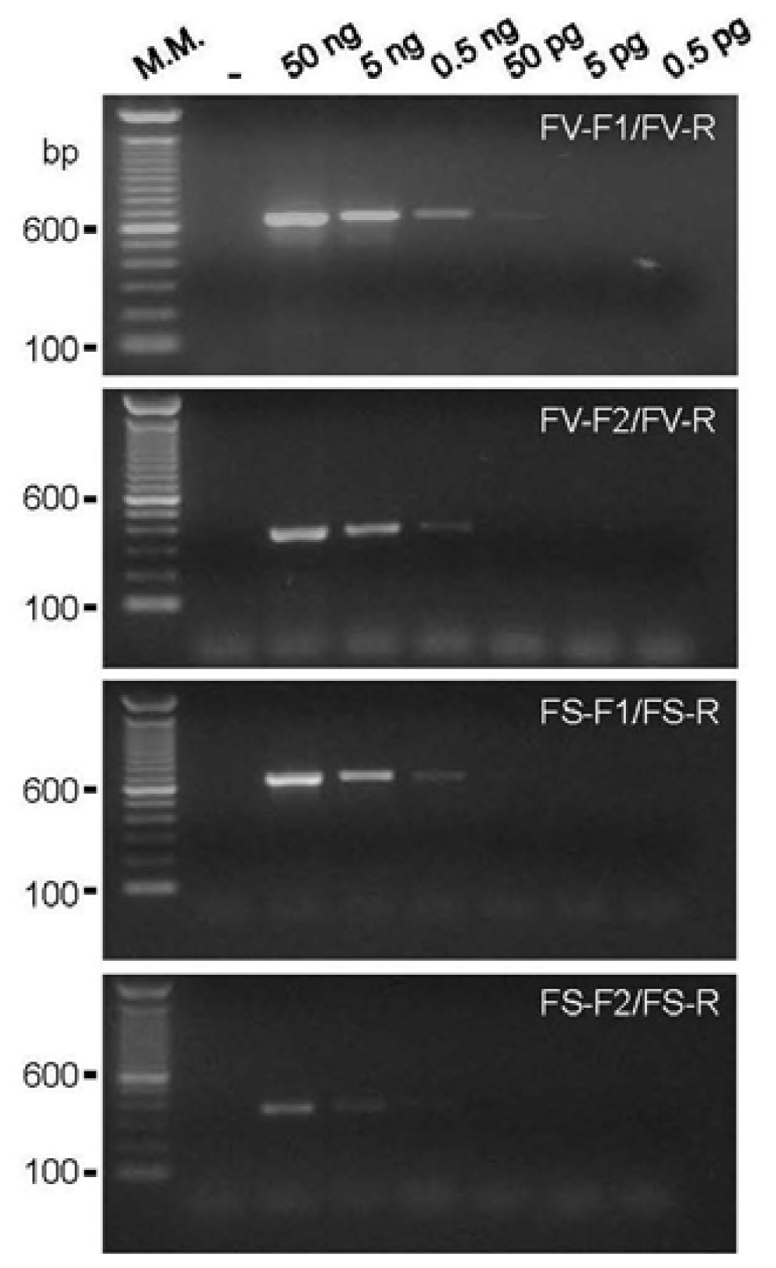
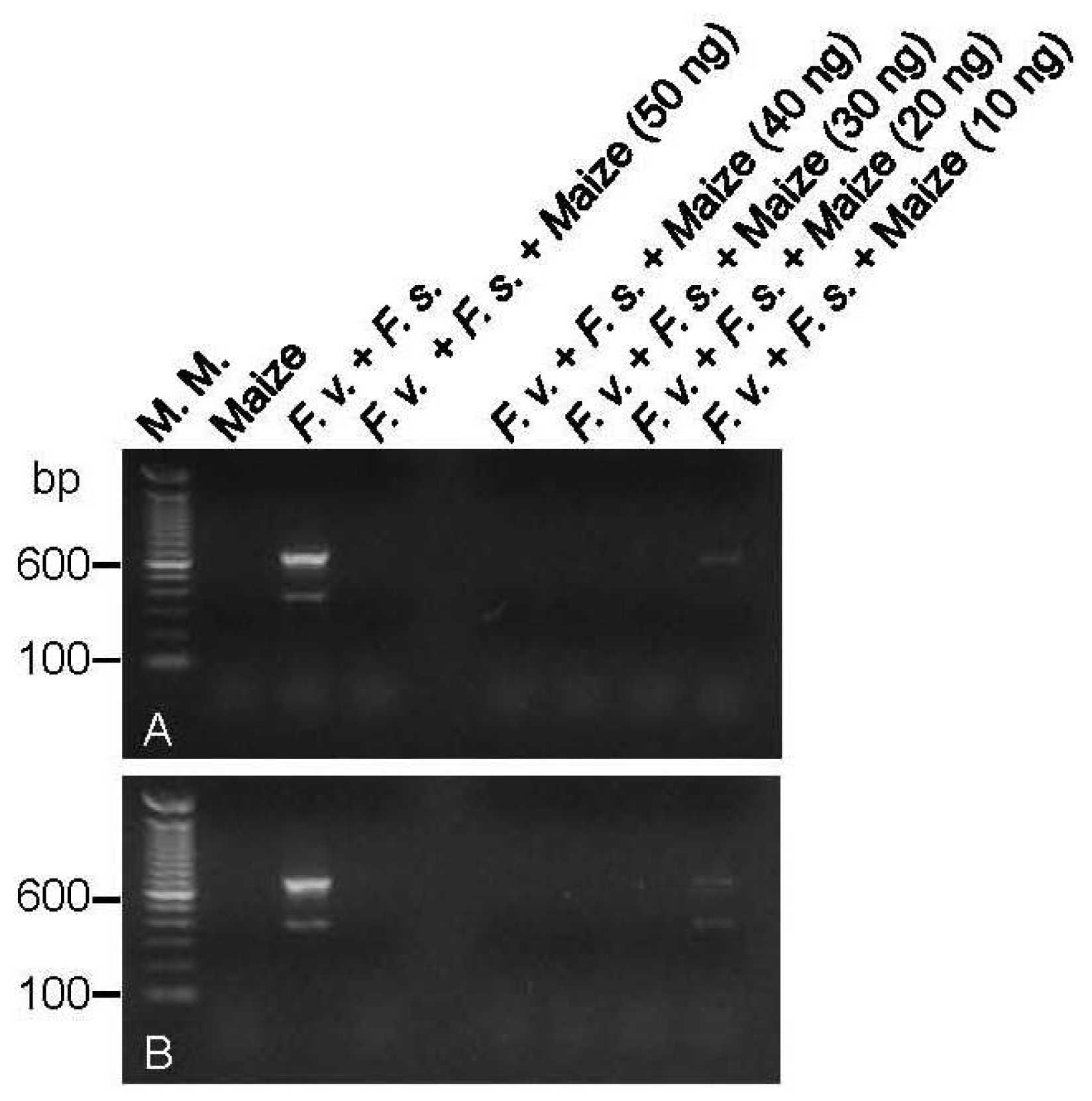
2.1. DNA Amplification and Primer Sensitivity Analysis

All of the new primer pairs amplified a unique band of the expected size with the genomic DNA from the control strains F. verticillioides (CML 767) or F. subglutinans (UnB 379). The lowest amount of genomic DNA of the control isolates that could generate a visible band in a conventional agarose gel stained with ethidium bromide was 50 pg (0.05 ng) for the primer pair FV-F1/FV-R and 500 pg (0.5 ng) for the other new primer pairs (Figure 1). This sensitivity is similar as that of other primers described in the literature [13,14,25].

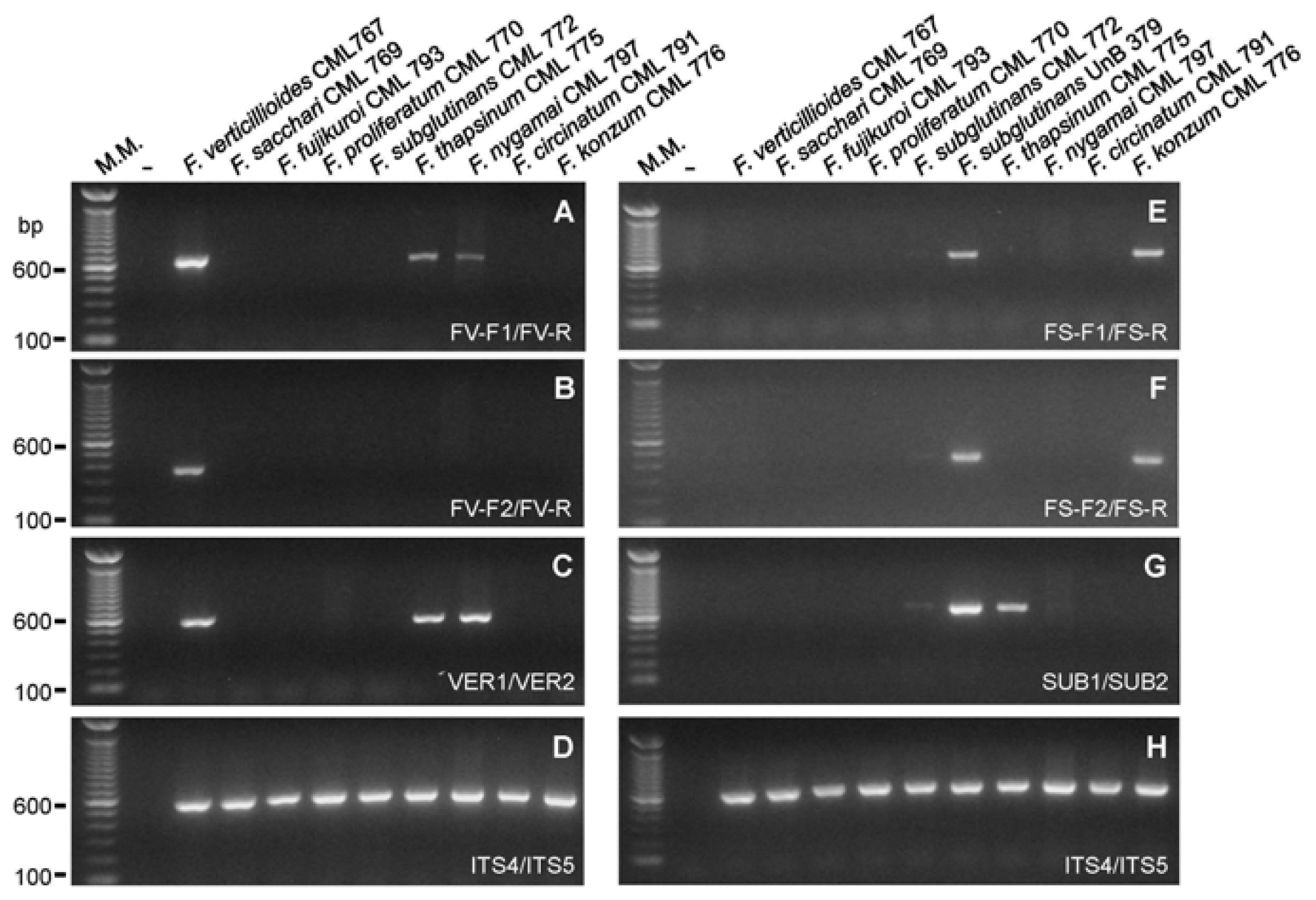
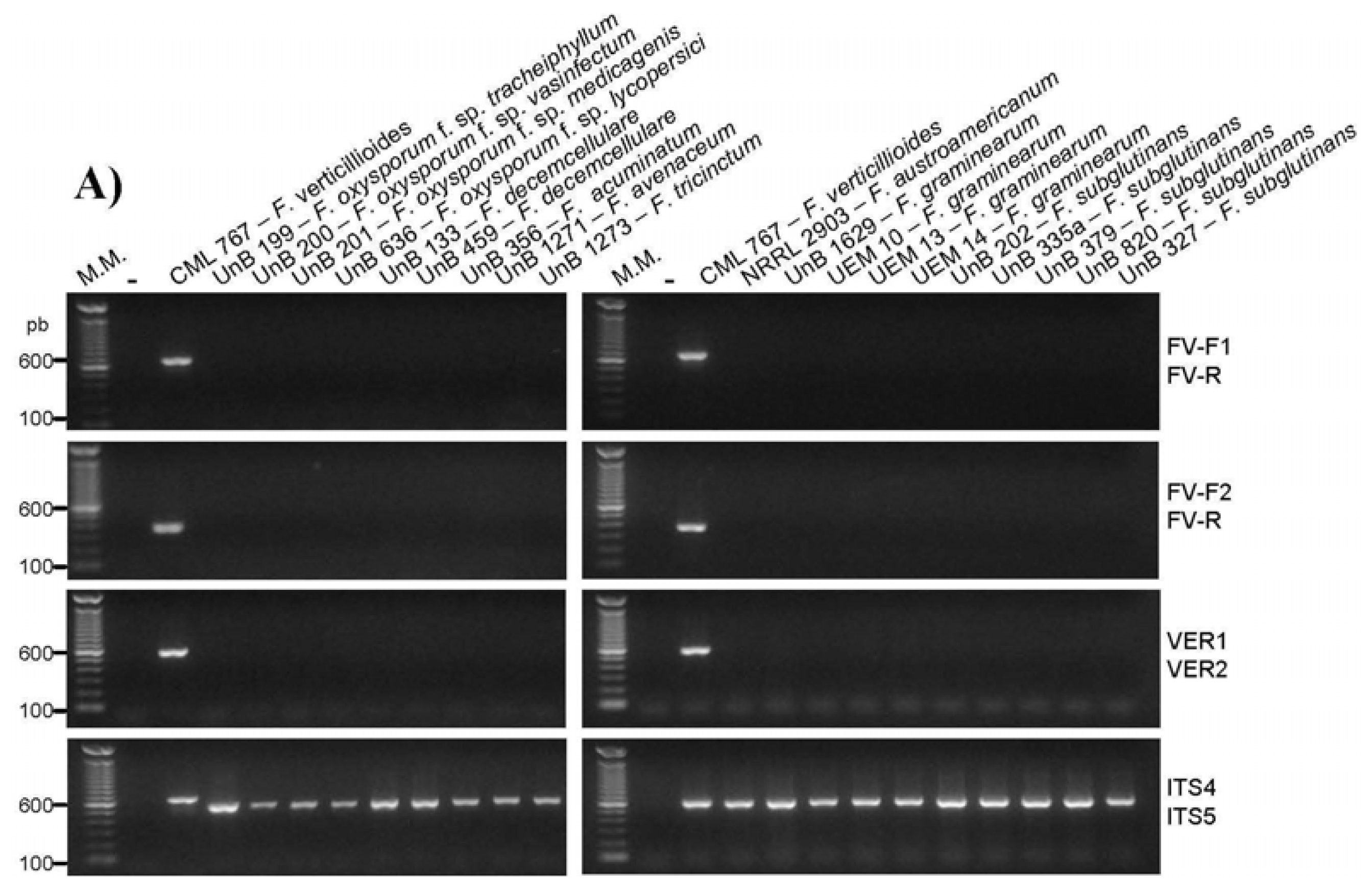
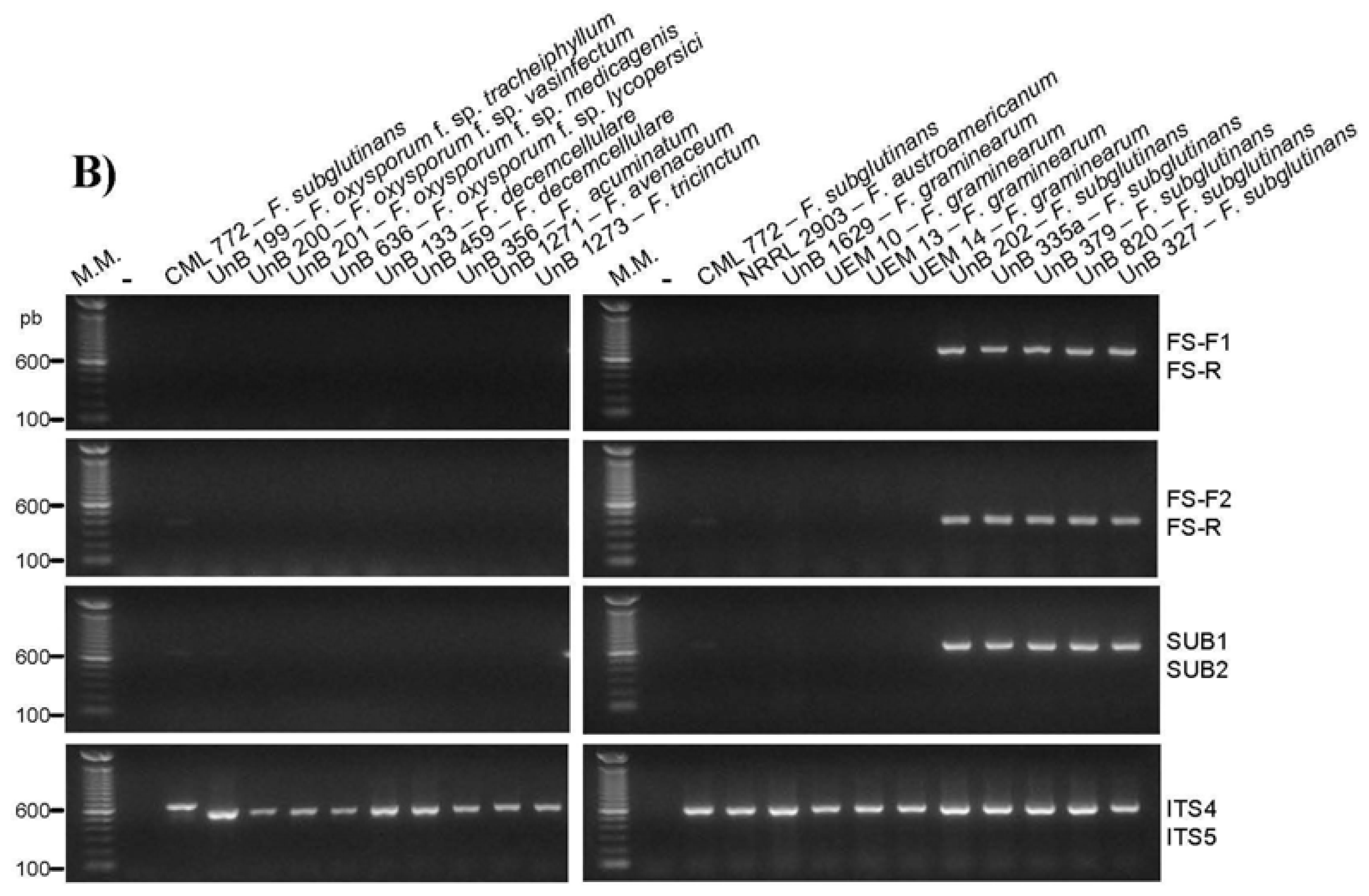
2.2. Primer Specificity Analysis

Regarding to the specificity analysis with the G. fujikuroi species, the primer pair FV-F1/FV-R targeting the gaoB gene from F. verticillioides amplified a DNA fragment of the expected size from the genomic DNA from F. verticillioides, but also from F. thapsinum and F. nygamai, two species phylogenetically related to F. verticillioides [2] (Figure 2A). The primer pair FV-F2/FV-R amplified a DNA fragment only from the F. verticillioides genomic DNA (Figure 2B). The two FS primer sets targeting the gaoB gene of F. subglutinans have amplified a DNA fragment of the expected size from the genomic DNA from the F. subglutinans strains used as control (UnB 379 and CML 772) and from F. konzum (Figure 2E,F), a species phylogenetic related to F. subglutinans [1]. The primer pair VER1/VER2 described by Mulè et al. [14] and targeting the calmodulin gene had specificity similar to the primer pair FV-F1/FV-R (Figure 2C). The primer set SUB1/SUB2 described by Mulè et al. [14] had a slightly different specificity of the FS primers, amplifying a DNA fragment from the genomic DNA from F. subglutinans and also from F. thapsinum (Figure 2G). The specificity of these primer pairs described by Mulè et al. [14] to the G. fujikuroi isolates had not been tested before.
All designed primer pairs were unable to amplify any DNA fragment, specific or nonspecific, from the genomic DNA of several species of the Fusarium genera and from several other fungal genera that could be associated with maize seeds (Table 1, Figure 3). In addition, the FV-F2/FV-R and FS primer pairs were also unable to recognize any target in the genomic DNA of several fungi that were isolated from maize seeds in this work as Penicillium purpurogenum, Penicillium digitatum, Penicillium sp., Stenocarpella sp., Curvularia sp., Cladosporium sp., Phlebiopsis sp., Pichia sp., Rhyzopus sp., Phoma sp., Thrichoderma sp., Epicocum sp., Cladosporium sp., Irpex sp., Microdochium nivale, Epicocum sp., Mucor sp., Fusarium graminearum, Aspergillus oryzae, Aspergillus candidus, Aspergillus flavus, and Aspergillus niger.
Concerning the primer sensitivity testing against Fusarium isolates found in maize seeds, the two FV primer pairs were able to amplify a DNA fragment from all 47 F. verticillioides isolates obtained from maize seeds (Table 2). The FS primer pairs were also able to molecularly identify the obtained F. subglutinans isolates. The DNA of other found Fusarium species was not recognized by the new primer pairs (Table 2). The genomic DNA of the isolate RV27-2 resulted in positive PCR reactions for the primer pairs FV-F1/FV-R and VER1/VER2 [14], indicating that it could be F. verticillioides, F. nygamai, or F. thapsinum. As it resulted in a negative PCR reaction with the more specific FV-F2/FV-R primer pair, it could not be F. verticillioides. In addition, it also resulted in a negative PCR reaction with the SUB1/SUB2 [14] primer pair, demonstrating that it is neither F. thapsinum nor F. subglutinans. Thus we have concluded that this isolate is F. nygamai, what was confirmed in the Fusarium-ID analysis. The identification of the isolate RV 27-2 as F. nygamai points out that the combination of individual PCR reactions with the primers FV-F2/FV-R (this work), FV-F1/FV-R (this work), and SUB1/SUB2 [14] can be used for the identification of F. nygamai. The information that the FV and FS primer pairs do not recognize the gaoB gene of F. andiyazi (Table 2) enhances the data about their specificity.

2.3. Multiplex PCR Reactions

The combination of the primer pairs FV-F2/FV-R and FS-F1/FS-R in only one PCR reaction could amplify a 370 bp DNA fragment from the F. verticillioides genomic DNA and a 649 bp DNA fragment from F. subglutinans genomic DNA (Figure 4A), what is consistent with the amplicon sizes obtained with the primer pairs used individually (Figure 1). In addition, this established reaction could also amplify DNA fragments of the expected size from genomic DNA extracted from fungi growing on and on the medium around the maize seeds that were artificially or naturally contaminated with F. verticillioides or F. subglutinans, or both species (Figure 4A). The naturally contaminated maize seed lots chosen for this analysis were: MGA 10 from which F. verticillioides was isolated, PG 1 from which F. subglutinans was isolated, and RV 27 from which neither of these fungi was initially isolated. The results obtained (Figure 4A) indicate the presence of F. subglutinans in the PG 1 sample and the presence of F. verticillioides in all three naturally contaminated seed samples tested, what is in accordance with the literature that reports the presence of F. verticillioides in 100% of the maize seeds in Brazil [26]. Möller et al. [13] have also set up a multiplex PCR to detect F. verticillioides and F. subglutinans simultaneously, but there are no data on the specificity of these primers with the genomic DNA of F. circinatum, F. konzum, and F. andiyazi. The present multiplex PCR reaction represents a significant advance for the simultaneous molecular detection of F. verticillioides and F. subglutinans.
When the primer pair FS-F2/FS-R was used in a multiplex reaction with the primer pair SUB1/SUB2 [14], two DNA fragments were amplified from the genomic DNA of F. subglutinans: one of 370 bp and one of 631 bp (Figure 4B). In addition, this set of primers could amplify a single DNA fragment of 370 bp when the genomic DNA from F. konzum was present in the reaction or a single DNA fragment of 631 bp when the genomic DNA from F. thapsinum was used in the reaction (Figure 4B). This reaction represents an improvement to the described reactions designed to detect F. subglutinans. The primers developed by Möller et al. [13] to identify F. subglutinans were not tested for all G. fujikuroi isolates, and the primers designed by Zheng and Ploetz [19] were specific for F. subglutinans and F. nygamai. In addition, there are few data in the literature about reactions to identify F. thapsinum, which is an important sorghum pathogen.
The isolation of DNA directly from the seeds, without previous culture to obtain mycelia, was also tried but resulted in no DNA amplification in the multiplex PCR reaction to detect F. verticillioides and F. subglutinans. This could have happened because the amount of fungal cells in the contaminated seeds is too low or because of the presence of inhibitors in the extracted DNA. To test for the presence of inhibitors in the DNA extracted from maize, the established multiplex PCR reaction to simultaneously detect F. verticillioides and F. subglutinans was performed with the lowest amount of fungi DNA that could result in amplification spiked with increasing amounts of genomic DNA extracted from healthy maize seeds. The results shown in Figure 5 evidenced that maize DNA only did not inhibit the reaction when present in a low amount.

3. Experimental Section

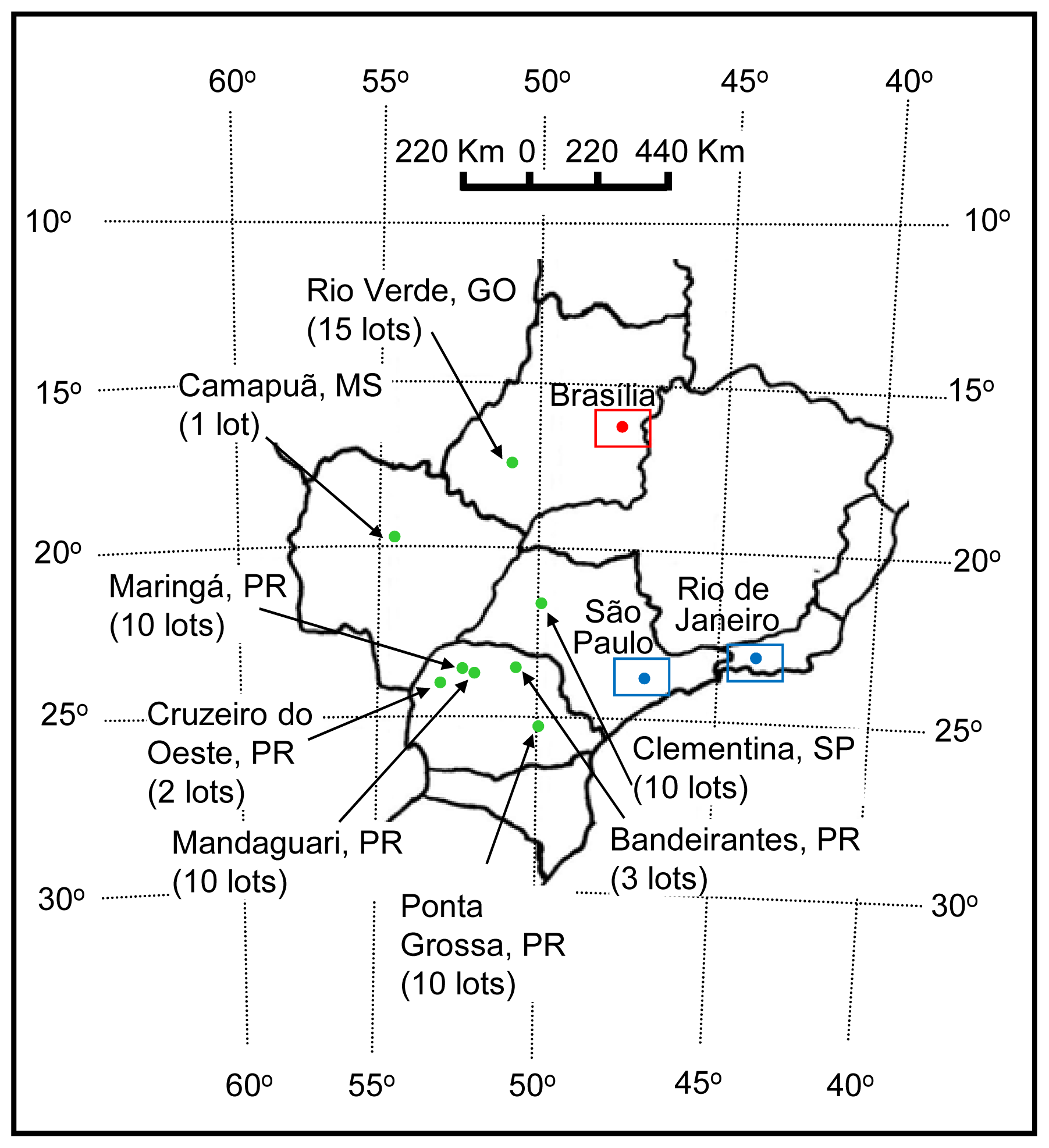
3.1. Fungal Isolates
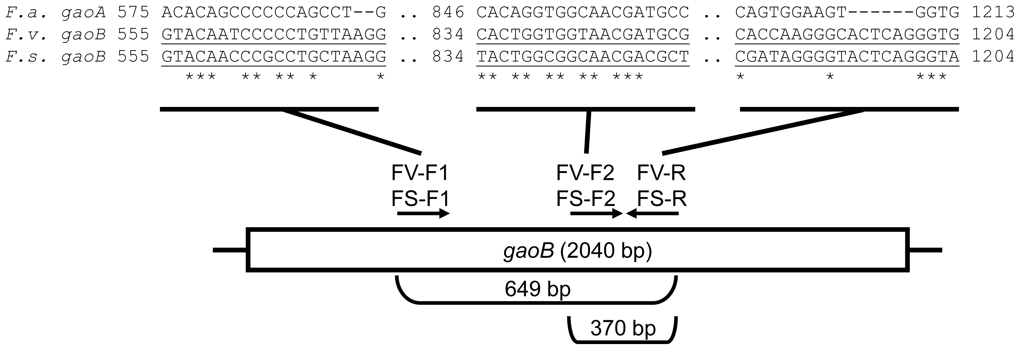
The fungal isolates used in this work are listed in Table 1. To obtain a Fusarium collection from maize seeds, corn spikes showing signs of rot were collected from January to October of 2009, in fields in several producing regions of Brazil (Figure 6). The seeds of each spike were denominated as a lot and were kept in paper bags at 4 °C after insecticide treatment. A total of 57 maize seed lots were collected and analyzed. Six maize seeds of each lot, in duplicate, were disinfected by one min incubation in a solution containing 0.2% active chlorine, washed in sterile distilled water, and inoculated in a 10 mm diameter dish containing Malaquite Green Agar-MGA [27] supplemented with 350,000 UI/L of penicillin and 145 UI/L of streptomycin. The seeds plated in the medium were incubated for four to five days at 25 °C, with a photoperiod of 12 h. After germination, mycelia and conidia of peach or violet colored colonies were transferred to Petri dishes containing Carnation Leaf-piece Agar-CLA [1], which were incubated for a period of seven days at 25 °C, with a photoperiod of 12 h. A well-colonized carnation leaf fragment in the CLA culture was used for monosporic isolation as described in Nelson et al. [28]. The isolates morphologic characteristics were analyzed in micro culture performed in a one-cm3 block of Spezieller Nährstoffarmer Agar-SNA [1]. The isolates with cultural and morphological characteristics of Fusarium were cultivated for DNA extraction and molecular identification with the specific primers described by Mulè et al. [14]. These primers were chosen because they target a protein coding gene as the primers designed in this work. The genomic DNA of Fusarium isolates that were not identified as F. verticillioides or F. subglutinans were used in PCR reactions with specific primers for F. proliferatum [14]. These fungi were also analyzed using the methodology described in Geiser et al. [29] in which a portion of the elongation factor α was amplified through PCR, purified with the ExoSap-IT Kit (GE HealthCare, USA), and sequenced in the Center for the Human Genome Studies (CEGH) in the University of São Paulo (USP), Brazil. For the isolate identification, the obtained sequences were compared with sequences deposited in the data bank Fusarium-ID [29]. All obtained isolates are being maintained in the laboratory collection on PDA and SNA media with trimestral passages and in SNA medium under mineral oil.
To obtain fungi other than F. verticillioides or F. subglutinans from maize seeds, to be used in the primers specificity analysis, six maize seeds of each lot, in triplicate, were disinfected as described above and inoculated in a 10 mm diameter dish containing PDA, pH 4.5 with lactic acid [6], supplemented with penicillin and streptomycin, as described above. The seeds plated in the medium were incubated for five to seven days at 25 °C, with a photoperiod of 12 h. The fungi growing on and on the medium around the maize seeds with culture characteristics different from G. fujikuroi were submitted to monosporic isolation [28]. The classification of Aspergillus and Penicillium isolates was performed as described in Pitt and Hocking [30] with cultures in specific media. The molecular identification of the isolates of other genera was performed by the amplification of an rRNA gene fragment with the ITS4/5 primers [31], purification of the amplified DNA fragment with the PureLink™ PCR purification kit (Invitrogen, USA), sequencing in the CEGH (USP, Brazil) and comparison with sequences deposited in data banks. Specific identification of F. graminearum was performed in PCR reactions with the GOF/R specific primers [21]. All isolates obtained are being maintained in the laboratory collection on PDA with trimestral passages and on PDA medium under mineral oil.

3.2. Primer Design

Initially, the gaoB genes from F. verticillioides and F. subglutinans [24] (GenBank AN HM069186 and HM069185, respectively) and the gaoA gene from F. austroamericanum [20] (GenBank AN M86819) were aligned with the ClustalW program. Low similarity regions were chosen for primer design. Four pairs of primers were designed; two directed to the gaoB gene from F. verticillioides (FV-F1/FV-R and FV-F2/FV-R) and two other for the gaoB gene from F. subglutinans (FS-F1/FS-R and FS-F2/FS-R) to amplify a DNA fragment of 649 bp and 370 bp, respectively (Figure 7).
The primer sensitivity was tested by a ten-fold serial dilution of the control isolates genomic DNA in the established PCR reactions. The primer specificity was tested in PCR reactions using genomic DNA from G. fujikuroi species isolates, other Fusarium spp., other fungal genera, and fungi isolated from maize seeds.

3.3. DNA Extraction

For the DNA extractions, an approximately one-cm3 fragment of a monosporic culture in inclined PDA was smashed and shaken in 5 mL of distilled water. Two mL of the obtained suspension were used as inoculum in Erlenmeyer flasks of 125 mL containing 25 mL of potato dextrose medium. These flasks were incubated for five days at 25 °C. The mycelia were collected by filtration in sterile gauze. The mycelia was macerated in a mortar with liquid nitrogen, and transferred to microcentrifuge tubes. The DNA was extracted from the macerated mycelia using the protocol described by Koenig et al. [32]. The DNA obtained was quantified by measuring the absorbance at 260 nm and/or by fluorometry using the Qubit Quantitation Fluorometer and the Quant-it™ dsDNA HS Assay Kit (Invitrogen, USA).

3.4. Polymerase Chain Reaction—PCR

The PCR with the primer pairs designed in this work was performed in a total volume of 25 μL of the following mixture: 1X enzyme buffer (20 mM Tris, pH 8.4, and 50 mM KCl), 1.5 mM MgCl2, 1.5 U of Platinum® Taq DNA polymerase (Invitrogen, USA), 0.2 mM of each dNTP, 25 pmol of each primer (FW and RV), and 50 ng of the genomic DNA. The reaction consisted of 25 cycles of 1.5 min at 94 °C, 1.5 min at the recommended annealing temperature for each pair of primers (Table 3), and 2 min at 72 °C in a Techne Thermocycler (England). Before the cycles, the samples were heated for 5 min at 94 °C, and after the cycles, the samples were incubated for 10 min at 72 °C. Negative controls (no DNA template) were used in each experiment to test for the presence of DNA contamination of reagents and reaction mixtures. Ten microliters of the PCR reaction were analyzed in an agarose gel containing ethidium bromide (0.25 μg/mL). Molecular weight markers were the 100 bp markers from Invitrogen (USA). The PCR products were visualized and photographed under UV light. All genomic DNA that resulted in nonamplification with the other used primers was tested for PCR amplification quality with the primers ITS4/5 [31]. All the PCR with the other primer pairs used in this work was performed as above with the exception with the primer pair ef1/ef2 [29] that was performed in a total volume of 25 μL with 50 ng of genomic DNA template and following the authors’ methodology.

3.5. Multiplex PCR Reactions

A first multiplex PCR reaction to simultaneously detect F. verticillioides and F. subglutinans was established with the primer pairs FS-F1/FS-R and FV-F2/FV-R. Another multiplex PCR reaction to identify F. subglutinans was established with the primer pairs FS-F2/FS-R (this work) and SUB1/SUB2 [14]. Genomic DNA from F. konzum and F. thapsinum gave a positive signal in this last multiplex PCR reaction as well.
The reaction established to simultaneously detect F. verticillioides and F. subglutinans was also tested with genomic DNA extracted directly from artificially or naturally contaminated seeds or from fungi growing on artificially or naturally contaminated maize seeds using the protocol described by Koenig et al. [32]. To artificially contaminate maize seeds, healthy maize seeds were disinfected as described above and incubated for 5 min in 5 mL of a suspension of 104 spores/mL of F. verticillioides, or F. subglutinans, and of F. verticillioides together with 104 spores/mL of F. subglutinans. The artificially contaminated seeds were inoculated in Petri dishes containing MGA-penicillin-streptomycin medium [27], prepared as described above. These seeds were incubated for five days at 25 °C on the medium, with a period of 12 h of light. Naturally infected maize seeds (Ponta Grossa-1, Rio Verde 27, and Maringá-10) showing signs of rot were disinfected and incubated in the same way. The mycelia growing on the seeds and on the agar around the seeds were scraped and used for the DNA extraction that was conducted with the method described by Koenig et al. [32]. To test if maize DNA could inhibit the reaction, maize DNA was also extracted from health maize seeds using the protocol described by Koenig et al. [32] and this DNA was added in multiplex PCR reactions with DNA from the reference isolates of F. verticillioides and F. subglutinans.
The multiplex reactions were performed as shown above, except that they contained 25 pmol of each primer. The annealing temperatures are listed in Table 3. The amplification of specific fragments was analyzed by running 10 μL of the PCR reaction on a 1.5% agarose gel that was stained with ethidium bromide (0.25 μg/mL).

4. Conclusions

In conclusion, in this work, primers to identify F. verticillioides and F. subglutinans were designed and tested in PCR reactions. Additionally, they could be combined in a multiplex reaction to simultaneously detect these two species in naturally and artificially contaminated maize seeds. Another multiplex reaction with a pair of primers developed in this work combined with a pre-existing pair of primers [14] has allowed identifying F. subglutinans, F. konzum, and F. thapsinum. In addition, the identification of F. nygamai was also possible using a combination of two different reactions described in this work and one described previously [14]. The results obtained in this work and the used experimental approaches have a great potential value for the molecular based identification of difficult fungal phytopathogens.

Acknowledgments

The authors are truly grateful to the International Foundation for Science (IFS—Sweden), National Council of Technological and Scientific Development (CNPq—Brazil), and Araucária Foundation (Paraná State, Brazil) for financial support; to the Coordination for the Improvement of the Higher Level Personnel (CAPES) for financial support of CB Faria and CN Silva; and to LH Pfenning (UFLA), JC Dianese (UnB), and C Kemmelmeier (UEM) for the kind donation of the isolates.

References

  1. Leslie, J.F.; Summerell, B.A. The Fusarium Laboratory Manual; Blackwell Publishing: Oxford, UK, 2006. [Google Scholar]
  2. O’Donnell, K.; Cigelnick, E.; Nirenberg, H.I. Molecular systematics and phylogeography of the Gibberella fujikuroi species complex. Mycologia 1998, 90, 465–493. [Google Scholar]
  3. Richard, J.L. Some major mycotoxins and their mycotoxicoses—An overview. Int. J. Food Microbiol 2007, 119, 3–10. [Google Scholar]
  4. Wilke, A.L.; Bronson, C.R.; Tomas, A.; Munkvold, G.P. Seed transmission of Fusarium verticillioides in maize plants grown under three different temperature regimes. Plant Dis 2007, 91, 1109–1115. [Google Scholar]
  5. Niessen, L. PCR-based diagnosis and quantification of mycotoxin producing fungi. Int. J. Food Microbiol 2007, 119, 38–46. [Google Scholar]
  6. Machado, J.C.; Langerak, C.J.; Jaccoud-Filho, D.S. Seed-Borne Fungi: A Contribution to Routine Seed Health Analysis; ISTA/UFLA: Bassersdorf, Switzerland, 2002. [Google Scholar]
  7. Baird, R.; Abbas, H.K.; Windham, G.; Williams, P.; Baird, S.; Ma, P.; Kelley, R.; Hawkins, L.; Scruggs, M. Identification of select fumonisin forming Fusarium species using PCR applications of the polyketide synthase gene and its relationship to fumonisin production in vitro. Int. J. Mol. Sci 2008, 9, 554–570. [Google Scholar]
  8. Beck, J.J.; Barnett, C.J. Detection of Fusarium species infecting corn using the polymerase chain reaction. U.S. Patent 6846631, 25 January 2005. [Google Scholar]
  9. Bluhm, B.M.; Flaherty, J.E.; Cousin, M.A.; Woloshuk, C.P. Multiplex polymerase chain reaction assay for the differential detection of trichothecene- and fumonisin-producing species of Fusarium in cornmeal. J. Food Prot 2002, 65, 1955–1961. [Google Scholar]
  10. González-Jaén, M.T.; Mirete, S.; Patiño, B.; López-Errasquín, E.; Vázquez, C. Genetic markers for the analysis of variability and for production of specific diagnostic sequences in fumonisin-producing strains of Fusarium verticillioides. Eur. J. Plant Pathol 2004, 110, 525–532. [Google Scholar]
  11. Grimm, C.; Geiser, R. A PCR-ELISA for the detection of potential fumonisin producing Fusarium species. Lett. Appl. Microbiol 1998, 26, 456–462. [Google Scholar]
  12. Hinojo, M.J.; Llorens, A.; Mateo, R.; Patiño, B.; Gonzáles-Jaén, M.T.; Jiménez, M. Utility of the polymerase chain reaction-restriction fragment length polymorphisms of the intergenic spacer region of the rDNA for characterizing Gibberella fujikuroi isolates. Syst. Appl. Microbiol 2004, 27, 681–688. [Google Scholar]
  13. Möller, E.M.; Chełkowski, J.; Geiger, H.H. Species-specific PCR assays for the fungal pathogens Fusarium moniliforme and Fusarium subglutinans and their application to diagnose maize ear rot disease. J. Phytopathol 1999, 147, 497–508. [Google Scholar]
  14. Mulè, G.; Susca, A.; Stea, G.; Moretti, A. A species-specific assay based on the calmodulin partial gene for identification of Fusarium verticillioides, Fusarium proliferatum and Fusarium subglutinans. Eur. J. Plant Pathol 2004, 110, 495–502. [Google Scholar]
  15. Murillo, I.; Cavallarin, L.; San-Segundo, B. The development of a rapid PCR assay for detection of Fusarium moniliforme. Eur. J. Plant Pathol 1998, 104, 301–311. [Google Scholar]
  16. Patiño, B.; Mirete, S.; González-Jaén, M.T.; Mulè, G.; Rodríguez, M.T.; Vázquez, C. PCR detection assay of fumonisin-producing Fusarium verticillioides strains. J. Food Prot 2004, 67, 1278–1283. [Google Scholar]
  17. Sanchez-Rangel, D.; SanJuan-Badillo, A.; Plasencia, J. Fumonisin production by Fusarium verticillioides strains isolated from maize in Mexico and development of a polymerase chain reaction to detect potential toxigenic strains in grains. J. Agric. Food Chem 2005, 53, 8565–857. [Google Scholar]
  18. Visentin, I.; Tamietti, G.; Valentino, D.; Portis, E.; Karlovsky, P.; Moretti, A.; Cardinale, F. The ITS region as a taxonomic discriminator between Fusarium verticillioides and Fusarium proliferatum. Mycol. Res 2009, 113, 1137–1145. [Google Scholar]
  19. Zheng, Q.; Ploetz, R. Genetic diversity in the mango malformation pathogen and development of a PCR assay. Plant Pathol 2002, 51, 208–216. [Google Scholar]
  20. McPherson, M.J.; Ogel, Z.B.; Stevens, C.; Yadav, K.D.S.; Keen, J.N.; Knowles, P.F. Galactose oxidase of Dactylium dendroides. Gene cloning and sequence analysis. J. Biol. Chem 1992, 267, 8146–8152. [Google Scholar]
  21. Biazio, G.R.; Leite, G.G.S.; Tessmann, D.J.; Barbosa-Tessmann, I.P. A new PCR approach for the identification of Fusarium graminearum. Br. J. Microbiol 2008, 39, 554–560. [Google Scholar]
  22. Niessen, M.L.; Vogel, R.F. Specific identification of Fusarium graminearum by PCR with gaoA targeted primers. Syst. Appl. Microbiol 1997, 20, 111–113. [Google Scholar]
  23. Whittaker, J.W. The radical chemistry of galactose oxidase. Arch. Biochem. Biophys 2005, 433, 227–239. [Google Scholar]
  24. Cordeiro, F.A.; Faria, C.B.; Barbosa-Tessmann, I.P. Identification of new galactose oxidase genes in Fusarium spp. J. Basic Microbiol 2010, 50, 527–537. [Google Scholar]
  25. Jurado, M.; Vásquez, C.; Marín, S.; Sanchis, V.; González-Jaén, M.T. PCR-based strategy to detect contamination with mycotoxigenic Fusarium species in maize. Syst. Appl. Microbiol 2006, 29, 681–689. [Google Scholar]
  26. Rocha, L.O.; Nakai, V.K.; Braghini, R.; Reis, T.A.; Kobashigawa, E.; Corrêa, B. Mycoflora and co-occurrence of fumonisins and aflatoxins in freshly harvested corn in different regions of Brazil. Int. J. Mol. Sci 2009, 10, 5090–5103. [Google Scholar]
  27. Castellá, G.; Bragulat, M.R.; Rubiales, M.V.; Cabañes, F.J. Malachite green agar, a new selective medium for Fusarium. Mycopathologia 1997, 137, 173–178. [Google Scholar]
  28. Nelson, P.E.; Touson, T.A.; Marasas, W.F.O. Fusarium Species, An Illustrated Manual for Identification; Pennsylvania State University Press: University Park, PA, USA, 1983. [Google Scholar]
  29. Geiser, D.M.; del Mar Jiménez-Gasco, M.; Kang, S.; Makalowska, I.; Veeraraghavan, N.; Ward, T.J.; Zhang, N.; Kuldau, G.A.; O’Donnell, K. FUSARIUM-ID v. 1.0: A DNA sequence database for identifying Fusarium. Eur. J. Plant Pathol 2004, 110, 473–479. [Google Scholar]
  30. Pitt, J.I.; Hocking, A.D. Fungi and Food Spoilage, 3rd ed; Springer: New York, NY, USA, 2009. [Google Scholar]
  31. White, T.J.; Bruns, T.; Lee, S.; Taylor, J. Innis, MA., Gelfand, H., Sninsky, J.J., White, T.J., Eds.; PCR Protocols, a Guide to Methods and Applications; Academic Press Inc: San Diego, CA, USA, 1990; pp. 315–322. [Google Scholar]
  32. Koenig, R.L.; Ploetz, R.C.; Kistler, H.C. Fusarium oxysporum f. sp. cubense consists of a small number of divergent and globally distributed clonal lineages. Phytopathology 1997, 87, 915–923. [Google Scholar]
Figure 1. Sensitivity analysis. Electrophoresis on a 1.5% agarose gel stained with ethidium bromide showing the PCR reactions for the genomic DNA of F. verticillioides CML 767 with a 10-fold serial dilution for the FV primer pairs of and from F. subglutinans UnB 379 also with a 10-fold serial dilution for the FS primer pairs. M.M. indicates the 100-bp molecular marker (Invitrogen, USA). The negative sign (−) indicates the negative control reaction with no DNA added.
Ijms 13 00115f1
Figure 2. Specificity analysis of the primer pairs FV and FS described in this work and the primer pairs VER1/VER2 and SUB1/SUB2 described by Mulè et al. [14] with the G. fujikuroi species complex isolates genomic DNA. The PCR reactions were analyzed in 1.5% agarose gel stained with ethidium bromide. M.M. indicates the 100-bp molecular marker from Invitrogen (USA). (−) indicates the negative control reaction in which no DNA was added.
Ijms 13 00115f2
Figure 3. Specificity analysis with the isolates of Fusarium spp. genomic DNA. (A) FV (this work) and VER1/VER2 [14] primer pairs. (B) FS (this work) and SUB1/SUB2 [14] primer pairs. The PCR reactions were analyzed in 1.5% agarose gels stained with ethidium bromide. M.M. indicates the 100-bp molecular marker from Invitrogen (USA). (−) indicates the negative control reaction.
Ijms 13 00115f3aIjms 13 00115f3b
Figure 4. Multiplex PCR reactions. (A) A multiplex PCR reaction for the simultaneous detection of F. verticillioides and F. subglutinans. The PCR reactions were performed with FV-F2/FV-R and FS-F1/FS-R primer pairs and the genomic DNA of only F. verticillioides (50 ng), only F. subglutinans (50 ng), or of F. verticillioides (25 ng) together with F. subglutinans (25 ng). The genomic DNA (50 ng) extracted from mycelia of fungi growing on the top of and around cultured maize seeds that were artificially or naturally contaminated (MGA 10, PG 1, and RV 27 seed lots) was also used in the reaction. (B) The multiplex reactions with primer pairs FS-F2/FS-R and SUB1/SUB2 [14] to identify F. subglutinans, F. thapsinum, and F. konzum. M.M. indicates the 100-bp molecular marker from Invitrogen (USA). (−) indicates the negative control reaction in which no DNA was added. F. v. is Fusarium verticillioides CML 767. F. s. is F. subglutinans UnB 379. F. k. is F. konzum CML 776. F. t. is F. thapsinum CML 775.
Ijms 13 00115f4
Figure 5. Multiplex PCR reactions for the simultaneous detection of F. verticillioides and F. subglutinans in the presence of maize DNA. The PCR reactions were performed with FV-F2/FV-R and FS-F1/FS-R primer pairs and the genomic DNA of maize (50 ng), of F. verticillioides together with F. subglutinans (0.5 ng of each in A and 5 ng of each in B) alone or with increasing amounts of maize DNA. M.M. indicates the 100-bp molecular marker from Invitrogen (USA). F. v. is Fusarium verticillioides CML 767. F. s. is F. subglutinans UnB 379.
Ijms 13 00115f5
Figure 6. Map of Brazil indicating the cities and states where the maize seeds were obtained. The collected and analyzed number lots in each city are indicated. The largest cities in Brazil, São Paulo and Rio de Janeiro, and the capital, Brasília, are indicated.
Ijms 13 00115f6
Figure 7. Primer design. The ClustalW alignment of portions of the galactose oxidase-coding gaoA gene from F. austroamericanum (F. a.—GenBank M86819) and the gaoB gene from F. verticillioides (F. v.—GenBank HM069186) and F. subglutinans (F. s.—GenBank HM069185) is shown above the positions of the designed primers regarding the gaoB gene from F. verticillioides and F. subglutinans. The primers sequences are underlined and the orientations are indicated by the arrows. The amplicon’s size is indicated on the bottom.
Ijms 13 00115f7
Table 1. Fungal isolates used in this study and PCR results with the indicated primers.
IsolatesReferencesPrimers

FV-F1
FV-R
FV-F2
FV-R
FS-F1
FS-R
FS-F2
FS-R
VER1
VER2
SUB1
SUB2

Fusarium verticillioides (MP A) *CML 767 a / KSU 999+++
Fusarium sacchari (MP B) *CML 769 a / KSU 3853
Fusarium fujikuroi (MP C) *CML 793 a / KSU 1994
Fusarium proliferatum (MP D) *CML 770 a / KSU 4853
Fusarium subglutinans (MP E) *CML 772 a / KSU 0990+++
Fusarium thapsinum (MP F) *CML 775 a / KSU 4094+++
Fusarium nygamai (MP G) *CML 797 a / KSU 5111++
Fusarium circinatum (MP H *CML 791 a / KSU 10850
Fusarium konzum (MP I) *CML 776 a / KSU 10653++
Fusarium oxysporum f. sp. tracheiphyllumUnB 199 b
Fusarium oxysporum f. sp. vasinfectumUnB 200 b
Fusarium oxysporum f. sp. medicagenisUnB 201 b
Fusarium oxysporum f. sp. lycopersiciUnB 636 b
Fusarium decemcellulareUnB 133 b
Fusarium decemcellulareUnB 459 b
Fusarium acuminatumUnB 326 b
Fusarium avenaceumUnB 1271 b
Fusarium tricinctumUnB 1273 b
Fusarium austroamericanumNRRL 2903 c / ATCC 46032
Fusarium graminearumUnB 1269 b
Fusarium graminearumUEM 10 d
Fusarium graminearumUEM 13 d
Fusarium graminearumUEM 14 d
Fusarium subglutinansUnB 202 b+++
Fusarium subglutinansUnB 335a b+++
Fusarium subglutinansUnB 379 b+++
Fusarium subglutinansUnB 820 b+++
Fusarium subglutinansUnB 327 b+++
Fusarium verticillioidesCMI 112801 c / NRRL 2284+++
Curvularia sp.UnB 64 b
Phoma sp.UnB 614 b
Glomerella sp.UnB 1067 b
Penicillium chrysogenumCMI 37767 c / ATCC 10002
Penicillium expansumc
Penicillium brevicompactumc
Peniclillium griseofulvumc
Colletotrichum truncatumUEPG 14 c
Cochliobolus sp.UnB 580 b
Ascochyta pisiUnB 617 b
Pyrenophora sp.UEPG 67 c
Cylindrocladium scopariumUEPG 16 c
Phomopsis sp.UnB 602 b
Macrophomina phaseolinac
Sordaria spp.UnB 37 b
Pestalotia sp.UnB 754 b
Alternaria alternataUnB 555 b
Aspergillus flavusc
Aspergillus fumigatus30R c
Rhyzopus arrhyzusCMI 83711 c / ATCC 2456
aDr. L. Pfenning, Federal University of Lavras, Brazil;
bDr. J. C. Dianese, Brasília University, Brazil;
cDr. C. Kemmelmeier, State University of Maringá, Brazil;
dMolecularly classified in a previous work [21];
*MP = G. Fukikuroi complex Mating Population. nt = non tested.
Table 2. Fusarium isolates obtained from maize seeds and the PCR results with the indicated primers.
IsolateGeographic origin (City, State)Fusarium speciesPrimers

FV-F1
FV-R
FV-F2
FV-R
FS-F1
FS-R
FS-F2
FS-R
VER1
VER2 *
SUB1
SUB2 *
PRO1
PRO2 *

MGA 2-2Maringá, PRF. verticillioides+++ntnt
MGA 5-1Maringá, PRF. verticillioides+++ntnt
MGA 6-1Maringá, PRF. verticillioides+++ntnt
MGA 9-2Maringá, PRF. verticillioides+++ntnt
MGA 10-1Maringá, PRF. verticillioides+++ntnt
MGA 17-2Maringá, PRF. verticillioides+++ntnt
MGA 19-2Maringá, PRF. verticillioides+++ntnt
MGA 42-1Maringá, PRF. verticillioides+++ntnt
MGA 45-1Maringá, PRF. verticillioides+++ntnt
MGA 49-2Maringá, PRF. verticillioides+++ntnt
MGI 1-1Mandaguari, PRF. verticillioides+++ntnt
MGI 3-2Mandaguari, PRF. verticillioides+++ntnt
MGI 5-2Mandaguari, PRF. verticillioides+++ntnt
MGI 6-1Mandaguari, PRF. verticillioides+++ntnt
MGI 7-1Mandaguari, PRF. verticillioides+++ntnt
MGI 10-2Mandaguari, PRF. verticillioides+++ntnt
MGI 18-1Mandaguari, PRF. verticillioides+++ntnt
MGI 19-2Mandaguari, PRF. verticillioides+++ntnt
MGI 20-2Mandaguari, PRF. verticillioides+++ntnt
PG-2-1Ponta Grossa, PRF. verticillioides+++ntnt
PG-3-1Ponta Grossa, PRF. verticillioides+++ntnt
PG-4-1Ponta Grossa, PRF. verticillioides+++ntnt
PG-5-2Ponta Grossa, PRF. verticillioides+++ntnt
PG-6-1Ponta Grossa, PRF. verticillioides+++ntnt
CO 2-2Cruzeiro do Oeste, PRF. verticillioides+++ntnt
CO 3-2Cruzeiro do Oeste, PRF. verticillioides+++ntnt
BAN 2-2Bandeirantes, PRF. verticillioides+++ntnt
BAN 4-2Bandeirantes, PRF. verticillioides+++ntnt
BAN 5-2Bandeirantes, PRF. verticillioides+++ntnt
CPÃ 1-1Camapuã, MSF. verticillioides+++ntnt
RV 8-1Rio Verde, GOF. verticillioides+++ntnt
RV 12-2Rio Verde, GOF. verticillioides+++ntnt
RV 14-1Rio Verde, GOF. verticillioides+++ntnt
RV 17-1Rio Verde, GOF. verticillioides+++ntnt
RV 21-2Rio Verde, GOF. verticillioides+++ntnt
RV 25-1Rio Verde, GOF. verticillioides+++ntnt
RV 26-1Rio Verde, GOF. verticillioides+++ntnt
RV 28-1Rio Verde, GOF. verticillioides+++ntnt
RV 29-2Rio Verde, GOF. verticillioides+++ntnt
CMA 2-1Clementina, SPF. verticillioides+++ntnt
CMA 3-1Clementina, SPF. verticillioides+++ntnt
CMA 4-1Clementina, SPF. verticillioides+++ntnt
CMA 6-2Clementina, SPF. verticillioides+++ntnt
CMA 7-2Clementina, SPF. verticillioides+++ntnt
CMA 8-1Clementina, SPF. verticillioides+++ntnt
CMA 9-1Clementina, SPF. verticillioides+++ntnt
CMA 10-2Clementina, SPF. verticillioides+++ntnt
PG-1-2Ponta Grossa, PRF. subglutinans+++
RV 23-2Rio Verde, GOF. subglutinans+++
PG-1-1Ponta Grossa, PRF. circinatum
RV 27-1Rio Verde, GOF. andiyazi
RV 27-2Rio Verde, GOF. nygamai++
RV 18-1Rio Verde, GOF. incarnatum-equiseti
CMA 1-2Clementina, SPF. incarnatum-equiseti
CMA 5-1Clementina, SPF. incarnatum-equiseti
nt = non tested.
*= Mulè et al. [14].
Table 3. Used primers.
Primer codeSequenceReferenceAnnealing temperature

VER15′-CTTCCTGCGATGTTTCTCCMulè et al. [14]56 °C
VER25′-AATTGGCCATTGGTATTATATATCTA

SUB15′-CTGTCGCTAACCTCTTTATCCAMulè et al. [14]56 °C
SUB25′-CAGTATGGACGTTGGTATTATATCTAA

PRO15′-CTTTCCGCCAAGTTTCTTCMulè et al. [14]56 °C
PRO25′-TGTCAGTAACTCGACGTTGTTG

ITS45′-TCCTCCGCTTATTGATATGCWhite et al. [31]50 °C
ITS55′-GAAGTAAAAGTCGTAACAAGG

FV-F15′-GTACAATCCCCCTGTTAAGGThis work62 °C
FV-R5′-CACCCTGAGTGCCCTTGGTG

FV-F25′-CACTGGTGGTAACGATGCGThis work64 °C
FV-R5′-CACCCTGAGTGCCCTTGGTG

FS-F15′-GTACAACCCGCCTGCTAAGGThis work62 °C
FS-R5′-TACCCTGAGTACCCCTATCG

FS-F25′-TACTGGCGGCAACGACGCTThis work62 °C
FS-R5′-TACCCTGAGTACCCCTATCG

ef15′-ATGGGTAAGGA(A/G)GACAAGACGeiser et al. [29]60 °C
ef25′-GGA(G/A)GTACCAGT(G/C)ATCATGTT

GOFW5′-ACCTCTGTTGTTCTTCCAGACGGBiazio et al. [21]55 °C
GORV5′-CTGGTCAGTATTAACCGTGTGTG

FV-F25′-CACTGGTGGTAACGATGCGThis work64 °C
FV-R5′-CACCCTGAGTGCCCTTGGTG
FS-F15′-GTACAACCCGCCTGCTAAGGThis work
FS-R5′-TACCCTGAGTACCCCTATCG

FS-F15′-TACTGGCGGCAACGACGCTThis work56 °C
FS-R5′-TACCCTGAGTACCCCTATCG
SUB15′-CTGTCGCTAACCTCTTTATCCAMulè et al. [14]
SUB25′-CAGTATGGACGTTGGTATTATATCTAA

