Article

RNA-Mediated Gene Silencing Signals Are Not Graft Transmissible from the Rootstock to the Scion in Greenhouse-Grown Apple Plants Malus sp.

1
Julius Kühn-Institute-Federal Research Centre for Cultivated Plants, Institute for Breeding Research on Horticultural and Fruit Crops, Dresden 01326, Germany
2
Plant Breeding Institute, Christian-Albrechts-University of Kiel, Kiel 24098, Germany
3
Division Surgical Research, Regenerative Medicine Program, University and University Hospital Zurich, Zurich 8001, Switzerland
4
Gregor Mendel Institute for Molecular Plant Biology GmbH, Vienna 2000-2419, Austria
5
Unit of Fruit Science, Department for Plant Sciences, Technical University Munich, Freising 85356, Germany
6
Plant Biochemistry and Physiology, Biocenter Botany, Ludwig-Maximilians-University Munich, Planegg-Martinsried 82152, Germany
*
Author to whom correspondence should be addressed.
These authors contributed equally to this work.
Int. J. Mol. Sci. 2012, 13(8), 9992-10009; https://doi.org/10.3390/ijms13089992
Submission received: 20 June 2012 / Revised: 18 July 2012 / Accepted: 25 July 2012 / Published: 10 August 2012
(This article belongs to the Special Issue Advances in Molecular Plant Biology)

Abstract

:
RNA silencing describes the sequence specific degradation of RNA targets. Silencing is a non-cell autonomous event that is graft transmissible in different plant species. The present study is the first report on systemic acquired dsRNA-mediated gene silencing of transgenic and endogenous gene sequences in a woody plant like apple. Transgenic apple plants overexpressing a hairpin gene construct of the gusA reporter gene were produced. These plants were used as rootstocks and grafted with scions of the gusA overexpressing transgenic apple clone T355. After grafting, we observed a reduction of the gusA gene expression in T355 scions in vitro, but not in T355 scions grown in the greenhouse. Similar results were obtained after silencing of the endogenous Mdans gene in apple that is responsible for anthocyanin biosynthesis. Subsequently, we performed grafting experiments with Mdans silenced rootstocks and red leaf scions of TNR31-35 in order to evaluate graft transmitted silencing of the endogenous Mdans. The results obtained suggested a graft transmission of silencing signals in in vitro shoots. In contrast, no graft transmission of dsRNA-mediated gene silencing signals was detectable in greenhouse-grown plants and in plants grown in an insect protection tent.

1. Introduction

Gene silencing is an idiom that combines transcription inhibition and RNA degradation. Transcription inhibition is called transcriptional gene silencing (TGS), whereas RNA degradation is called post-transcriptional gene silencing (PTGS) or “RNA interference” [1]. RNA silencing controls development, maintains chromatin, and defends many eukaryotic organisms against viruses [2]. It is based on the existence of partially or perfectly double stranded RNAs (dsRNAs), which are recognized by an RNAse III-like nuclease called DICER-like (DCL) and then become processed into small RNAs (sRNA). At least four different types of silencing pathways that involve different types of sRNAs seem to exist in plants [3]. Based on their origin and biosynthesis, these sRNAs are categorized as micro-RNA (miRNA), trans-acting short interfering (si) RNA (tasiRNA), heterochromatin-associated siRNA (hc-siRNA), and viral siRNA (reviewed in [3]).
In plants sRNAs are incorporated into the RNA induced silencing complex (RISC) containing ARGONAUTE-like (AGO) proteins. After strand separation, the single stranded RNA guides the RISC complex to homologous sequences resulting in transcriptional regulation via DNA/histone methylation or post-transcriptional regulation via mRNA cleavage/destabilization, or translational inhibition of the target sequence (reviewed in [2,3]). In addition, targeted mRNAs can be converted by RNA dependent RNA-polymerases (RDRs) into dsRNAs, which are then processed by DCL to secondary sRNAs [4].
RNA silencing is a non-cell autonomous, mobile process. This is especially the case for siRNAs (e.g., tasiRNAs), which are produced by DCL4 in contrast to many miRNAs, which are mostly produced by DCL1 and not enabled to act non-cell autonomously [5]. After local induction, the silencing effect can spread to adjacent cells or over the whole organism. It can spread from cell to cell over short distances (less than 15 cells), extensive locally (more than 15 cells) or systemically via phloem (reviewed in [1]). The cell to cell transport occurs through plasmodesmata. For spreading over distances exceeding the 15 cell limit, the RNA silencing signal and therewith the silencing effect is amplified by RDRs [68]. In contrast to short distance and extensive local silencing, systemic RNA silencing affects the whole plant. Systemic RNA silencing is mediated through signals likely to be transported within the phloem sap. Thereby, the transport through the phloem occurs strictly from source to sink [9]. Systemic silencing can spread from rootstocks to scion [1012] and vice versa [9,13]. It can pass tissues without complementary sequences and signal amplification [10]. Once the signal reaches the destination (sink) tissue, it is taken from the phloem, amplified and finally transported symplastically between adjacent cells, resulting in gene silencing [6,8].
Graft-transmission of RNA silencing could become of practical importance in horticulture, especially for fruit crops (e.g., apple, pear, grape, and sweet cherry), which are propagated vegetatively by grafting scions of superior clones/cultivars onto clonally propagated rootstocks [14]. The idea to graft non-transgenic scions onto silencing transmitter rootstocks that affect traits within non-transgenic parts of the tree like fruits, seems promising. Beside the advantage of being a straightforward approach to improve individual traits of well-established cultivars such as self-fertility, resistance, flavor, or sugar content, the use of graft-transmissible gene silencing would avoid the possibility of spreading transgenes by outcrossing through pollen and/or seeds. The graft-transmissible manipulation of specific traits provoked by sRNAs has not been demonstrated in apple until now, but it was several times reported in other horticultural plants. For example, wild-type potatoes grafted as stocks with scions overexpressing miR172 showed induced tuberization [15].
Two different approaches allowing a sensitive and visual evaluation of the systemic spread of silencing were developed to prove the possibility of graft-transmitted gene silencing in apple. The first approach is based on silencing of a transgenic reporter gene encoding a β-glucuronidase of Escherichia coli. Transgenic apple plants expressing a hairpin (hrp) gene construct of the gusA reporter gene were produced, used as rootstocks, and grafted with scions of the transgenic gusA overexpressing apple clone T355 [16]. The second approach is based on silencing of an endogenous gene encoding Malus domestica anthocyanidin synthase (Mdans), which catalyzes the penultimate step in anthocyanin biosynthesis. Transgenic plants expressing a hairpin gene construct of the Mdans gene (hrp-Mdans) were produced, used as rootstocks, and grafted with the red leaf genotype TNR31-35 of Malus sieversii var. sieversii f. Niedzwetzkyana [17]. Transgenic plants expressing the hrp-gusA or the hrp-Mdans gene construct were used for grafting experiments in vitro and in the greenhouse.
Depending on the gene to be silenced the grafted plants were evaluated by reverse transcription quantitative PCR (RT-qPCR), visually for anthocyanidin coloration, and by histochemical GUS staining, to determine the degree of silencing of the gusA transgene and the endogenous Mdans, respectively.

2. Results

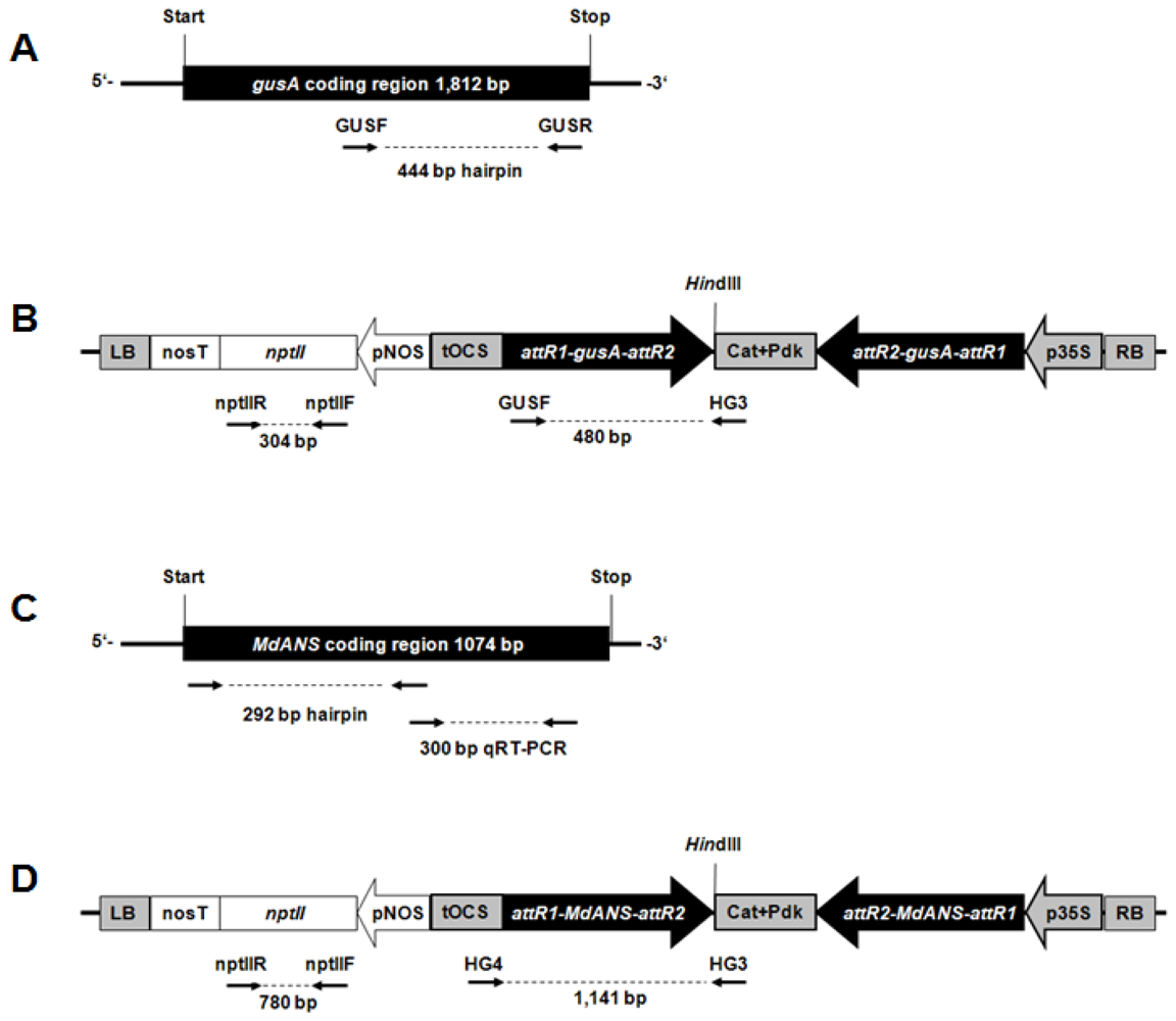
2.1. Generation of Hrp-Gusa Transgenic and Hrp-Mdans Transgenic Apple Clones

A total of three transformation experiments were performed to transform the apple genotype “PinS” with the binary plasmid vector pHELLSGATE8::hrp-gus (Figure 1). In total 19 independent putative transgenic plants were obtained after Agrobacterium tumefaciens-mediated gene transfer. Thirteen out of them were successfully propagated to establish transgenic clones. Genomic DNA of these clones was isolated from young leaves, in order to examine the integration of the hrp-gusA gene construct. For all clones DNA fragments of hrp-gusA and nptII were amplified by PCR using the primers nptIIF/R for nptII and GUSF/HG3 for hrp-gusA (Figure S1). Total RNA of each clone was isolated and reverse transcribed into cDNA to determine transgene transcription. RT-PCR analysis using the primers nptIIF/R and GUSF/HG3 showed that all clones transcribed both the nptII gene and the hrp-gusA gene construct (Figure S1). Southern blot analyses were performed using a labeled probe specific for the hrp-gusA gene construct. In all transgenic clones hybridization signals were detected indicating the integration of the gene construct.
In total, 26 putative transgenic apple plants were obtained after transformation of “PinS” using Agrobacterium tumefaciens strain EHA105 containing the binary plasmid vector pHELLSGATE8::hrp-Mdans (Figure 1). These clones were subsequently tested by PCR, Southern blot and RT-PCR on the presence, the integration and expression of the transferred genes. Only seven of these (T1295, T1296, T1297, T1302, T1303, T1308, and T1379) showed consistent results for transgene integration and expression. An example of this investigation is shown in Figure S2. The remaining clones showed no transgene integration except of clone T1378. A fragment of the transferred hrp-Mdans gene construct could be amplified by PCR for this clone, but integration of nptII was not supported by Southern hybridization. This clone seems to have an imperfect T-DNA integration or a chimeric character. All clones which did not show consistent results in all tests were excluded from further experiments. Clone T1300 which was obviously not transformed was used as additional non-transgenic control.
Most of the hrp-Mdans transgenic clones died within the next eight months. In vitro leaves of these clones went brown (Figure 2A) and the shoots were unable to grow. DAB (3,3-diaminobenzidine) staining suggested a high level of oxidative stress on in vitro leaves of the hrp-Mdans transgenic clones (Figure 2(B,C)). After transfer to the greenhouse, plants of these clones became necrotic (Figure 2D). Only the two transgenic clones T1297 and T1308, the non-transformed “PinS”, and the non-transgenic clone T1300 were successfully established in the greenhouse.

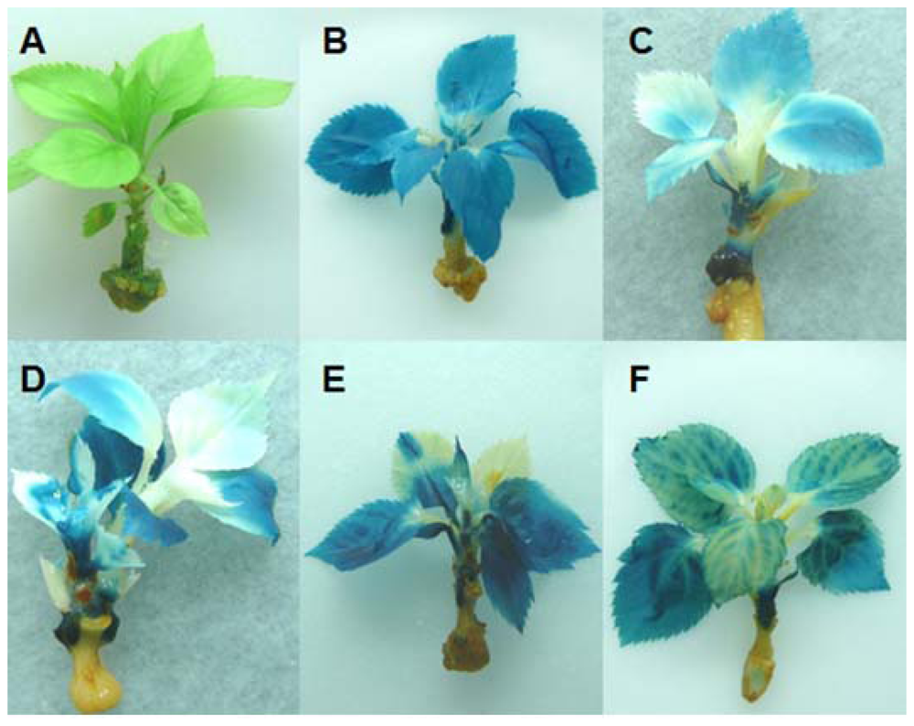
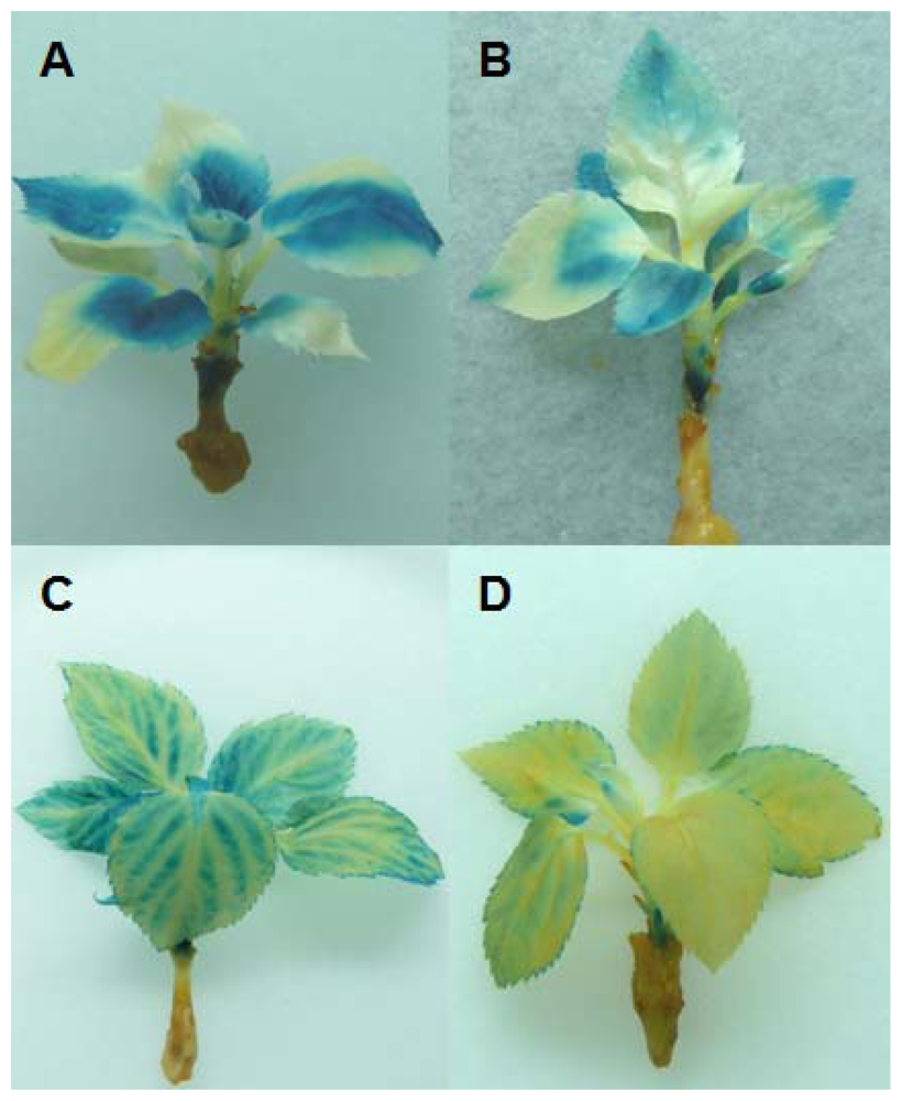
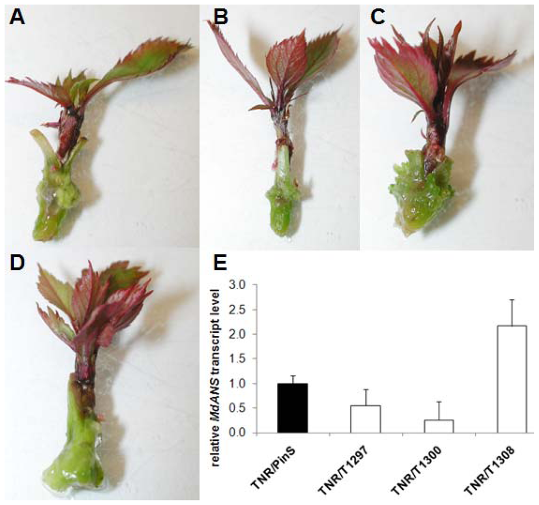
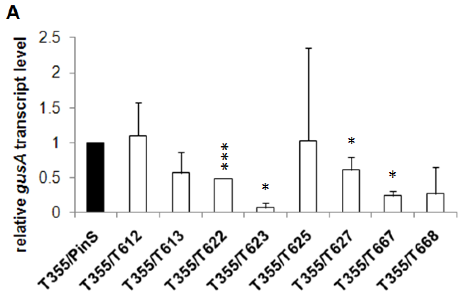
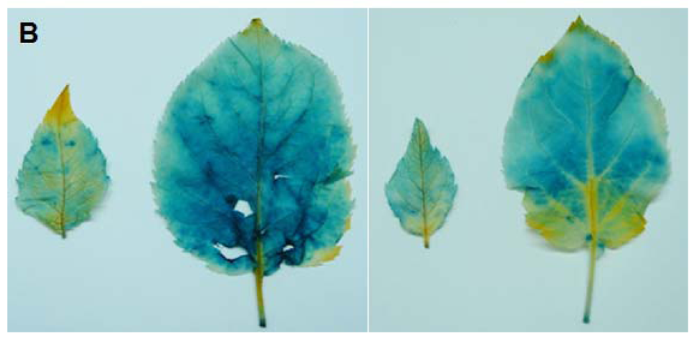
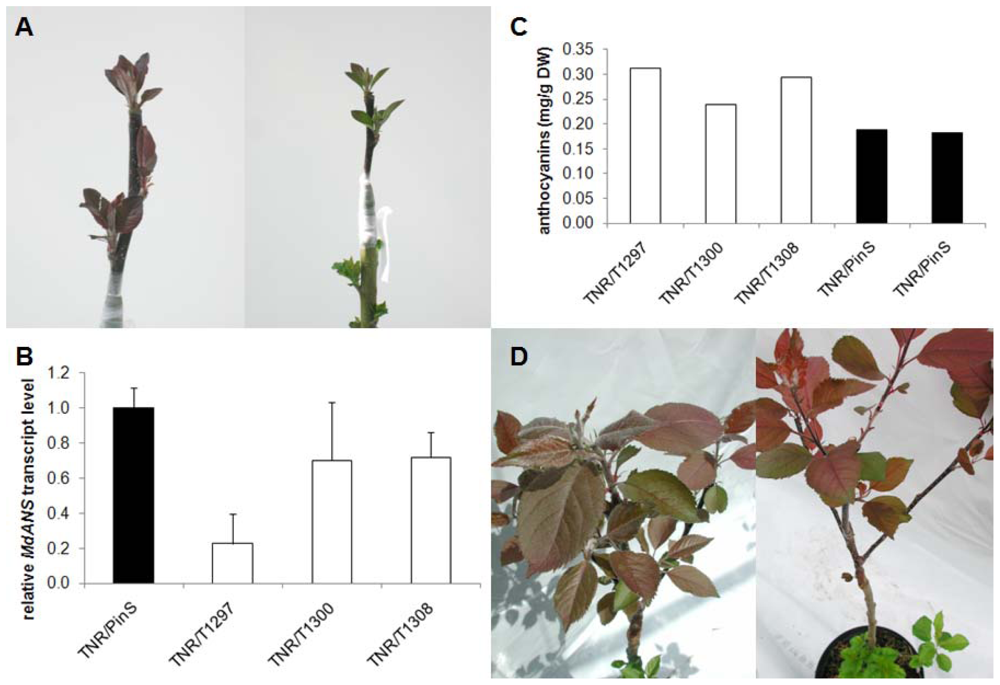
2.2. In Vitro Grafting Experiments

Micrografting experiments were performed using in vitro shoots of the hrp-gusA transgenic clones as rootstocks and shoots of the CaMV35S::gusA transgenic clone T355 as scions. Furthermore, scions of T355 were also grafted onto non-transgenic “PinS” used as a control (Figure 3A). A total of three to 12 grafted plants per rootstock genotype were established. At the time when the T355 scions had developed five to seven new leaves, young leaf material was collected. Total RNA of these leaves was extracted, reversely transcribed and tested on the level of gusA gene transcripts by RT-qPCR. The transcript level of gusA was reduced in all in vitro micrograftings grown on hrp-gusA transgenic rootstocks compared to those grown onto non-transgenic “PinS” (Figure 3G).
Four to five weeks after grafting, the grafted plants were evaluated on gusA gene expression by histochemical GUS staining. Leaves showing sclerosis or necrosis were excluded from the experiment to ensure that non-blue colored leaf areas are based on silencing. Grafted control plants showed a blue colored scion (T355) and a white rootstock (“PinS”) as expected. Leaves of the T355 scions grafted onto non-transformed “PinS” were nearly blue colored (Figure 3B), resulting the percentage of white (unstained) leaf areas was 7.4 ± 2.2% (mean of 27 individual leaves). In contrast, T355 scions grafted onto hrp-gusA transgenic rootstocks were only partial blue colored (Figure 3C–F). Between 19.4 ± 1.3% (clone T610, mean of 15 individual leaves) and 90 ± 12.0% (clone T668, mean of 12 individual leaves) of the entire leaf laminas of the T355 scions were white. The percentage of white colored leaf areas of T355 grafted onto “PinS” and T355 grafted onto the silencing transmitter clones was significantly different at α ≤ 0.001. Silencing of gusA affected young and old leaves, spread over entire leaf blades or different parts of the leaves. Two distinct phenotypes of silencing were detected. Type 1 plants showed only parts of leaves that were silenced. Non-silenced parts of these plants/leaves were dark blue colored (Figure 4A,B). The silencing effect was not restricted to a specific leaf area. In type 2 plants, the silencing effect entered the veins and expanded from the veins throughout the whole leaf lamina (Figure 4C,D). Shoots grafted onto the same silencing transmitter genotype (e.g., clone T668) showed always the same phenotype of silencing. The results were reproducible in independent grafting experiments, comprising of two to four grafted plants per experiment.
In vitro shoots of the natural red leaf genotype TNR31-35 were used as scion and grafted onto shoots of the hrp-Mdans transgenic clones T1297 and T1308 used as rootstocks. In parallel, shoots of TNR31-35 were also grafted onto non-transgenic “PinS” and clone T1300 used as controls. A total of 30 micrografted plants per genotype were established. After three to four weeks, young leaves of the grafted TNR31-35 were evaluated for leaf coloration or the presence of other visible silencing effects. Surprisingly, no phenotypic changes were detectable. Leaves of TNR31-35 grown on hrp-Mdans transgenic clones were normally red colored and could not be distinguished by visible means from those grown on “PinS” (Figure 5(A–D)). Subsequently, young leaves of the grafted TNR31-35 were collected for total RNA extraction. The relative Mdans transcript level was determined by RT-qPCR using the primers MdANS_MB1/MB2. The Mdans transcript level seemed not to be reduced in leaves of the TNR31-35 shoots grown on hrp-Mdans transgenic rootstocks compared to those grown on non-transgenic “PinS” and T1300 (Figure 5E).

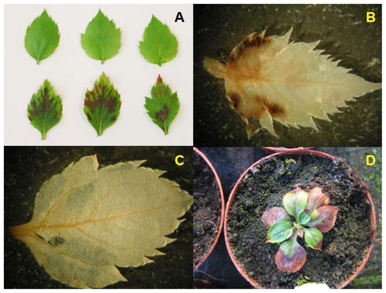
2.3. Grafting Experiments on Greenhouse Plants

In vitro shoots of hrp-gusA transgenic clones and non-transformed “PinS” were rooted and transferred to the greenhouse. After one year of cultivation all plants were pruned and grafted with scions of the gusA transgenic clone T355. Leaves of each grafted shoot were collected several times within the next two years and tested on gusA gene expression by RT-qPCR, histochemically and by colorimetric GUS assay. An example of this is shown in Figure 6. The gusA mRNA transcript level often seemed to be reduced (Figure 6A), but this was not supported by the results of histochemical staining (Figure 6B) and colorimetric GUS assay (data not presented). We never found silencing effects that were comparable to those obtained on in vitro plants (see Figures 3 and 4).
Plants of hrp-Mdans transgenic clones and the non-transformed “PinS” were rooted as described and transferred to the greenhouse. After one year of greenhouse cultivation these plants were pruned and used as rootstocks. The rootstocks were grafted with scions of TNR31-35 in order to evaluate the graft transmissible silencing effect of endogenous genes in woody plants. The first leaves, which became visible four to five weeks after grafting, were evaluated on the presence of silencing effects. Leaves of TNR31-35 grown on hrp-Mdans transgenic clones appeared to be less intensive red colored then leaves of TNR31-35 grown on “PinS” (Figure 7A). The results obtained by RT-qPCR were not consistent. TNR31-35 shoots grown on the silencing clone T1297 showed a strong reduction of the Mdans transcript level, whereas the transcript level was slightly increased in shoots grown on the control T1300 (Figure 7B). The anthocyanin coloration of TNR31-35 shoots grown on hrp-Mdans transgenic rootstocks was comparable to that grown on “PinS”. No reduction of the anthocyanin content was detectable (Figure 7C). No differences in leaf coloration were detectable on adult leaves (Figure 7D).

3. Discussion

Systemically induced gene silencing from genetically modified (gm) rootstocks to non-gm scions and its application to practical fruit breeding would offer a straightforward approach with significant impact on fruit production and horticulture. Grafting of scions onto silencing transmitter rootstocks for improving individual traits (e.g., self-fertility, resistance or flavor) of cultivars, which are well established in fruit production and on the market place would open a new door for the horticultural practice. The existence of graft-transmissibility of silencing inducing signals has been demonstrated several times in different plant species [912,1821]. Long-distance transport between grafted partners was demonstrated for transcriptional [21] and post-transcriptional gene silencing [22], for transgenes [9,10,20], and for endogenous gene sequences as well [22]. The silencing inducing signal, which moves systemically, is still unknown. There are a number of indications which argue for sRNAs and/or sRNA precursors [3]. In apple Malus × domestica, where gene silencing has been used effectively several times [2325], there is no evidence for its systemic spread until today.
Graft-transmission of post-transcriptional gene silencing in apple was tested in the present study using two different approaches. In the first approach, the β-glucuronidase encoding gusA gene of Escherichia coli was effectively silenced in grafted scions of the transgenic gusA overexpressing apple clone T355 of in vitro plants. The observed different types of silencing patterns (either complete or partially along vascular tissues, but not gradually in cells in both cases) are evidence for the existence of the whole silencing machinery in apple, which is necessary for its long-distance transport. Furthermore, they may indicate the existence of a critical threshold of siRNAs in cells of grafted scions to switch towards gene silencing. In contrast, for later stages of growth after transfer to greenhouse conditions graft-transmission of silencing was not effective.
In the second approach, the endogenous Mdans gene was silenced in transgenic plants of genotype “PinS” used as rootstocks. Several transgenic clones of the apple genotype “PinS” overexpressing a hairpin gene construct of the MdANS gene showed a sub-lethal phenotype. This is surprising as this genotype does neither produce anthocyanins in leaves nor in wooden tissue. With vital lines, a systemic spread of silencing was not detectable, neither in vitro nor in the greenhouse. Similar results were also obtained in other studies (for review see [1]). Endogenes, like the Mdans gene in our study, are known to be protected from the amplification abilities of RDRs. The exclusion from the amplification by RDRs is assumed to be rather correlated with the relatively low steady-state level of mRNA of the endogenes compared to transgenes as with the gene-sequence specificity itself. Therefore, spreading of endogene silencing depends on the long distance transport of the original silencing signal produced in the gm rootstock [1].
The lack of systemic spread of the Mdans gene silencing on in vitro plants may either be due to the relative low level of Mdans gene expression in grafted scions of TNR31-35 in which the Mdans gene is actually up-regulated by the MdMYB10 transcription factor [26,27], or by the lack (or low level) of the original silencing inducing signal.
The systemic transport of signals in plants occurs via the vascular system comprising the phloem and the xylem. A xylem transport of silencing signals would possibly explain the lack of graft-transmission of gusA gene silencing in woody plants. The differentiated xylem of trees consists of dead instead of living cells. However, the long distance movement of silencing signals is generally from source to sink through a bulk flow process that is characteristic of the phloem (reviewed in [3]). Based on this fact and the fact that the xylem sap is free of RNA [28] it is generally supposed that silencing is rather transported via phloem than via xylem [3].
The transfer of apple plants from in vitro to ex vitro conditions is accompanied by lignification of rootstock and scions. Lignification may influence active or passive cell-to-cell transport of siRNAs in living cells. In contrast to in vitro plants, ex vitro plants possess roots and their habit is multi-branched resulting in a shift in the source to sink relationship. The source to sink direction of in vitro shoots is exclusively from older to younger leaves and to the shoot apex whereas the roots of ex vitro plants can also represent very strong sinks. Such complex physiological changes associated with the ex vitro transfer or lignification can currently not be excluded as a cause for the lack of a systemic silencing effect. Investigation in other woody crop plants for systemic silencing effects seems essential to decide upon a putative general influence of lignification. In case that lignification is generally detrimental for systemic silencing, the use of grafting as a tool to transfer silencing effects from rootstocks to non-transgenic scions seems to be excluded. This would limit the range of applications of silencing in horticulture on the one hand. On the other hand, it would open the possibility to down-regulate specific genes in rootstocks (e.g., growth parameters) without influencing scions. The spatial restriction of the genetic modification within a plant consisting of genetically different partners (e.g., rootstock and scion) is an important point in the current biosafety assessment of gm plants. It is intensively discussed in the study “New Plant Breeding Techniques: State-of-the-Art and Prospects for Commercial Development” which was carried out to respond to an initial request from the Directorate General for the Environment (DG ENV) of the European Commission [29].

4. Experimental Section

4.1. Vector Design

For cloning of the hrp-gusA silencing gene construct a 444 bp fragment of the gene β-glucuronidase (gusA) was amplified by PCR using the primers gus-attB1 and gus-attB2 (Table 1). For cloning of the hrp-Mdans silencing gene construct a 292 bp fragment of the Mdans gene of apple was amplified by PCR using the primers ANS_attB1 and ANS_attB2 (Table 1). Both PCR fragments contained the recombination sites attB1 and attB2, respectively. They were separately cloned into the attR1 and attR2 sites of the binary vector pHELLSGATE8. The cloning into pHELLSGATE8 was performed via the intermediate vector pDONRTM 207 (Invitrogen) containing attP sites in a two step process. PCR, in vitro BP and LR clonase recombination reactions were carried out according to the manufacturer’s instructions (Invitrogen). The correct sequence and orientation of the introduced fragments were confirmed by sequencing.

4.2. Plant Material and Transformation

Leaves of in vitro axillary shoots of a descendant of the apple (Malus × domestica BORKH.) cultivar “Pinova” (“PinS”) were used for plant transformation. Plant transformation was done as described in [30] using the A. tumefaciens strain EHA105 containing the plasmid pHELLSGATE8::hrp-gus and the plasmid pHELLSGATE8::hrp-Mdans, respectively. Cultivation and micropropagation of in vitro shoots were also realized as described in [30]. Furthermore, the CaMV35S::gusA transgenic apple clone T355 described by Flachowsky et al. [16] and the red leaf genotype TNR31-35 (hybrid of Malus sieversii var. sieversii f. niedzwetzkyana, JKI collection Pillnitz, Germany) were used.

4.3. Molecular Evaluation of Transgenic Plants

Genomic DNA was extracted from leaf tissue with the DNeasy® Plant Mini Kit (Qiagen). Standard PCR assays were performed in a total volume of 25 μL containing 50 ng DNA, 1 × NH4-buffer, 1.5 mM MgCl2, 0.2 mM dNTPs, 0.5 μM of each primer and 0.5 U Taq DNA polymerase (Invitrogen). The PCR conditions were 35 cycles of 30 s denaturation at 94 °C, 1 min annealing (temperature depended on the primers used) and 1 min elongation at 72 °C. The length of PCR products was determined by electrophoresis on a 1% agarose gel with a 100 bp molecular size marker (MBI Fermentas). All primers used in this study are listed in Table 1.
For transcription analyses, total RNA was isolated from leaves using the Invisorb® Spin Plant RNA Mini Kit (Invitek), followed by a DNaseI treatment (Ambion). Presence of DNA contamination was tested by standard PCR using 2 μL RNA as template and the primers EF_F/EF_R, which are specific for the elongation factor gene EF1α of apple. The remaining RNA was reverse transcribed using oligo(dT)18 primers and the RevertAid™ First Strand cDNA Synthesis Kit (MBI Fermentas). The subsequent PCR reactions were performed with 1 μL cDNA and gene specific primers (Table 1).
Southern hybridization was performed as described by Flachowsky et al. [31]. Transgene integration was detected with digoxygenin (DIG)-labeled probes generated by PCR using the primer pairs GusF/HG3 for the hrp-gusA gene construct and nptIIF/nptIIR for the selectable marker gene nptII (Table 1).

4.4. Reverse Transcription Quantitative PCR (RT-qPCR)

RT-qPCR was performed as described by Flachowsky et al. [31]. Amplification and correlation efficiencies of each PCR assay were determined on diluted plasmid DNA of the transformation vectors. The expression of the transgenes as well as for the endogenous genes was studied using the gene specific primers listed in Table 1. All expression data were normalized using apple β-actin or RNA polymerase subunit II (RNApolII) as internal control for each sample.

4.5. Grafting Experiments

Micrografting of in vitro grown plants was performed as described by Tränkner et al. [32]. For grafting experiments in the greenhouse, micropropagated in vitro shoots were rooted and acclimatized to greenhouse conditions as described by Flachowsky et al. [30]. Grafting of greenhouse plants was performed using the whip-and-tongue grafting technique as described by Crasweller [33].

4.6. Histochemical GUS Assay

For GUS staining assays, the plant material was completely covered with X-Gluc solution containing 50 mM sodium phosphate pH 7.0, 1 mM EDTA, 0.2% Triton X 100, 0.05% SDS, 0.033% N-Lauryl-Sarcosine, 1 mM potassium ferricyanide and 50 mg X-Gluc (MBI Fermentas). After 1 h vacuum infiltration, the plants were incubated over night at 37 °C. The chlorophyll was removed with ethanol-acetic acid solution in a ratio of 3:1 for 16 h at room temperature. The software program Carnoy 2.0 (Alnini, Inc.: Petaluma, CA, USA, 2012) was used to measure silenced and non-silenced areas after GUS staining.

4.7. Anthocyanin Coloration

Anthocyanidin coloration was evaluated visually for this study. Analytical verification of color by apple anthocyanins after overexpression or direct silencing of anthocyanidin synthase in a red-leaved cultivar has been performed as described by Li et al. [13] and Szankowski et al. [23].

4.8. Statistical Analysis

Quantitative data were subjected to statistical analysis (ANOVA and Duncan’s multiple range test) using the SAS® 9.1 software (SAS Institute: Cary, NC, USA, 2004).

5. Conclusions

It has been demonstrated for the first time that a silencing signal is transported from a transgenic rootstock of apple (Malus × domestica) to in vitro grafted scions over expressing the heterologous reporter gene β-glucuronidase. This transport of a silencing signal in in vitro graftings is not seen when the endogenous anthocyanidin synthase gene is used as a reporter gene. Ex vitro under greenhouse conditions the transport of the silencing signal for the heterologous reporter gene β-glucuronidase is also not observed. Hypothetically, this is correlated with the lignification process in tissues under these conditions. For final conclusion, this should be compared with results to be obtained with other woody plant systems.

Supplementary Materials

ijms-13-09992-s001.pdf

Acknowledgments

The authors acknowledge the financial support for substantial parts of this work by the German Federal Ministry for Education and Research (BMBF) and by the European research project ISAFRUIT. The ISAFRUIT project was funded by the European Commission under the Thematic Priority 5-Food Quality and Safety of the 6th Framework Programme of RTD (Contract No. FP6-FOOD-CT-2006-016279). The views and opinions expressed in this publication are purely those of the authors and may not in any circumstances be regarded as stating an official position of the European Commission. We further acknowledge Ines Hiller, Katrin Winkler, Ines Polster, Uta Hille, P. Wolff, and Volkmar Vogt for their technical assistance. The work was performed within the frame of COST 864.

Abbreviations

ans
anthocyanidin synthase
gus
β-glucuronidase

References

  1. Kalantidis, K.; Schumacher, H.T.; Alexiadis, T.; Helm, J.M. RNA silencing movement in plants. Biol. Cell 2008, 100, 13–26. [Google Scholar]
  2. Molnar, A.; Melnyk, C.; Baulcombe, D.C. Silencing signals in plants: A long journey for small RNAs. Genome Biol 2011, 12. [Google Scholar] [CrossRef]
  3. Melnyk, C.W.; Molnar, A.; Baulcombe, D.C. Intercellular and systemic movement of RNA silencing signals. EMBO J 2011, 30, 3553–3563. [Google Scholar]
  4. Voinnet, O. Use, tolerance and avoidance of amplified RNA silencing by plants. Trends Plant Sci 2008, 13, 317–328. [Google Scholar]
  5. Schwab, R.; Maizel, A.; Ruiz-Ferrer, V.; Garcia, D.; Bayer, M.; Crespi, M.; Voinnet, O.; Martienssen, R.A. Endogenous tasiRNAs mediate non-cell autonomous effects on gene regulation in Arabidopsis thaliana. PLoS One 2009, 4, e5980. [Google Scholar]
  6. Himber, C.; Dunoyer, P.; Moissiard, G.; Ritzenthaler, C.; Voinnet, O. Transitivity-dependent and -independent cell-to-cell movement of RNA silencing. EMBO J 2003, 22, 4523–4533. [Google Scholar]
  7. Wassenegger, M.; Krzcal, G. Nomenclature and functions of RNA-directed RNA polymerases. Trends Plant Sci 2006, 11, 142–151. [Google Scholar]
  8. Smith, L.M.; Pontes, O.; Searle, L.; Yelina, N.; Yousafzai, F.K.; Herr, A.J.; Pikaard, C.S.; Baulcombe, D.C. An SNF2 protein associated with nuclear RNA silencing and the spread of a silencing signal between cells in Arabidopsis. Plant Cell 2007, 19, 1507–1521. [Google Scholar]
  9. Tournier, B.; Tabler, M.; Kalantidis, K. Phloem flow strongly influences the systemic spread of silencing in GFP Nicotiana benthamiana plants. Plant J 2006, 47, 383–394. [Google Scholar]
  10. Palauqui, J.-C.; Elmayan, T.; Pollien, J.-M.; Vaucheret, H. Systemic acquired silencing: Transgene-specific post-transcriptional silencing is transmitted by grafting from silenced stocks to non-silenced scions. EMBO J 1997, 16, 4738–4745. [Google Scholar]
  11. Mallory, A.C.; Mlotshwa, S.; Bowman, L.H.; Vance, V.B. The capacity of transgenic tobacco to send a systemic RNA silencing signal depends on the nature of the inducing transgene locus. Plant J 2003, 35, 82–92. [Google Scholar]
  12. Yoo, B.-C.; Kragler, F.; Varkonyi-Gasic, E.; Haywood, V.; Archer-Evans, S.; Lee, Y.M.; Lough, T.J.; Lucas, W.J. A systemic small RNA signaling system in plants. Plant Cell 2004, 16, 1979–2000. [Google Scholar]
  13. Li, H.; Flachowsky, H.; Fischer, T.C.; Hanke, V.; Forkmann, G.; Treutter, D.; Schwab, W.; Hofmann, T.; Szankowski, I. Maize Lc transcription factor enhances biosynthesis of anthocyanins, distinct proanthocyanidins and phenylpropanoids in apple (Malus domestica Borkh.). Planta 2007, 226, 1243–1254. [Google Scholar]
  14. Harada, T. Grafting and RNA transport via phloem tissue in horticultural plants. Sci. Hortic. Amst 2010, 125, 545–550. [Google Scholar]
  15. Martin, A.; Adam, H.; Diaz-Mendoza, M.; Zurczak, M.; González-Schain, D.; Suárez-López, P. Graft-transmissible induction of potato tuberization by the microRNA miR172. Development 2009, 136, 2873–2881. [Google Scholar]
  16. Flachowsky, H.; Riedel, M.; Reim, S.; Hanke, M.-V. Evaluation of the uniformity and stability of T-DNA integration and gene expression in transgenic apple plants. Electron. J. Biotechnol 2008, 11. [Google Scholar] [CrossRef]
  17. Lespinasse, Y.; Godicheau, M. Creation et description d’une plante haploide de Pommier (Malus pumila Mill.). Ann. Amelior. Plante 1980, 30, 39–44. (in French). [Google Scholar]
  18. Hewezi, T.; Alibert, G.; Kallerhoff, J. Local infiltration of high- and low-molecular-weight RNA from silenced sunflower (Helianthus annuus L.) plants triggers post-transcriptional gene silencing in non-silenced plants. Plant Biotechnol. J 2005, 3, 81–89. [Google Scholar]
  19. Li, M.; Jiang, S.; Wang, Y.; Liu, G. Post-transcriptional gene silencing signal could move rapidly and bidirectionally in grafted Arabidopsis thaliana. Chin. Sci. Bull 2006, 51, 313–319. [Google Scholar]
  20. Shaharuddin, N.A.; Han, Y.; Li, H.; Grierson, D. The mechanism of graft transmission of sense and antisense gene silencing in tomato plants. FEBS Lett 2006, 580, 6579–6586. [Google Scholar]
  21. Bai, S.; Kasai, A.; Yamada, K.; Li, T.; Harada, T. A mobile signal transported over a long distance induces systemic transcriptional gene silencing in a grafted partner. J. Exp. Bot 2011, 62, 4561–4570. [Google Scholar]
  22. Kasai, A.; Bai, S.; Li, T.; Harada, T. Graft-transmitted siRNA signal from the root induces visual manifestation of endogenous post-transcriptional gene silencing in the scion. PLoS One 2011, 6, e16895. [Google Scholar]
  23. Szankowski, I.; Flachowsky, H.; Li, H.; Halbwirth, H.; Treutter, D.; Regos, I.; Hanke, M.-V.; Stich, K.; Fischer, T.C. Shift in polyphenol profile and sublethal phenotype caused by silencing of anthocyanidin synthase in apple (Malus sp.). Planta 2009, 229, 681–692. [Google Scholar]
  24. Broothaerts, W.; Keulemans, J.; Nerum, I.V. Self-fertile apple resulting from S-RNase gene silencing. Plant Cell Rep 2004, 22, 497–501. [Google Scholar]
  25. Gilissen, L.J.W.J.; Bolhaar, S.T.H.P.; Matos, C.I.; Rouwendal, G.J.A.; Boone, M.J.; Krens, F.A.; Zuidmeer, L.; van Leeuwen, A.; Akkerdaas, J.; Hoffmann-Sommergruber, K.; et al. Silencing the major apple allergen Mal d 1 by using the RNA interference approach. J. Allergy Clin. Immunol 2005, 115, 364–369. [Google Scholar]
  26. Ban, Y.; Honda, C.; Hatsuyama, Y.; Igarashi, M.; Bessho, H.; Moriguchi, T. Isolation and functional analysis of a MYB transcription factor gene that is a key regulator for the development of red coloration in apple skin. Plant Cell Physiol 2007, 48, 958–970. [Google Scholar]
  27. Espley, R.V.; Hellens, R.P.; Putterill, J.; Stevenson, D.E.; Kutty-Amma, S.; Allan, A.C. Red colouration in apple fruit is due to the activity of the MYB transcription factor, MdMYB10. Plant J 2007, 49, 414–427. [Google Scholar]
  28. Buhtz, A.; Springer, F.; Chappell, L.; Baulcombe, D.C.; Kehr, J. Identification and characterization of small RNAs from the phloem of Brassica napus. Plant J 2008, 53, 739–749. [Google Scholar]
  29. Lusser, M.; Parisi, C.; Plan, D.; Rodríguez-Cerezo, E. New Plant Breeding Techniques, State-of-The-Art and Prospects for Commercial Development; EUR Number: 24760 EN; European Union: Seville, Spain, 2011. [Google Scholar]
  30. Flachowsky, H.; Peil, A.; Sopanen, T.; Elo, A.; Hanke, V. Overexpression of BpMADS4 from silver birch (Betula pendula Roth.) induces early flowering in apple (Malus × domestica Borkh.). Plant Breed 2007, 126, 137–145. [Google Scholar]
  31. Flachowsky, H.; Hättasch, C.; Höfer, M.; Peil, A.; Hanke, M.-V. Overexpression of LEAFY in apple leads to a columnar phenotype with shorter internodes. Planta 2010, 231, 251–263. [Google Scholar]
  32. Tränkner, C.; Lehmann, S.; Hoenicka, H.; Hanke, M.-V.; Fladung, M.; Lenhardt, D.; Dunemann, F.; Gau, A.; Schlangen, K.; Malnoy, M.; et al. Over-expression of an FT-homologous gene of apple induces early flowering in annual and perennial plants. Planta 2010, 232, 1309–1324. [Google Scholar]
  33. Crasweller, R.M. Grafting and propagating fruit trees. Penn State Coll. Agric. Sci 2005, 1–12. [Google Scholar]
Figure 1. Schematic application of the T-DNAs of the vectors used for plant transformation. (A) coding region of the gusA gene including the region used for silencing vector construction; (B) T-DNA of the gusA silencing vector; (C) coding region of the Mdans gene including the region used for silencing vector construction; (D) T-DNA of the Mdans silencing vector. Black arrows indicate the position of primers.
Ijms 13 09992f1
Figure 2. Stress symptoms on leaves of hrp-Mdans transgenic apple clones. (A) in vitro leaves of the non-transformed “PinS” (upper row) without and clone T1302 (lower row) with necrosis; (B) leaves of clone T1302 after DAB staining; (C) leaves of the non-transformed “PinS” after DAB staining; (D) greenhouse plant of clone T1302 showing strong necrosis.
Ijms 13 09992f2
Figure 3. Micrografting experiments with gusA transgenic scions of T355 grafted onto non-transformed “PinS” (A and B) and hrp-gusA transgenic rootstocks of clones T612 (C), T613 (D), T623 (E), and T627 (F). (A) Graftings of T355 onto “PinS” before histochemical β-glucuronidase (GUS) staining; (BF) Graftings of different genotypes after histochemical GUS staining; (G) Detection of the relative gusA transcript level in T355 grafted onto “PinS” and hrp-gusA transgenic shoots used as rootstocks. The gusA transcript levels were determined by Reverse Transcription Quantitative PCR (RT-qPCR). Young leaves of three individual plants were pooled and used for RNA extraction representing one biological replicate. The values are expressed in comparison to T355 grafted onto “PinS”, which was set to be one. The values of T355 grafted onto “PinS” are the mean of three biological replicates of which each was measured in three technical replicates. The values of T355 grafted onto the silencing transmitter clones T610, T612, T613, T615, T622, T623, T625, T626, T628 and T638 are the mean of one biological replicate per clone each measured with three technical replications. The values of T355 grafted onto the silencing transmitter clones T627, T667 and T668 are the mean of two biological replicates each measured with three technical replications. Bars represent standard errors. Mean, mean of all transgenic silencing transmitter clones (T610–T668); * significantly different compared to T355/PinS at α ≤ 0.05.
Ijms 13 09992f3aIjms 13 09992f3b
Figure 4. Different types of silencing detected on gusA transgenic scions of clone T355 grafted onto hrp-gusA transgenic silencing transmitter rootstocks of clones T625 (A and B) and T668 (C and D). Shoots grafted onto rootstocks of clone T625 showed locally restricted silencing (type 1). Shoots grafted onto rootstocks of clone T668 showed systemic silencing (type 2).
Ijms 13 09992f4
Figure 5. Micrografting experiments with non-transgenic scions of the red leaf Malus genotype TNR31-35 grafted onto non-transformed “PinS” (A), the hrp-Mdans transgenic clones T1297 and T1308 (B and D), as well as the non-transgenic clone T1300 (C). Relative quantification of the Mdans mRNA transcript levels in TNR31-35 grafted onto hrp-Mdans transgenic apple clones used as rootstocks (E). The Mdans transcript levels were determined by quantitative real-time PCR (mean of two biological replicates, each three technical repetitions, bars represent standard errors).
Ijms 13 09992f5
Figure 6. Grafting experiments with greenhouse-grown gusA transgenic scions of T355 grafted onto non-transformed “PinS” and hrp-gusA transgenic rootstocks of clones T612, T613, T622, T623, T625, T627, T667 and T668. (A) Relative quantification of gusA mRNA transcript levels in T355 scions grafted onto non-transgenic and hrp-gusA transgenic rootstocks, respectively (values mean of three biological replicates, each three technical repetitions, bars represent standard errors). * significantly different to T355/PinS at α ≤ 0.05; *** significantly different to T355/PinS at α ≤ 0.001; (B) Histochemical GUS staining of leaves of T335 grafted onto non-transformed “PinS” (left) and silencing clone T627 (right).
Ijms 13 09992f6aIjms 13 09992f6b
Figure 7. Grafting experiments in the greenhouse with non-transgenic scions of red-leave TNR31-35 grafted onto non-transformed “PinS” and hrp-Mdans transgenic rootstocks of clones T1297 and T1308. The non-transformed clone T1300 and “PinS” were used as controls. (A) TNR31-35 scions four to five weeks after grafting onto “PinS” (left) and T1297 (right); (B) relative quantification of the Mdans mRNA transcript levels in TNR31-35 grafted onto non-transgenic and hrp-Mdans transgenic rootstocks, respectively. The Mdans transcript levels were determined by quantitative real-time PCR (values mean of three biological replicates, each three technical repetitions, bars represent standard errors); (C) Mdans silencing dependent changes in anthocyanin contents of TNR31-35 scions grafted onto non-transformed “PinS”, T1300 and hrp-Mdans transgenic clones; (D) TNR31-35 grafted shoots after one year of greenhouse cultivation. Left: TNR31-35 grafted onto “PinS”; Right: TNR31-35 grafted onto T1297.
Ijms 13 09992f7
Table 1. Sequences of the primers used in this study.
PrimerSequence 5′→3′
gus-attBB1GGGGACAAGTTTGTACAAAAAAGC
AGGCTGTTCTGCGACGCTCACACCGATACC
B2GGGACCACTTTGTACAAGAAAGCT
GGGTTCACCGAAGTTCATGCCAGTCCAG
ANS_attBB1GGGGACAAGTTTGTACAAAAAAGC
AGGCTCTGTGAGCTCTGATTCAGTGA
B2GGGACCACTTTGTACAAGAAAGCT
GGGTACCTTGTCCATGAGCTCGTCA
nptIIFGGTTCTCCGGCCGCTTGGGTG
RCGGCAGGAGCAAGGTGAGATGAC
GUSFGTTCTGCGACGCTCACACCGATACC
RTCACCGAAGTTCATGCCAGTCCAG
EFFATTGTGGTCATTGG(CT)CA(CT)GT
RCCAATCTTGTA(AGC)ACATCCTG
ActFGTGAGGCTCTATTCCAACCATC
RGGAACACAAATTGGGCAAGTAT
HG1GCAAGTGGATTGATGTGACATCTCC
3CGTCTGTGATGGCTTCCATGTCGGC
3nGGATCCTCTAGACCACTTTGTAC
4CGAAACCGGCGGTAAGGATCTGAGC
EF1aFATTGTGGTCATTGGYCAYGT
RCCAATCTTGTAVACATCCTG
RNApolIIFATATGCCACCCCGTTCTCTACT
RCACGTTCCATTTGTCCAAACTT
MdANSMB1CACCTTCATCCTCCACAACAT
MB2ATGTGCTCAGCAAAAGTTCGT
FGTGAGCTCTGATTCAGTGA
RCCTTGTCCATGAGCTCGTCA

