Soluble Receptor for Advanced Glycation End-Product (sRAGE)/Pentosidine Ratio: A Potential Risk Factor Determinant for Type 2 Diabetic Retinopathy

Abstract
:
1. Introduction
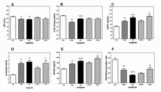
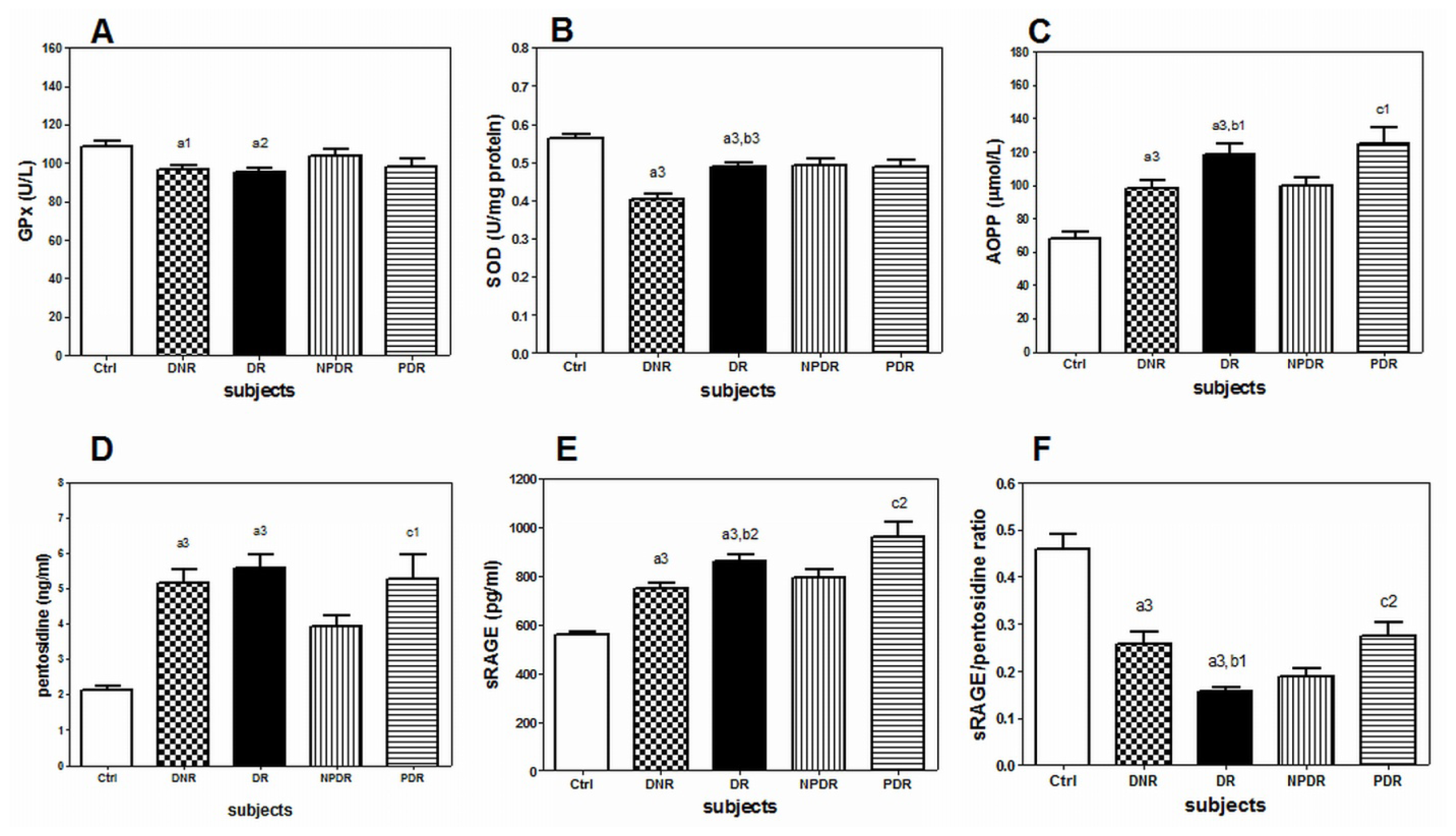
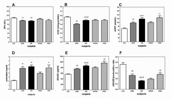
2. Results
3. Discussion
4. Experimental Section
4.1. Ethic Statement
4.2. Study Population
4.3. Sample Collection and Preparation
4.4. Measurement of Plasma Pentosidine
4.5. Measurement of Plasma sRAGE
4.6. Measurement of Plasma AOPP
4.7. Measurement of Plasma GPx Activity
4.8. Measurement of PBMC SOD Activity
4.9. Statistical Analysis
5. Conclusions
Acknowledgments
Conflict of Interest
References
- Ooyub, S.; Ismail, F.; Daud, N.A. Diabetes program in Malaysia-current and future. NCD Malays 2004, 3, 6–12. [Google Scholar]
- Zanariah, H.; Chandran, L.R.; wan Mohamad, W.B.; wan Nazaimoon, W.M.; Letchuman, G.R.; Jamaiyah, H.; Fatanah, I.; Nurain, M.N.; Helen Tee, G.H.; Mohd Rodi, I. DWP1-3 Prevalence of diabetes mellitus in Malaysia in 2006—Results of the 3rd National Health and Morbidity Survey (NHMS III). Diabetes Res. Clin. Pract 2008, 79, S21. [Google Scholar]
- Uthra, S.; Raman, R.; Mukesh, B.N.; Rajkumar, S.A.; Padmaja, K.R.; Paul, P.G.; Lakshmipathy, P.; Gnanamoorthy, P.; Sharma, T.; McCarty, C.A.; et al. Association of VEGF gene polymorphisms with diabetic retinopathy in a south Indian cohort. Ophthalmic Genet 2008, 29, 11–15. [Google Scholar]
- Goh, P.P. Status of diabetic retinopathy among diabetics registered to the Diabetic Eye Registry, National Eye Database, 2007. Med. J. Malays 2008, 63, 24–28. [Google Scholar]
- Betteridge, D.J. What is oxidative stress? Metabolism 2000, 49, 3–8. [Google Scholar]
- Valko, M.; Leibfritz, D.; Moncol, J.; Cronin, M.T.; Mazur, M.; Telser, J. Free radicals and antioxidants in normal physiological functions and human disease. Int. J. Biochem. Cell Biol 2007, 39, 44–84. [Google Scholar]
- Costagliola, C. Oxidative state of glutathione in red blood cells and plasma of diabetic patients: In vivo and in vitro study. Clin. Physiol. Biochem 1990, 8, 204–210. [Google Scholar]
- Droge, W. Free radicals in the physiological control of cell function. Physiol. Rev 2002, 82, 47–95. [Google Scholar]
- Kankova, K.; Marova, I.; Zahejsky, J.; Muzik, J.; Stejskalova, A.; Znojil, V.; Vacha, J. Polymorphisms 1704G/T and 2184A/G in the RAGE gene are associated with antioxidant status. Metabolism 2001, 50, 1152–1160. [Google Scholar]
- Hudson, B.I.; Hofmann, M.; Bucciarelli, L.; Wendt, T.; Moser, B.; Lu, Y.; Qu, W.; Stern, D.; D’Agati, V.; Yan, S.D.; et al. Glycation and diabetes: The RAGE connection. Curr. Sci 2002, 83, 1515–1521. [Google Scholar]
- Kalousova, M.; Jachymova, M.; Mestek, O.; Hodkova, M.; Kazderova, M.; Tesar, V.; Zima, T. Receptor for advanced glycation end products–soluble form and gene polymorphisms in chronic haemodialysis patients. Nephrol. Dial. Transplant 2007, 22, 2020–2026. [Google Scholar]
- Grossin, N.; Wautier, M.P.; Meas, T.; Guillausseau, P.J.; Massin, P.; Wautier, J.L. Severity of diabetic microvascular complications is associated with a low soluble RAGE level. Diabetes Metab 2008, 34, 392–395. [Google Scholar]
- Nazratun, N.; Mahmood, A.A.; Kuppusamy, U.R.; Ahmad, T.S.; Tan, S.Y. Diabetes mellitus exacerbates advanced glycation end product accumulation in the veins of end-stage renal failure patients. Vasc. Med 2006, 11, 245–250. [Google Scholar]
- Challier, M.; Jacqueminet, S.; Benabdesselam, O.; Grimaldi, A.; Beaudeux, J.L. Increased serum concentrations of soluble receptor for advanced glycation endproducts in patients with type 1 diabetes. Clin. Chem 2005, 51, 1749–1750. [Google Scholar]
- Al-Mesallamy, H.O.; Hammad, L.N.; El-Mamoun, T.A.; Khalil, B.M. Role of advanced glycation end product receptors in the pathogenesis of diabetic retinopathy. J. Diabetes Complicat 2010, 25, 168–174. [Google Scholar]
- Nakamura, K.; Yamagishi, S.I.; Matsui, T.; Adachi, H.; Takeuchi, M.; Imaizumi, T. Serum levels of soluble form of receptor for advanced glycation end products (sRAGE) are correlated with AGEs in both diabetic and non-diabetic subjects. Clin. Exp. Med 2007, 7, 188–190. [Google Scholar]
- Forbes, J.M.; Thorpe, S.R.; Thallas-Bonke, V.; Pete, J.; Thomas, M.C.; Deemer, E.R.; Bassal, S.; El-Osta, A.; Long, D.M.; Panagiotopoulos, S.; et al. Modulation of soluble receptor for advanced glycation end products by angiotensin-converting enzyme-1 inhibition in diabetic nephropathy. J. Am. Soc. Nephrol 2005, 16, 2363–2372. [Google Scholar]
- Salman, A.G.; Mansour, D.E.; Swelem, A.H.; Al-Zawahary, W.M.; Radwan, A.A. Pentosidine—A new biochemical marker in diabetic retinopathy. Ophthalmic Res 2009, 42, 96–98. [Google Scholar]
- Stitt, A.W. AGEs and diabetic retinopathy. Invest. Ophthalmol. Vis. Sci 2010, 51, 4867–4874. [Google Scholar]
- Mates, J.M.; Perez-Gomez, C.; Nunez de Castro, I. Antioxidant enzymes and human diseases. Clin. Biochem 1999, 32, 595–603. [Google Scholar]
- Ramakrishna, V.; Jailkhani, R. Oxidative stress in non-insulin-dependent diabetes mellitus (NIDDM) patients. Acta Diabetol 2008, 45, 41–46. [Google Scholar]
- Kalousova, M.; Sulkova, S.; Fialova, L.; Soukupova, J.; Malbohan, I.M.; Spacek, P.; Braun, M.; Mikulikova, L.; Fortova, M.; Horejsi, M.; et al. Glycoxidation and inflammation in chronic haemodialysis patients. Nephrol. Dial. Transplant 2003, 18, 2577–2581. [Google Scholar]
- Witko-Sarsat, V.; Friedlander, M.; Nguyen Khoa, T.; Capeillere-Blandin, C.; Nguyen, A.T.; Canteloup, S.; Dayer, J.M.; Jungers, P.; Drueke, T.; Descamps-Latscha, B. Advanced oxidation protein products as novel mediators of inflammation and monocyte activation in chronic renal failure. J. Immunol 1998, 161, 2524–2532. [Google Scholar]
- Hammes, H.P.; Brownlee, M.; Lin, J.; Schleicher, E.; Bretzel, R.G. Diabetic retinopathy risk correlates with intracellular concentrations of the glycoxidation product Nepsilon-(carboxymethyl) lysine independently of glycohaemoglobin concentrations. Diabetologia 1999, 42, 603–607. [Google Scholar]
- Boehm, B.O.; Schilling, S.; Rosinger, S.; Lang, G.E.; Lang, G.K.; Kientsch-Engel, R.; Stahl, P. Elevated serum levels of N(epsilon)-carboxymethyl-lysine, an advanced glycation end product, are associated with proliferative diabetic retinopathy and macular oedema. Diabetologia 2004, 47, 1376–1379. [Google Scholar]
- Kalousova, M.; Zima, T.; Tesar, V.; Dusilova-Sulkova, S.; Skrha, J. Advanced glycoxidation end products in chronic diseases-clinical chemistry and genetic background. Mutat. Res 2005, 579, 37–46. [Google Scholar]
- Diabetic retinopathy study. Report Number 6. Design, methods, and baseline results. Report Number 7. A modification of the Airlie House classification of diabetic retinopathy. Prepared by the Diabetic Retinopathy. Invest. Ophthalmol. Vis. Sci. 1981, 21, 1–226.
- Early Treatment Diabetic Retinopathy Study Research Group. Fundus photographic risk factors for progression of diabetic retinopathy. ETDRS report number 12. Ophthalmology 1991, 98, 823–833.
- Early Treatment Diabetic Retinopathy Study Research Group. Grading diabetic retinopathy from stereoscopic color fundus photographs–An extension of the modified Airlie House classification. ETDRS report number 10. Ophthalmology 1991, 98, 786–806.
- Boyum, A. Separation of leukocytes from blood and bone marrow. Introduction. Scand. J. Clin. Lab. Invest. Suppl 1968, 97, 7. [Google Scholar]
- Bradford, M.M. A rapid and sensitive method for the quantitation of microgram quantities of protein utilizing the principle of protein-dye binding. Anal. Biochem 1976, 72, 248–254. [Google Scholar]
- Witko-Sarsat, V.; Friedlander, M.; Capeillere-Blandin, C.; Nguyen-Khoa, T.; Nguyen, A.T.; Zingraff, J.; Jungers, P.; Descamps-Latscha, B. Advanced oxidation protein products as a novel marker of oxidative stress in uremia. Kidney Int 1996, 49, 1304–1313. [Google Scholar]

| Demography | Ctrl (n = 235) | DNR (n = 171) | DR (n = 200) | NPDR (n = 125) | PDR (n = 75) |
|---|---|---|---|---|---|
| Gender (male/female) | 134/101 | 100/71 | 110/90 | 67/58 | 43/32 |
| Races (Malay/Chinese/Indian) (n) | 106/90/39 | 63/28/80 a | 70/47/83 a | 40/29/56 | 30/18/27 |
| Age (years) | 57.1 ± 4.1 | 59.2 ± 9.6 | 57.2 ± 9.8 | 59.0 ± 9.9 | 53.1 ± 8.8 c |
| BMI (kg/m2) | 25.6 ± 4.8 | 27.2 ± 4.4 | 26.3 ± 5.0 | 26.2 ± 4.5 | 26.6 ± 5.9 |
| HbA1c (%) | 5.6 ± 0.4 | 7.9 ± 1.8 a | 8.9 ± 2.1 a,b | 9.1 ± 2.3 | 8.5 ± 1.8 |
| Total cholesterol (mmol/L) | 3.8 ± 0.6 | 4.5 ± 1.0 a | 4.8 ± 1.5 a,b | 4.8 ± 1.4 | 4.9 ± 1.5 |
| Triglyceride (mmol/L) | 1.8 ± 1.3 | 1.6 ± 0.7 | 1.7 ± 1.0 | 1.6 ± 0.9 | 2.0 ± 1.1 c |
| HDL-C (mmol/L) | 1.0 ± 0.3 | 1.2 ± 0.3 a | 1.2 ± 0.3 a | 1.3 ± 0.3 | 1.1 ± 0.3 c |
| LDL-C (mmol/L) | 2.1 ± 0.5 | 2.5 ± 0.9 a | 2.8 ± 1.2 a | 2.8 ± 1.2 | 2.9 ± 1.3 |
| HDL-C/LDL-C ratio | 0.6 ± 0.2 | 0.5 ± 0.2 a | 0.5 ± 0.2 a | 0.5 ± 0.2 | 0.4 ± 0.2 |
| SBP (mmHg) | 124.0 ± 8.0 | 136.5 ± 19.5 a | 139.3 ± 22.4 a | 136.6 ± 21.4 | 146.6 ± 23.0 c |
| DBP (mmHg) | 83.0 ± 7.0 | 79.0 ± 10.5 a | 78.4 ± 13.1 a | 77.2 ± 11.9 | 81.6 ± 15.6 |
| ALT (IU/L) | – | 37.8 ± 17.5 | 36.8 ± 24.6 | 38.1 ± 27.0 | 33.9 ± 18.3 |
| AST (IU/L) | – | 22.0 ± 14.0 | 22.8 ± 16.4 | 22.7 ± 17.2 | 23.0 ± 15.0 |
| Creatinine (μmol/L) | – | 94.9 ± 13.4 | 98.1 ± 12.8 | 90.0 ± 15.4 | 100.1 ± 13.6 |
| eGFR (ml/min/1.73m2) | – | 82.6 ± 9.6 | 90.2 ± 10.4 b | 92.2 ± 7.3 | 88.3 ± 12.7 |
| Diabetes duration (years) | – | 10.4 ± 7.9 | 15.7 ± 9.1 b | 16.3 ± 9.4 | 14.6 ± 8.2 |
| Retinopathy duration d (years) | – | – | 5.0 ± 3.6 | 3.4 ± 2.0 | 7.4 ± 4.3 c |
| Current smoker (n) | 43 | 29 | 13 a,b | 7 | 6 |
| Hypertension (n) | 11 | 104 a | 119 a | 80 | 39 |
| Antihyperglycemic treatment e (n) | 0 | 107 a | 130a | 93 | 37 c |
| Duration of antihyperglycemic treatment (years) | – | 9.5 ± 5.5 | 11.5 ± 7.5 | 9.9 ± 3.6 | 11.0 ± 5.2 |
| Antihypertensive treatment f (n) | 0 | 104 a | 119 a | 79 | 40 |
| Duration of antihypertensive treatment (years) | – | 7.0 ± 3.5 | 8.5 ± 4.0 | 7.7 ± 2.8 | 8.3 ± 3.5 |
| Variables | AOPP (μmol/L) | Pentosidine (ng/mL) | sRAGE (pg/mL) | |||
|---|---|---|---|---|---|---|
| r | p value | r | p value | r | p value | |
| HbA1c (%) | 0.20 | 0.017 | 0.07 | NS | 0.03 | NS |
| Total cholesterol (mmol/L) | 0.19 | 0.026 | −0.05 | NS | −0.17 | 0.047 |
| Triglycerides (mmol/L) | 0.24 | 0.004 | −0.11 | NS | −0.10 | NS |
| GPx (U/L) | −0.17 | 0.043 | −0.31 | 0.0002 | −0.19 | 0.028 |
| SOD (U/mg) | −0.20 | 0.015 | −0.19 | 0.027 | 0.02 | NS |
| Diabetes duration (years) | 0.13 | NS | 0.31 | 0.0002 | 0.25 | 0.003 |
| Variables | AOPP (μmol/L) | Pentosidine (ng/mL) | sRAGE (pg/mL) | sRAGE/pentosidine ratio | ||||
|---|---|---|---|---|---|---|---|---|
| r | p value | r | p value | r | p value | r | p value | |
| HbA1c (%) | 0.08 | NS | 0.18 | 0.021 | −0.10 | NS | −0.23 | 0.003 |
| Total cholesterol (mmol/L) | 0.10 | NS | 0.05 | NS | 0.02 | NS | −0.04 | NS |
| Triglycerides (mmol/L) | 0.30 | 0.0002 | −0.10 | NS | 0.11 | NS | −0.08 | NS |
| GPx (U/L) | −0.35 | <0.0001 | −0.26 | 0.0006 | −0.31 | <0.0001 | −0.05 | NS |
| SOD (U/mg) | −0.16 | 0.033 | −0.21 | 0.008 | −0.03 | NS | 0.02 | NS |
| Diabetes duration (years) | 0.06 | NS | 0.18 | 0.013 | 0.15 | NS | − 0.20 | 0.004 |
| Retinopathy duration (years) | 0.11 | NS | 0.12 | NS | −0.15 | 0.043 | 0.48 | 0.010 |
| Characteristics | Unadjusted model | Adjusted model a | |||
|---|---|---|---|---|---|
| OR (95% CI) | p value | OR (95% CI) | p value | ||
| Age (years) | 0.98 (0.96–1.00) | 0.022 | 0.96 (0.93–1.00) | 0.052 | |
| Gender | Female | 1.00 (reference) | – | – | – |
| Male | 0.87 (0.58–1.31) | 0.511 | – | – | |
| Race | Malay | 1.00 (reference) | – | – | – |
| Chinese | 1.30 (0.74–2.30) | 0.365 | – | – | |
| Indian | 0.79 (0.50–1.23) | 0.297 | – | – | |
| Smoker | No | 1.00 (reference) | – | – | – |
| Yes | 0.51 (0.30–0.87) | 0.013 | 0.40 (0.15–1.03) | 0.057 | |
| Hypertension | No | 1.00 (reference) | – | – | – |
| Yes | 0.79 (0.49–1.28) | 0.347 | – | – | |
| BMI (kg/m2) | 0.96 (0.92–1.01) | 0.099 | – | – | |
| SBP (mmHg) | 1.01 (1.00–1.02) | 0.274 | – | – | |
| DBP (mmHg) | 1.00 (0.98–1.02) | 0.736 | – | – | |
| Total cholesterol (mmol/L) | 1.25 (1.04–1.49) | 0.018 | 1.46 (0.76–2.79) | 0.251 | |
| Triglyceride (mmol/L) | 1.25 (0.96–1.63) | 0.100 | – | – | |
| HDL-C (mmol/L) | 1.02 (0.53–1.95) | 0.952 | – | – | |
| LDL-C (mmol/L) | 1.25 (1.01–1.54) | 0.045 | 0.68 (0.33–1.42) | 0.308 | |
| ALT (IU/L) | 1.00 (0.99–1.01) | 0.998 | – | – | |
| AST (IU/L) | 1.00 (0.99–1.02) | 0.615 | – | – | |
| Diabetes duration (years) | 1.08 (1.05–1.11) | 0.000 | 1.08 (1.04–1.13) | 0.000 | |
| HbA1c (%) | 1.34 (1.18–1.53) | 0.000 | 1.20 (1.00–1.44) | 0.046 | |
| GPx (U/L) | 1.00 (1.00–1.01) | 0.586 | – | – | |
| SOD (U/mg) | 7.28 (4.42–17.57) | 0.000 | 3.48 (0.55–22.18) | 0.187 | |
| AOPP (μmol/L) | 1.00 (1.00–1.01) | 0.212 | – | – | |
| Pentosidine (ng/mL) | 0.99 (0.97–1.01) | 0.168 | – | – | |
| sRAGE (pg/mL) | 1.00 (1.00–1.01) | 0.090 | – | – | |
| sRAGE/pentosidine ratio | 0.08 (0.02–0.37) | 0.001 | 0.08 (0.01–0.98) | 0.048 | |
© 2013 by the authors; licensee MDPI, Basel, Switzerland This article is an open access article distributed under the terms and conditions of the Creative Commons Attribution license (http://creativecommons.org/licenses/by/3.0/).
Share and Cite
Ng, Z.X.; Chua, K.H.; Iqbal, T.; Kuppusamy, U.R. Soluble Receptor for Advanced Glycation End-Product (sRAGE)/Pentosidine Ratio: A Potential Risk Factor Determinant for Type 2 Diabetic Retinopathy. Int. J. Mol. Sci. 2013, 14, 7480-7491. https://doi.org/10.3390/ijms14047480
Ng ZX, Chua KH, Iqbal T, Kuppusamy UR. Soluble Receptor for Advanced Glycation End-Product (sRAGE)/Pentosidine Ratio: A Potential Risk Factor Determinant for Type 2 Diabetic Retinopathy. International Journal of Molecular Sciences. 2013; 14(4):7480-7491. https://doi.org/10.3390/ijms14047480
Chicago/Turabian StyleNg, Zhi Xiang, Kek Heng Chua, Tajunisah Iqbal, and Umah Rani Kuppusamy. 2013. "Soluble Receptor for Advanced Glycation End-Product (sRAGE)/Pentosidine Ratio: A Potential Risk Factor Determinant for Type 2 Diabetic Retinopathy" International Journal of Molecular Sciences 14, no. 4: 7480-7491. https://doi.org/10.3390/ijms14047480