Article

Effects of Melatonin on the Proliferation and Apoptosis of Sheep Granulosa Cells under Thermal Stress

1
Key Laboratory of Animal Genetics and Breeding of the Ministry of Agriculture, National Engineering Laboratory for Animal Breeding, Beijing Key Laboratory for Animal Genetic Improvement, National Key Laboratory of Animal Nutrition, College of Animal Science and Technology, China Agricultural University, Beijing 100193, China
2
College of Animal Science, Jilin University, Changchun 130062, China
3
College of Animal Science, Xinjiang Agricultural University, Wulumuqi 830052, China
4
Department of Cellular & Structural Biology, The UT Health Science Center, San Antonio, TX 78229, USA
*
Author to whom correspondence should be addressed.
These authors contributed equally to this work.
Int. J. Mol. Sci. 2014, 15(11), 21090-21104; https://doi.org/10.3390/ijms151121090
Submission received: 30 July 2014 / Revised: 3 November 2014 / Accepted: 3 November 2014 / Published: 14 November 2014
(This article belongs to the Special Issue Advances in the Research of Melatonin 2014)

Abstract

:
The cross-talk between oocyte and somatic cells plays a crucial role in the regulation of follicular development and oocyte maturation. As a result, granulosa cell apoptosis causes follicular atresia. In this study, sheep granulosa cells were cultured under thermal stress to induce apoptosis, and melatonin (MT) was examined to evaluate its potential effects on heat-induced granulosa cell injury. The results demonstrated that the Colony Forming Efficiency (CFE) of granulosa cells was significantly decreased (heat 19.70% ± 1.29% vs. control 26.96% ± 1.81%, p < 0.05) and the apoptosis rate was significantly increased (heat 56.16% ± 13.95%vs. control 22.80% ± 12.16%, p < 0.05) in granulosa cells with thermal stress compared with the control group. Melatonin (10−7 M) remarkably reduced the negative effects caused by thermal stress in the granulosa cells. This reduction was indicated by the improved CFE and decreased apoptotic rate of these cells. The beneficial effects of melatonin on thermal stressed granulosa cells were not inhibited by its membrane receptor antagonist luzindole. A mechanistic exploration indicated that melatonin (10−7 M) down-regulated p53 and up-regulated Bcl-2 and LHR gene expression of granulosa cells under thermal stress. This study provides evidence for the molecular mechanisms of the protective effects of melatonin on granulosa cells during thermal stress.

1. Introduction

Thermal stress disrupts spermatogenesis, follicle development, oocyte maturation, early embryonic development, fetal and placental growth and lactation. It has negative impacts on human health and also causes serious problems in the livestock industry [1]. The effects of high temperature on gametes and early embryos may involve an increased production of reactive oxygen species (ROS). Under physiological conditions, ROS formation and elimination is a dynamic balance. Thermal stress disturbs this balance and promotes ROS production in cells, which, in turn, causes cellular oxidative stress [2]. These effects include DNA, protein and lipid damage, which ultimately leads to cell apoptosis or necrosis [3,4]. Melatonin (MT), a tryptophan derivative first identified in the pineal gland of vertebrates, has an important role in the control of seasonal reproduction in photoperiodic animals, the promotion of sleep in some species and the regulation of body temperature [5,6]. In addition, melatonin also functions as an anti-tumor and anti-aging agent and provides protective effects for the gastrointestinal and cardiovascular systems [7,8,9]. In the peripheral reproductive organs, melatonin maintains normal physiology and functional integrity [10,11]. It is widely believed that the membrane receptors MT1/MT2, the cytosolic binding site MT3 and the nuclear receptor ROR partially mediate the physiological functions of melatonin, MT1 and MT2, which are primarily located on cells of the pituitary pars tuberalis (PT) and suprachiasmatic nucleus (SCN) and distributed in peripheral tissues [12,13,14]. Nevertheless, recent research has provided evidence that ROR is not a receptor for melatonin [15].
Melatonin is a potent free radical scavenger and antioxidant [16]. Because it is amphiphilic, melatonin can reach any cellular compartment, including the membrane, cytosol and mitochondria, with ease. Importantly, it inhibits peroxidation, which is a common feature of other antioxidants. Regarding the free radical scavenging capacity, melatonin is 5-fold more potent than glutathione (GSH) and 8-fold more potent than mannitol [17]. In addition, the anti-stress effects of melatonin on hypoxia, burning injury, noise, and light disturbance have been extensively studied [18,19,20,21,22,23,24,25,26,27]. The results demonstrate that melatonin effectively protects organisms against theses environmental insults. The anti-stress activity of melatonin, at least in part, contributes to its anti-aging and health beneficial effects under adverse circumstances [28,29]. The role of melatonin in cell proliferation and apoptosis are cell type dependent [30,31]. In tumor cells, such as human hepatoma, breast cancer, osteosarcoma and neural tumor cells, melatonin inhibits cell proliferation and promotes apoptosis [32,33]. In contrast, it stimulates the proliferation, differentiation and maturation of a variety of normal cells, including human bone cells and rat embryonic neural stem cells [34]. In mesencephalic neural stem cells (NSCs), MT stimulates the proliferation and differentiation of dopaminergic neurons and inhibits their differentiation to astrocyte cells [35].
Little is known regarding the effects of MT on cellular proliferation and apoptosis in reproductive supporting cells. In the current study, sheep granulosa cells were used to address this question.
Granulosa cells are the somatic cells that surround oocytes. In mammals, oocytes undergo a prolonged and carefully regulated developmental process as a result of instructive paracrine and junctional interactions with granulosa cells [36]. It has been demonstrated that follicular selection and atresia depend on granulosa cell apoptosis [37,38]. The aim of this study was to explore the effects and mechanisms of melatonin on granulosa cell proliferation and apoptosis under thermal stress. The results will provide basic knowledge regarding the role of melatonin in follicular development and atresia-related functions.

2. Results

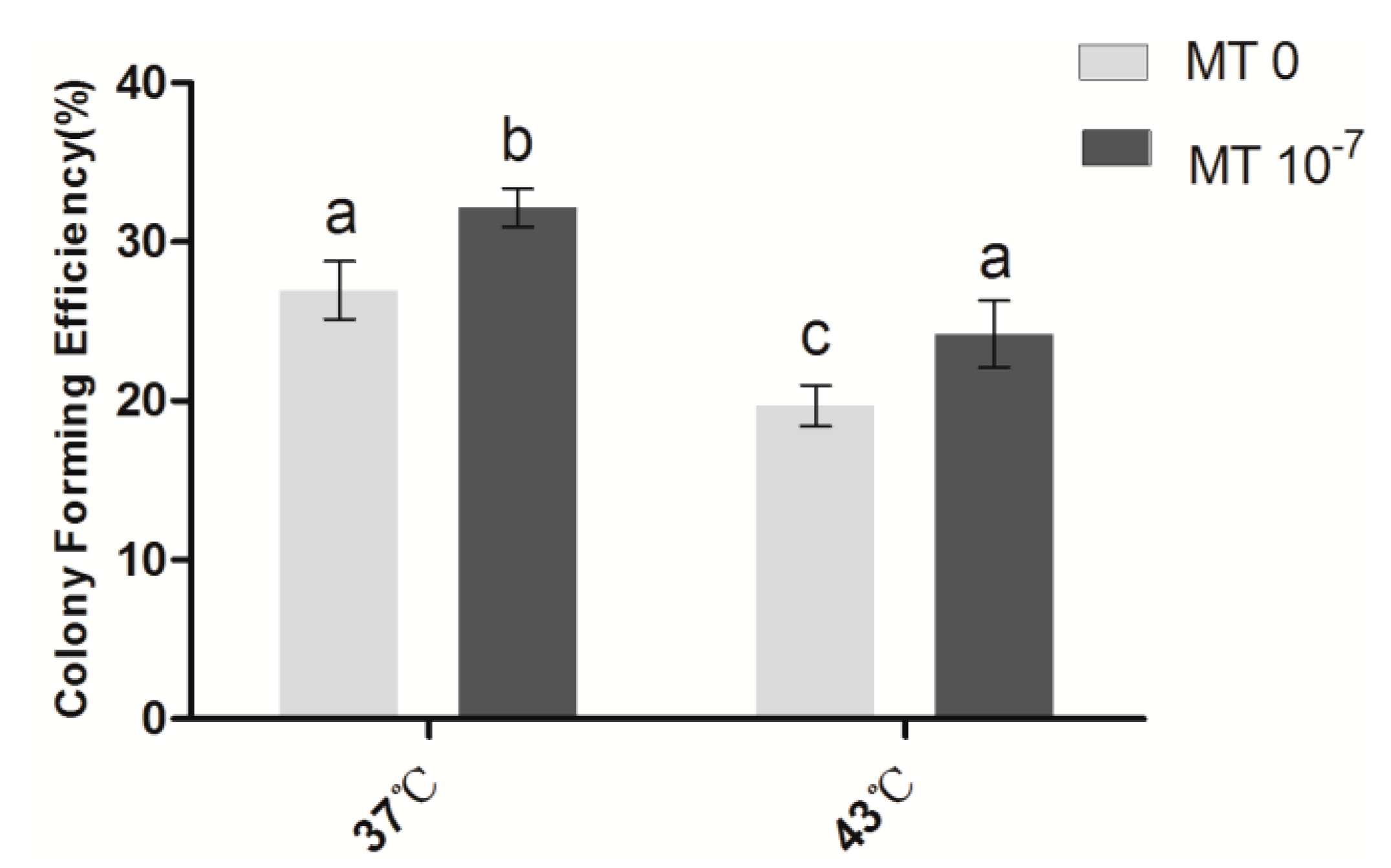
2.1. Effects of Melatonin on the Cloning Efficiency of Granulosa Cells

As shown in Figure 1, the colony forming efficiency (CFE) of sheep granulosa cells in the group with thermal stress (43 °C) (19.7% ± 1.29%) was significantly lower than the control group (37 °C) (27.0% ± 1.81%) (p < 0.05). Following melatonin (10−7 M) treatment, the CFEs of both the thermal stressed and control groups were significantly increased compared with their melatonin-free counterparts (p < 0.05).
Figure 1. Effects of melatonin on the CFE of granulosa cells. MT: melatonin. Each bar represents the mean ± SEM for experiments performed in triplicate. Different letters indicate significant differences, p < 0.05.
Ijms 15 21090 g001

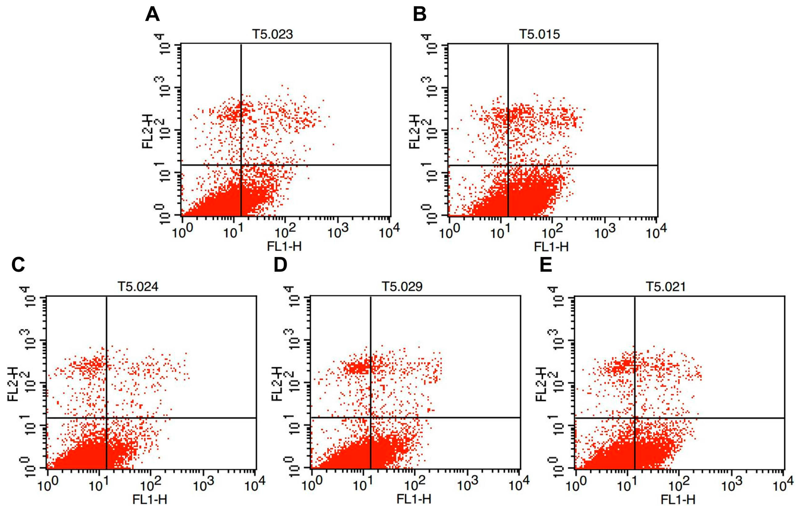
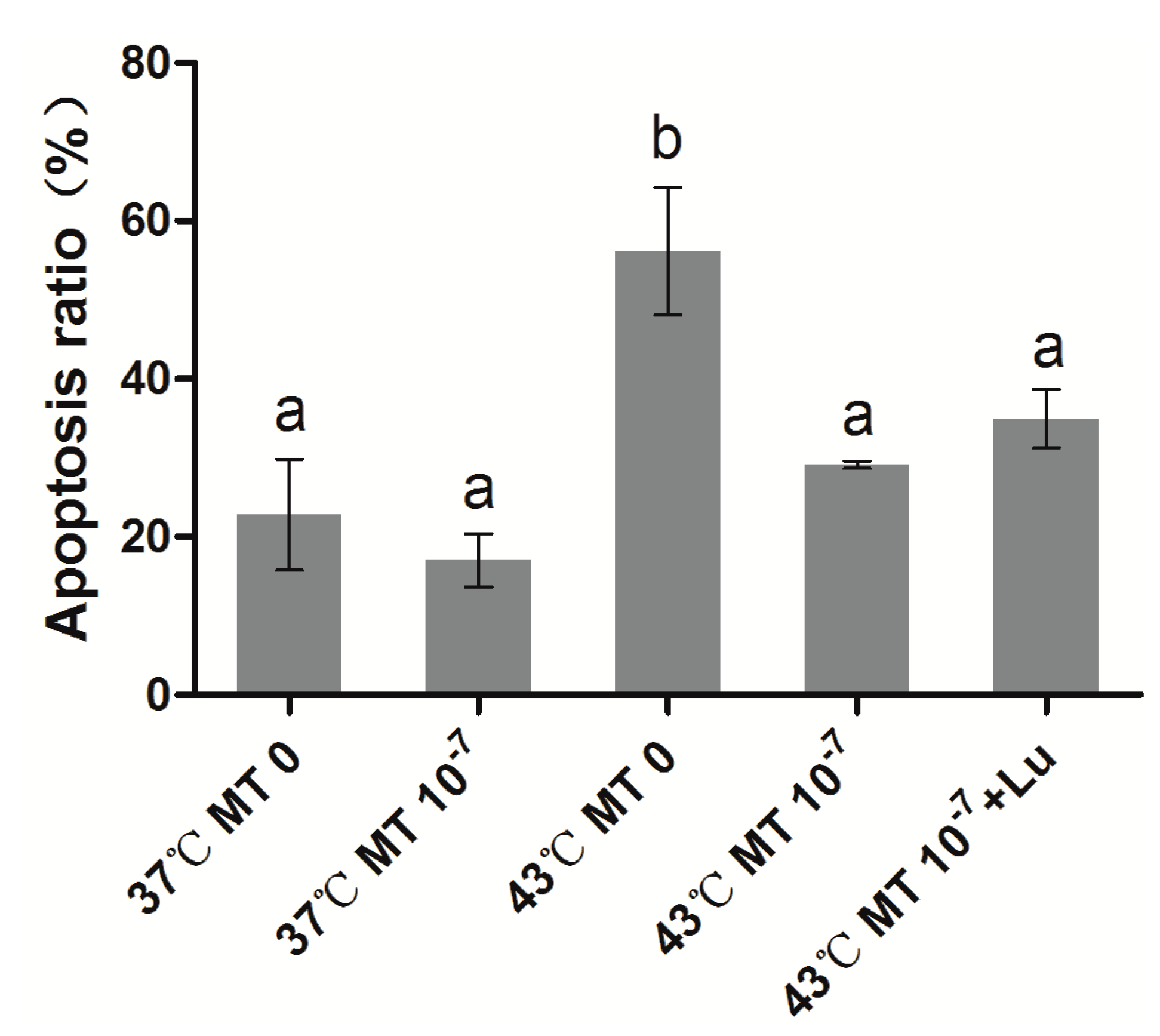
2.2. Effects of Melatonin on Granulosa Cell Apoptosis

As shown in Figure 2 and Figure 3, the percentage of apoptotic granulosa cells in the groups subjected to thermal stress (43 °C) (56.2% ± 13.94%) was significantly higher than the control group (37 °C) (22.8% ± 12.16%) (p < 0.05). The percentage of apoptotic cells in the thermal stressed group with 10−7 M melatonin was significantly lower than the group without melatonin, and it was not significantly different (p > 0.05) from the control group. It appears that the melatonin receptor antagonist luzindole does not influence the antiapoptotic effects of melatonin on granulosa cells under thermal stress.
Figure 2. Low cytometry analysis of cell apoptosis. (A) Control cells (37 °C); (B) Control cells (37 °C) with MT (10−7 M); (C) Thermal stressed cells (43 °C) without MT; (D) Thermal stressed cells (43 °C) with MT (10−7 M); (E) Thermal stressed cells (43 °C) with MT(10−7 M) and Luzindole (10−6 M). The Upper Left Quadrant: necrotic cells; The Upper Right Quadrant: late apoptotic cells; The Lower Left Quadrant: normal cells; The Lower Right Quadrant: early apoptotic cells.
Ijms 15 21090 g002
Figure 3. Effects of melatonin on granulosa cell apoptosis. MT: melatonin; Lu: luzindole. Each bar represents the mean ± SEM for experiments performed in triplicate. Different letters indicate significant differences, p < 0.05.
Ijms 15 21090 g003

2.3. Effects of Melatonin on the Expression of Apoptosis Genes in Sheep Granulosa Cells

As shown in Figure 4, the expression level of p53 in the sheep granulosa cells under thermal stress (43 °C) was significantly higher than the control group (37 °C); however, Bcl-2 gene expression was not significantly different between the thermal stressed and control groups. The expression level of p53 was significantly lower in the thermal stressed group treated with melatonin (10−7 M). The phenomenon in Bcl-2 expression was not similar to p53. The expression level of Bcl-2 treated with melatonin (10−7 M) was significantly increased not only in the group at 37 °C but also in the 43 °C treated group.
Figure 4. Effects of melatonin on the relative expression levels of p53 and Bcl-2 in sheep granulosa cells. (A) The relative expression of p53 at different treatments; (B) The relative expression of Bcl-2 at different treatments. MT: melatonin. Each bar represents the mean ± SEM for experiments performed in triplicate. Different letters in the same column represent significant differences, p < 0.05.
Ijms 15 21090 g004

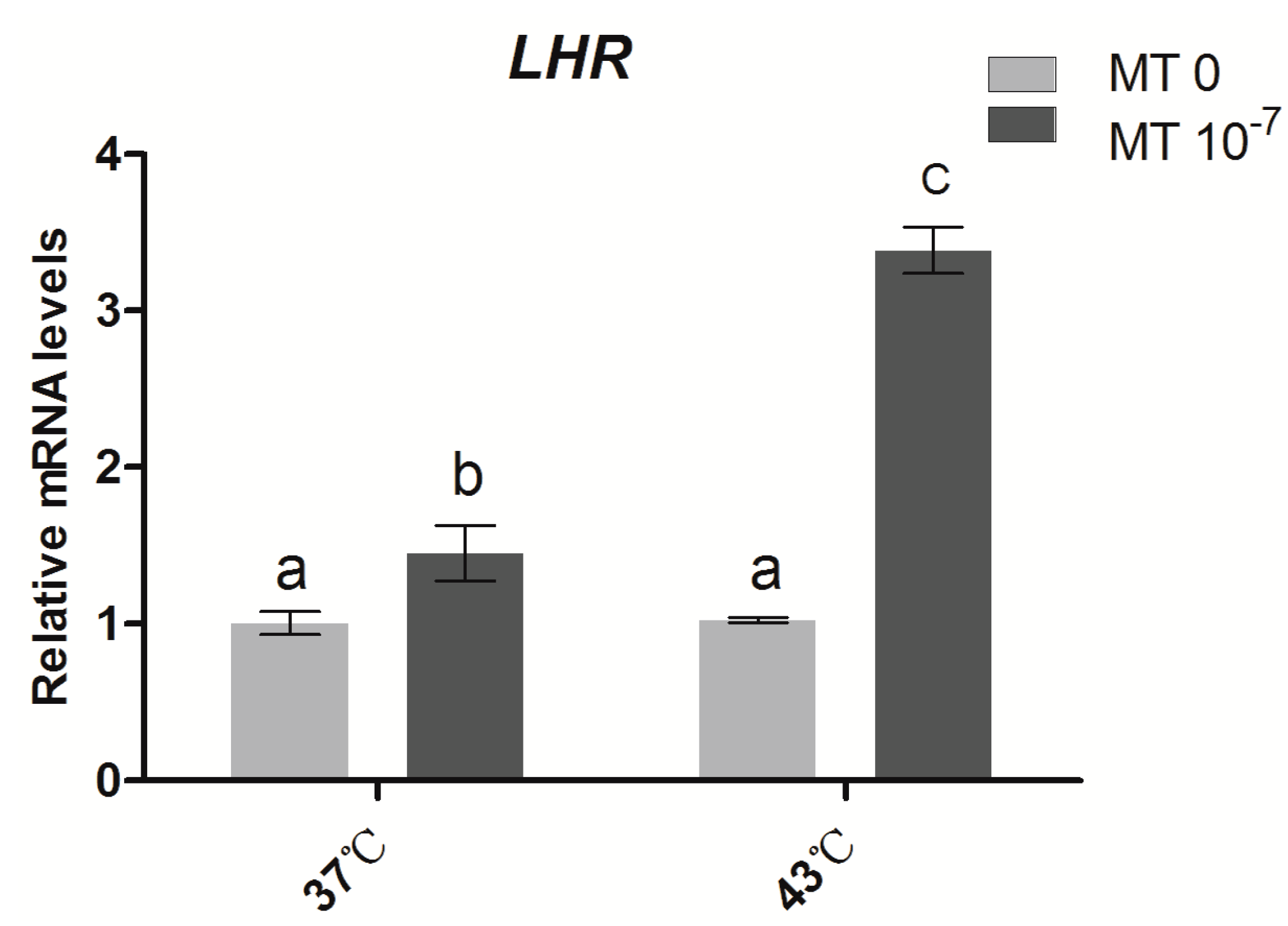
2.4. Effects of Melatonin on the Gene Expression of the Gonadotropin Receptor LHR in Sheep Granulosa Cells

As shown in Figure 5, under thermal stress (43 °C), the mRNA expression level of LHR in granulosa cells was not significantly different from the control groups (37 °C). When cells were incubated at 37 °C and supplemented with melatonin (10−7 M), the LHR expression level was higher than the controls. Moreover, a significant increase in LHR gene expression was observed in the thermal stressed groups treated with melatonin (10−7 M).
Figure 5. Effects of melatonin on the relative expression of LHR in sheep granulosa cells MT: melatonin. Each bar represents the mean ± SEM for experiments performed in triplicate. Different letters in the same column represent significant differences, p < 0.05.
Ijms 15 21090 g005

3. Discussion

Antioxidants play a protective role against oxidative damage caused by thermal stress in the cells and tissues of organisms. The positive effects of antioxidants have been reported regarding several reproductive aspects [39,40,41,42,43,44,45,46,47,48] and the recovery of injuries induced by thermal stresses [49,50]. Melatonin treatment for high-yielding dairy cows during a dry period under thermal stress improved their reproductive performance and reduced the rates of breeding syndrome and pregnancy loss [51,52].
In physiological conditions, cells can maintain their dynamic balance of ROS production and elimination. In contrast, thermal stress can disrupt this balance and lead to oxidative damage in cells. In this study, we observed that thermal stress significantly reduced the CFE and elevated the apoptosis rate in sheep granulose cells. These results are consistent with other findings in mouse [52]. It has been observed that melatonin at the appropriate concentration (10−4 M) promoted bovine blastocyst development [53]. Several recent studies have shown that melatonin promotes oocyte maturation and embryo development in the mouse, bovine and porcine. When a culture medium of porcine and mouse embryos was supplemented with melatonin (10−7 M), the cleavage rate, blastocyst rate and cell number of blastocytes were significantly increased [54,55,56,57]. More importantly, melatonin(10−7 M) reduces ROS production and cellular apoptosis during in vitro embryo development and improves the quality of blastocysts, up-regulates the relative expression of the antioxidant enzyme superoxide dismutase (SOD) and the anti-apoptotic factor Bcl-2 and down-regulates the pro-apoptotic gene p53 [55]. Based on these previous reports, 10−7 M melatonin was selected as the optimal concentration in the current study. It was observed that melatonin at this concentration (10−7 M) significantly increased the CFE and decreased the apoptotic rate of sheep granulosa cells caused by thermal stress (43 °C) (Figure 1). These results suggest that melatonin plays an important role in the protection of sheep granulosa cells from the harmful effects caused by thermal stress and this protection is most likely related to its antioxidant capacity.
It is well-known that in apoptosis, cytochrome C (cytC) released from mitochondria binds to Apaf-1 (a cytoplasmic protein that contains a caspase binding domain). This combination increases the binding affinity of Apaf-1 to dATP/ATP. dATP/ATP then binds to the cytC/Apaf-1 complex and forms a programmed death body (apoptosome); apoptosome further activates downstream factors through enzyme digestion to guide programmed cell death [58]. The release of cytC is suppressed by Bcl-2, an important member of the anti-apoptotic family. Bcl-2 plays a critical role in the regulation of antral follicle atresia. Bcl-2 knockout animals have a reduced number of healthy follicles, and local over-expression of the Bcl-2 gene in the granulosa cells of developing follicles decreases apoptosis [59,60]. Melatonin has been reported to inhibit the release of cytC from mitochondria, and thereby reduces apoptosis in neural hippocampal cells [61,62]. In the current study, we observed that the Bcl-2 expression level of granulosa cells under thermal stress was significantly up-regulated by melatonin treatment. This finding was consistent with the results previously discussed [55,56]. Thus, we speculate that melatonin may directly regulate Bcl-2 and subsequently inhibit cytC release from mitochondria. We also recognized that p53 is another important factor in the regulation of Bcl-2. In general, p53 is regarded as a key player in tumor suppression because it promotes growth arrest, apoptosis and cellular senescence. Most importantly, the phosphorylation sites on p53 are Ser-15, which promotes accumulation and activation of p53 and DNA repair, and Ser-46, which regulates apoptosis following DNA damage. The former can be up-regulated by melatonin in a stress-induced system [63]. p53 also has the ability to regulate the transcription of various apoptotic genes, including the Bcl-2 family. The inhibition of p53 expression can up-regulate Bcl-2 proteins in a rat model of cholestasis [64,65,66,67,68]. In this study, p53 expression, which was elevated by thermal stress in sheep granulose cells, was significantly reduced by melatonin treatment (Figure 4). The results also suggested that p53 participated in the anti-apoptotic function of melatonin via the Bcl-2 pathway [56].
Luteinizing hormone (LH) is an important hormone in the differentiation process of granulosa cells, and it regulates the development process of preantral follicles to ovulation follicles. It is an obligatory step in the differentiation and maturation of granulosa cells and is also essential for the initiation of luteinization [69,70,71,72]. It was demonstrated that during follicular atresia and granulosa cell apoptosis, LH receptors in the ovary significantly decreased. When follicles or granulosa cells were treated with FSH or LH, it inhibited follicular atresia and cell apoptosis [73,74]. A recent study has shown that melatonin treatment significantly increased the mRNA expression of the LH receptor but not of FSH; furthermore, melatonin was thought to be involved in maintaining the appropriate level of LHR expression for ovarian function [11,75,76,77]. Similar results were observed in the current study. As shown in Figure 5, the expression of LHR in granulosa cells under thermal stress was not significantly different from the control. This finding suggests that LHR gene expression is not significantly affected by increased temperature. However, melatonin treatments remarkably up-regulated the expression level of LHR in both control and thermal stressed groups. It appears that melatonin can induce the expression of the LHR gene, which thereby improves the quality of granulosa cells and enhances their ability to protect against thermal-stress.
In conclusion, melatonin at 10−7 M was demonstrated to effectively protect sheep granulosa cells from the harmful effects caused by thermal stress. This effect is indicated by an increase in the formation of CFE and a decrease in the apoptotic rate. The anti-apoptotic effects of melatonin in thermal stressed granulose cells are primarily attributed to its activities that down-regulated p53 and up-regulated Bcl-2 and LHR gene expression. These effects of melatonin may involve its antioxidant capacity, since many naturally occurring antioxidants exhibit similar functions.

4. Materials and Methods

4.1. Materials

DMEM, FBS and TCM199 were products of GIBCO Company (Carlsbad, CA, USA). Trypsin and PBS were purchased from Beijing Maichen Technology Company (Beijing, China). An Annexin V-FITC Apoptosis Assay Kit was obtained from Beyotime Institute of Biotechnology (Beijing, China). Melatonin and all other chemicals were of the highest analytical and tissue culture grades and were purchased primarily from Sigma Aldrich Chemical Company (St. Louis, MO, USA). The sheep ovaries were collected from the local abattoir.

4.2. Granular Cell Separation and Culture

Adult ovine ovaries were collected, stored in physiological saline and transported to the laboratory within 3–4 h. The ovaries were cleaned repeatedly with physiological saline that contained antibiotics. The follicles were cut to a 2–6 mm size in diameter using a surgical knife blade. Granule cells were aspirated from the follicle fluid and washed with Dulbecco’s phosphate-buffered saline three times. The suspended cells were cultured with DMEM/F12 that contained 10% FBS in the cell culture plate. They were incubated at 37 °C with 5% CO2 in humidified air.

4.3. Measurement of Colony Forming Efficiency

The granular cells were divided into normal temperature (37 °C) and thermal stressed groups (43 °C). MT was added to the medium with a final concentration of 10−7 M. After the cells adhered, in the thermal stressed group, the culture temperature was increased from 37 to 43 °C with 5% CO2 in humidified air for 2 h; the temperature was then decreased to 37 °C. This procedure was repeated every 24 h. The colony-forming efficiency (CFE) was evaluated on the 12th day of culture. The colony efficiency of the isolated cells was evaluated by inoculating single-cell suspensions at a density of 1000 cells/well in a 10 mm cell culture plate. The cells were incubated in DMED/F12 medium supplemented with 10% fetal bovine serum and 1 × 10⁻⁷ M MT 10 mL per well. The medium was replaced every two days. Colony formation was monitored by microscopy and analyzed on day 12 after removal of the medium. The cells were fixed in methanol for 5 min and stained with 50% Giemsa staining at room temperature for 15 min. The colony-forming efficiency was calculated as the number of clones/total number of cells seeded per well.

4.4. Flow Cytometric Analysis of Apoptotic Cells

The granular cells were divided into 4 groups: control group (37 °C); thermal stress group (43 °C); thermal stress plus MT 10−7 group; thermal stress plus MT 10−7 and luzindole 10−6 group. After exposed to 43 °C for 2 h, the granulosa cells were cultured at 37 °C for 12 h; flow cytometry was subsequently performed to analyze the apoptotic cells. The apoptotic cells were differentiated from viable or necrotic cells by the combined application of annexinV-FLUOS and propidium iodide (PI). The three parallel samples were washed twice. The cells were harvested via the method of 0.25% trypsin + 0.02% EDTA and centrifuged at 1500 r/min for 5 min. The pellet was re-suspended and washed twice with cold PBS. The cell suspension was added to 195 µL binding buffer and 5 µL Annexin V-FITC and incubated at room temperature for 10 min in darkness. The cells were centrifuged at 1500 r/min for 5 min, and the supernatant was discarded. Finally, 200 µL binding buffer that contained 10 µL PI was added to each tube. The samples were immediately analyzed using FACS (Becton, Dickinson and Company, Franklin Lake, NJ, USA).

4.5. RNA Isolation and Quantitative RT-PCR

Ovine granulose cells were divided into normal temperature (37 °C) and thermal stressed groups (43 °C). The thermal stressed group was incubated with melatonin (10−7 M) in culture medium. In the thermal stressed groups, the cells were exposed to 43 °C for 2 h; these cells were subsequently cultured at a normal temperature (37 °C) for an additional 12 h. Finally, the cells were harvested. The harvested cells were washed twice with D-PBS solution and centrifuged at 1500 rpm for 5 min. The pellet was stored at 80 °C until the RNA was extracted. The total RNA was extracted using TRIzol reagent (Invitrogen Inc., Carlsbad, CA, USA), and it was quantified by measuring the absorbance at 260 nm. The extracted RNA was stored at 80 °C until use. The levels of relevant mRNAs, including the apoptosis-related genes p53 and Bcl-2 and the gonadotropic hormone receptor LFR, were detected by quantitative RT-PCR using a One Step SYBR PrimeScript RT-PCR Kit (TaKaRa Bio., Inc., Tokyo, Japan) in a Light Cycler instrument (Roche Applied Science, Mannheim, Germany). The levels of accumulated fluorescence were analyzed using the second-derivative method after the melting-curve analysis was complete. The relative expression levels of the target genes were calculated with the 2−ΔΔCt method. The results were normalized to the GAPDH expression level in each sample. The primer pairs for the analyzed mRNAs are listed in Table 1.
Table 1. Primers used in this study.
GenesAccession NumberPrimersSequence (5'–3')Product Size (bp)
GAPDHHM043737ForwardGTGTCTGTTGTGGATCTGACCTG162
ReverseAGAAGAGTGAGTGTCGCTGTTGAAGT
p53FJ855223ForwardGCACGACCATCCACTACAACTTC148
ReverseGGACAGGCACAAACACGCAC
Bcl-2DQ152929ForwardACTTCGCCGAGATGTCCAG138
ReverseCGACACCTCCGAACTCAAAG
LHRL36329ForwardTCTGCTCACCCAAGACACTCC247
ReverseGAGGCAATGAGTAGCAGGTAGAG

4.6. Statistical Analysis

All data are expressed as the mean ± SEM. The data were subjected to multiple comparison analyses using GLM(General Linear Model) analysis for the intergroup comparison with SPSS 19.0 statistical software (SPSS Inc., Chicago, IL, USA). The correlations were analyzed using the Correlations procedure. p < 0.05 was considered statistically significant.

Acknowledgments

This work was supported, in part, by the National Natural Science Foundation of China (31172177) and other national grants as follows: 2011ZX08008-005, 2014ZX0800802B and 2014CB138500.

Author Contributions

Yao Fu contributed to the study design, data collection and analysis, and manuscript writing. Chang-jiu He contributed to the data collection, analysis and manuscript writing and revisions. Peng-yun Ji, Xiu-zhi Tian, and Feng Wang contributed to the experimentation. Guo-shi Liu developed the idea of the study, was responsible for its design and coordination, and contributed to the analysis and interpretation of the data, as well as the manuscript writing and revisions to the manuscript. All authors read and approved the final manuscript.

Conflicts of Interest

The authors declare there is no conflict of interest.

References

  1. Mustafi, S.B.; Chakraborty, P.K.; Dey, R.S.; Raha, S. Heat stress up-regulates chaperone heat shock protein 70 and antioxidant manganese superoxide dismutase through reactive oxygen species (ROS), p38MAPK, and Akt. Cell Stress Chaperones 2009, 14, 579–589. [Google Scholar]
  2. Farinati, F.; Piciocchi, M.; Lavezzo, E.; Bortolami, M.; Cardin, R. Oxidative stress and inducible nitric oxide synthase induction in carcinogenesis. Dig. Dis. 2010, 28, 579–584. [Google Scholar]
  3. Ziech, D.; Franco, R.; Pappa, A.; Panayiotidis, M.I. Reactive oxygen species (ROS)-induced genetic and epigenetic alterations in human carcinogenesis. Mut.Res.Fundam. Mol. Mechan. Mutagen. 2011, 711, 167–173. [Google Scholar]
  4. Hansen, P.J. Effects of heat stress on mammalian reproduction. Philos. Trans. R. Soc. B 2009, 364, 3341–3350. [Google Scholar]
  5. Reiter, R.J. The melatonin rhythm: Both a clock and a calendar. Experientia 1993, 49, 654–664. [Google Scholar]
  6. Reiter, R.J.; Rosales-Corral, S.; Coto-Montes, A.; Boga, J.A.; Tan, D.X.; Davis, J.M.; Konturek, P.C.; Konturek, S.J.; Brzozowski, T. The photoperiod, circadian regulation and chronodisruption: The requisite interplay between the suprachiasmatic muclei and the pineal and gut melatonin. J. Physiol. Pharmacol. 2011, 62, 269–274. [Google Scholar]
  7. Anisimov, V.N.; Popovich, I.G.; Zabezhinski, M.A.; Anisimov, S.V.; Vesnushkin, G.M.; Vinogradova, I.A. Melatonin as antioxidant, geroprotector and anticarcinogen. Biochim. Biophys. Acta Bioenerg. 2006, 1757, 573–589. [Google Scholar]
  8. Hardeland, R.; Pandi-Perumal, S. Melatonin, a potent agent in antioxidative defense: Actions as a natural food constituent, gastrointestinal factor, drug and prodrug. Nutr. Metab. 2005, 2, 22. [Google Scholar]
  9. Tengattini, S.; Reiter, R.J.; Tan, D.X.; Terron, M.P.; Rodella, L.F.; Rezzani, R. Cardiovascular diseases: Protective effects of melatonin. J. Pineal Res. 2008, 44, 16–25. [Google Scholar]
  10. Sirotkin, A.; Schaeffer, H. Direct regulation of mammalian reproductive organs by serotonin and melatonin. J. Endocrinol. 1997, 154, 1–5. [Google Scholar]
  11. Tamura, H.; Nakamura, Y.; Korkmaz, A.; Manchester, L.C.; Tan, D.X.; Sugino, N.; Reiter, R.J. Melatonin and the ovary: Physiological and pathophysiological implications. Fertil. Steril. 2009, 92, 328–343. [Google Scholar]
  12. Von Gall, C.; Stehle, J.H.; Weaver, D.R. Mammalian melatonin receptors: Molecular biology and signal transduction. Cell Tissue Res. 2002, 309, 151–162. [Google Scholar]
  13. Cutando, A.C.; Fernandez, J.A.; Valverde, A.L.; Santiago, S.A.; Cachaza, J.A.; Reiter, R.J. A new perspective in oral health: Potential importance and actions of melatonin receptors MT1, MT2, MT3, and RZR/ROR in the oral cavity. Arch. Oral Biol. 2011, 56, 944–950. [Google Scholar]
  14. Slominski, R.M.; Reiter, R.J.; Loutsevitch, N.S.; Ostrom, R.S.; Slominski, A.T. Melatonin membrane receptors in peripheral tissues: Distrbution and functions. Mol. Cell. Endocrinol. 2012, 351, 152–166. [Google Scholar]
  15. Slominski, A.T.; Kim, T.K.; Takeda, Y.; Janjetovic, Z.; Brozyna, A.A.; Skobowiat, C.; Wang, J.; Postlethwaite, A.; Li, W.; Tuckey, R.C.; et al. RORα and RORγ are expressed in human skin and serve as receptors for endogenously produced noncalcemic 20-hydroxy- and 20,23-dihydroxy vitamin D. Faseb. J. 2014, 28, 2775–2789. [Google Scholar]
  16. Reiter, R. Interactions of the pineal hormone melatonin with oxygen-centered free radicals: A brief review. Braz. J. Med. Biol. Res. 1993, 26, 1141–1155. [Google Scholar]
  17. Reiter, R.J.; Guerrero, J.M.; Escames, G.; Pappolla, M.A.; Acuña-Castroviejo, D. Prophylactic actions of melatonin in oxidative neurotoxicity. Ann. N. Y. Acad. Sci. 1997, 825, 70–78. [Google Scholar]
  18. Reiter, R.J.; Tan, D.X. Melatonin: A novel protective agent against oxidative injury of the ischemic/reperfused heart. Cardiovasc. Res. 2003, 58, 10–19. [Google Scholar]
  19. Carloni, S.; Perrone, S.; Buonocore, G.; Longini, M.; Proietti, F.; Balduini, W. Melatonin protects from the long-term consequences of a neonatal hypoxic-ischemic brain injury in rats. J. Pineal Res. 2008, 44, 157–164. [Google Scholar]
  20. Hung, M.W.; Tipoe, G.L.; Poon, A.M.S.; Reiter, R.J.; Fung, M.L. Protective effect of melatonin against hippocampal injury of rats with intermittent hypoxia. J. Pineal Res. 2008, 44, 214–221. [Google Scholar]
  21. Maldonado, M.D.; Murillo-Cabezas, F.; Calvo, J.R.; Lardone, P.J.; Tan, D.X.; Guerrero, J.M.; Reiter, R.J. Melatonin as pharmacologic support in burn patients: A proposed solution to thermal injury-related lymphocytopenia and oxidative damage. Crit. Care Med. 2007, 35, 1177–1185. [Google Scholar]
  22. Şener, G.; Şehirli, A.Ö.; Şatıroğlu, H.; Keyer-Uysal, M.Ç.; Yeğen, B. Melatonin improves oxidative organ damage in a rat model of thermal injury. Burns 2002, 28, 419–425. [Google Scholar]
  23. Karlidağ, T.; Yalçin, Ş.; Öztürk, A.; Üstündağ, B.; Gök, Ü.; Kaygusuz, İ.; Susaman, N. The role of free oxygen radicals in noise induced hearing loss: Effects of melatonin and methylprednisolone. Auris Nasus Larynx 2002, 29, 147–152. [Google Scholar]
  24. Bas, E.; Martinez-Soriano, F.; Láinez, J.M.; Marco, J. An experimental comparative study of dexamethasone, melatonin and tacrolimus in noise-induced hearing loss. Acta Oto-Laryngol. 2009, 129, 385–389. [Google Scholar]
  25. Seggie, J.; Campbell, L.; Brown, G.M.; Grota, L.J. Melatonin and N-acetylserotonin stress responses: Effects of type of stimulation and housing conditions. J. Pineal Res. 1985, 2, 39–49. [Google Scholar]
  26. Mahlberg, R.; Kunz, D.; Sutej, I.; Kühl, K.P.; Hellweg, R. Melatonin treatment of day-night rhythm disturbances and sundowning in Alzheimer disease: An open-label pilot study using actigraphy. J. Clin. Psychopharmacol. 2004, 24, 456–459. [Google Scholar]
  27. Blask, D.E. Melatonin, sleep disturbance and cancer risk. Sleep Med. Rev. 2009, 13, 257–264. [Google Scholar]
  28. Armstrong, S.; Redman, J. Melatonin: A chronobiotic with anti-aging properties? Med Hypotheses 1991, 34, 300–309. [Google Scholar]
  29. Reiter, R.J.; Tan, D.X.; Poeggeler, B.; Menendez-Pelaez, A.; Chen, L.D.; Saarela, S. Melatonin as a free radical scavenger: Implications for aging and age-related diseases. Ann. N. Y. Acad.Sci. 1994, 719, 1–12. [Google Scholar]
  30. Bizzarri, M.; Proietti, S.; Cucina, A.; Reiter, R.J. Molecular mechanisms of the pro-apoptotic actions of melatonin in cancer: A review. Expert Opin. Ther. Targets 2013, 17, 1483–1496. [Google Scholar]
  31. Sainz, R.; Mayo, J.; Rodriguez, C.; Tan, D.X.; Lopez-Burillo, S.; Reiter, R. Melatonin and cell death: Differential actions on apoptosis in normal and cancer cells. Cell Mol. Life Sci. 2003, 60, 1407–1426. [Google Scholar]
  32. Liu, L.; Xu, Y.; Reiter, R.J. Melatonin inhibits the proliferation of human osteosarcoma cell line MG-63. Bone 2013, 55, 432–438. [Google Scholar]
  33. Wang, J.; Xiao, X.; Zhang, Y.; Shi, D.; Chen, W.; Fu, L.; Liu, L.; Xie, F.; Kang, T.; Huang, W.; et al. Simultaneous modulation of COX-2, p300, Akt, and Apaf-1 signaling by melatonin to inhibit proliferation and induce apoptosis in breast cancer cells. J. Pineal Res. 2012, 53, 77–90. [Google Scholar]
  34. Roth, J.A.; Kim, B.-G.; Lin, W.-L.; Cho, M.-I. Melatonin promotes osteoblast differentiation and bone formation. J. Biol. Chem. 1999, 274, 22041–22047. [Google Scholar]
  35. Kong, X.; Li, X.; Cai, Z.; Yang, N.; Liu, Y.; Shu, J.; Pan, L.; Zuo, P. Melatonin regulates the viability and differentiation of rat midbrain neural stem cells. Cell Mol. Neurobiol. 2008, 28, 569–579. [Google Scholar]
  36. Li, R.; Albertini, D.F. The road to maturation: Somatic cell interaction and self-organization of the mammalian oocyte. Nat. Rev. Mol. Cell Biol. 2013, 14, 141–152. [Google Scholar]
  37. Manabe, N.; Goto, Y.; Matsuda-Minehata, F.; Inoue, N.; Maeda, A.; Sakamaki, K.; Miyano, T. Regulation mechanism of selective atresia in porcine follicles: Regulation of granulosa cell apoptosis during atresia. J. Reprod. Dev. 2004, 50, 493–514. [Google Scholar]
  38. Nakayama, M.; Manabe, N.; Nishihara, S.; Miyamoto, H. Species-specific differences in apoptotic cell localization in granulosa and theca interna cells during follicular atresia in porcine and bovine ovaries. J. Reprod. Dev. 2000, 46, 147–156. [Google Scholar]
  39. Krisher, R.L. In vivo and in vitro environmental effects on mammalian oocyte quality. Annu. Rev. Anim. Biosci. 2013, 1, 393–417. [Google Scholar]
  40. Agarwal, A.; Aponte-Mellado, A.; Premkumar, B.J.; Shaman, A.; Gupta, S. The effects of oxidative stress on female reproduction: A review. Reprod. Biol. Endocrinol. 2012, 10, 49. [Google Scholar]
  41. Agarwal, A.; Virk, G.; Ong, C.; du Plessis, S.S. Effect of oxidative stress on male reproduction. World J. Menʼs Health 2014, 32, 1–17. [Google Scholar]
  42. Ishikawa, S.; Machida, R.; Hiraga, K.; Hiradate, Y.; Suda, Y.; Tanemura, K. Hanging drop monoculture for selection of optimal antioxidants during in vitro maturation of porcine oocytes. Reprod. Domest. Anim. 2014, 49, e26–e30. [Google Scholar]
  43. Lian, H.Y.; Gao, Y.; Jiao, G.Z.; Sun, M.J.; Wu, X.F.; Wang, T.Y.; Li, H.; Tan, J.H. Antioxidant supplementation overcomes the deleterious effects of maternal restraint stress-induced oxidative stress on mouse oocytes. Reproduction 2013, 146, 559–568. [Google Scholar]
  44. Dong, Y.; Bai, Y.; Liu, G.; Wang, Z.; Cao, J.; Chen, Y.; Yang, H. The immunologic and antioxidant effects of L-phenylalanine on the uterine implantation of mice embryos during early pregnancy. Histol. Histopathol. 2014, 29, 1335–1242. [Google Scholar]
  45. Liu, M.; Yin, Y.; Ye, X.; Zeng, M.; Zhao, Q.; Keefe, D.L.; Liu, L. Resveratrol protects against age-associated infertility in mice. Hum. Reprod. 2013. [Google Scholar] [CrossRef]
  46. Chen, Z.G.; Luo, L.L.; Xu, J.J.; Zhuang, X.L.; Kong, X.X.; Fu, Y.C. Effects of plant polyphenols on ovarian follicular reserve in aging rats. Biochem. Cell Biol. 2010, 88, 737–745. [Google Scholar]
  47. Wang, F.; Tian, X.; Zhang, L.; He, C.; Ji, P.; Li, Y.; Tan, D.; Liu, G. Beneficial effect of resveratrol on bovine oocyte maturation and subsequent embryonic development after in vitro fertilization. Fertil. Steril. 2014, 101, 577–586. [Google Scholar]
  48. Sharma, S.; Ramesh, K.; Hyder, I.; Uniyal, S.; Yadav, V.; Panda, R.P.; Maurya, V.P.; Singh, G.; Kumar, P.; Mitra, A.; et al. Effect of melatonin administration on thyroid hormones, cortisol and expression profile of heat shock proteins in goats (Capra hircus) exposed to heat stress. Small Rumin. Res. 2013, 112, 216–223. [Google Scholar]
  49. Niu, Z.; Liu, F.; Yan, Q.; Li, W. Effects of different levels of vitamin E on growth performance and immune responses of broilers under heat stress. Poult. Sci. 2009, 88, 2101–2107. [Google Scholar]
  50. Xue, S.; Mu, D.; Dai, J.; Wang, Z.; Zhao, H.; Yuan, Y.; Peng, G. Preventive effect of glutamine combined with vitamin C on intestinal endotoxemia induced by heat stress in rats. J. Third Milit. Med. Univ. 2011, 17, 1779–1782. (In Chinese) [Google Scholar]
  51. Garcia-Ispierto, I.; Abdelfatah, A.; López-Gatius, F. Melatonin treatment at dry-off improves reproductive performance postpartum in high-producing dairy cows under heat stress conditions. Reprod. Domest. Anim. 2013, 28, 577–583. [Google Scholar]
  52. Wang, H.; Gao, G. Effect of hyperthermia on ovary antioxidant capacity and granulosa cells apoptosis of mouse. Heilongjiang Anim. Sci. Vet. Med. 2012, 11, 154–157. (In Chinese) [Google Scholar]
  53. Cebrian-Serrano, A.; Salvador, I.; Raga, E.; Dinnyes, A.; Silvestre, M. Beneficial effect of melatonin on blastocyst in vitro production from heat-stressed bovine oocytes. Reprod. Domest. Anim. 2013, 48, 738–746. [Google Scholar]
  54. Gao, C.; Han, H.B.; Tian, X.Z.; Tan, D.X.; Wang, L.; Zhou, G.B.; Zhu, S.E.; Liu, G.S. Melatonin promotes embryonic development and reduces reactive oxygen species in vitrified mouse 2-cell embryos. J. Pineal Res. 2012, 52, 305–311. [Google Scholar]
  55. Wang, F.; Tian, X.; Zhang, L.; Tan, D.; Reiter, R.J.; Liu, G. Melatonin promotes the in vitro development of pronuclear embryos and increases the efficiency of blastocyst implantation in murine. J. Pineal Res. 2013, 55, 267–274. [Google Scholar]
  56. Wang, F.; Tian, X.; Zhang, L.; Gao, C.; He, C.; Fu, Y.; Ji, P.Y.; Li, Y.; Liu, G.S. Beneficial effects of melatonin on in vitro bovine embryonic development are mediated by melatonin receptor 1. J. Pineal Res. 2014, 56, 333–342. [Google Scholar]
  57. Shi, J.M.; Tian, X.Z.; Zhou, G.B.; Wang, L.; Gao, C.; Zhu, S.E.; Zeng, S.-M.; Tian, J.-H.; Liu, G.-S. Melatonin exists in porcine follicular fluid and improves in vitro maturation and parthenogenetic development of porcine oocytes. J. Pineal Res. 2009, 47, 318–323. [Google Scholar]
  58. Jiang, X.; Wang, X. Cytochrome c promotes caspase-9 activation by inducing nucleotide binding to Apaf-1. J. Biol. Chem. 2000, 275, 31199–31203. [Google Scholar]
  59. Ratts, V.; Flaws, J.; Kolp, R.; Sorenson, C.; Tilly, J. Ablation of bcl-2 gene expression decreases the numbers of oocytes and primordial follicles established in the post-natal female mouse gonad. Endocrinology 1995, 136, 3665–3668. [Google Scholar]
  60. Hsu, S.Y.; Lai, R.; Finegold, M.; Hsueh, A. Targeted overexpression of Bcl-2 in ovaries of transgenic mice leads to decreased follicle apoptosis, enhanced folliculogenesis, and increased germ cell tumorigenesis. Endocrinology 1996, 137, 4837–4843. [Google Scholar]
  61. Baydas, G.; Reiter, R.; Akbulut, M.; Tuzcu, M.; Tamer, S. Melatonin inhibits neural apoptosis induced by homocysteine in hippocampus of rats via inhibition of cytochrome c translocation and caspase-3 activation and by regulating pro-and anti-apoptotic protein levels. Neuroscience 2005, 135, 879–886. [Google Scholar]
  62. Guha, M.; Maity, P.; Choubey, V.; Mitra, K.; Reiter, R.J.; Bandyopadhyay, U. Melatonin inhibits free radical-mediated mitochondrial-dependent hepatocyte apoptosis and liver damage induced during malarial infection. J. Pineal Res. 2007, 43, 372–381. [Google Scholar]
  63. Janjetovic, Z.; Zachary, P.; Nahmias, H.S.; Jarrett, S.G.; Kim, T.K.; Reiter, R.J.; Slominski, A.T. Melatonin and its metabolites ameliorate ultraviolet B-induced damage in human epidermal keratinocytes. J. Pineal Res. 2014, 57, 90–102. [Google Scholar]
  64. Sultana, H.; Kigawa, J.; Kanamori, Y.; Itamochi, H.; Oishi, T.; Sato, S.; Ohwada, M.; Suzuki, M.; Terakawa, N. Chemosensitivity and p53–Bax pathway-mediated apoptosis in patients with uterine cervical cancer. Ann. Oncol. 2003, 14, 214–219. [Google Scholar]
  65. Toshiyuki, M.; Reed, J.C. Tumor suppressor p53 is a direct transcriptional activator of the human bax gene. Cell 1995, 80, 293–299. [Google Scholar]
  66. Oh, S.H.; Nan, J.X.; Sohn, D.H.; Kim, Y.C.; Lee, B.H. Salvia miltiorrhiza inhibits biliary obstruction-induced hepatocyte apoptosis by cytoplasmic sequestration of p53. Toxicol. Appl. Pharmacol. 2002, 182, 27–33. [Google Scholar]
  67. Amaral, J.D.; Xavier, J.M.; Steer, C.J.; Rodrigues, C. The role of p53 in apoptosis. Discov. Med. 2010, 9, 145–152. [Google Scholar]
  68. Amaral, J.D.; Xavier, J.M.; Steer, C.J.; Rodrigues, C.M. Targeting the p53 pathway of apoptosis. Curr. Pharm. Des. 2010, 16, 2493–2503. [Google Scholar]
  69. Hsyeh, A.J.; Adashi, E.; Jones, P.B.; Welsh, J.R. Hormonal regulation of the differentiation of cultured ovarian granulosa cells. Endocrine Rev. 1984, 5, 76–127. [Google Scholar]
  70. Richards, J.S. Maturation of ovarian follicles: Actions and interactions of pituitary and ovarian hormones on follicular cell differentiation. Physiol. Rev. 1980, 60, 51–89. [Google Scholar]
  71. Jia, X.C.; Hhuen, J. Homologous regulation of hormone receptors: Luteinizing hormone increases its own receptors in cultured rat granulosa cells. Endocrinology 1984, 115, 2433–2439. [Google Scholar]
  72. Yao, N.; Yang, B.Q.; Liu, Y.; Tan, X.Y.; Lu, C.L.; Yuan, X.H.; Ma, X. Follicle-stimulating hormone regulation of microRNA expression on progesterone production in cultured rat granulosa cells. Endocrine 2010, 38, 158–166. [Google Scholar]
  73. Maizels, E.T.; Cottom, J.; Jones, J.C.; Hunzicker-Dunn, M. Follicle stimulating hormone (FSH) activates the p38 mitogen-activated protein kinase pathway, inducing small heat shock protein phosphorylation and cell rounding in immature rat ovarian granulosa cells. Endocrinology 1998, 139, 3353–3356. [Google Scholar]
  74. Boone, D.L.; Carnegie, J.A.; Rippstein, P.U.; Tsang, B.K. Induction of apoptosis in equine chorionic gonadotropin (eCG)-primed rat ovaries by anti-eCG antibody. Biol. Reprod. 1997, 57, 420–427. [Google Scholar]
  75. Webb, R.; Nicholas, B.; Gong, J.G.; Campbell, B.K.; Gutierrez, C.G.; Garverick, H.A.; Armstrong, D.G. Mechanisms regulating follicular development and selection of the dominant follicle. Reprod. Suppl. 2002, 61, 71–90. [Google Scholar]
  76. Woo, M.M.; Tai, C.J.; Kang, S.K.; Nathwani, P.S.; Pang, S.F.; Leung, P.C. Direct action of melatonin in human granulosa-luteal cells. J. Clin. Endocrinol. Metab. 2001, 86, 4789–4797. [Google Scholar]
  77. Lombardo, F.; Gioacchini, G.; Fabbrocini, A.; Candelma, M.; DʼAdamo, R.; Giorgini, E.; Carnevali, O. Melatonin-mediated effects on killifish reproductive axis. Comp. Biochem. Physiol. A 2014, 172, 31–38. [Google Scholar]

