Article

Ginsenoside Rd Attenuates Mitochondrial Permeability Transition and Cytochrome c Release in Isolated Spinal Cord Mitochondria: Involvement of Kinase-Mediated Pathways

1
Department of Orthopedics, the Second Affiliated Hospital of Medical School, Xi'an Jiaotong University, Xi'an 710004, China
2
Department of Neurosurgery, the Second Affiliated Hospital of Shaanxi University of Chinese Medicine, Xi'an 712000, China
3
Department of Orthopedics, Hong Hui Hospital, Xi'an Jiaotong University College of Medicine, Xi'an 710054, China
4
Department of Orthopedics, Shaanxi University of Chinese Medicine, Xi'an 712046, China
5
Department of Orthopedics, Xi'an No. 5 Hospital, Xi'an 710082, China
*
Author to whom correspondence should be addressed.
Int. J. Mol. Sci. 2014, 15(6), 9859-9877; https://doi.org/10.3390/ijms15069859
Submission received: 25 April 2014 / Revised: 8 May 2014 / Accepted: 21 May 2014 / Published: 3 June 2014
(This article belongs to the Special Issue Neuroprotective Strategies 2014)

Abstract

:
Ginsenoside Rd (Rd), one of the main active ingredients in Panax ginseng, has multifunctional activity via different mechanisms and neuroprotective effects that are exerted probably via its antioxidant or free radical scavenger action. However, the effects of Rd on spinal cord mitochondrial dysfunction and underlying mechanisms are still obscure. In this study, we sought to investigate the in vitro effects of Rd on mitochondrial integrity and redox balance in isolated spinal cord mitochondria. We verified that Ca2+ dissipated the membrane potential, provoked mitochondrial swelling and decreased NAD(P)H matrix content, which were all attenuated by Rd pretreatment in a dose-dependent manner. In contrast, Rd was not able to inhibit Ca2+ induced mitochondrial hydrogen peroxide generation. The results of Western blot showed that Rd significantly increased the expression of p-Akt and p-ERK, but had no effects on phosphorylation of PKC and p38. In addition, Rd treatment significantly attenuated Ca2+ induced cytochrome c release, which was partly reversed by antagonists of Akt and ERK, but not p-38 inhibitor. The effects of bisindolylmaleimide, a PKC inhibitor, on Rd-induced inhibition of cytochrome c release seem to be at the level of its own detrimental activity on mitochondrial function. Furthermore, we also found that pretreatment with Rd in vivo (10 and 50 mg/kg) protected spinal cord mitochondria against Ca2+ induced mitochondrial membrane potential dissipation and cytochrome c release. It is concluded that Rd regulate mitochondrial permeability transition pore formation and cytochrome c release through protein kinases dependent mechanism involving activation of intramitochondrial Akt and ERK pathways.

1. Introduction

Paraplegia is defined as the impairment in motor or sensory function of the lower extremities. It is usually caused by traumatic or ischemic spinal cord injury that affects the neural elements in the thoracic, lumbar or sacral regions. It is reported that the incidence of spinal cord ischemia after thoracic and thoraco-abdominal aortic surgery is approximately 5%–8%, and there are more than 50 new traumatic spinal cord injury cases per million people each year [1,2]. The aetiology of spinal cord injury induced paraplegia is multi-factorial, and in the past few years various efforts to reduce this clinical problem have been made, including improving surgical technique [3,4], progress in monitoring and anaesthesia [5,6,7], and breakthrough in the prevention of complications [8,9]. However, there is still no effective treatment that leads to the full functional recovery of patients, whose deficits are generally permanent and irreversible [10,11,12].
Mitochondria are double-membraned organelles that primarily function as the powerhouses of cells through oxidative phosphorylation. Neuronal damage following spinal cord injury has been shown to be related to multiple intracellular changes, including toxic elevations of excitatory and inhibitory amino acids, neuronal edema, inflammation, vascular dysfunction, and activation of proteases, such as calpains and caspases, which are all associated with mitochondrial dysfunction in spinal cord neurons [13,14]. Inhibition or promotion of particular steps in the above mentioned molecular signaling cascades via targeting the entire organelle mitochondria might provide optimal neuronal protection or functional recovery following spinal cord injury. In support of this notion, mitochondria-targeted antioxidants, such as 5,5-dimethyl-pyrroline N-oxide, Mito-Q, and Mito-CP, have been shown to reduce oxidative damage and attenuate motor neuron degeneration in experimental spinal cord injury models [15]. Furthermore, targeting mitochondrial dysfunction in both primary insult and followed secondary injury events, many pharmacological agents, especially natural products extracted from medicinal plants, are suggested to have neuroprotective effects against traumatic and ischemic spinal cord injury [16,17,18].
Ginseng, the root of Panax ginseng C.A. Meyer, has been used as rejuvenating tonic for more than 2000 years in China [19]. In traditional Chinese medicine, it is known as the king of herbs because of the various pharmacological effects in the nervous system and cardiovascular system [20,21]. Ginsenosides are a special group of triterpenoid saponins that are found nearly exclusively in ginseng, and are considered to be responsible for most functions of ginseng. Previous studies have isolated more than 150 ginsenosides, with similar basic structure of a gonane steroid nucleus with 17 carbon atoms arranged in four rings [22,23]. Among the various ginsenosides, such as Rb, Rc, Rd, Re, Rf, and Rg, ginsenoside Rd (Rd) is one of the most abundant ingredients in the ginseng root and consequently has been accepted as one of the marker compounds of ginseng quality [24]. There is evidence that Rd exerts neuroprotective effects against excitotoxicity- and oxidative stress-induced injury in cultured neurons [25,26,27]. More recently, Rd was reported to ameliorate the histological and functional outcome after focal cerebral ischemia in rats [28,29,30,31]. However, the efficacy of Rd has not been established in animal models of spinal cord injury, and the molecular mechanism of Rd-induced neuroprotective activity has not been fully understood. In the present study, we sought to investigate the potential protective effects of Rd in isolated spinal cord mitochondria and the underlying mechanism with focus on mitochondrial permeability transition and cytochrome c release.

2. Results and Discussion

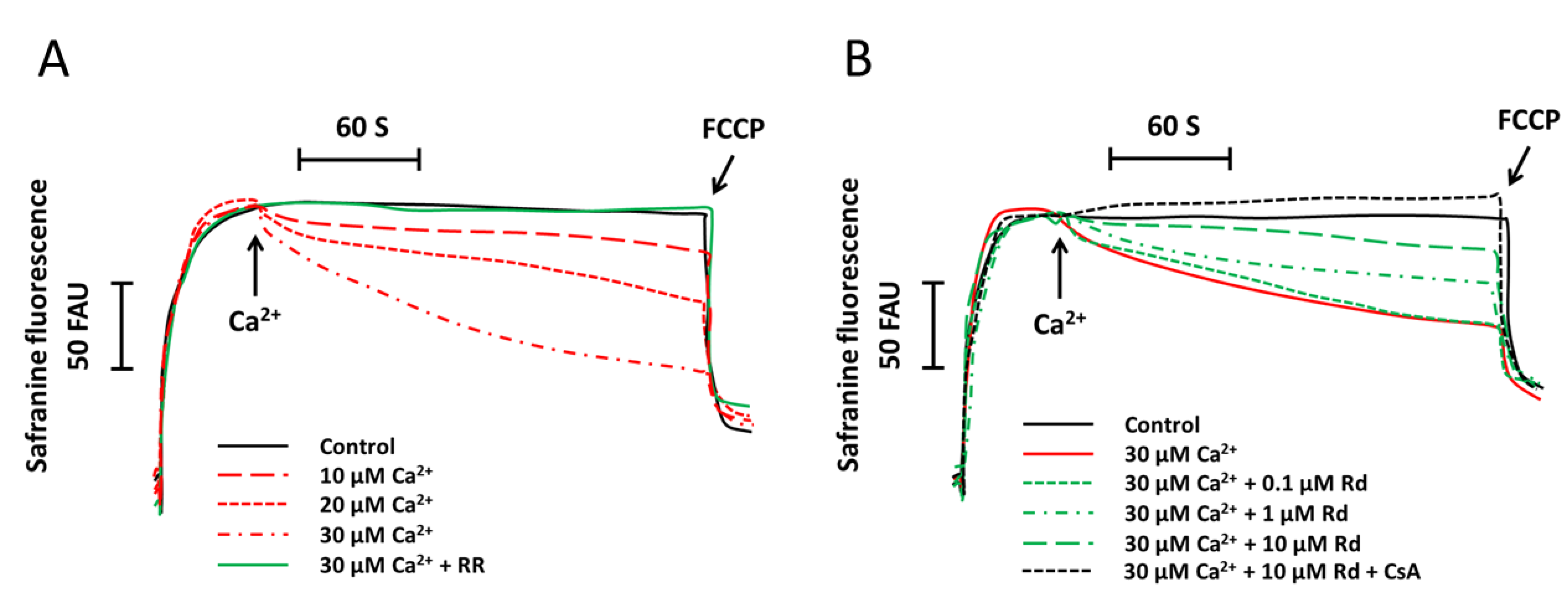
2.1. Rd (Ginsenoside Rd) Protects Isolated Spinal Cord Mitochondria against Ca2+ Induced Mitochondrial Membrane Potential Dissipation

We first investigated the influence of increasing concentrations of Ca2+ (10–30 μM) on mitochondrial membrane potential in succinate supported spinal cord mitochondria. As shown in Figure 1A, Ca2+ treatment induced a dose-dependent increase of safranine fluorescence, which indicated the dissipation of mitochondrial membrane potential. It was also found that ruthenium (RR), an inhibitor of Ca2+ uptake by mitochondria, completely blocked the reduction of mitochondrial membrane potential, suggesting that the mitochondrial membrane depolarization was Ca2+ dependent. We next tested the effects of Rd on Ca2+ induced mitochondrial membrane depolarization. The results showed that Rd significantly attenuated the Ca2+ induced reduction of mitochondrial membrane potential in a dose-dependent fashion, although 0.1 μM Rd was not effective as compared with that in isolated mitochondria without Rd pretreatment (p > 0.05) (Figure 1B). Pretreatment with Rd plus cyclosporin A (CsA), a mitochondrial permeability transition (MPT) inhibitor, further prevented Ca2+ induced mitochondrial membrane depolarization as compared to Rd pretreatment alone.
Figure 1. Rd (ginsenoside Rd) protects isolated spinal cord mitochondria against Ca2+ induced mitochondrial membrane potential dissipation. (A) Isolated spinal cord mitochondria (0.5 mg protein/mL supported by succinate) were treated with or without 1 μM ruthenium (RR) for 60 s before the incubation with increasing concentrations of Ca2+ (10, 20, or 30 μM). The mitochondrial membrane potential was measured up to 300 s; (B) Isolated spinal cord mitochondria (0.5 mg protein/mL supported by succinate) were treated with Rd at different concentrations (0.1, 1, or 10 μM) in the presence or absence of 1 μM cyclosporin A (CsA) for 60 s before the incubation with 30 μM Ca2+. The mitochondrial membrane potential was measured up to 300 s. Controls were performed in the absence of Ca2+, and 1 μM p-trifluoromethoxy-phenylhydrazone (FCCP) was added to induce maximal depolarization at the end of the measurements. Traces are representative of three independent experiments performed in duplicate and were expressed as fluorescence arbitrary units (FAU).
Ijms 15 09859 g001

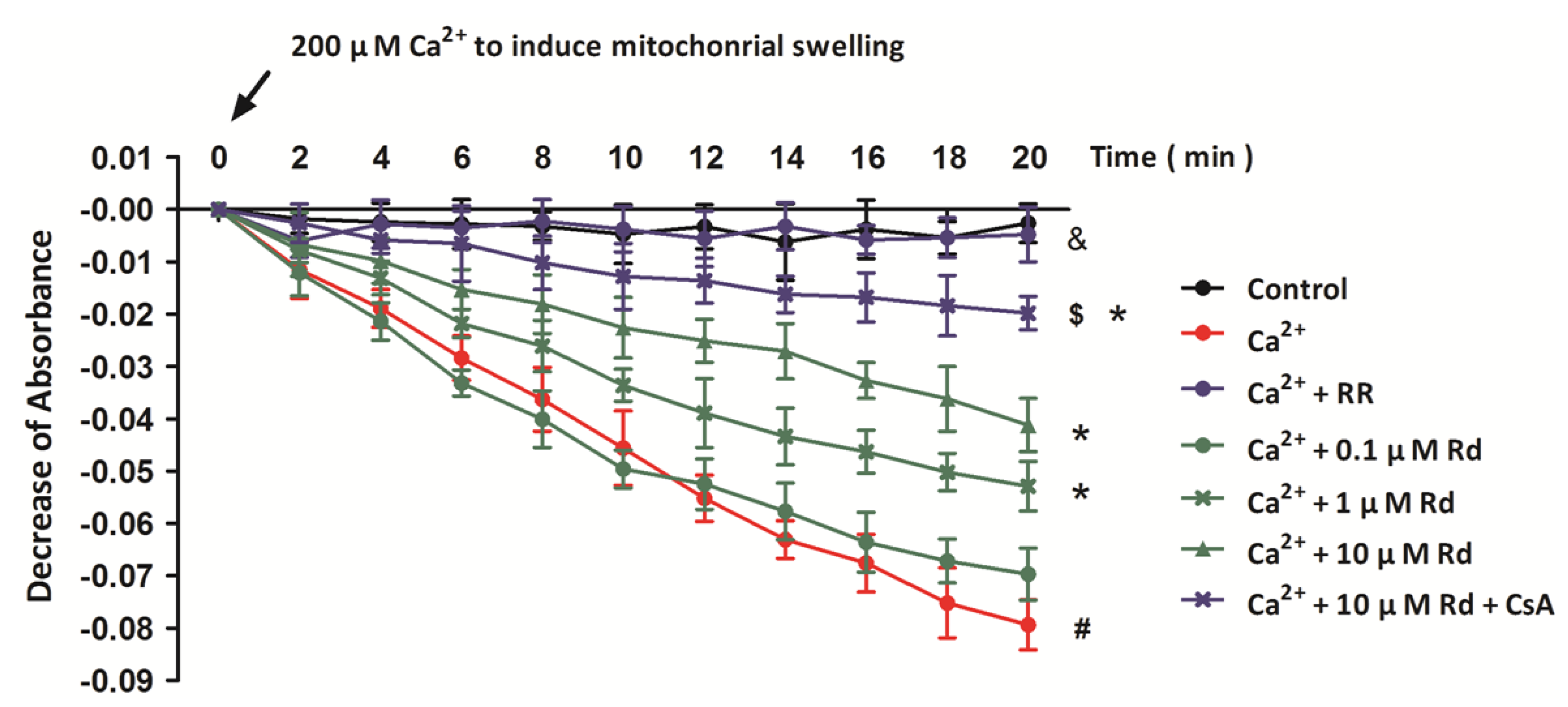
2.2. Rd Protects Isolated Spinal Cord Mitochondria against Ca2+ Induced Mitochondrial Swelling

To further verify the involvement of MPT inhibition in Rd induced inhibition of mitochondrial membrane depolarization after Ca2+ treatment, we also examined mitochondrial swelling in succinate supported spinal cord mitochondria. It has been shown previously that mitochondrial swelling is one of the common biomarkers of MPT pore opening, and in our in vitro model, 200 μM Ca2+ was added into the medium to induce mitochondrial swelling (Figure 2). We verified that 200 μM Ca2+ provoked extensive swelling in succinate supported spinal cord mitochondria, as represented by the significant decrease of absorbance at 540 nm. It was shown that RR fully prevented, whereas CsA attenuated Ca2+ induced mitochondrial swelling. The partial protective effect of CsA under our conditions is in accordance with the observation that CsA is less effective as MPT inhibitor in central nervous system than in isolated liver or heart mitochondria [32]. Furthermore, pretreatment with Rd at 1 or 10 μM in the medium partly reversed the Ca2+ induced mitochondrial swelling, but 0.1 μM Rd was not effective.
Figure 2. Rd protects isolated spinal cord mitochondria against Ca2+ induced mitochondrial swelling. Isolated spinal cord mitochondria (0.5 mg protein/mL supported by succinate) were treated with Rd at different concentrations (0.1, 1, or 10 μM) in the presence or absence of 1 μM RR and 1 μM CsA. Mitochondrial swelling was examined by monitoring the absorbance at 540 nm induced by 200 μM Ca2+. The control was measured without Ca2+. The data were represented as means ± SD from five experiments. # p < 0.05 vs. control. * p < 0.05 vs. Ca2+. $ p < 0.05 vs. 10 μM + Ca2+. & p < 0.05 Ca2+ + RR vs. Ca2+.
Ijms 15 09859 g002

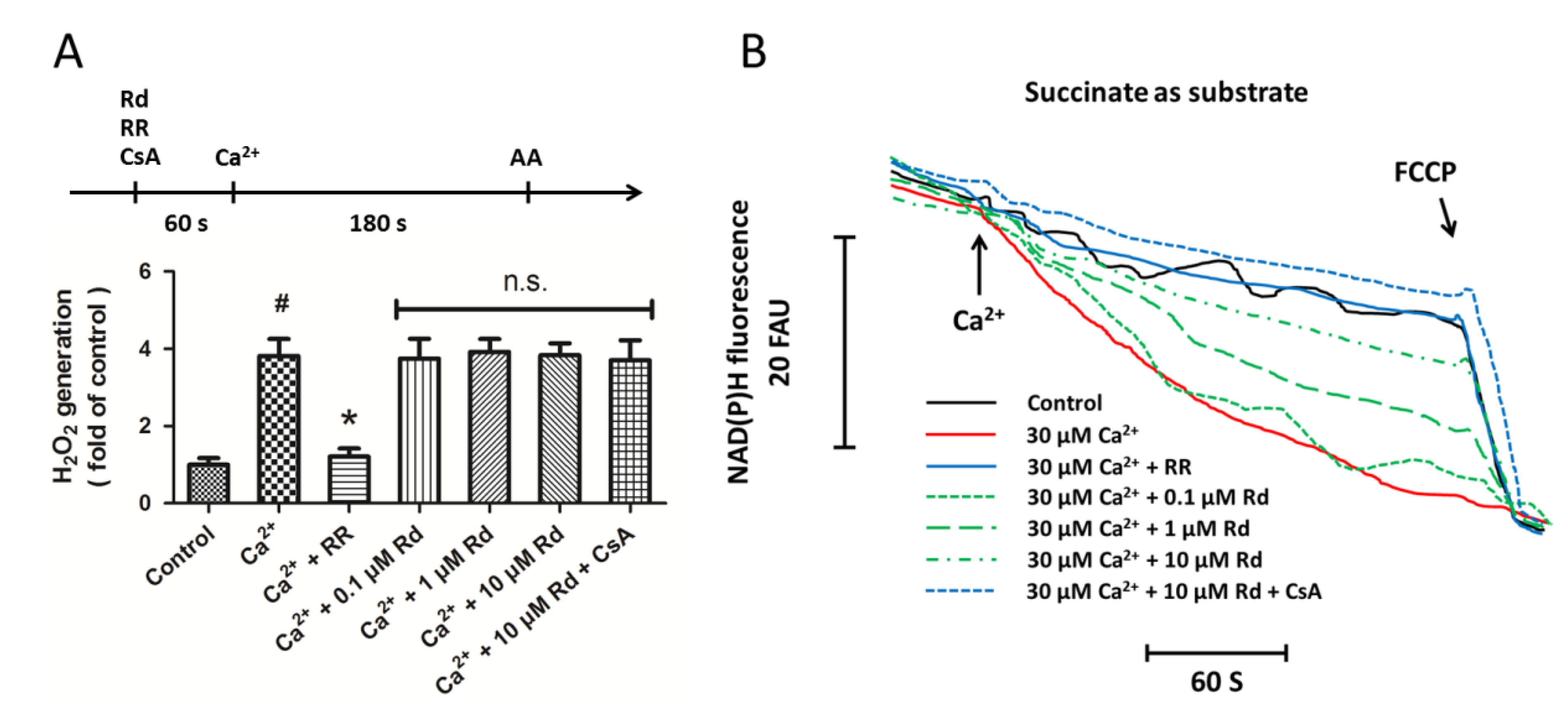
2.3. Effects of Rd on Mitochondrial H2O2 Generation and NAD(P)H Content after Ca2+ Treatment in Isolated Spinal Cord Mitochondria

In order to determine whether inhibition of reactive oxygen species (ROS) generation may be an underlying mechanism of Rd-induced protection in isolated spinal cord mitochondria, we detected the mitochondrial H2O2 generation under our in vitro experimental conditions. Figure 3A shows that Ca2+ significantly increased the mitochondrial H2O2 generation that were totally prevented by RR pretreatment. However, pretreatment with Rd at all concentrations used in our study did not alter H2O2 generation in succinate supported spinal cord mitochondria in the presence or absence of the MPT inhibitor CsA. These results suggest that H2O2 related ROS generation might not be involved in Rd induced protection against MPT pore formation. We also measured the influence of Rd and Ca2+ on NAD(P)H matrix content in succinate supported spinal cord mitochondria (Figure 3B). The results showed that Ca2+ addition significantly decreased the NAD(P)H content, which was fully prevented by RR pretreatment. Rd at 1 or 10 μM significantly attenuated the Ca2+ induced reduction of NAD(P)H content, and pretreatment with CsA further preserved the NAD(P)H content as compared to Rd alone.
Figure 3. Effects of Rd on mitochondrial H2O2 generation and NAD(P)H content after Ca2+ treatment in isolated spinal cord mitochondria. (A) Isolated spinal cord mitochondria (0.5 mg protein/mL supported by succinate) were treated with Rd at different concentrations (0.1, 1, or 10 μM) in the presence or absence of 1 μM RR and 1 μM CsA. The mitochondrial H2O2 generation was measured, and 0.1 μg/mL Antimycin A (AA) was added to induce maximal H2O2 generation at the end of the measurements; (B) Isolated spinal cord mitochondria (0.5 mg protein/mL supported by succinate) were treated with Rd at different concentrations (0.1, 1, or 10 μM) in the presence or absence of 1 μM RR and 1 μM CsA. The mitochondrial NADH and NADPH content was measured, and 1 μM FCCP was added to induce maximal NAD(P)H oxidation at the end of the measurements. The data were represented as means ± SD from five experiments. Traces are representative of three independent experiments performed in duplicate and were expressed as fluorescence arbitrary units (FAU). # p < 0.05 vs. control. * p < 0.05 vs. Ca2+. n.s., not statistically significant.
Ijms 15 09859 g003

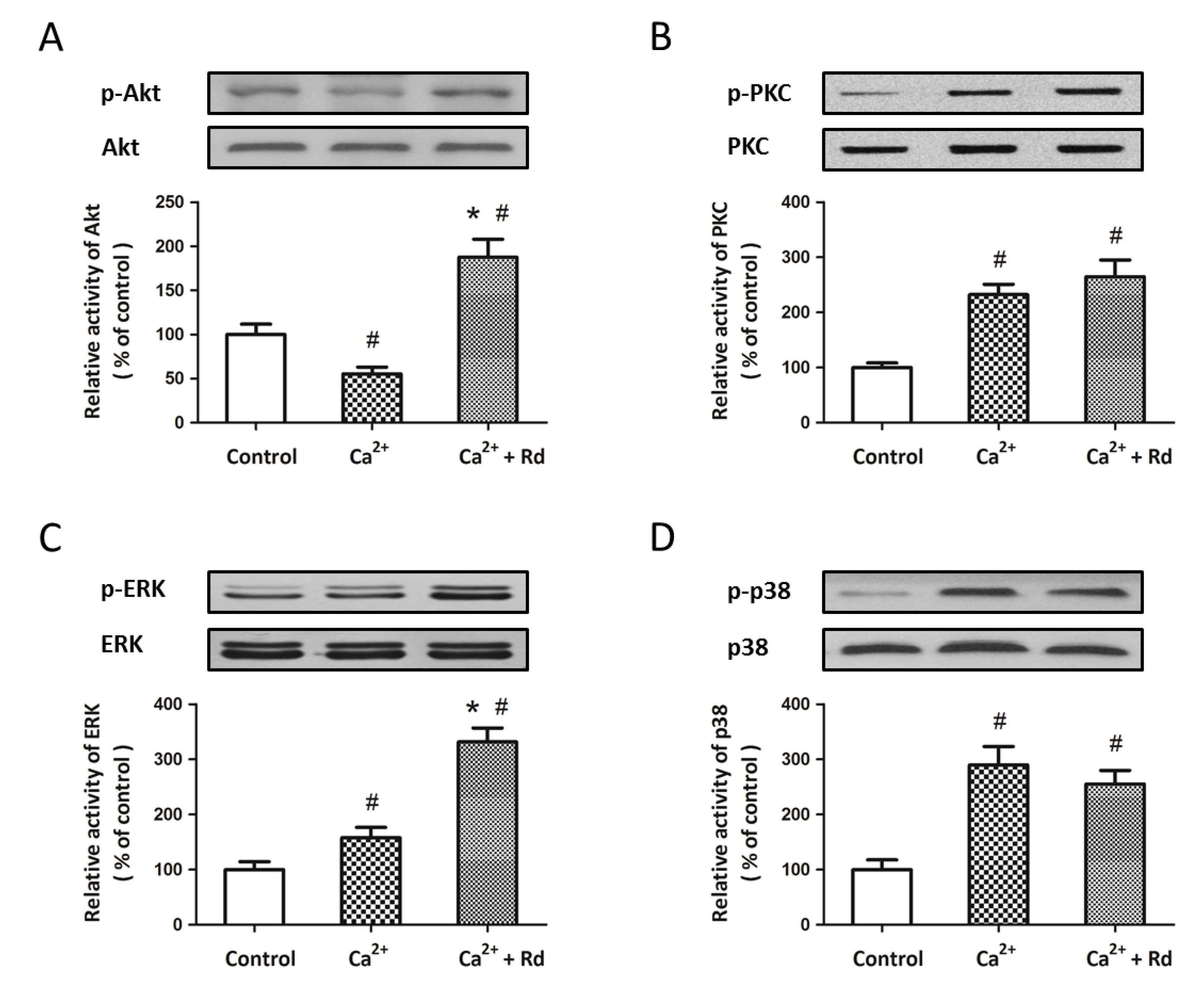
2.4. Effects of Rd on the Phosphorylation of Mitochondrial Protein Kinases after Ca2+ Treatment in Isolated Spinal Cord Mitochondria

To elucidate signaling pathways underlying Rd induced protection in spinal cord mitochondria, we consider the possible involvement of mitochondrial kinases. Recent published studies have verified the presence of various protein kinases in mitochondria and their connection to MPT regulation [33,34]. We used Western blot analysis to detect the activation of Akt, PKC, ERK and p38 after Ca2+ treatment in the presence or absence of 10 μM Rd. As shown in Figure 4A, Ca2+ decreased the expression of p-Akt, whereas Rd pretreatment significantly increased the expression of p-Akt as compared to not only Ca2+ alone, but also control group. The results also showed that phosphorylation of ERK was increased by Ca2+ and further enhanced by Rd pretreatment (Figure 4C). As shown in Figure 4B and 4D, the expression of p-PKC and p-p38 were upregulated by Ca2+, but Rd pretreatment had no effect on p-PKC and p-p38 expression. In addition, neither Ca2+ nor Rd treatment had any effect on the total protein levels of Akt, PKC, ERK, and p-38.
Figure 4. Effects of Rd on the phosphorylation of mitochondrial protein kinases after Ca2+ treatment in isolated spinal cord mitochondria. Isolated spinal cord mitochondria (0.5 mg protein/mL supported by succinate) were pretreated with 10 μM Rd for 10 min before the incubation with 30 μM Ca2+. The activation of Akt (A); PKC (B); ERK (C) and p38 (D) were detected by Western blotting analysis. The results were represented as the percentage of control. The data were represented as means ± SD from five experiments. # p < 0.05 vs. control. * p < 0.05 vs. Ca2+.
Ijms 15 09859 g004

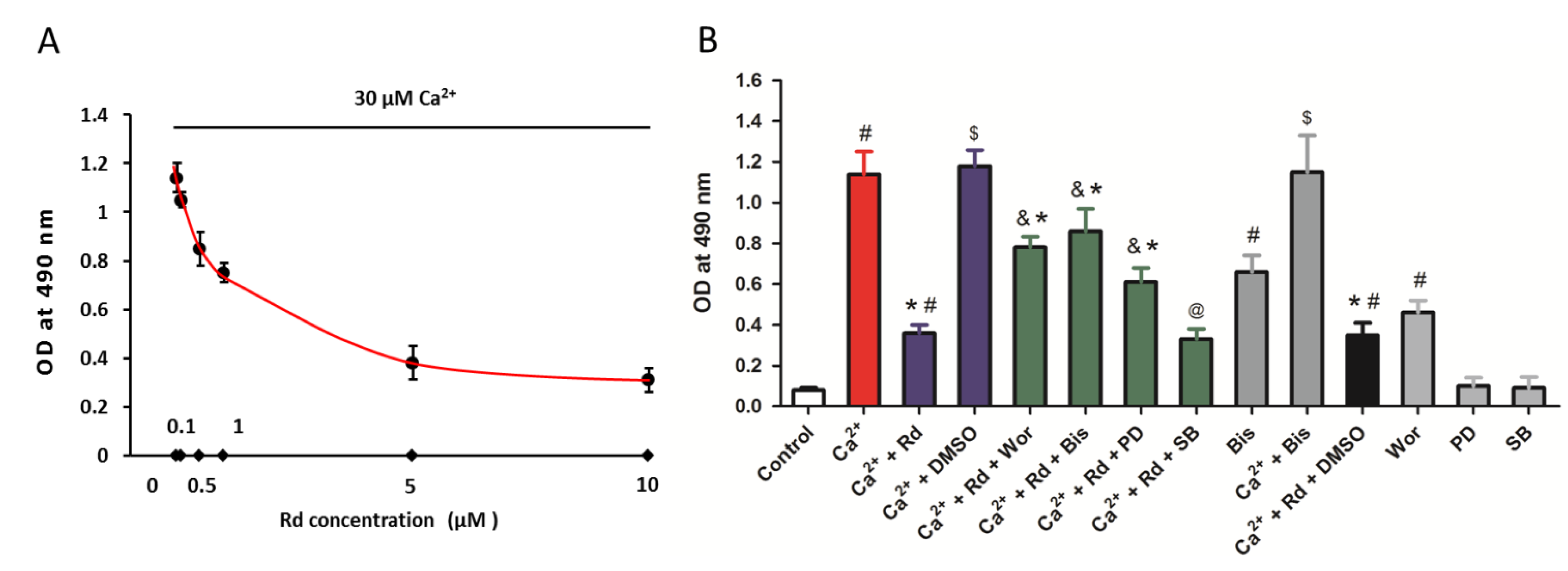
2.5. Effects of Rd and Protein Kinase Inhibitors on Cytochrome c Release after Ca2+ Treatment in Isolated Spinal Cord Mitochondria

MPT pore opening and followed cytochrome c release are facilitated by Ca2+ overload under various neuropathological conditions. Thus, we next detected the cytochrome c release in isolated spinal cord mitochondria after 30 μM Ca2+ treatment in the presence or absence of Rd at different concentrations (0.1, 0.5, 1, 5, or 10 μM) (Figure 5A). The results showed that Rd pretreatment inhibited cytochrome c release from mitochondria stimulated by Ca2+ in a dose-dependent manner. To further shed light on the relationship between the protection induced by Rd against mitochondrial cytochrome c release and its action on protein kinases regulation, the phosphorylation of Akt, PKC, ERK, and p38 were blocked, respectively, using the specific inhibitors, wortmannin (1 μM), bisindolylmaleimide (50 nM), PD98059 (10 μM), or SB202190 (10 μM). As shown in Figure 5B, the Rd induced reduction of mitochondrial cytochrome c release was partly reversed by Akt, PKC, and ERK inhibitors, but not p38 inhibitor. By using inhibitors alone, we found that treatment with Akt inhibitor alone increased the mitochondrial cytochrome c release, but PD98059 and SB 202190 had no effects on mitochondrial cytochrome c release. It was also observed that treatment with PKC inhibitor alone significantly increased the mitochondrial cytochrome c release, whereas bisindolylmaleimide could not further promote Ca2+ induced mitochondrial cytochrome c release.
Figure 5. Effects of Rd and protein kinase inhibitors on cytochrome c release after Ca2+ treatment in isolated spinal cord mitochondria. (A) Isolated spinal cord mitochondria (0.5 mg protein/mL supported by succinate) were pretreated with Rd at different concentrations (0.1, 0.5, 1, 5, or 10 μM) for 10 min before the incubation with 30 μM Ca2+. The cytochrome c release induced by Ca2+ was measured; (B) Isolated spinal cord mitochondria (0.5 mg protein/mL supported by succinate) were pretreated with 1 μM wortmannin (Wor, PI3K inhibitor), 50 nM bisindolylmaleimide (Bis, PKC inhibitor), 10 μM PD98059 (PD, ERK inhibitor), or 10 μM SB202190 (SB, p38 inhibitor) in the presence or absence of 10 μM Rd for 10 min before the incubation with 30 μM Ca2+. The cytochrome c release induced by Ca2+ was measured. The data were represented as means ± SD from five experiments. # p < 0.05 vs. control. * p < 0.05 vs. Ca2+. & p < 0.05 vs. Ca2+ + Rd. @ not statistically significant vs. Ca2+ + Rd. $ not statistically significant vs. Ca2+.
Ijms 15 09859 g005

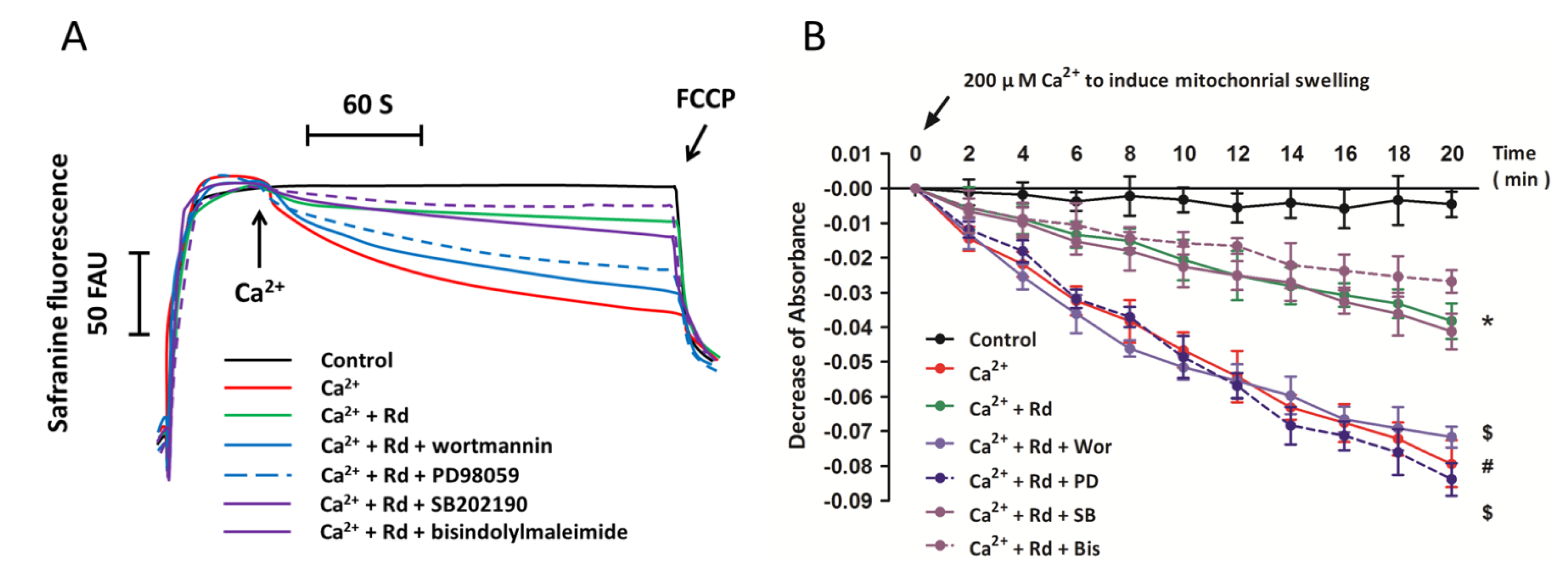
2.6. Effects of Protein Kinase Inhibitors on Mitochondrial Membrane Potential Dissipation and Swelling after Ca2+ Treatment in Rd Pretreated Spinal Cord Mitochondria

To further confirm the involvement of protein kinases in Rd-induced regulation of mitochondrial membrane potential dissipation and mitochondrial swelling, the phosphorylation of Akt, PKC, ERK, and p38 were blocked, respectively, using the specific inhibitors, wortmannin (Wor, 1 μM), bisindolylmaleimide (Bis, 50 nM), PD98059 (PD, 10 μM), or SB202190 (SB, 10 μM), and the mitochondrial membrane potential dissipation and mitochondrial swelling were measured. As shown in Figure 6A, Rd-induced attenuation of mitochondrial membrane potential dissipation was prevented by pretreatment with Akt and ERK inhibitors, but not PKC and p38 inhibitors. The results of mitochondrial swelling assay also showed that Akt and ERK inhibitors reversed the regulatory effects of Rd on Ca2+-induced mitochondrial swelling, but PKC and p38 inhibitors had not such effects.
Figure 6. Effects of protein kinase inhibitors on mitochondrial membrane potential dissipation and swelling after Ca2+ treatment in Rd pretreated spinal cord mitochondria. Isolated spinal cord mitochondria (0.5 mg protein/mL supported by succinate) were pretreated with 1 μM wortmannin (Wor, PI3K inhibitor), 50 nM bisindolylmaleimide (Bis, PKC inhibitor), 10 μM PD98059 (PD, ERK inhibitor), or 10 μM SB202190 (SB, p38 inhibitor) in the presence or absence of 10 μM Rd for 10 min before the incubation with 30 μM Ca2+. (A) The mitochondrial membrane potential was measured up to 300 s. Controls were performed in the absence of Ca2+, and 1 μM FCCP was added to induce maximal depolarization at the end of the measurements. Traces are representative of three independent experiments performed in duplicate and were expressed as fluorescence arbitrary units (FAU); (B) Mitochondrial swelling was examined by monitoring the absorbance at 540 nm induced by 200 μM Ca2+. The control was measured without Ca2+. The data were represented as means ± SD from five experiments. # p < 0.05 vs. control. * p < 0.05 vs. Ca2+. $ p < 0.05 vs. Ca2+ + Rd.
Ijms 15 09859 g006

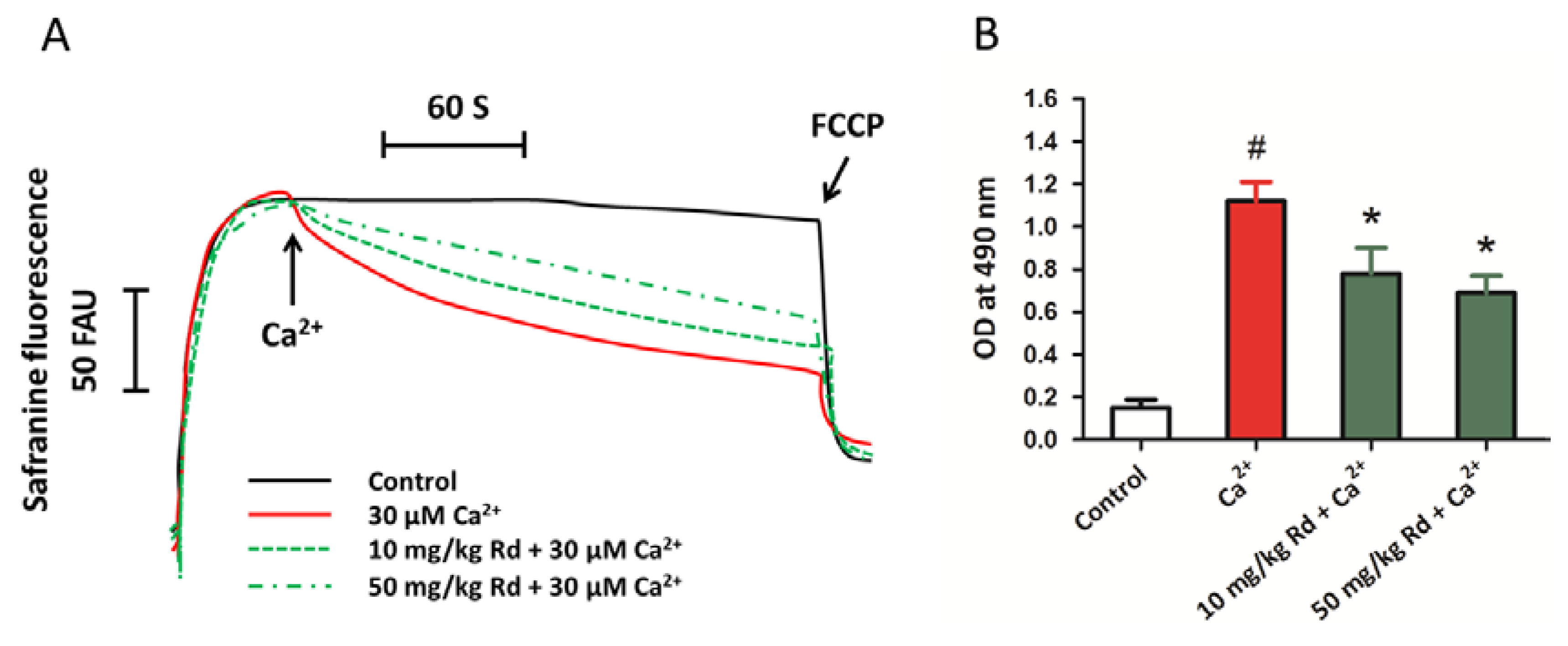
2.7. Protective Activity of Rd against Mitochondrial Permeability Transition and Cytochrome c Release through in Vivo Administration

To further confirm the protective effects of Rd in spinal cord tissues, we also determined mitochondrial membrane potential and mitochondrial cytochrome c release in isolated spinal cord mitochondria after in vivo administration of Rd at the dose of 10 and 50 mg/kg, which was chosen according to the previous studies [28,30]. The results showed that Rd pretreated spinal cord mitochondria were less vulnerable to Ca2+ induced mitochondrial membrane potential dissipation (Figure 7A). It was also found that pretreatment with Rd in vivo (both at the dose of 10 and 50 mg/kg) protected spinal cord mitochondria against Ca2+ induced cytochrome c release, although the effect was not dose-dependent as observed in in vitro experiments.
Figure 7. Protective activity of Rd against mitochondrial permeability transition and cytochrome c release through in vivo administration. Rats were pretreated with Rd at the concentrations of 10 or 50 mg/kg for 7 days before the spinal cord mitochondria were isolated, and then the isolated spinal cord mitochondria (0.5 mg protein/mL supported by succinate) were incubated with 30 μM Ca2+. The mitochondrial membrane potential was measured up to 300 s (A); and the cytochrome c release induced by Ca2+ was detected (B). The data were represented as means ± SD from five experiments. Traces are representative of three independent experiments performed in duplicate and were expressed as fluorescence arbitrary units (FAU). # p < 0.05 vs. control. * p < 0.05 vs. Ca2+.
Ijms 15 09859 g007

2.8. Discussion

Natural products found in herbs, fruits, vegetables, and plant extracts, are important parts of human health care in both developing and developed countries, with increasing commercial values. They are chemical compounds that usually have therapeutic activities for both treatable and incurable diseases, and many of the modern pharmaceutical industries have been developed on the basis of medicinal plants [35]. It is reported that more than 25% of the drugs prescribed at present come from medical plants, and approximately 60% of anti-infectious drugs on the market or under clinical trials are of natural origin [36]. Ginseng species have been used as rejuvenating tonic for more than 2000 years in China, and the protective effects of ginsenosides, the principle active ingredients, in central nervous system and cardiovascular system have been deeply investigated in rodents, dogs, and humans [20,24]. Ginsenoside Rd was shown to protect against oxidative stress and apoptotic cell death in several in vitro neuronal injury models, and attenuate neuronal damage in in vivo traumatic, ischemic or degenerative neurological diseases [24,25,26,29,30]. In the present study, we extended the protective effects of Rd to Ca2+ induced mitochondrial dysfunction in isolated spinal cord mitochondria, which could be involved in several related spinal cord related diseases, such as spinal cord ischemia and amyotrophic lateral sclerosis [37,38].
Although many compounds with antioxidant activities are demonstrated to have neuroprotective effects against mitochondrial dysfunction in spinal cord, Rd exerts unique advantages. Ginsenosides have already had thousands of years of human exposure with little reported toxicity [24]. Rd is a small lipophilic molecule that freely permeates through the phospholipids bilayers and interacts with various compounds to form different thiol-contained beneficial metabolites. In addition, the brain/serum ratio of Rd after intravenous injection was significantly higher than that of blood-brain barrier (BBB) impermeable albumin, even in energy deficient conditions [28,29], indicating an effective passage of Rd through the intact BBB. The mean plasma elimination half-life was approximately 14 h in mice, 24 h in dogs, and more than 19 h in healthy humans [39,40,41]. The relative long half-life as compared to other neuroprotective agents is useful for avoiding repeated dosing in patients. More importantly, Rd is being developed to treat patients with acute ischemic stroke, and a Phase III randomized, double-blind, placebocontrolled, multicenter clinical trial showed that Rd improves outcome of ischemic stroke in patients up to 90 days after stroke onset [42].
Most of the previous experiments on neuroprotective agents were performed in animals or using neuronal cell lines. In order to confirm the involvement of mitochondria and to elucidate the potential molecular mechanism underlying the protective activity of Rd, we used an in vitro system of isolated spinal cord mitochondria. Importantly, this system allowed us to investigate the roles of MPT, mitochondrial oxidative phosphorylation, and mitochondrial cytochrome c release in Rd induced beneficial effects and their cross talk, which might be an important supplement to the animal experiments. Our results showed that Ca2+ treatment induced mitochondrial membrane potential dissipation and significantly provoked mitochondrial swelling, which were fully prevented by RR, an inhibitor of the mitochondrial Ca2+ uniporter [43], but only partly attenuated by CsA. This is in accord with the data obtained in the isolated brain mitochondria where CsA failed to prevent Ca2+-induced depolarization or repolarize mitochondria when mitochondria were depolarized excessively [32]. It is emphasized that the MPT induction causes mitochondrial depolarization, organelle swelling, and dysregulation of metabolites, including Ca2+, Mg2+, glutathione, NAD(P)H, and H2O2 [44,45]. Interestingly, our data showed that Rd significantly attenuated Ca2+ induced reduction of NAD(P)H content in a dose-dependent manner, however, neither Rd treatment nor CsA had any effect on Ca2+ induced H2O2 generation in isolated mitochondria, indicating a H2O2 metabolism independent mechanism. This might be explained by that Rd could directly blocked the opening of MPT pore, thereby preserving NAD(P)H content and inhibiting cytochrome c release, with no effects on the H2O2 metabolism related enzymes, such as catalase (CAT) or superoxide dismutase (SOD) [46,47]. In accordance with our hypothesis, we found that Rd and CsA exerted synergetic effects on inhibition of Ca2+ induced mitochondrial cytochrome c release. In our in vitro conditions, MPT induction in the presence of Ca2+ seems to be a consequence of mitochondrial depolarization, since a significant decrease in membrane potential was observed. However, the exact mechanisms by which Rd affect the mitochondrial Ca2+ threshold for MPT opening, which makes spinal cord mitochondria less susceptible to insults, need to be further determined.
Proteins kinases are a group of kinase enzymes that modify other proteins through chemically adding phosphates to amino acid residues (phosphorylation), which result in a functional of the target substrate proteins by regulating enzymatic activity, subcellular location or interaction with other proteins [48]. Protein kinases mediate most of the signal transduction form the cell membrane to the nucleus in eukaryotic, but it is by no means the only pathway that signaling molecules can take. Numerous studies have shown that several proteins, such as Akt, PKC, and MAPKs [49,50], can translocated into the mitochondria to regulate phosphorylation of respiratory chain proteins, control the release of mitochondrial components, and thereby affect the whole cell responses [34]. We confirmed the expression and phosphorylation of Akt, PKC, ERK, and p38 in isolated spinal cord mitochondria in the present study, and found that their enzymatic activities were differently regulated by Ca2+ treatment, indicating the involvement of these kinases in MPT opening, cytochrome c release, and followed mitochondrial dysfunction. This point of view was further supported by that obvious increase of cytochrome c release was observed after treating isolated mitochondria with PKC inhibitor Bis alone, which was consistent with a recent study on purified liver mitochondria [51]. Many studies using pharmacologic inhibitors of MAPKs have demonstrated that ERK and p38 can modulate mitochondrial functions, especially those associated with cell death, and increased activation of Akt and PKC were also shown to exert pro-survival effects against mitochondrial related apoptotic cell death [52,53,54]. More recently, Rd was demonstrated to play protective roles via regulating PI3K/Akt and ERK pathways in both glutamate-induced excitotoxicity and myocardial ischemia/reperfusion injury [55,56]. Akt exerts its protective effects via phosphorylation of diverse target molecules (such as Bcl-2 family and GSK-3), preserving mitochondrial integrity [57,58]. A recent study showed that Rd attenuates myocardial ischemia/reperfusion injury via a Akt-mediated mitochondrial-dependent apoptotic pathway [56], which was also confirmed in transient forebrain ischemia models [59]. The role of ERK activation in the Ca2+ related neuronal death was disputable. Despite the volume of evidence supporting the p-ERK elevation after ischemic injury as detrimental effects that were essential for oxidative stress and inflammation-related cell death, numerous studies demonstrated that ERK activation contributed to protective effects of many neuroprotectants. For example, TGF-β1 blocked neuronal injuries by increasing ERK phosphorylation and the activated ERK pathway suppressed Bad protein levels and caspase-3 activation [60]. As a well-known neuroprotectant, estrogen also elicited rapid ERK activation against β-amyloid toxicity and quinolinic acid-induced cell death [61,62]. Data from our study might be another evidence for the protective role of ERK activation in Ca2+-induced mitochondrial dysfunction. Our present data using kinases inhibitors showed that Ca2+-induced MPT opening and cytochrome c release were partly reversed by Akt and ERK inhibitors, but not p38 inhibitor. Because Rd has no effects on PKC activation in isolated mitochondria as shown in Figure 4, the effects of Bis on Rd-induced protection (inhibition of cytochrome c release) seemed to be at the level of its own detrimental activity on mitochondrial function. All these data suggest that Rd attenuates Ca2+-induced mitochondrial dysfunction through different regulation of mitochondrial protein kinases, the targets of which might be different from those of cytosolic ones [34,51].

3. Experimental Section

3.1. Animals

We used male C57BL/6J mice (3-month old, 25–28 g). The animals had continuous access to food and water and were housed in cages in a room maintained at 20–22 °C with a 12 h light/12 h dark cycle. All experimental protocols and animal handling procedures were performed in accordance with the National Institutes of Health (NIH) guidelines for the use of experimental animals (NIH Publications No. 80-23, revised 1996). All efforts were made to minimize animal number and their suffering.

3.2. Mitochondria Purification

Mitochondria were isolated from the mouse spinal cord tissues and purification of their outer membranes was performed by differential ultracentrifugation. To prepare detergent lysates, the spinal cord tissues were frozen at −70 °C, thawed and treated with the lysing buffer with protease inhibitors cocktail for 2 h. The suspensions were centrifuged at 800× g for 10 min at 4 °C. The resulting lysate was cleared by centrifugation at 25,000× g for 20 min and dialyze. The final pellet was gently washed and resuspended in isolation buffer without ethyleneglycol bistetraacetic acid (EGTA), at an approximate protein concentration of 0.5 mg/mL. Total protein concentration was determined using Enhanced BCA Protein Assay Kit (Beijing Biosynthesis Biotechnology, Beijing, China).

3.3. Rd Treatment

Rd with a purity of 98% was obtained from Tai-He Biopharmaceutical Co. Ltd (Guangzhou, China). Stock solutions of Rd were prepared in saline containing 10% 1,3-propanediol (v/v). In in vitro experiments, isolated spinal cord mitochondria (0.5 mg protein/mL supported by succinate) were pretreated with Rd at different concentrations (0.1, 1, or 10 μM) for 60 s (for the measurement of mitochondrial membrane potential, mitochondrial swelling, H2O2 production and NAD(P)H content) or for 10 min (for the measurement of mitochondrial kinases expression) before the incubation with Ca2+. In in vivo experiments, Rd at the dose of 10 or 50 mg/kg was administered intraperitoneally twice a day for 3 days before mitochondria purification.

3.4. Mitochondrial Membrane Potential Measurement

The mitochondrial membrane potential was measured by using the cationic fluorescent probe safranine O as described previously [63]. The measurements were conducted on a temperature-controlled spectro-fluorometer with magnetic stirring operating at excitation and emission wavelengths of 495 and 586 nm, respectively. Mitochondrial preparations (0.5 mg protein/mL) were incubated at 37 °C with Rd for 60 s, and incubated with CaCl2 (10, 20, or 30 μM) afterward. In the end of the measurements, maximal depolarization was induced by 1 μM FCCP. The results were expressed as FAU.

3.5. Determination of Mitochondrial Swelling

Mitochondria swelling was measured following a previously published protocol [64]. Briefly, isolated mitochondria were suspended in fresh swelling buffer (0.2 M sucrose, 10 mM Tris-MOPS, pH 7.4, 5 mM succinate, 1 mM phosphate, 2 μM rotenone, and 1 μM EGTA-Tris, pH 7.4) at 0.5 mg/mL, and the swelling of mitochondria was monitored by a decrease in absorbance at 540 nm in the presence of CaCl2 (200 μM).

3.6. Measurement of H2O2 Production

Mitochondrial H2O2 production was assessed on a temperature-controlled spectro-fluorometer with magnetic stirring operating at excitation of 563 and emission of 587 nm by using 5 mM succinate plus 4 μM rotenone as substrate. Mitochondrial preparations (0.5 mg protein/mL) were incubated at 37 °C with Rd for 60 s, and incubated with CaCl2 (30 μM) afterward. Antimycin A (AA, 0.1 μg/mL) was added at the end of the measurements for maximal H2O2 generation. The results were represented as the fold of control.

3.7. Measurement of NAD(P)H Content

Matrix mitochondrial NAD(P)H content was measured on a temperature-controlled spectro-fluorometer with magnetic stirring operating at an excitation wavelengths of 366 nm and emission wavelength of 450 nm by using 5 mM succinate as substrate. Mitochondrial preparations (0.5 mg protein/mL) were incubated at 37 °C with Rd for 60 s, and incubated with CaCl2 (30 μM) afterward. In the end of the measurements, the maximal NAD(P)H oxidation was induced by 1 μM FCCP. The results were expressed as FAU.

3.8. Western Blot Analysis

Equivalent amounts of protein (40 μg per lane) were loaded and separated by 10% SDS-PAGE gels, and transferred to polyvinylidene difluoride (PVDF) membranes. Membranes were blocked with 5% nonfat milk solution in tris-buffered saline with 0.1% Triton X-100 (TBST) for 1 h, and then incubated overnight at 4 °C with the following primary antibody dilutions in TBST: anti-p-Akt (1:800), Akt (1:1000), p-PKC (1:500), PKC (1:1200), p-ERK (1:600), ERK (1:1000), p-p38 (1:500) and p38 (1:1200). After that the membranes were washed and incubated with secondary antibody for 1 h at room temperature. An analysis software named Image J was used to quantify the optical density of each band. The relative activity of Akt, PKC, ERK and p-38 were calculated by dividing the intensity of phosphorylated kinase bands by the intensity of the total kinase bands, and the data are presented as ratios to control values.

3.9. Determination of Cytochrome c Release

The purified mitochondria (0.5 mg protein/mL) were incubated with 30 μM CaCl2 in the presence or absence of Rd for 10 min at room temperature and were immediately pelleted by centrifugation (10 min, 7000× g) at 4 °C. To investigate the potential involvement of mitochondrial protein kinases, the kinases inhibitors (wortmannin, Wor, 1 μM; bisindolylmaleimide, Bis, 50 nM; PD98059, PD, 10 μM or SB202190, SB, 10 μM) were applied 10 min prior CaCl2 treatment. The mitochondria supernatants were collected and tested for the cytochrome c release by sandwich assay as described previously [65]. Experimental values of optical density (OD 490 nm) were calculated within the linear part of the calibration curve built with bovine cytochrome c.

3.10. Statistical Analysis

Statistical analysis was performed using SPSS 16.0, a statistical software package. Statistical evaluation of the data was performed by one-way analysis of variance (ANOVA) followed by Bonferroni’s multiple comparisons or unpaired t test (two groups). A value of p < 0.05 was considered statistically significant.

4. Conclusions

In conclusion, data from our study revealed that Rd protects isolated spinal cord mitochondria against Ca2+ induced MPT and cytochrome c release in a mitochondrial protein kinases-dependent manner. These effects of Rd in our in vitro system were mediated by the activation of Akt and ERK, but not related to the activities of PKC and p38 kinases. These results may implicate the possible therapeutic potential of Rd in the management of mitochondrial dysfunction in spinal cord injury, which needs to be further determined in clinical trials.

Conflicts of Interest

The authors declare no conflict of interest.

References

  1. Svensson, L.G.; Crawford, E.S.; Hess, K.R.; Coselli, J.S.; Safi, H.J. Experience with 1509 patients undergoing thoracoabdominal aortic operations. J. Vasc. Surg. 1993, 17, 357–368. [Google Scholar] [CrossRef]
  2. Zurita, M.; Aguayo, C.; Bonilla, C.; Otero, L.; Rico, M.; Rodriguez, A.; Vaquero, J. The pig model of chronic paraplegia: A challenge for experimental studies in spinal cord injury. Prog. Neurobiol. 2012, 97, 288–303. [Google Scholar]
  3. Chiesa, R.; Melissano, G.; Civilini, E.; de Moura, M.L.; Carozzo, A.; Zangrillo, A. Ten years experience of thoracic and thoracoabdominal aortic aneurysm surgical repair: lessons learned. Ann. Vasc. Surg. 2004, 18, 514–520. [Google Scholar] [CrossRef]
  4. Etz, C.D.; Homann, T.M.; Luehr, M.; Kari, F.A.; Weisz, D.J.; Kleinman, G.; Plestis, K.A.; Griepp, R.B. Spinal cord blood flow and ischemic injury after experimental sacrifice of thoracic and abdominal segmental arteries. Eur. J. Cardiothorac. Surg. 2008, 33, 1030–1038. [Google Scholar] [CrossRef]
  5. Chang, C.K.; Chuter, T.A.; Reilly, L.M.; Ota, M.K.; Furtado, A.; Bucci, M.; Wintermark, M.; Hiramoto, J.S. Spinal arterial anatomy and risk factors for lower extremity weakness following endovascular thoracoabdominal aortic aneurysm repair with branched stent-grafts. J. Endovasc. Ther. 2008, 15, 356–362. [Google Scholar] [CrossRef]
  6. Backes, W.H.; Nijenhuis, R.J.; Mess, W.H.; Wilmink, F.A.; Schurink, G.W.; Jacobs, M.J. Magnetic resonance angiography of collateral blood supply to spinal cord in thoracic and thoracoabdominal aortic aneurysm patients. J. Vasc. Surg. 2008, 48, 261–271. [Google Scholar] [CrossRef]
  7. Ogino, H.; Sasaki, H.; Minatoya, K.; Matsuda, H.; Yamada, N.; Kitamura, S. Combined use of adamkiewicz artery demonstration and motor-evoked potentials in descending and thoracoabdominal repair. Ann. Thorac. Surg. 2006, 82, 592–596. [Google Scholar] [CrossRef]
  8. Jacobs, M.J.; Mess, W.; Mochtar, B.; Nijenhuis, R.J.; Statius van Eps, R.G.; Schurink, G.W. The value of motor evoked potentials in reducing paraplegia during thoracoabdominal aneurysm repair. J. Vasc. Surg. 2006, 43, 239–246. [Google Scholar] [CrossRef]
  9. Melissano, G.; Chiesa, R. Advances in imaging of the spinal cord vascular supply and its relationship with paraplegia after aortic interventions. Eur. J. Vasc. Endovasc. Surg. 2009, 38, 567–577. [Google Scholar] [CrossRef]
  10. Silver, J.; Miller, J.H. Regeneration beyond the glial scar. Neuroscience 2004, 5, 146–156. [Google Scholar]
  11. Saxena, P.; Abu Hasan, F.; Merry, C. Spinal cord ischemia following coronary artery bypass surgery. J. Card. Surg. 2012, 27, 45–46. [Google Scholar] [CrossRef]
  12. De Haan, P.; Kalkman, C.J.; Jacobs, M.J. Pharmacologic neuroprotection in experimental spinal cord ischemia: A systematic review. J. Neurosurg. Anesthesiol. 2001, 13, 3–12. [Google Scholar] [CrossRef]
  13. Ahn, Y.H.; Lee, G.; Kang, S.K. Molecular insights of the injured lesions of rat spinal cords: Inflammation, apoptosis, and cell survival. Biochem. Biophys. Res. Commun. 2006, 348, 560–570. [Google Scholar] [CrossRef]
  14. Genovese, T.; Cuzzocrea, S. Role of free radicals and poly (ADP-ribose) polymerase-1 in the development of spinal cord injury: New potential therapeutic targets. Curr. Med. Chem. 2008, 15, 477–487. [Google Scholar] [CrossRef]
  15. Cassina, P.; Cassina, A.; Pehar, M.; Castellanos, R.; Gandelman, M.; de Leon, A.; Robinson, K.M.; Mason, R.P.; Beckman, J.S.; Barbeito, L.; et al. Mitochondrial dysfunction in SOD1G93A-bearing astrocytes promotes motor neuron degeneration: prevention by mitochondrial-targeted antioxidants. J. Neurosci. 2008, 28, 4115–4122. [Google Scholar] [CrossRef]
  16. Zhou, Y.F.; Li, L.; Feng, F.; Yuan, H.; Gao, D.K.; Fu, L.A.; Fei, Z. Osthole attenuates spinal cord ischemia-reperfusion injury through mitochondrial biogenesis-independent inhibition of mitochondrial dysfunction in rats. J. Surg. Res. 2013, 185, 805–814. [Google Scholar] [CrossRef]
  17. Zhu, J.W.; Chen, T.; Guan, J.; Liu, W.B.; Liu, J. Neuroprotective effects of allicin on spinal cord ischemia-reperfusion injury via improvement of mitochondrial function in rabbits. Neurochem. Int. 2012, 61, 640–648. [Google Scholar] [CrossRef]
  18. Fu, J.; Fan, H.B.; Guo, Z.; Wang, Z.; Li, X.D.; Li, J.; Pei, G.X. Salvianolic acid B attenuates spinal cord ischemia-reperfusion-induced neuronal injury and oxidative stress by activating the extracellular signal-regulated kinase pathway in rats. J. Surg. Res. 2014, 188, 222–230. [Google Scholar] [CrossRef]
  19. Jia, L.; Zhao, Y. Current evaluation of the millennium phytomedicine-ginseng (I): Etymology, pharmacognosy, phytochemistry, market and regulations. Curr. Med. Chem. 2009, 16, 2475–2484. [Google Scholar] [CrossRef]
  20. Peng, L.; Sun, S.; Xie, L.H.; Wicks, S.M.; Xie, J.T. Ginsenoside Re: Pharmacological effects on cardiovascular system. Cardiovasc. Ther. 2012, 30, 183–188. [Google Scholar] [CrossRef]
  21. Lu, J.M.; Yao, Q.; Chen, C. Ginseng compounds: An update on their molecular mechanisms and medical applications. Curr. Vasc. Pharmacol. 2009, 7, 293–302. [Google Scholar] [CrossRef]
  22. Christensen, L.P.; Jensen, M. Biomass and content of ginsenosides and polyacetylenes in American ginseng roots can be increased without affecting the profile of bioactive compounds. J. Nat. Med. 2009, 63, 159–168. [Google Scholar] [CrossRef]
  23. Christensen, L.P. Ginsenosides chemistry, biosynthesis, analysis, and potential health effects. Adv. Food Nutr. Res. 2009, 55, 1–99. [Google Scholar] [CrossRef]
  24. Ye, R.; Zhao, G.; Liu, X. Ginsenoside Rd for acute ischemic stroke: translating from bench to bedside. Expert Rev. Neurother. 2013, 13, 603–613. [Google Scholar] [CrossRef]
  25. Zhang, C.; Du, F.; Shi, M.; Ye, R.; Cheng, H.; Han, J.; Ma, L.; Cao, R.; Rao, Z.; Zhao, G. Ginsenoside Rd protects neurons against glutamate-induced excitotoxicity by inhibiting Ca2+ influx. Cell. Mol. Neurobiol. 2012, 32, 121–128. [Google Scholar] [CrossRef]
  26. Ye, R.; Han, J.; Kong, X.; Zhao, L.; Cao, R.; Rao, Z.; Zhao, G. Protective effects of ginsenoside Rd on PC12 cells against hydrogen peroxide. Biol. Pharm. Bull. 2008, 31, 1923–1927. [Google Scholar] [CrossRef]
  27. Ye, R.; Li, N.; Han, J.; Kong, X.; Cao, R.; Rao, Z.; Zhao, G. Neuroprotective effects of ginsenoside Rd against oxygen-glucose deprivation in cultured hippocampal neurons. Neurosci. Res. 2009, 64, 306–310. [Google Scholar] [CrossRef]
  28. Ye, R.; Kong, X.; Yang, Q.; Zhang, Y.; Han, J.; Zhao, G. Ginsenoside Rd attenuates redox imbalance and improves stroke outcome after focal cerebral ischemia in aged mice. Neuropharmacology 2011, 61, 815–824. [Google Scholar] [CrossRef]
  29. Ye, R.; Kong, X.; Yang, Q.; Zhang, Y.; Han, J.; Li, P.; Xiong, L.; Zhao, G. Ginsenoside rd in experimental stroke: Superior neuroprotective efficacy with a wide therapeutic window. Neurotherapeutics 2011, 8, 515–525. [Google Scholar] [CrossRef]
  30. Ye, R.; Zhang, X.; Kong, X.; Han, J.; Yang, Q.; Zhang, Y.; Chen, Y.; Li, P.; Liu, J.; Shi, M.; et al. Ginsenoside Rd attenuates mitochondrial dysfunction and sequential apoptosis after transient focal ischemia. Neuroscience 2011, 178, 169–180. [Google Scholar] [CrossRef]
  31. Ye, R.; Yang, Q.; Kong, X.; Han, J.; Zhang, X.; Zhang, Y.; Li, P.; Liu, J.; Shi, M.; Xiong, L.; Zhao, G. Ginsenoside Rd attenuates early oxidative damage and sequential inflammatory response after transient focal ischemia in rats. Neurochem. Int. 2011, 58, 391–398. [Google Scholar] [CrossRef]
  32. Brustovetsky, N.; Dubinsky, J.M. Limitations of cyclosporin A inhibition of the permeability transition in CNS mitochondria. J. Neurosci. 2000, 20, 8229–8237. [Google Scholar]
  33. Kowalczyk, J.E.; Zablocka, B. [Protein kinases in mitochondria]. Postep. Biochem. 2008, 54, 209–216. [Google Scholar]
  34. Antico Arciuch, V.G.; Alippe, Y.; Carreras, M.C.; Poderoso, J.J. Mitochondrial kinases in cell signaling: Facts and perspectives. Adv. Drug Deliv. Rev. 2009, 61, 1234–1249. [Google Scholar] [CrossRef]
  35. Lopez, D.; Fischbach, M.A.; Chu, F.; Losick, R.; Kolter, R. Structurally diverse natural products that cause potassium leakage trigger multicellularity in Bacillus subtilis. Proc. Natl. Acad. Sci. USA 2009, 106, 280–285. [Google Scholar] [CrossRef]
  36. Zhang, A.; Sun, H.; Wang, X. Recent advances in natural products from plants for treatment of liver diseases. Eur. J. Med. Chem. 2013, 63, 570–577. [Google Scholar] [CrossRef]
  37. McEwen, M.L.; Sullivan, P.G.; Rabchevsky, A.G.; Springer, J.E. Targeting mitochondrial function for the treatment of acute spinal cord injury. Neurotherapeutics 2011, 8, 168–179. [Google Scholar] [CrossRef]
  38. Shi, P.; Wei, Y.; Zhang, J.; Gal, J.; Zhu, H. Mitochondrial dysfunction is a converging point of multiple pathological pathways in amyotrophic lateral sclerosis. J. Alzheimer’s Dis. 2010, 20, S311–S324. [Google Scholar]
  39. Sun, D.; Wang, B.; Shi, M.; Zhang, Y.X.; Zhou, L.F.; Liu, Z.R.; Wu, Z.L.; Jiang, W.; Han, J.L.; Xiong, L.Z.; et al. Pharmacokinetic, tissue distribution and excretion of ginsenoside-Rd in rodents. Phytomedicine 2012, 19, 369–373. [Google Scholar] [CrossRef]
  40. Wang, W.; Wang, G.J.; Xie, H.T.; Sun, J.G.; Zhao, S.; Jiang, X.L.; Li, H.; Lv, H.; Xu, M.J.; Wang, R. Determination of ginsenoside Rd in dog plasma by liquid chromatography-mass spectrometry after solid-phase extraction and its application in dog pharmacokinetics studies. J. Chromatogr. B 2007, 852, 8–14. [Google Scholar] [CrossRef]
  41. Deng, Y.H.; Feng, Y.; Zeng, X.; Yang, L.; Liu, Y.M.; Huang, Y.; Sun, J.; Zhou, D.; Liang, W.X. Determination of ginsenoside rd in human plasma by LC/MS/MS. Zhong yao cai/Zhongyaocai/J. Chin. Med. Mater. 2006, 29, 928–931. (In Chinese) [Google Scholar]
  42. Liu, X.; Xia, J.; Wang, L.; Song, Y.; Yang, J.; Yan, Y.; Ren, H.; Zhao, G. Efficacy and safety of ginsenoside-Rd for acute ischaemic stroke: A randomized, double-blind, placebo-controlled, phase II multicenter trial. Eur. J. Neurol. 2009, 16, 569–575. [Google Scholar] [CrossRef]
  43. Moore, C.L. Specific inhibition of mitochondrial Ca++ transport by ruthenium red. Biochem. Biophys. Res. Commun. 1971, 42, 298–305. [Google Scholar] [CrossRef]
  44. Bernardi, P.; von Stockum, S. The permeability transition pore as a Ca2+ release channel: New answers to an old question. Cell Calcium 2012, 52, 22–27. [Google Scholar] [CrossRef]
  45. Crompton, M. The mitochondrial permeability transition pore and its role in cell death. Biochem. J. 1999, 341, 233–249. [Google Scholar] [CrossRef]
  46. Armogida, M.; Nistico, R.; Mercuri, N.B. Therapeutic potential of targeting hydrogen peroxide metabolism in the treatment of brain ischaemia. Br. J. Pharmacol. 2012, 166, 1211–1224. [Google Scholar] [CrossRef]
  47. Gough, D.R.; Cotter, T.G. Hydrogen peroxide: A Jekyll and Hyde signalling molecule. Cell Death Dis. 2011, 2. [Google Scholar] [CrossRef]
  48. Manning, G.; Whyte, D.B.; Martinez, R.; Hunter, T.; Sudarsanam, S. The protein kinase complement of the human genome. Science 2002, 298, 1912–1934. [Google Scholar] [CrossRef]
  49. Bijur, G.N.; Jope, R.S. Rapid accumulation of Akt in mitochondria following phosphatidylinositol 3-kinase activation. J. Neurochem. 2003, 87, 1427–1435. [Google Scholar] [CrossRef]
  50. Galli, S.; Arciuch, V.G.A.; Poderoso, C.; Converso, D.P.; Zhou, Q.; de Kier Joffe, E.B.; Cadenas, E.; Boczkowski, J.; Carreras, M.C.; Poderoso, J.J. Tumor cell phenotype is sustained by selective MAPK oxidation in mitochondria. PLoS One 2008, 3, e2379. [Google Scholar] [CrossRef]
  51. Gergalova, G.; Lykhmus, O.; Komisarenko, S.; Skok, M. α7 nicotinic acetylcholine receptors control cytochrome c release from isolated mitochondria through kinase-mediated pathways. Int. J. Biochem. Cell Biol. 2014, 49, 26–31. [Google Scholar] [CrossRef]
  52. Chao, X.; Zhou, J.; Chen, T.; Liu, W.; Dong, W.; Qu, Y.; Jiang, X.; Ji, X.; Zhen, H.; Fei, Z. Neuroprotective effect of osthole against acute ischemic stroke on middle cerebral ischemia occlusion in rats. Brain Res. 2010, 1363, 206–211. [Google Scholar] [CrossRef]
  53. Chen, T.; Cao, L.; Dong, W.; Luo, P.; Liu, W.; Qu, Y.; Fei, Z. Protective effects of mGluR5 positive modulators against traumatic neuronal injury through PKC-dependent activation of MEK/ERK pathway. Neurochem. Res. 2012, 37, 983–990. [Google Scholar] [CrossRef]
  54. Chen, T.; Zhang, L.; Qu, Y.; Huo, K.; Jiang, X.; Fei, Z. The selective mGluR5 agonist CHPG protects against traumatic brain injury in vitro and in vivo via ERK and Akt pathway. Int. J. Mol. Med. 2012, 29, 630–636. [Google Scholar]
  55. Zhang, X.; Shi, M.; Bjoras, M.; Wang, W.; Zhang, G.; Han, J.; Liu, Z.; Zhang, Y.; Wang, B.; Chen, J.; et al. Ginsenoside Rd promotes glutamate clearance by up-regulating glial glutamate transporter GLT-1 via PI3K/AKT and ERK1/2 pathways. Front. Pharmacol. 2013, 4. [Google Scholar] [CrossRef]
  56. Wang, Y.; Li, X.; Wang, X.; Lau, W.; Xing, Y.; Zhang, X.; Ma, X.; Gao, F. Ginsenoside Rd attenuates myocardial ischemia/reperfusion injury via Akt/GSK-3beta signaling and inhibition of the mitochondria-dependent apoptotic pathway. PLoS One 2013, 8, e70956. [Google Scholar]
  57. Sussman, M.A. Mitochondrial integrity: Preservation through Akt/Pim-1 kinase signaling in the cardiomyocyte. Expert Rev. Cardiovasc. Ther. 2009, 7, 929–938. [Google Scholar] [CrossRef]
  58. Miyamoto, S.; Murphy, A.N.; Brown, J.H. Akt mediated mitochondrial protection in the heart: metabolic and survival pathways to the rescue. J. Bioenerg. Biomembr. 2009, 41, 169–180. [Google Scholar] [CrossRef]
  59. Zhang, X.; Shi, M.; Ye, R.; Wang, W.; Liu, X.; Zhang, G.; Han, J.; Zhang, Y.; Wang, B.; Zhao, J.; et al. Ginsenoside Rd attenuates Tau protein phosphorylation via the PI3K/AKT/GSK-3β pathway after transient forebrain ischemia. Neurochem. Res. 2014. [Google Scholar] [CrossRef]
  60. Zhu, Y.; Yang, G.Y.; Ahlemeyer, B.; Pang, L.; Che, X.M.; Culmsee, C.; Klumpp, S.; Krieglstein, J. Transforming growth factor-beta 1 increases bad phosphorylation and protects neurons against damage. J. Neurosci. 2002, 22, 3898–3909. [Google Scholar]
  61. Kuroki, Y.; Fukushima, K.; Kanda, Y.; Mizuno, K.; Watanabe, Y. Neuroprotection by estrogen via extracellular signal-regulated kinase against quinolinic acid-induced cell death in the rat hippocampus. Eur. J. Neurosci. 2001, 13, 472–476. [Google Scholar] [CrossRef]
  62. Guerra, B.; Diaz, M.; Alonso, R.; Marin, R. Plasma membrane oestrogen receptor mediates neuroprotection against beta-amyloid toxicity through activation of Raf-1/MEK/ERK cascade in septal-derived cholinergic SN56 cells. J. Neurochem. 2004, 91, 99–109. [Google Scholar] [CrossRef]
  63. Cecatto, C.; Amaral, A.U.; Leipnitz, G.; Castilho, R.F.; Wajner, M. Ethylmalonic acid induces permeability transition in isolated brain Mitochondria. Neurotox. Res. 2014. [Google Scholar] [CrossRef]
  64. Yu, Z.; Liu, N.; Li, Y.; Xu, J.; Wang, X. Neuroglobin overexpression inhibits oxygen-glucose deprivation-induced mitochondrial permeability transition pore opening in primary cultured mouse cortical neurons. Neurobiol. Dis. 2013, 56, 95–103. [Google Scholar] [CrossRef]
  65. Gergalova, G.; Lykhmus, O.; Kalashnyk, O.; Koval, L.; Chernyshov, V.; Kryukova, E.; Tsetlin, V.; Komisarenko, S.; Skok, M. Mitochondria express α7 nicotinic acetylcholine receptors to regulate Ca2+ accumulation and cytochrome c release: study on isolated mitochondria. PLoS One 2012, 7, e31361. [Google Scholar] [CrossRef]

