Review

The Importance of Thrombin in Cerebral Injury and Disease

1
Institute for Neurosurgical Pathophysiology, University Medicine Mainz, Langenbeckstr. 1, Mainz 55101, Germany
2
Clinic for Neurosurgery, HELIOS Dr. Horst Schmidt Clinic (HSK) Wiesbaden, Ludwig-Erhard-Strasse 100, Wiesbaden 65197, Germany
3
Division of Newborn Medicine, Boston Children’s Hospital, 300 Longwood Avenue, Boston, MA 02115, USA
4
German Centre for Cardiovascular Research (DZHK), University Medicine Mainz, Langenbeckstr. 1, Mainz 55101, Germany
5
Institute for Clinical Chemistry and Laboratory Medicine University, Medical Center Mainz, Langenbeckstr. 1, Mainz 55101, Germany
6
Center for Thrombosis and Hemostasis (CTH), University Medical Center Mainz, Langenbeckstr. 1, Mainz 55101, Germany
*
Author to whom correspondence should be addressed.
Int. J. Mol. Sci. 2016, 17(1), 84; https://doi.org/10.3390/ijms17010084
Submission received: 23 November 2015 / Revised: 28 December 2015 / Accepted: 30 December 2015 / Published: 11 January 2016
(This article belongs to the Special Issue Neuroprotective Strategies 2015)

Abstract

:
There is increasing evidence that prothrombin and its active derivative thrombin are expressed locally in the central nervous system. So far, little is known about the physiological and pathophysiological functions exerted by thrombin in the human brain. Extra-hepatic prothrombin expression has been identified in neuronal cells and astrocytes via mRNA measurement. The actual amount of brain derived prothrombin is expected to be 1% or less compared to that in the liver. The role in brain injury depends upon its concentration, as higher amounts cause neuroinflammation and apoptosis, while lower concentrations might even be cytoprotective. Its involvement in numerous diseases like Alzheimer’s, multiple sclerosis, cerebral ischemia and haemorrhage is becoming increasingly clear. This review focuses on elucidation of the cerebral thrombin expression, local generation and its role in injury and disease of the central nervous system.

Graphical Abstract

1. Introduction

Thrombin is a trypsin-like allosteric serine protease active within the coagulation cascade. It belongs to the chymotrypsin family and is generated by proteolytic cleavage of its inactive precursor prothrombin by the prothrombinase complex. This activation complex consists of activated factor V (FVa), FX (Stuart-Prower factor), Ca2+ and phospohlipids [1,2]. Thrombin mediates the conversion of fibrinogen to fibrin, the main constituent of a blood clot. Furthermore, thrombin activates the blood coagulation factors V, VIII, XI, XIII and the anticoagulant protein C [3].
Beside the central role in the coagulation cascade, the generation of thrombin leads to receptor mediated inflammatory responses, cell proliferation/modulation, cell protection and apoptosis [4,5]. The irreversible inactivation of thrombin in vivo is mainly mediated by the serpin (serine protease inhibitor) antithrombin (AT). Other thrombin inhibitors, including hirudin and heparin, bind to either of two substrate binding sites (exosites) [6]. Thrombin consists of a light A-chain and a heavy B-chain with two extended exosites for increased substrate affinity. Exosite I recognises fibrinogen, fibrin, FVa, thrombomodulin and hirudin, while exosite II is responsible for heparin, platelet integrin membrane receptors such as glycoprotein Ibα (GPIbα) and glycosaminoglycan binding [7,8]. Thus, thrombin acts as a polyfunctional signalling molecule binding to several substrates with a broad structural diversity [9].
The vast majority of prothrombin is produced in the liver and released into the plasma. It circulates within the bloodstream until it is converted into mature thrombin in the the coagulation cascade [10]. Thrombin is a large, spherical molecule, with a major groove around its equatorial axis, that is unable to pass the blood-brain barrier (BBB) [9]. In cases of a BBB breakdown, e.g., during head trauma, severe epilepsy, inflammation and other pathologic conditions, thrombin enters the brain and reaches high concentrations [11]. Yet, thrombin has been demonstrated within the central nervous system (CNS) in rat and human specimens under physiological conditions [12]. Additionally, main thrombin regulatory factors such as FX, protease nexin-1 (PN-1), AT III and thrombin-activated receptors have been identified in these specimens, indicating a potential role of thrombin in the CNS [13].
Prothrombin has been found on neuronal cells and astrocytes via mRNA measurement in rat and human nervous system tissue, but the actual amount of brain derived prothrombin is expected to be 1% or less compared to hepatic concentrations [14]. Prothrombin has been found in the olfactory bulb, cortex, colliculus superior and inferior, corpus striatum, thalamus and hippocampus in rat brain [14]. Almost all regions express prothrombin transcripts except for white matter areas [14]. The physiological importance of this brain-derived generation is mostly unknown. It has been found that the expression of prothrombin mRNA follows a developmental pattern with a strong increase post partum in the rat brain [15]. In later stages, during adolescence, neuronal cells express the prothrombinase complex. At this time point, FX immune-reactivity was found in microglia and brainstem neurons [16,17].
Besides the involvement of thrombin in developmental processes, there has been evidence of various other functions within the CNS, exerted in a dosage dependent manner [18,19]. At low concentrations thrombin causes neuron and astrocyte modification, induces glial cell proliferation and exerts a neuroprotective influence [18]. At high concentrations, a neurotoxic effect with disruption of the BBB, oedema and inflammation, has been reported [20]. Thrombin inhibitors such as PN-1, AT III, a1-antitrypsin, a2-macroglobulin, C1-inhibitor and thrombomodulin are also locally expressed in the brain [12,21,22,23,24,25]. In order to control undesired thrombin effects in case of a BBB breakdown, PN-1 is predominantly expressed around the intraparenchymal blood vessels [26].
This local expression of prothrombin activators and inhibitors in the CNS (in the presence of an intact BBB) suggests that the neuronal prothrombin, expressed in neurons and glia cells, might be the primary source of the brain-derived thrombin and might reflect its yet not fully understood physiological significance [12,23,27]. This reviews aim is to outline the available literature and to shed light onto possible functions in health and disease.

2. Thrombin Signalling in Health and Disease

The serine protease thrombin exerts its physiological function through soluble target proteins and G-protein-coupled receptors. These protease-activated receptors (PARs) belong to a family of seven transmembrane domain receptors, activated through a di-phasic cleavage process of the extracellular N-terminus [28,29]. The PAR receptor family consists of 4 members so far. PAR-1, PAR-3 and PAR-4 are assumed to interact with thrombin, while PAR-2 is activated by other serine proteases such as trypsin and mast cell tryptase [30,31,32,33]. Depending on each cell type, PAR-1 is coupled to heterotrimeric G proteins including pertussis toxin (PTX)-sensitive G proteins (Gi/Go) and/or PTX-insensitive G proteins (Gq) [18,34]. Intracellular signal transduction via activation of phospholipase C (PLC), generation of inositol trisphosphate and diacylglycerol leads to consecutive Ca2+ release and protein kinase C (PKC) activation. Further activation mechanisms via non-receptor tyrosine kinases like Src and focal adhesion kinase, rat sarcoma protein (Ras) and Ras-related protein Rho, activation of phosphatidylinositol 3-kinase (PI 3-kinase) and mitogen- activated protein kinase (MAPK) have been reported [35]. While PARs are well characterized within the blood vascular system, their importance in the CNS is not completely understood. PARs are expressed throughout the brain in neurons, microglia, astro- and oligodendrocytes [36,37].
PAR-1 and prothrombin mRNA expression are co-localized and follow similar developmental patterns in rat brain [38]. PAR-1 mRNA first occurs in the late embryonic and early postnatal nervous system. It becomes more abundantly expressed in the adult brain [13]. The intriguing pattern of prothrombin mRNA and PAR-1 expression throughout the pre- and postnatal period suggests a role for thrombin in neuronal development [15]. PAR-1 is abundant in the pyramidal cell layers of the hippocampus, while strong PAR-3 and PAR-4 expression is observed in all cortical layers and the thalamus in rat brains [36]. Multiple receptor subtypes of the PAR family coexist in neuronal cells. In rat neurons and astrocytes all 4 PAR receptors are present [34,39]. The simultaneous expression of prothrombin, FX, PAR-1 and PN-1 in intact nervous tissue suggests a PAR mediated thrombin effect under physiological conditions. However, the thrombin effect is more dominant when blood-derived factors are supplemented, e.g., under conditions when the BBB is disrupted [40].
Increasing evidence has surfaced that thrombin, mainly via PAR-1 activation, influences neurons and glia cells. Although thrombin uses PAR-1, PAR-3 and PAR-4 for signal transduction, it appears that modification and proliferation are predominantly mediated via PAR-1 activation. Thrombin changes the cell morphology of astrocytes, fetal neurons and neuroblastoma cells, where it causes neurite retraction and reverse stellation [41,42,43]. At times, thrombin induced shape changes involve the activation of Rho GTPases, which link plasma membrane receptors to the organization of the actin cytoskeleton [43,44]. Furthermore, thrombin is considered a strong mitogenic agent, exerting its function on astrocytes and microglia through PAR-1 activated tyrosine kinase activity [34,45,46,47]. Cellular proliferation and differentiation signals are regulated through MAPKs such as extracellular signal-regulated protein kinase 1 (ERK1, p44 MAPK) and ERK2 (p42 MAPK) [48]. Further pathways involve the PI 3-kinase pathway and the PLC/Ca2+/PKC pathway, however these signalling cascades appear to be cell type specific [18]. Cytoskeletal rearrangements are necessary for growth cone guidance, cell migration and neuronal plasticity. Beyond solely morphological changes, thrombin mediates nerve growth factor and endothelin-1 synthesis, which causes astrocyte proliferation [42,43,44,49,50].
In contrast to neuronal plasticity and regeneration, the occurrence of astrogliosis, in response to a proliferation stimulus, is common in both head trauma and neurodegenerative disease [51]. The activation of PARs is also known to potentiate N-methyl-d-aspartate (NMDA) receptor responses, such as glutamate-mediated cell death or provoke post traumatic seizures [52,53]. In exerting a direct influence on astrocyte shape and function, thrombin might contribute to maintaining the BBB integrity and proper brain function.
A cytokine-induced nitric oxide (NO) release and the increased expression of inducible nitric oxide synthase (iNOS) in the presence of thrombin, indicates a possible involvement of a PAR activated pathway in brain inflammation [54,55]. This assumption is further strengthened by the finding that PARs are expressed in microglial cells activated in the wake of brain injury and stimulates their proliferation [56]. Microglia constitute the innate immune system of the central nervous system and represent cellular mediators of neuroinflammatory processes. As thrombin is capable of activating glia cells, including astrocytes and microglia, in vitro and in vivo, it has long been considered a proinflammatory agent and is associated with glia scar formation [34,39,46,57]. In microglia p38 MAPK and c-Jun N-terminal kinase (JNK) are activated by thrombin. Microglial activation involves PAR-1, PAR-3 and PAR-4 [46,57]. Thrombin facilitates the release of cyclooxygenase 2 (COX-2), iNOS, tumor necrosis factor-α (TNF-α), interleukin-1α/β (IL-1α/β), IL-6, IL-12, and boosts CD40 expression, all potent pro-inflammatory factors [46]. It has been shown that COX-2 and iNOS expression by microglial cells causes cell death of dopaminergic neurons in the substantia nigra of rat brains. Cell death is averted by iNOS, COX-2 (inhibitor DuP-697) or MAPKs inhibition [58,59]. Astrocytes, likewise activated by thrombin, are known to act as immune effector cells alongside the microglia through release of further proinflammatory mediators such as arachidonic acid, the chemokine growth-regulated oncogene/cytokine-induced neutrophil chemoattractant-1 (GRO/CINC-1), IL-8 and NO [60,61]. Additionally, IL-6, IL-1α/β and TNF-α mRNA expression, exaggerating the proinflammatory environment has been reported in murine glioma cells [55]. Conversely, these cytokines might be capable of a prothrombin activation themselves [62].
In the light of these findings an ever-increasing role of thrombin in health and disease in our brain becomes evident. In neurodegenerative diseases such as Alzheimer’s disease (AD) thrombin has been found in the neurofibrillary tangles (NFTs) and senile plaques of affected patients [63]. Microglia activation is not solely mediated via PAR and is not triggered by PAR specific agonists [64]. Furthermore, elevated thrombin levels of the CNS have not only been linked to neurodegenerative diseases, but also to multiple sclerosis (MS), human immunodeficiency virus (HIV)-related dementia (HIVD) and Parkinson’s disease (PD). In addition to its impact on many neuroinflammatory and neurodegenerative diseases, experiments using organotypic slice cultures demonstrate that thrombin exerts influence on oedema formation and the amount of secondary damage after cerebral ischemia and haemorrhage. This influence is exerted in a dosage dependent manner [65]. Paradoxically, endogenous neuroprotective mechanisms can be triggered by intracerebral infusion of a low dose of thrombin (thrombin preconditioning (TPC)), which fails to produce marked brain oedema by itself, leading to tolerance to cerebral ischemia or haemorrhage [65,66]. Thus, the modulation of thrombin activity has become of increasing interest in the quest for new therapeutic targets. The following chapters seek to elucidate the contemporary knowledge of these links between thrombin and ischemic, neuroinflammatory and neurodegenerative diseases that affect our central nervous system.

3. Thrombin in Neurodegenerative Disease

Parkinson’s disease: PD is characterized by a progressive loss of dopaminergic neurons within the substantia nigra pars compacta (SNpc) as well as a microglia activation [67]. Increasingly, thrombin derived from blood or brain prothrombin has been associated with injury modulation and progression in PD. This influence seems likewise dose dependent, as described in other pathological conditions and increases with higher concentrations (~20U) [68]. In brain slices of diseased patients, prothrombin and PAR-1 protein are up-regulated in glial fibrillary acidic protein (GFAP) positive astrocytes. Also, thrombin is increased in the vessel wall within the SNpc [69]. Studies demonstrated thrombin induced dopaminergic cell loss in vitro and a decrease of dopaminergic neurons after thrombin injection with the substantia nigra in vitro [58,68,70,71]. The key histo-pathological characteristics of PD, neuroinflammation and oxidative stress, were observed after thrombin injection into the substantia nigra [72]. Microglia activation by thrombin led to expression of proinflammatory factors such as NO, IL-1β, IL-6 and TNF-α, resembling the pathophysiology of PD (Figure 1). The pro-apoptotic proteins caspase-3 and p53 are similarly up-regulated in nigral dopaminergic neurons [58,71,72,73]. Thus, microglia activation by thrombin lead to neuronal death in vivo and in vitro, mimicking pathophysiological changes likewise observed in PD [74]. Intriguingly, despite the pro-inflammatory capacity of the PAR-1 pathway, thrombin appears to exert its neurotoxic effects on dopaminergic neurons in a PAR-1 independent manner [73]. Furthermore, PAR-1 activation seems to possess neuroprotective effects in rat models. PAR-1 agonists mimic these effects, while PAR-1 antagonists increase neurological deficits secondary to 6-hydroxydopamine (6-OHDA) administration [75,76]. As PAR-1 is indeed up-regulated on astrocytes within the SNpc in PD patients, but seemingly absent from microglia, a neuroprotective influence appears more likely and might outweigh a possible neurotoxic capacity.
In contrast, other studies demonstrate the constitutive expression of PAR-1 on murine microglia [46]. The expression of PAR-1 is discussed as a restorative measurement taken by the brain to provide neuroprotection against neuronal degeneration and cell death of dopaminergic neurons, caused by noxious insults during the progression of PD pathology [69]. At the same time, distant thrombin effects might be held accountable for other detrimental processes, as it has been found in rat brain [77]. Similar to the findings observed in ischemic stroke, mild TPC and, much more surprising, delayed thrombin infusion, protects against 6-OHDA induced cell damage in PD models [78]. The preconditioning effects were abolished in animals previously treated with PAR-1 antagonists. In contrast to these findings, neuronal damage and resulting behavioural deficits were much more pronounced, if thrombin or PAR-1 agonists were simultaneously applied with 6-OHDA [78]. These findings stress the importance of time and concentration of thrombin occurrence in terms of its pathophysiological implication. Furthermore, it is noteworthy, that Parkinson like-effects such as tremors, chorea or asterixis, which are responsive to L-dopamine, have been observed in patients which previously suffered from intracerebral haemorrhage with consecutive thrombin influx at the time of ictus [79]. Thus, haemorrhages near the nigrostriatal tract might represent a potential way in which thrombin exerts influence on PD. Furthermore, the activation of microglia, in a yet not fully understood manner, might lead to Ca2+ triggered NO synthesis, contributing to the degeneration of dopaminergic neurons in vivo [58,78].
Figure 1. Parkinson’s disease. In PD a progressive loss of dopaminergic neurons and microglia activation in the substantia nigra pars compact (SNpc), is characteristic. Microglia activation by thrombin leads to the expression of NO, IL-1β, IL-6 and TNF-α, which causes a release of caspase-3 and p53 from the dopaminergic neurons and results in neuronal death. On the other hand, PAR-1 activation in astrocytes seems to exert a more neuroprotective influence.
Ijms 17 00084 g001
Alzheimer’s disease: Thrombin was first associated with the pathophysiology of AD in 1992 [63]. The hallmarks of AD comprise the accumulation of intracellular tau protein aggregates and the occurrence of neurotoxic amyloid beta (Aβ). Thrombin is present in senile plaques, amyloid deposits, microvessels and NFTs in brains of patients with AD. Prothrombin mRNA is expressed in neuronal cells [27,80]. In a rat model for AD, PAR-1, and to a lesser extent PAR-3 and PAR-4, are up-regulated in the hippocampus [81]. The finding, that the activity of the thrombin inhibitor PN-1 is reduced due to thrombin-PN-1 complex formation, falls in line with the previously mentioned results [82]. Thrombin is known to proteolyze the microtubule-associated tau protein in vitro, but fails to process phosphorylated tau protein, causing intracellular aggregates in hippocampal neurons [83]. The activation of PAR-1/4 and the ERK1/2 pathway is involved in thrombin induced hyperphosphorylation and tau aggregation [57,83]. These tau deposits have been shown to be neurotoxic leading to apoptosis of predominantly hippocampal neurons (Figure 2) [83]. Thrombin further leads to Aβ accumulation (amyloid plaques) by cleavage of amyloid-β precursor protein (β-APP) in vitro [54,84]. The neurotoxic capacity of Aβ is further increased by thrombin via intracellular Ca2+ influx and increase of oxidative stress, while other reports suggest that thrombin might alleviate the neurotoxic Aβ effect [85,86]. It is worth to mention that PN-1 seems to protect neurons against Aβ induced neurotoxicity [86].
Further effects of thrombin lead to generation of reactive oxygen species (ROS) and the expression of proinflammatory proteins such as IL-8 and integrins. The production of ROS is triggered via activation of microglial NADPH oxidase, while the expression of proinflammatory proteins is a further source of oxidative stress and cell death in vitro [80,87]. Both, oxidative stress and inflammation, are strongly associated with the pathomechanisms in AD and therefore, further strengthen the connection between the pro-neuroinflammatory agent thrombin and this disease. Intriguingly, thrombin itself is released from brain endothelia cells under pathological conditions [80]. The transcription factor hypoxia inducing factor 1α (HIF-1α) is constitutionally up-regulated in brains with AD and decreases after direct thrombin inhibition (e.g., dabigatran) [80,88,89]. Nevertheless, all these interrelations of thrombin, PAR and the accumulation of tau, were so far only shown in vitro. In rats, intracerebral administration of thrombin increased the levels of apolipoprotein E that facilitates β-amyloid deposition and cognitive deficits [90]. The involvement of thrombin in the formation of amyloid plagues, NFTs and neuronal loss remains to be proven in patients with Alzheimer’s disease.
Figure 2. Alzheimer’s disease. In brains of patients with AD, thrombin is present in senile plaques, amyloid deposits, microvessels and neurofibrillary tangles. Thrombin-dependent activation of PAR1/4 results in hyperphosphorylation and aggregation of tau protein leading to apoptosis of predominantly hippocampal neurons. Furthermore, thrombin induces the cleavage of amyloid-β precursor protein (β-APP) leading to the amyloid β (Aβ) accumulation (amyloid plaques), which have neurotoxic capacity. Additionally, microglia activation by thrombin leads to the generation of reactive oxygen species (ROS) and proinflammatory proteins e.g., IL-8.
Ijms 17 00084 g002
Multiple sclerosis: MS is a neurological disease in which inflammation and demyelination lead to progressive disability. The consecutive axonal loss is associated with the activation of the coagulation cascade and accumulation of fibrin in the cerebral vasculature in animal models [91]. Experimental autoimmune encephalitis (EAE) models in mice demonstrated that fibrin depositions correlate with axonal damage and demyelination [92,93]. Although thrombin inhibitors and thrombin-AT III complexes are elevated in the EAE, no elevation of thrombin has been found in the CNS of MS patients [94,95]. At the peak of EAE, thrombin activity is dramatically increased in the spinal cord, before any motor impairment was apparent. In these lesions, a strong correlation between thrombin activity and fibrin, increased microglial activation and the extent of demyelination has been described (Figure 3) [96]. Thrombin activation begins early at disease onset, prior to demyelination, and correlates with disease progression. This strong association between thrombin activity and BBB disruption, microglial activation, inflammatory demyelination, and axonal damage predestines thrombin as a putative disease marker in neuroinflammation and MS [96].
Oligodendrocytes express PAR-1 receptors, which have been previously demonstrated to mediate proinflammatory factor expression after activation by thrombin [97]. Thus, they might play an important role in MS pathophysiology. Macrophage associated PAR-2 activation leads to oligodendrocyte death in the EAE animal model [98]. Furthermore, a BBB breakdown has been found in EAE animals in early clinical stages [99]. A recent study confirmed the potential of PAR-1 inhibitors in EAE mice by stabilizing the BBB and by suppressing demyelination and infiltration of inflammatory cells in the spinal cord and brain [100]. These findings were accompanied by a significant reduction of thrombin, TNF-α, as well as a down-regulation of matrix metalloproteinase-9 (MMP-9) expression and preserved expression of occluding/zonula occludens (ZO)-1 in the brain. Analogous results were obtained in astrocyte cultures in vitro further strengthening the interrelation of MS and thrombin as well as placing PAR inhibitors in the limelight of pharmacological research [100]. Nevertheless, it remains uncertain if thrombin influx is mediated solely via BBB breakdown or if local expression of prothrombin and consecutive thrombin generation contributes to the pathophysiology of neuroinflammation.
Figure 3. Multiple sclerosis. In MS fibrin deposits are related to axonal damage and demyelination. Thrombin-dependent PAR-1 activation on oligodendrocytes mediates the release of proinflammatory factors such as TNF-α or matrix metalloproteinase-9 (MMP-9). Furthermore, increased thrombin activity in the spinal cord was associated with increased microglia activation and neuronal demyelination, which correlate with a disease progression and neurological deficits.
Ijms 17 00084 g003
HIV-induced dementia and HIV-associated ischemia: HIV-associated encephalitis and dementia are characterized by neuroinflammation and consecutive neuronal damage. The level of prothrombin protein and mRNA is increased in brains of patients with HIV-induced encephalitis. Likewise, the expression of PAR-1 is up-regulated on astrocytes [94]. PAR-2 is up-regulated on neurons of HIV patients indicating a possible involvement in HIVD [101]. HIVD has been associated with neurite retraction including dendritic simplification and reduced synaptic density, potentially reflecting PAR-induced cell shape change, neurite retraction and/or neurotoxicity. Recent evidence suggests that neuroinflammation in HIV patients might be triggered in a PAR-1-dependent manner and might even influence the viral infectiosity [102]. Peripheral blood effector memory CD4(+) and CD8(+) T-lymphocytes were found to express PAR-1 and this expression is increased in CD8(+) T-cells from HIV-infected patients. Thrombin enhances cytokine secretion in CD8(+) T-cells from both, healthy controls and HIV-infected patients, which ultimately leads to structural changes, including cell polarization, enhanced cytokine gene expression and consecutive release of interferon-γ. Thus, one might speculate, that thrombin mediates the cross-talk between the coagulation system and the adaptive immune system at sites of vascular injury through increased T-cell motility and production of proinflammatory cytokines [103]. To further aggravate the negative thrombin effect in HIV infections, direct regulation of the GP120 epitope and 2F5 antigen on HIV infected cells has been demonstrated [104]. Thus, thrombin might facilitate the HIV-induced cell-fusion rate in a concentration dependent manner. Using computational models, a consistent increase in thrombin and concomitant decrease in key anticoagulants (e.g., lower AT and protein C), during untreated HIV replication, has been demonstrated. In these models, thrombin generation rose by 24% to 48% [105]. Beyond that, HIV infection leads to a breakdown of the BBB which might facilitate the influx of prothrombin/thrombin into the brain and further fostering the above mentioned mechanisms [106]. Despite the increasing data, these findings are still under debate, as some studies claim an unaltered thrombin generation in HIV patients [107]. On a daily clinical level, HIV infection leads to activation of coagulation, with a resulting increased risk for atherosclerosis, venous thromboembolic events and ischemic cerebral infarction [108].

4. Thrombin in Stroke and Intracerebral Haemorrhage

Symptoms brought about by a sudden loss of cerebral blood supply, are summarized as stroke. Focal ischemia due to a vascular narrowing or blockage might lead to local hypoperfusion and consecutive stroke symptoms. Possible reasons include vasospasms, thrombosis, and thromboembolic events. Intracerebral haemorrhage (ICH) is a subtype of stroke accounting for approximately 15% of all deaths from stroke [109]. Recent evidence indicates that thrombin plays a modulatory role in traumatic brain injury as well as in ischemic and haemorrhagic stroke.
An increase of thrombin and its precursor prothrombin has been demonstrated in rat brains after focal ischemia [110]. Activated factor X (FXa) leads to consecutive thrombin generation in rat hippocampal slice cultures secondary to oxygen or glucose deprivation [111]. In addition to local thrombin formation, BBB disruption secondary to ischemia and haemorrhage leads to thrombin influx, originating from the bloodstream. In parallel, an up-regulation of PAR-1 and PAR-3 secondary to hypoxia and glucose deprivation (OGD) has been demonstrated in rat hippocampal slices [36]. In contrast, PAR-1 is down-regulated after transient focal ischemia induced through microinjection of the vasoconstrictor endothelin-1. PAR-3 and PAR-4 are regulated inversely in a time dependent manner [112]. Ischemia-dependent PAR-1 and PAR-3 expression were found in microglial cells, while PAR-4 signalling was increased within the penumbra [113]. Despite the regulation of different PAR receptors, the effect of thrombin appears to be largely mediated via PAR-1.
The instillation of thrombin concentrations in nanomolar and in micromolar ranges (10 nM–10 μM) induces cell death in hippocampal slice cultures and in motor neurons (Figure 4) [65,114]. In contrast, thrombin protects hippocampal neurons and astrocytes at concentrations within the picomolar to the nanomolar range (10 pM–10 nM) against a variety of cellular insults, such as OGD, hypoglycemia and ROS [51,65]. PN-1 is secreted in vitro, but its expression levels remain unchanged under ischemic conditions in vivo due to triggered de novo synthesis [115]. It is thus hypothesized, that the balance between thrombin and PN-1 might be essential to optimize modulatory and repair processes in stroke [116]. This, together with recent data strongly suggests the influence and importance of thrombin signalling and modulation under ischemic conditions within the CNS.
Increasing data on protective effects of thrombin in pathological conditions such as stroke and haemorrhage becomes available. It was shown that intracerebral thrombin administration (thrombin preconditioning, TPC) leads to oedema reduction in the wake of intracerebral haemorrhage [117]. TPC reduces the infarct volume after cerebral ischemia, whereas Hirudin and PAR-1 antagonists reverse the neuroprotective TPC effect in animal models [118,119,120]. In contrast, other authors describe a neuroprotective effect of the thrombin inhibitor hirudin, with significantly reduced infarct volumes, when instilled after middle cerebral artery occlusion [121]. Extending the morphological phenotype to clinical conditions, TPC enhances the capacity of mice in motor performance tests [122]. It is hypothesized that TPC-induced ceruloplasmin up-regulation might be one possible mechanism in thrombin-induced brain tolerance to cerebral oedema [123]. Another mechanism might involve JNK as their inhibitors reverse the neuroprotective thrombin effects after focal cerebral oedema [122,124]. Adverse effects of thrombin have been documented at 10-fold higher concentrations of about 100 nM. In such high concentrations, thrombin has been shown to increase neuronal death after ischemic conditions or induce cellular damage de novo [125]. In slice cultures the administration of thrombin led to delayed neuronal injury [126]. The mechanisms underlying the PAR-1 mediated neurotoxic effects, after cerebral thrombin accumulation, are still unknown.
Figure 4. Stroke/intracerebral haemorrhage. Ischemia of brain-tissue leads to an increase in local thrombin and to thrombin influx via BBB breakdown. Thrombin concentrations in pico- to nanomolar range (10 pM–10 nM) are shown to protect hippocampal neurons and astrocytes from a variety of cellular insults. In sharp contrast, for higher concentrations in nano- to micromolar range (10 nM–10 μM) adverse effects, such as increased death of motor neurons and cells of the hippocampus, are documented. Oedema formation after ICH is linked with BBB breakdown and increased thrombin infusion leading to TNF-α and MMP-9 up-regulation. The release of proinflammatory factors contributes to neuroinflammation and neurodegeneration.
Ijms 17 00084 g004
ICH is associated with severe oedema formation due to BBB breakdown. The formation of brain oedema has been linked to an increase of thrombin during ICH and focal ischemia [19,127,128,129]. Intracerebral infusion of thrombin leads to TNF-α up-regulation and consecutive increase of brain oedema and neurological deficits in mice [129]. Furthermore, thrombin-induced release of pro-inflammatory factors (e.g., TNF-α and NO) contributes to neuroinflammation, neurodegeneration or, to certain degrees, to neuroprotection [46,58,130,131,132]. The neuroprotective effects exerted by thrombin in lower concentrations are compromised by iron-loaded transferrin and lead to larger cerebral oedemas [133]. On the other hand, it has been shown that intracerebral thrombin administration prior to the occurrence of a haemorrhagic lesion, leads to oedema reduction similar to the effects of TPC observed in ischemic stroke [134]. Iron has been established as an independent factor contributing to brain oedema formation after ICH [135]. It has been shown that neurodegeneration and death depends on PAR-1 and MMP-9 activation by thrombin [136]. The latter leads to extracellular matrix degradation and consecutive BBB disruption. MMP-9-deficient mice are less vulnerable to thrombin-induced brain damage [137]. PAR-1-deficient mice are likewise less vulnerable to focal ischemia while PAR-1 activation leads to an increase in infarct volume and neuronal damage [138]. Motor behavioural impairment was less pronounced in PAR-1-deficient mice secondary to focal ischemic events [139]. The activation of PAR-1 leads to amino acid glutamate release from astrocytes and was increased under hypoosmotic in vitro conditions [140]. An increase in cerebral glutamate levels after traumatic or ischemic brain injury exerts receptor mediated excitotoxicity and influences synaptic plasticity [141,142]. NMDA receptor increase might contribute to BBB breakdown and is partially reversed by NMDA receptor antagonist MK-801 or the simultaneous presence of thrombin [52,94,143]. Neuronal degeneration under ischemic conditions has been linked to thrombin-induced apoptosis. Cyclin D1 and Cyclin-dependent kinase 4 (CDK 4), two cell cycle proteins, are increased under hypoxic conditions in vivo and in vitro. CDK 4 leads to the expression of the proapoptotic protein Bim linking cell cycle control to thrombin-induced apoptosis [144,145,146]. As mentioned above, PAR-1 activation leads to astrogliosis via the MAPK pathway and glial scar formation [39,46,61,147]. The occurrence of glial scars poses an obstacle for neuroregeneration and leads to further neuronal loss after ischemia and ICH [148].
It seems that similar receptors and signalling pathways are deployed by thrombin after hypoxia or haemorrhage induced activation leading to both, neuroprotective and neurotoxic effects, in a time and dosage dependent manner.

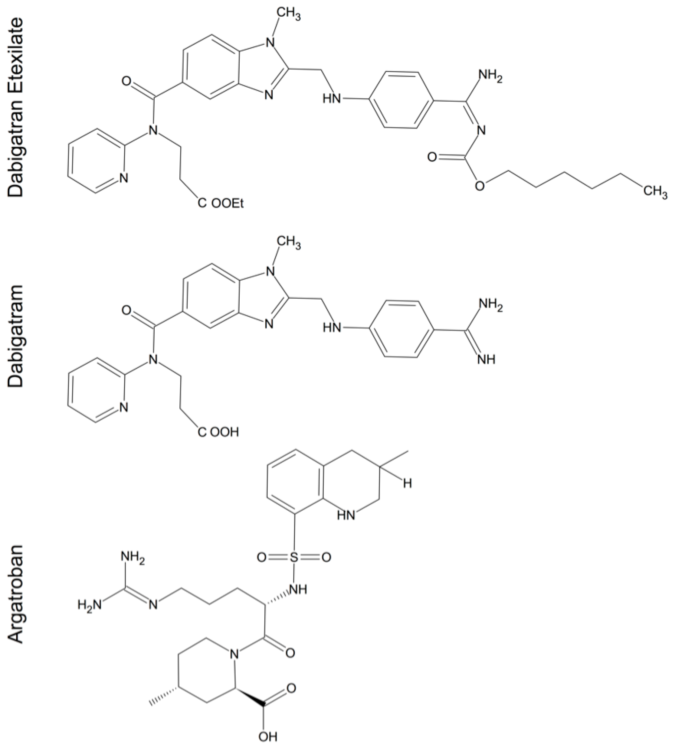
5. Thrombin Inhibitors in Cerebral Injury and Disease

The increasing awareness of thrombin’s involvement in various pathophysiological CNS conditions suggests a potential therapeutic implication. Different pharmacological substances that interfere with thrombin activity and production are available. Vitamin-K antagonists (e.g., warfarin) slow the activation of the coagulation cascade, while heparin acts as a molecular bridge between AT III and thrombin and thus increases the inhibitory potential of AT III. Hirudin acts as a direct thrombin inhibitor that irreversibly binds to the active site of thrombin and blocks its clotting activity [149]. More recently, new direct acting thrombin inhibitors (DTI) with a lower molecular weight, such as argatroban and dabigatran, have been introduced as alternatives to the classic anticoagulants (Figure 5) [150]. The majority of studies are focused on the validation of DTIs in the therapy and prevention of ischemia. A pilot trial, designed using argatroban for its anti-thrombotic property in secondary stroke prevention, has been completed [151,152]. So far, there has been no larger study investigating DTIs for their potential role in alleviating ischemia or haemorrhage induced neuronal loss and secondary clinical deficits.
Still, argatroban has been shown to reduce the latency time, increase the learning curve and reduce primary errors during standardized neurobehavioral testing in rats after unilateral ischemic stroke [153]. A simultaneous blockade of the thrombin and C5a-receptors had a synergistic neuroprotective effect after ICH in mice. The administration of both substances after ICH reduced the occurrence of inflammatory factors and brain oedema, thus leading to improvements in neurofunctional outcome. A small preliminary study reported a reduction of oedema and improved neurological outcome in 4 patients presenting with spontaneous ICH due to hypertension when treated with intravenous argatroban [154]. Furthermore, the use of argatroban after subarachnoid hemorrhage (SAH) in a rabbit model prevents an enhancement and prolongation of the contractile response leading to cerebral vasospasm [155]. Beside that, thrombin inhibition improved the neurological outcome after experimental SAH in rats [156].
As thrombin has been shown to worsen cognitive deficits in AD, the administration of donepezil, a reversible acetylcholinesterase inhibitor used to improve cognition and behaviour, was compared to a simultaneous treatment with donepezil and hirudin. Patients receiving the dual regiment demonstrate improvements in activities of daily living, behaviour, psychological symptoms of dementia as well as cognition compared to those treated with donepezil alone [157]. These findings might be transferrable to patients suffering from CNS disease influenced by thrombin and warrants the necessity of further trials with thrombin inhibitors in these diseases.
Figure 5. 2D chemical structure of common direct acting thrombin inhibitors (DIT). The given structures depict the low molecular weight DTIs: the prodrug dabigatran etexilate, the active principle dabigatran and argatroban [158,159].
Ijms 17 00084 g005

6. Conclusions

During the last decade, a rising number of reports emerged, revealing the influence of thrombin not only for our coagulation system but also for neuronal signalling, development and plasticity. With our increasing knowledge about the importance of thrombin for the maintenance of functionality of the CNS, we simultaneously discover its importance for various neuronal diseases. High concentrations of thrombin seem to contribute to the pathological processes during neurodegenerative diseases, including stroke, haemorrhage, multiple sclerosis, Alzheimer’s- and Parkinson’s disease. Even rare conditions such as HIV-related dementia seem to develop under conditions influenced by thrombin signalling. In contrast to these detrimental effects, low concentrations of thrombin and thrombin preconditioning yield the potential of rescuing cells and to induce survival of neurons and astrocytes exposed to various ischemic insults. The effects of thrombin are largely mediated via PAR-1 and, to a lesser extent, via PAR-3 and PAR-4. Convincing evidence on the distinguished role of each receptor remains scarce. While PAR-1 seems to influence infarct volume, BBB integrity, neuronal damage and motor function in vivo, PAR-3 and PAR-4 are increasingly linked to neurodegenerative and neuroinflammatory diseases. However, conflicting data on the extent and importance of these receptors for neuropathophysiology exists. So far, only little is known about the importance of the cerebral capability to generate thrombin from its precursor prothrombin. Under most circumstances, the origin of the observed thrombin remains elusive. This underlines the dire need of further research to determine the origin of thrombin in the CNS, in order to elucidate its signalling pathways and thus helping to identify future therapeutic targets.

Author Contributions

Harald Krenzlin, Beat Alessandri and Oliver Kempski conceived and designed the review article; Harald Krenzlin, Viola Lorenz and Sven Danckwardt did the literature research and analyzed the data; Harald Krenzlin and Viola Lorenz designed the illustrations; Harald Krenzlin wrote the paper.

Conflicts of Interest

The authors declare no conflict of interest.

Abbreviations

β-APP
amyloid-β precursor protein
amyloid beta
AD
Alzheimer’s disease
AT
antithrombin
BBB
blood-brain-barrier
CDK 4
Cyclin-dependent kinase 4
CINC-1
cytokine-induced neutrophil chemoattractant-1
CNS
central nervous system
DTI
direct acting thrombin inhibitors
EAE
experimental autoimmune encephalitis
ERK1
extracellular signal-regulated protein kinase 1
F
factor
GFAP
glial fibrillary acidic protein
GP
glycoprotein
GRO
growth-regulated oncogene
HIF-1α
hypoxia inducing factor 1α
HIV
human immunodeficiency virus
HIVD
HIV dementia
ICH
intracerebral haemorrhage
IL
interleukin
iNOS
inducible nitric oxide synthase
JNK
N-terminal kinase
MAPK
mitogen- activated protein kinase
MMP-9
matrix metalloproteinase-9
MS
multiple sclerosis
NFTs
neurofibrillary tangles
NMDA
N-methyl-d-aspartate
NO
nitric oxid
OGD
glucose deprivation
6-OHDA
6-hydroxydopamine
PARs
protease-activated receptors
PD
Parkinson’s disease
PI 3-kinase
phosphatidylinositol 3-kinase
PKC
protein kinase C
PN-1
protease nexin-1
PTX
pertussis toxin
RAS
rat sarcoma protein
ROS
reactive oxygen species
SNpc
substantia nigra pars compacta
TNF-α
tumor necrosis factor-α
TPC
thrombin preconditioning
ZO
zonula occludens

References

  1. Fenton, J.W., 2nd. Thrombin. Ann. N. Y. Acad. Sci. 1986, 485, 5–15. [Google Scholar] [CrossRef] [PubMed]
  2. Grand, R.J.; Turnell, A.S.; Grabham, P.W. Cellular consequences of thrombin-receptor activation. Biochem. J. 1996, 313, 353–368. [Google Scholar] [CrossRef] [PubMed]
  3. Strukova, S.M. Role of platelets and serine proteinases in coupling of blood coagulation and inflammation. Biochem. Biokhimiia 2004, 69, 1067–1081. [Google Scholar] [CrossRef]
  4. Ossovskaya, V.S.; Bunnett, N.W. Protease-activated receptors: Contribution to physiology and disease. Physiol. Rev. 2004, 84, 579–621. [Google Scholar] [CrossRef] [PubMed]
  5. Steinhoff, M.; Buddenkotte, J.; Shpacovitch, V.; Rattenholl, A.; Moormann, C.; Vergnolle, N.; Luger, T.A.; Hollenberg, M.D. Proteinase-activated receptors: Transducers of proteinase-mediated signaling in inflammation and immune response. Endocr. Rev. 2005, 26, 1–43. [Google Scholar] [CrossRef] [PubMed]
  6. Bock, P.E.; Olson, S.T.; Bjork, I. Inactivation of thrombin by antithrombin is accompanied by inactivation of regulatory exosite I. J. Biol. Chem. 1997, 272, 19837–19845. [Google Scholar] [CrossRef] [PubMed]
  7. Stubbs, M.T.; Bode, W. The clot thickens: Clues provided by thrombin structure. Trends Biochem. Sci. 1995, 20, 23–28. [Google Scholar] [CrossRef]
  8. Lechtenberg, B.C.; Freund, S.M.; Huntington, J.A. GpIbalpha interacts exclusively with exosite II of thrombin. J. Mol. Biol. 2014, 426, 881–893. [Google Scholar] [CrossRef] [PubMed]
  9. Walker, C.P.; Royston, D. Thrombin generation and its inhibition: A review of the scientific basis and mechanism of action of anticoagulant therapies. Br. J. Anaesth. 2002, 88, 848–863. [Google Scholar] [CrossRef] [PubMed]
  10. Davie, E.W.; Fujikawa, K.; Kisiel, W. The coagulation cascade: Initiation, maintenance, and regulation. Biochemistry 1991, 30, 10363–10370. [Google Scholar] [CrossRef] [PubMed]
  11. Sokolova, E.; Reiser, G. Prothrombin/thrombin and the thrombin receptors PAR-1 and PAR-4 in the brain: Localization, expression and participation in neurodegenerative diseases. Thromb. Haemost. 2008, 100, 576–581. [Google Scholar] [CrossRef] [PubMed]
  12. Deschepper, C.F.; Bigornia, V.; Berens, M.E.; Lapointe, M.C. Production of thrombin and antithrombin III by brain and astroglial cell cultures. Brain Res. Mol. Brain Res. 1991, 11, 355–358. [Google Scholar] [CrossRef]
  13. Niclou, S.P.; Suidan, H.S.; Pavlik, A.; Vejsada, R.; Monard, D. Changes in the expression of protease-activated receptor 1 and protease nexin-1 mRNA during rat nervous system development and after nerve lesion. Eur. J. Neurosci. 1998, 10, 1590–1607. [Google Scholar] [CrossRef] [PubMed]
  14. Dihanich, M.; Kaser, M.; Reinhard, E.; Cunningham, D.; Monard, D. Prothrombin mRNA is expressed by cells of the nervous system. Neuron 1991, 6, 575–581. [Google Scholar] [CrossRef]
  15. Jamison, C.S.; Degen, S.J. Prenatal and postnatal expression of mRNA coding for rat prothrombin. Biochim. Biophys. Acta 1991, 1088, 208–216. [Google Scholar] [CrossRef]
  16. Yamada, T.; Nagai, Y. Immunohistochemical studies of human tissues with antibody to factor Xa. Histochem. J. 1996, 28, 73–77. [Google Scholar] [CrossRef] [PubMed]
  17. Shikamoto, Y.; Morita, T. Expression of factor X in both the rat brain and cells of the central nervous system. FEBS Lett. 1999, 463, 387–389. [Google Scholar] [CrossRef]
  18. Wang, H.; Reiser, G. Thrombin signaling in the brain: The role of protease-activated receptors. Biol. Chem. 2003, 384, 193–202. [Google Scholar] [CrossRef] [PubMed]
  19. Lee, K.R.; Colon, G.P.; Betz, A.L.; Keep, R.F.; Kim, S.; Hoff, J.T. Edema from intracerebral hemorrhage: The role of thrombin. J. Neurosurg. 1996, 84, 91–96. [Google Scholar] [CrossRef] [PubMed]
  20. Lee, K.R.; Kawai, N.; Kim, S.; Sagher, O.; Hoff, J.T. Mechanisms of edema formation after intracerebral hemorrhage: Effects of thrombin on cerebral blood flow, blood-brain barrier permeability, and cell survival in a rat model. J. Neurosurg. 1997, 86, 272–278. [Google Scholar] [CrossRef] [PubMed]
  21. Kalaria, R.N.; Golde, T.; Kroon, S.N.; Perry, G. Serine protease inhibitor antithrombin III and its messenger RNA in the pathogenesis of Alzheimer’s disease. Am. J. Pathol. 1993, 143, 886–893. [Google Scholar] [PubMed]
  22. Walker, D.G.; Yasuhara, O.; Patston, P.A.; McGeer, E.G.; McGeer, P.L. Complement C1 inhibitor is produced by brain tissue and is cleaved in Alzheimer disease. Brain Res. 1995, 675, 75–82. [Google Scholar] [CrossRef]
  23. Mansuy, I.M.; van der Putten, H.; Schmid, P.; Meins, M.; Botteri, F.M.; Monard, D. Variable and multiple expression of Protease Nexin-1 during mouse organogenesis and nervous system development. Development 1993, 119, 1119–1134. [Google Scholar] [PubMed]
  24. Pindon, A.; Hantai, D.; Jandrot-Perrus, M.; Festoff, B.W. Novel expression and localization of active thrombomodulin on the surface of mouse brain astrocytes. Glia 1997, 19, 259–268. [Google Scholar] [CrossRef]
  25. Hua, Y.; Xi, G.; Keep, R.F.; Wu, J.; Jiang, Y.; Hoff, J.T. Plasminogen activator inhibitor-1 induction after experimental intracerebral hemorrhage. J. Cereb. Blood Flow Metab. 2002, 22, 55–61. [Google Scholar] [CrossRef] [PubMed]
  26. Choi, B.H.; Suzuki, M.; Kim, T.; Wagner, S.L.; Cunningham, D.D. Protease nexin-1. Localization in the human brain suggests a protective role against extravasated serine proteases. Am. J. Pathol. 1990, 137, 741–747. [Google Scholar] [PubMed]
  27. Arai, T.; Miklossy, J.; Klegeris, A.; Guo, J.P.; McGeer, P.L. Thrombin and prothrombin are expressed by neurons and glial cells and accumulate in neurofibrillary tangles in Alzheimer disease brain. J. Neuropathol. Exp. Neurol. 2006, 65, 19–25. [Google Scholar] [CrossRef] [PubMed]
  28. Coughlin, S.R. Protease-activated receptors start a family. Proc. Natl. Acad. Sci. USA 1994, 91, 9200–9202. [Google Scholar] [CrossRef] [PubMed]
  29. Coughlin, S.R. Molecular mechanisms of thrombin signaling. Semin. Hematol. 1994, 31, 270–277. [Google Scholar] [PubMed]
  30. Vu, T.K.; Hung, D.T.; Wheaton, V.I.; Coughlin, S.R. Molecular cloning of a functional thrombin receptor reveals a novel proteolytic mechanism of receptor activation. Cell 1991, 64, 1057–1068. [Google Scholar] [CrossRef]
  31. Coughlin, S.R. Thrombin signalling and protease-activated receptors. Nature 2000, 407, 258–264. [Google Scholar] [CrossRef] [PubMed]
  32. Ishihara, H.; Connolly, A.J.; Zeng, D.; Kahn, M.L.; Zheng, Y.W.; Timmons, C.; Tram, T.; Coughlin, S.R. Protease-activated receptor 3 is a second thrombin receptor in humans. Nature 1997, 386, 502–506. [Google Scholar] [CrossRef] [PubMed]
  33. Nystedt, S.; Emilsson, K.; Wahlestedt, C.; Sundelin, J. Molecular cloning of a potential proteinase activated receptor. Proc. Natl. Acad. Sci. USA 1994, 91, 9208–9212. [Google Scholar] [CrossRef] [PubMed]
  34. Wang, H.; Ubl, J.J.; Reiser, G. Four subtypes of protease-activated receptors, co-expressed in rat astrocytes, evoke different physiological signaling. Glia 2002, 37, 53–63. [Google Scholar] [CrossRef] [PubMed]
  35. Macfarlane, S.R.; Seatter, M.J.; Kanke, T.; Hunter, G.D.; Plevin, R. Proteinase-activated receptors. Pharmacol. Rev. 2001, 53, 245–282. [Google Scholar] [PubMed]
  36. Striggow, F.; Riek-Burchardt, M.; Kiesel, A.; Schmidt, W.; Henrich-Noack, P.; Breder, J.; Krug, M.; Reymann, K.G.; Reiser, G. Four different types of protease-activated receptors are widely expressed in the brain and are up-regulated in hippocampus by severe ischemia. Eur. J. Neurosci. 2001, 14, 595–608. [Google Scholar] [CrossRef] [PubMed]
  37. Ubl, J.J.; Vohringer, C.; Reiser, G. Co-existence of two types of [Ca2+]i-inducing protease-activated receptors (PAR-1 and PAR-2) in rat astrocytes and C6 glioma cells. Neuroscience 1998, 86, 597–609. [Google Scholar] [CrossRef]
  38. Weinstein, J.R.; Gold, S.J.; Cunningham, D.D.; Gall, C.M. Cellular localization of thrombin receptor mRNA in rat brain: Expression by mesencephalic dopaminergic neurons and codistribution with prothrombin mRNA. J. Neurosci. 1995, 15, 2906–2919. [Google Scholar] [PubMed]
  39. Wang, H.; Ubl, J.J.; Stricker, R.; Reiser, G. Thrombin (PAR-1)-induced proliferation in astrocytes via MAPK involves multiple signaling pathways. Am. J. Physiol. Cell Physiol. 2002, 283, C1351–C1364. [Google Scholar] [CrossRef] [PubMed]
  40. Sinnreich, M.; Meins, M.; Niclou, S.P.; Suidan, H.S.; Monard, D. Prothrombin overexpressed in post-natal neurones requires blood factors for activation in the mouse brain. J. Neurochem. 2004, 88, 1380–1388. [Google Scholar] [CrossRef] [PubMed]
  41. Cunningham, D.D.; Gurwitz, D. Proteolytic regulation of neurite outgrowth from neuroblastoma cells by thrombin and protease nexin-1. J. Cell. Biochem. 1989, 39, 55–64. [Google Scholar] [CrossRef] [PubMed]
  42. Grabham, P.; Cunningham, D.D. Thrombin receptor activation stimulates astrocyte proliferation and reversal of stellation by distinct pathways: Involvement of tyrosine phosphorylation. J. Neurochem. 1995, 64, 583–591. [Google Scholar] [CrossRef] [PubMed]
  43. Pai, K.S.; Mahajan, V.B.; Lau, A.; Cunningham, D.D. Thrombin receptor signaling to cytoskeleton requires Hsp90. J. Biol. Chem. 2001, 276, 32642–32647. [Google Scholar] [CrossRef] [PubMed]
  44. Jalink, K.; van Corven, E.J.; Hengeveld, T.; Morii, N.; Narumiya, S.; Moolenaar, W.H. Inhibition of lysophosphatidate- and thrombin-induced neurite retraction and neuronal cell rounding by ADP ribosylation of the small GTP-binding protein Rho. J. Cell Biol. 1994, 126, 801–810. [Google Scholar] [CrossRef] [PubMed]
  45. Laskowski, A.; Reiser, G.; Reymann, K.G. Protease-activated receptor-1 induces generation of new microglia in the dentate gyrus of traumatised hippocampal slice cultures. Neurosci. Lett. 2007, 415, 17–21. [Google Scholar] [CrossRef] [PubMed]
  46. Suo, Z.; Wu, M.; Ameenuddin, S.; Anderson, H.E.; Zoloty, J.E.; Citron, B.A.; Andrade-Gordon, P.; Festoff, B.W. Participation of protease-activated receptor-1 in thrombin-induced microglial activation. J. Neurochem. 2002, 80, 655–666. [Google Scholar] [CrossRef] [PubMed]
  47. Debeir, T.; Gueugnon, J.; Vige, X.; Benavides, J. Transduction mechanisms involved in thrombin receptor-induced nerve growth factor secretion and cell division in primary cultures of astrocytes. J. Neurochem. 1996, 66, 2320–2328. [Google Scholar] [CrossRef] [PubMed]
  48. Marshall, C.J. Specificity of receptor tyrosine kinase signaling: Transient versus sustained extracellular signal-regulated kinase activation. Cell 1995, 80, 179–185. [Google Scholar] [CrossRef]
  49. Jalink, K.; Moolenaar, W.H. Thrombin receptor activation causes rapid neural cell rounding and neurite retraction independent of classic second messengers. J. Cell Biol. 1992, 118, 411–419. [Google Scholar] [CrossRef] [PubMed]
  50. Ehrenreich, H.; Costa, T.; Clouse, K.A.; Pluta, R.M.; Ogino, Y.; Coligan, J.E.; Burd, P.R. Thrombin is a regulator of astrocytic endothelin-1. Brain Res. 1993, 600, 201–207. [Google Scholar] [CrossRef]
  51. Vaughan, P.J.; Pike, C.J.; Cotman, C.W.; Cunningham, D.D. Thrombin receptor activation protects neurons and astrocytes from cell death produced by environmental insults. J. Neurosci. 1995, 15, 5389–5401. [Google Scholar] [PubMed]
  52. Gingrich, M.B.; Junge, C.E.; Lyuboslavsky, P.; Traynelis, S.F. Potentiation of NMDA receptor function by the serine protease thrombin. J. Neurosci. 2000, 20, 4582–4595. [Google Scholar] [PubMed]
  53. Gingrich, M.B.; Traynelis, S.F. Serine proteases and brain damage—Is there a link? Trends Neurosci. 2000, 23, 399–407. [Google Scholar] [CrossRef]
  54. Davis-Salinas, J.; Saporito-Irwin, S.M.; Donovan, F.M.; Cunningham, D.D.; van Nostrand, W.E. Thrombin receptor activation induces secretion and nonamyloidogenic processing of amyloid β-protein precursor. J. Biol. Chem. 1994, 269, 22623–22627. [Google Scholar] [PubMed]
  55. Meli, R.; Raso, G.M.; Cicala, C.; Esposito, E.; Fiorino, F.; Cirino, G. Thrombin and PAR-1 activating peptide increase iNOS expression in cytokine-stimulated C6 glioma cells. J. Neurochem. 2001, 79, 556–563. [Google Scholar] [CrossRef] [PubMed]
  56. Moller, T.; Hanisch, U.K.; Ransom, B.R. Thrombin-induced activation of cultured rodent microglia. J. Neurochem. 2000, 75, 1539–1547. [Google Scholar] [CrossRef] [PubMed]
  57. Suo, Z.; Wu, M.; Citron, B.A.; Gao, C.; Festoff, B.W. Persistent protease-activated receptor 4 signaling mediates thrombin-induced microglial activation. J. Biol. Chem. 2003, 278, 31177–31183. [Google Scholar] [CrossRef] [PubMed]
  58. Choi, S.H.; Joe, E.H.; Kim, S.U.; Jin, B.K. Thrombin-induced microglial activation produces degeneration of nigral dopaminergic neurons in vivo. J. Neurosci. 2003, 23, 5877–5886. [Google Scholar] [PubMed]
  59. Choi, S.H.; Lee, D.Y.; Chung, E.S.; Hong, Y.B.; Kim, S.U.; Jin, B.K. Inhibition of thrombin-induced microglial activation and NADPH oxidase by minocycline protects dopaminergic neurons in the substantia nigra in vivo. J. Neurochem. 2005, 95, 1755–1765. [Google Scholar] [CrossRef] [PubMed]
  60. Wang, Y.; Luo, W.; Reiser, G. Activation of protease-activated receptors in astrocytes evokes a novel neuroprotective pathway through release of chemokines of the growth-regulated oncogene/cytokine-induced neutrophil chemoattractant family. Eur. J. Neurosci. 2007, 26, 3159–3168. [Google Scholar] [CrossRef] [PubMed]
  61. Ryu, J.; Pyo, H.; Jou, I.; Joe, E. Thrombin induces NO release from cultured rat microglia via protein kinase C, mitogen-activated protein kinase, and NF-κB. J. Biol. Chem. 2000, 275, 29955–29959. [Google Scholar] [CrossRef] [PubMed]
  62. Li, D.Q.; Zhou, Y.P.; Yang, H. Donepezil combined with natural hirudin improves the clinical symptoms of patients with mild-to-moderate Alzheimer’s disease: A 20-week open-label pilot study. Int. J. Med. Sci. 2012, 9, 248–255. [Google Scholar] [CrossRef] [PubMed]
  63. Akiyama, H.; Ikeda, K.; Kondo, H.; McGeer, P.L. Thrombin accumulation in brains of patients with Alzheimer’s disease. Neurosci. Lett. 1992, 146, 152–154. [Google Scholar] [CrossRef]
  64. Boche, D.; Perry, V.H.; Nicoll, J.A. Review: Activation patterns of microglia and their identification in the human brain. Neuropathol. Appl. Neurobiol. 2013, 39, 3–18. [Google Scholar] [CrossRef] [PubMed]
  65. Striggow, F.; Riek, M.; Breder, J.; Henrich-Noack, P.; Reymann, K.G.; Reiser, G. The protease thrombin is an endogenous mediator of hippocampal neuroprotection against ischemia at low concentrations but causes degeneration at high concentrations. Proc. Natl. Acad. Sci. USA 2000, 97, 2264–2269. [Google Scholar] [CrossRef] [PubMed]
  66. Mirante, O.; Price, M.; Puentes, W.; Castillo, X.; Benakis, C.; Thevenet, J.; Monard, D.; Hirt, L. Endogenous protease nexin-1 protects against cerebral ischemia. Int. J. Mol. Sci. 2013, 14, 16719–16731. [Google Scholar] [CrossRef] [PubMed] [Green Version]
  67. Olanow, C.W.; Tatton, W.G. Etiology and pathogenesis of Parkinson’s disease. Annu. Rev. Neurosci. 1999, 22, 123–144. [Google Scholar] [CrossRef] [PubMed]
  68. Carreno-Muller, E.; Herrera, A.J.; de Pablos, R.M.; Tomás-Camardiel, M.; Venero, J.L.; Cano, J.; Machado, A. Thrombin induces in vivo degeneration of nigral dopaminergic neurones along with the activation of microglia. J. Neurochem. 2003, 84, 1201–1214. [Google Scholar] [CrossRef] [PubMed]
  69. Ishida, Y.; Nagai, A.; Kobayashi, S.; Kim, S.U. Upregulation of protease-activated receptor-1 in astrocytes in Parkinson disease: Astrocyte-mediated neuroprotection through increased levels of glutathione peroxidase. J. Neuropathol. Exp. Neurol. 2006, 65, 66–77. [Google Scholar] [CrossRef] [PubMed]
  70. Katsuki, H.; Okawara, M.; Shibata, H.; Kume, T.; Akaike, A. Nitric oxide-producing microglia mediate thrombin-induced degeneration of dopaminergic neurons in rat midbrain slice culture. J. Neurochem. 2006, 97, 1232–1242. [Google Scholar] [CrossRef] [PubMed] [Green Version]
  71. Lee, D.Y.; Oh, Y.J.; Jin, B.K. Thrombin-activated microglia contribute to death of dopaminergic neurons in rat mesencephalic cultures: Dual roles of mitogen-activated protein kinase signaling pathways. Glia 2005, 51, 98–110. [Google Scholar] [CrossRef] [PubMed]
  72. Choi, S.H.; Lee, D.Y.; Ryu, J.K.; Kim, J.; Joe, E.H.; Jin, B.K. Thrombin induces nigral dopaminergic neurodegeneration in vivo by altering expression of death-related proteins. Neurobiol. Dis. 2003, 14, 181–193. [Google Scholar] [CrossRef]
  73. Jenner, P. Oxidative stress in Parkinson’s disease. Ann. Neurol. 2003, 53, S26–S36. [Google Scholar] [CrossRef] [PubMed]
  74. Lee da, Y.; Park, K.W.; Jin, B.K. Thrombin induces neurodegeneration and microglial activation in the cortex in vivo and in vitro: Proteolytic and non-proteolytic actions. Biochem. Biophys. Res. Commun. 2006, 346, 727–738. [Google Scholar] [CrossRef] [PubMed]
  75. Cannon, J.R.; Keep, R.F.; Hua, Y.; Richardson, R.J.; Schallert, T.; Xi, G. Thrombin preconditioning provides protection in a 6-hydroxydopamine Parkinson’s disease model. Neurosci. Lett. 2005, 373, 189–194. [Google Scholar] [CrossRef] [PubMed]
  76. Cannon, J.R.; Keep, R.F.; Schallert, T.; Hua, Y.; Richardson, R.J.; Xi, G. Protease-activated receptor-1 mediates protection elicited by thrombin preconditioning in a rat 6-hydroxydopamine model of Parkinson’s disease. Brain Res. 2006, 1116, 177–186. [Google Scholar] [CrossRef] [PubMed]
  77. Herrera, A.J.; de Pablos, R.M.; Carreño-Müller, E.; Villarán, R.F.; Venero, J.L.; Tomás-Camardiel, M.; Cano, J.; Machado, A. The intrastriatal injection of thrombin in rat induced a retrograde apoptotic degeneration of nigral dopaminergic neurons through synaptic elimination. J. Neurochem. 2008, 105, 750–762. [Google Scholar] [CrossRef] [PubMed]
  78. Cannon, J.R.; Hua, Y.; Richardson, R.J.; Xi, G.; Keep, R.F.; Schallert, T. The effect of thrombin on a 6-hydroxydopamine model of Parkinson’s disease depends on timing. Behav. Brain Res. 2007, 183, 161–168. [Google Scholar] [CrossRef] [PubMed]
  79. Inoue, H.; Udaka, F.; Takahashi, M.; Nishinaka, K.; Kameyama, M. Secondary parkinsonism following midbrain hemorrhage. Clin. Neurol. 1997, 37, 266–269. [Google Scholar]
  80. Grammas, P.; Samany, P.G.; Thirumangalakudi, L. Thrombin and inflammatory proteins are elevated in Alzheimer’s disease microvessels: Implications for disease pathogenesis. J. Alzheimer Dis. 2006, 9, 51–58. [Google Scholar]
  81. Pompili, E.; Nori, S.L.; Geloso, M.C.; Guadagni, E.; Corvino, V.; Michetti, F.; Fumagalli, L. Trimethyltin-induced differential expression of PAR subtypes in reactive astrocytes of the rat hippocampus. Brain Res. Mol. Brain Res. 2004, 122, 93–98. [Google Scholar] [CrossRef] [PubMed]
  82. Vaughan, P.J.; Su, J.; Cotman, C.W.; Cunningham, D.D. Protease nexin-1, a potent thrombin inhibitor, is reduced around cerebral blood vessels in Alzheimer’s disease. Brain Res. 1994, 668, 160–170. [Google Scholar] [CrossRef]
  83. Suo, Z.; Wu, M.; Citron, B.A.; Palazzo, R.E.; Festoff, B.W. Rapid tau aggregation and delayed hippocampal neuronal death induced by persistent thrombin signaling. J. Biol. Chem. 2003, 278, 37681–37689. [Google Scholar] [CrossRef] [PubMed]
  84. Brewer, G.J. Thrombin causes cell spreading and redistribution of beta-amyloid immunoreactivity in cultured hippocampal neurons. J. Neurochem. 1996, 67, 119–130. [Google Scholar] [CrossRef] [PubMed]
  85. Smith-Swintosky, V.L.; Zimmer, S.; Fenton, J.W., 2nd; Mattson, M.P. Opposing actions of thrombin and protease nexin-1 on amyloid β-peptide toxicity and on accumulation of peroxides and calcium in hippocampal neurons. J. Neurochem. 1995, 65, 1415–1418. [Google Scholar] [CrossRef] [PubMed]
  86. Pike, C.J.; Vaughan, P.J.; Cunningham, D.D.; Cotman, C.W. Thrombin attenuates neuronal cell death and modulates astrocyte reactivity induced by β-amyloid in vitro. J. Neurochem. 1996, 66, 1374–1382. [Google Scholar] [CrossRef] [PubMed]
  87. Choi, S.H.; Lee, D.Y.; Kim, S.U.; Jin, B.K. Thrombin-induced oxidative stress contributes to the death of hippocampal neurons in vivo: Role of microglial NADPH oxidase. J. Neurosci. 2005, 25, 4082–4090. [Google Scholar] [CrossRef] [PubMed]
  88. Thirumangalakudi, L.; Samany, P.G.; Owoso, A.; Wiskar, B.; Grammas, P. Angiogenic proteins are expressed by brain blood vessels in Alzheimer’s disease. J. Alzheimer Dis. 2006, 10, 111–118. [Google Scholar]
  89. Tripathy, D.; Sanchez, A.; Yin, X.; Luo, J.; Martinez, J.; Grammas, P. Thrombin, a mediator of cerebrovascular inflammation in AD and hypoxia. Front. Aging Neurosci. 2013, 5, 19. [Google Scholar] [CrossRef] [PubMed]
  90. Van der Poll, T.; Büller, H.R.; ten Cate, H.; Wortel, C.H.; Bauer, K.A.; van Deventer, S.J.; Hack, C.E.; Sauerwein, H.P.; Rosenberg, R.D.; ten Cate, J.W. Activation of coagulation after administration of tumor necrosis factor to normal subjects. N. Engl. J. Med. 1990, 322, 1622–1627. [Google Scholar] [CrossRef] [PubMed]
  91. Paterson, P.Y.; Koh, C.S.; Kwaan, H.C. Role of the clotting system in the pathogenesis of neuroimmunologic disease. Fed. Proc. 1987, 46, 91–96. [Google Scholar] [PubMed]
  92. Kwon, E.E.; Prineas, J.W. Blood-brain barrier abnormalities in longstanding multiple sclerosis lesions. An immunohistochemical study. J. Neuropathol. Exp. Neurol. 1994, 53, 625–636. [Google Scholar] [CrossRef] [PubMed]
  93. Davalos, D.; Ryu, J.K.; Merlini, M.; Baeten, K.M.; Le Moan, N.; Petersen, M.A.; Deerinck, T.J.; Smirnoff, D.S.; Bedard, C.; Hakozaki, H.; et al. Fibrinogen-induced perivascular microglial clustering is required for the development of axonal damage in neuroinflammation. Nat. Commun. 2012, 3, 1227. [Google Scholar] [CrossRef] [PubMed]
  94. Boven, L.A.; Vergnolle, N.; Henry, S.D.; Silva, C.; Imai, Y.; Holden, J.; Warren, K.; Hollenberg, M.D.; Power, C. Up-regulation of proteinase-activated receptor 1 expression in astrocytes during HIV encephalitis. J. Immunol. 2003, 170, 2638–2646. [Google Scholar] [CrossRef] [PubMed]
  95. Inaba, Y.; Ichikawa, M.; Inoue, A.; Itoh, M.; Kyogashima, M.; Sekiguchi, Y.; Nakamura, S.; Komiyama, A.; Koh, C. Plasma thrombin-antithrombin III complex is associated with the severity of experimental autoimmune encephalomyelitis. J. Neurol. Sci. 2001, 185, 89–93. [Google Scholar] [CrossRef]
  96. Davalos, D.; Baeten, K.M.; Whitney, M.A.; Mullins, E.S.; Friedman, B.; Olson, E.S.; Ryu, J.K.; Smirnoff, D.S.; Petersen, M.A.; Bedard, C.; et al. Early detection of thrombin activity in neuroinflammatory disease. Ann. Neurol. 2014, 75, 303–308. [Google Scholar] [CrossRef] [PubMed]
  97. Wang, Y.; Richter-Landsberg, C.; Reiser, G. Expression of protease-activated receptors (PARs) in OLN-93 oligodendroglial cells and mechanism of PAR-1-induced calcium signaling. Neuroscience 2004, 126, 69–82. [Google Scholar] [CrossRef] [PubMed]
  98. Noorbakhsh, F.; Tsutsui, S.; Vergnolle, N.; Boven, L.A.; Shariat, N.; Vodjgani, M.; Warren, K.G.; Andrade-Gordon, P.; Hollenberg, M.D.; Power, C. Proteinase-activated receptor 2 modulates neuroinflammation in experimental autoimmune encephalomyelitis and multiple sclerosis. J. Exp. Med. 2006, 203, 425–435. [Google Scholar] [CrossRef] [PubMed]
  99. Juhler, M.; Barry, D.I.; Offner, H.; Konat, G.; Klinken, L.; Paulson, O.B. Blood-brain and blood-spinal cord barrier permeability during the course of experimental allergic encephalomyelitis in the rat. Brain Res. 1984, 302, 347–355. [Google Scholar] [CrossRef]
  100. Kim, H.N.; Kim, Y.R.; Ahn, S.M.; Lee, S.K.; Shin, H.K.; Choi, B.T. Protease activated receptor-1 antagonist ameliorates the clinical symptoms of experimental autoimmune encephalomyelitis via inhibiting breakdown of blood brain barrier. J. Neurochem. 2015. [Google Scholar] [CrossRef] [PubMed]
  101. Noorbakhsh, F.; Vergnolle, N.; McArthur, J.C.; Silva, C.; Vodjgani, M.; Andrade-Gordon, P.; Hollenberg, M.D.; Power, C. Proteinase-activated receptor-2 induction by neuroinflammation prevents neuronal death during HIV infection. J. Immunol. 2005, 174, 7320–7329. [Google Scholar] [CrossRef] [PubMed]
  102. Kim, W.; Zekas, E.; Lodge, R.; Susan-Resiga, D.; Marcinkiewicz, E.; Essalmani, R.; Mihara, K.; Ramachandran, R.; Asahchop, E.; Gelman, B.; et al. Neuroinflammation-induced interactions between protease-activated receptor 1 and proprotein convertases in HIV-associated neurocognitive disorder. Mol. Cell. Biol. 2015. [Google Scholar] [CrossRef] [PubMed]
  103. Hurley, A.; Smith, M.; Karpova, T.; Hasley, R.B.; Belkina, N.; Shaw, S.; Balenga, N.; Druey, K.M.; Nickel, E.; Packard, B.; et al. Enhanced effector function of CD8(+) T cells from healthy controls and HIV-infected patients occurs through thrombin activation of protease-activated receptor 1. J. Infect. Dis. 2013, 207, 638–650. [Google Scholar] [CrossRef] [PubMed]
  104. Ling, H.; Xiao, P.; Usami, O.; Hattori, T. Thrombin activates envelope glycoproteins of HIV type 1 and enhances fusion. Microbes Infect. 2004, 6, 414–420. [Google Scholar] [CrossRef] [PubMed]
  105. Baker, J.V.; Brummel-Ziedins, K.; Neuhaus, J.; Duprez, D.; Cummins, N.; Dalmau, D.; DeHovitz, J.; Lehmann, C.; Sullivan, A.; Woolley, I.; et al. HIV replication alters the composition of extrinsic pathway coagulation factors and increases thrombin generation. J. Am. Heart Assoc. 2013, 2, e000264. [Google Scholar] [CrossRef] [PubMed]
  106. Ivey, N.S.; MacLean, A.G.; Lackner, A.A. Acquired immunodeficiency syndrome and the blood-brain barrier. J. Neurovirol. 2009, 15, 111–122. [Google Scholar] [CrossRef] [PubMed]
  107. Hsue, P.Y.; Scherzer, R.; Grunfeld, C.; Nordstrom, S.M.; Schnell, A.; Kohl, L.P.; Nitta, E.; Martin, J.N.; Deeks, S.G.; Weiss, E.J. HIV infection is associated with decreased thrombin generation. Clin. Infect. Dis. 2012, 54, 1196–1203. [Google Scholar] [CrossRef] [PubMed]
  108. Singer, E.J.; Valdes-Sueiras, M.; Commins, D.L.; Yong, W.; Carlson, M. HIV stroke risk: Evidence and implications. Ther. Adv. Chronic Dis. 2013, 4, 61–70. [Google Scholar] [CrossRef] [PubMed]
  109. Qureshi, A.I.; Mendelow, A.D.; Hanley, D.F. Intracerebral haemorrhage. Lancet 2009, 373, 1632–1644. [Google Scholar] [CrossRef]
  110. Riek-Burchardt, M.; Striggow, F.; Henrich-Noack, P.; Reiser, G.; Reymann, K.G. Increase of prothrombin-mRNA after global cerebral ischemia in rats, with constant expression of protease nexin-1 and protease-activated receptors. Neurosci. Lett. 2002, 329, 181–184. [Google Scholar] [CrossRef]
  111. Thevenet, J.; Angelillo-Scherrer, A.; Price, M.; Hirt, L. Coagulation factor Xa activates thrombin in ischemic neural tissue. J. Neurochem. 2009, 111, 828–836. [Google Scholar] [CrossRef] [PubMed]
  112. Rohatgi, T.; Henrich-Noack, P.; Sedehizade, F.; Goertler, M.; Wallesch, C.W.; Reymann, K.G.; Reiser, G. Transient focal ischemia in rat brain differentially regulates mRNA expression of protease-activated receptors 1 to 4. J. Neurosci. Res. 2004, 75, 273–279. [Google Scholar] [CrossRef] [PubMed]
  113. Henrich-Noack, P.; Riek-Burchardt, M.; Baldauf, K.; Reiser, G.; Reymann, K.G. Focal ischemia induces expression of protease-activated receptor1 (PAR1) and PAR3 on microglia and enhances PAR4 labeling in the penumbra. Brain Res. 2006, 1070, 232–241. [Google Scholar] [CrossRef] [PubMed]
  114. Wu, X.; Zhang, W.; Li, J.Y.; Chai, B.X.; Peng, J.; Wang, H.; Mulholland, M.W. Induction of apoptosis by thrombin in the cultured neurons of dorsal motor nucleus of the vagus. Neurogastroenterol. Motil. 2011, 23, 279–285. [Google Scholar] [CrossRef] [PubMed]
  115. Cunningham, D.D.; Pulliam, L.; Vaughan, P.J. Protease nexin-1 and thrombin: Injury-related processes in the brain. Thromb. Haemost. 1993, 70, 168–171. [Google Scholar] [PubMed]
  116. Hoffmann, M.C.; Nitsch, C.; Scotti, A.L.; Reinhard, E.; Monard, D. The prolonged presence of glia-derived nexin, an endogenous protease inhibitor, in the hippocampus after ischemia-induced delayed neuronal death. Neuroscience 1992, 49, 397–408. [Google Scholar] [CrossRef]
  117. Xi, G.; Keep, R.F.; Hua, Y.; Xiang, J.; Hoff, J.T. Attenuation of thrombin-induced brain edema by cerebral thrombin preconditioning. Stroke 1999, 30, 1247–1255. [Google Scholar] [CrossRef] [PubMed]
  118. Henrich-Noack, P.; Striggow, F.; Reiser, G.; Reymann, K.G. Preconditioning with thrombin can be protective or worsen damage after endothelin-1-induced focal ischemia in rats. J. Neurosci. Res. 2006, 83, 469–475. [Google Scholar] [CrossRef] [PubMed]
  119. Masada, T.; Xi, G.; Hua, Y.; Keep, R.F. The effects of thrombin preconditioning on focal cerebral ischemia in rats. Brain Res. 2000, 867, 173–179. [Google Scholar] [CrossRef]
  120. Jiang, Y.; Wu, J.; Hua, Y.; Keep, R.F.; Xiang, J.; Hoff, J.T.; Xi, G. Thrombin-receptor activation and thrombin-induced brain tolerance. J. Cereb. Blood Flow Metab. 2002, 22, 404–410. [Google Scholar] [CrossRef] [PubMed]
  121. Karabiyikoglu, M.; Hua, Y.; Keep, R.F.; Ennis, S.R.; Xi, G. Intracerebral hirudin injection attenuates ischemic damage and neurologic deficits without altering local cerebral blood flow. J. Cereb. Blood Flow Metab. 2004, 24, 159–166. [Google Scholar] [CrossRef] [PubMed]
  122. Granziera, C.; Thevenet, J.; Price, M.; Wiegler, K.; Magistretti, P.J.; Badaut, J.; Hirt, L. Thrombin-induced ischemic tolerance is prevented by inhibiting c-jun N-terminal kinase. Brain Res. 2007, 1148, 217–225. [Google Scholar] [CrossRef] [PubMed]
  123. Yang, S.; Hua, Y.; Nakamura, T.; Keep, R.F.; Xi, G. Up-regulation of brain ceruloplasmin in thrombin preconditioning. Acta Neurochir. Suppl. 2006, 96, 203–206. [Google Scholar] [PubMed]
  124. Wang, Y.; Luo, W.; Stricker, R.; Reiser, G. Protease-activated receptor-1 protects rat astrocytes from apoptotic cell death via JNK-mediated release of the chemokine GRO/CINC-1. J. Neurochem. 2006, 98, 1046–1060. [Google Scholar] [CrossRef] [PubMed]
  125. Xi, G.; Reiser, G.; Keep, R.F. The role of thrombin and thrombin receptors in ischemic, hemorrhagic and traumatic brain injury: Deleterious or protective? J. Neurochem. 2003, 84, 3–9. [Google Scholar] [CrossRef] [PubMed]
  126. Fujimoto, S.; Katsuki, H.; Kume, T.; Akaike, A. Thrombin-induced delayed injury involves multiple and distinct signaling pathways in the cerebral cortex and the striatum in organotypic slice cultures. Neurobiol. Dis. 2006, 22, 130–142. [Google Scholar] [CrossRef] [PubMed] [Green Version]
  127. Hua, Y.; Keep, R.F.; Schallert, T.; Hoff, J.T.; Xi, G. A thrombin inhibitor reduces brain edema, glioma mass and neurological deficits in a rat glioma model. Acta Neurochir. Suppl. 2003, 86, 503–506. [Google Scholar] [PubMed]
  128. Hua, Y.; Wu, J.; Keep, R.F.; Hoff, J.T.; Xi, G. Thrombin exacerbates brain edema in focal cerebral ischemia. Acta Neurochir. Suppl. 2003, 86, 163–166. [Google Scholar] [PubMed]
  129. Hua, Y.; Wu, J.; Keep, R.F.; Nakamura, T.; Hoff, J.T.; Xi, G. Tumor necrosis factor-alpha increases in the brain after intracerebral hemorrhage and thrombin stimulation. Neurosurgery 2006, 58, 542–550. [Google Scholar] [CrossRef] [PubMed]
  130. Trendelenburg, G.; Dirnagl, U. Neuroprotective role of astrocytes in cerebral ischemia: Focus on ischemic preconditioning. Glia 2005, 50, 307–320. [Google Scholar] [CrossRef] [PubMed]
  131. Coughlin, S.R.; Camerer, E. PARticipation in inflammation. J. Clin. Investig. 2003, 111, 25–27. [Google Scholar] [CrossRef] [PubMed]
  132. Masada, T.; Hua, Y.; Xi, G.; Yang, G.Y.; Hoff, J.T.; Keep, R.F.; Nagao, S. Overexpression of interleukin-1 receptor antagonist reduces brain edema induced by intracerebral hemorrhage and thrombin. Acta Neurochir. Suppl. 2003, 86, 463–467. [Google Scholar] [PubMed]
  133. Nakamura, T.; Xi, G.; Park, J.W.; Hua, Y.; Hoff, J.T.; Keep, R.F. Holo-transferrin and thrombin can interact to cause brain damage. Stroke 2005, 36, 348–352. [Google Scholar] [CrossRef] [PubMed]
  134. Xi, G.; Keep, R.F.; Hua, Y.; Hoff, J.T. Thrombin preconditioning, heat shock proteins and thrombin-induced brain edema. Acta Neurochir. Suppl. 2000, 76, 511–515. [Google Scholar] [PubMed]
  135. Hua, Y.; Keep, R.F.; Hoff, J.T.; Xi, G. Brain injury after intracerebral hemorrhage: The role of thrombin and iron. Stroke 2007, 38, 759–762. [Google Scholar] [CrossRef] [PubMed]
  136. Svedin, P.; Hagberg, H.; Savman, K.; Zhu, C.; Mallard, C. Matrix metalloproteinase-9 gene knock-out protects the immature brain after cerebral hypoxia-ischemia. J. Neurosci. 2007, 27, 1511–1518. [Google Scholar] [CrossRef] [PubMed]
  137. Xue, M.; Hollenberg, M.D.; Yong, V.W. Combination of thrombin and matrix metalloproteinase-9 exacerbates neurotoxicity in cell culture and intracerebral hemorrhage in mice. J. Neurosci. 2006, 26, 10281–10291. [Google Scholar] [CrossRef] [PubMed]
  138. Junge, C.E.; Sugawara, T.; Mannaioni, G.; Alagarsamy, S.; Conn, P.J.; Brat, D.J.; Chan, P.H.; Traynelis, S.F. The contribution of protease-activated receptor 1 to neuronal damage caused by transient focal cerebral ischemia. Proc. Natl. Acad. Sci. USA 2003, 100, 13019–13024. [Google Scholar] [CrossRef] [PubMed]
  139. Olson, E.E.; Lyuboslavsky, P.; Traynelis, S.F.; McKeon, R.J. PAR-1 deficiency protects against neuronal damage and neurologic deficits after unilateral cerebral hypoxia/ischemia. J. Cereb. Blood Flow Metab. 2004, 24, 964–971. [Google Scholar] [CrossRef] [PubMed]
  140. Ramos-Mandujano, G.; Vazquez-Juarez, E.; Hernandez-Benitez, R.; Pasantes-Morales, H. Thrombin potently enhances swelling-sensitive glutamate efflux from cultured astrocytes. Glia 2007, 55, 917–925. [Google Scholar] [CrossRef] [PubMed]
  141. Liu, S.J.; Zukin, R.S. Ca2+-permeable AMPA receptors in synaptic plasticity and neuronal death. Trends Neurosci. 2007, 30, 126–134. [Google Scholar] [CrossRef] [PubMed]
  142. Kwak, S.; Weiss, J.H. Calcium-permeable AMPA channels in neurodegenerative disease and ischemia. Curr. Opin. Neurobiol. 2006, 16, 281–287. [Google Scholar] [CrossRef] [PubMed]
  143. Gorbacheva, L.R.; Storozhevykh, T.P.; Pinelis, V.G.; Ishiwata, S.; Strukova, S.M. Modulation of hippocampal neuron survival by thrombin and factor Xa. Biochem. Biokhimiia 2006, 71, 1082–1089. [Google Scholar] [CrossRef]
  144. Putcha, G.V.; Moulder, K.L.; Golden, J.P.; Bouillet, P.; Adams, J.A.; Strasser, A.; Johnson, E.M. Induction of BIM, a proapoptotic BH3-only BCL-2 family member, is critical for neuronal apoptosis. Neuron 2001, 29, 615–628. [Google Scholar] [CrossRef]
  145. Rao, H.V.; Thirumangalakudi, L.; Desmond, P.; Grammas, P. Cyclin D1, cdk4, and Bim are involved in thrombin-induced apoptosis in cultured cortical neurons. J. Neurochem. 2007, 101, 498–505. [Google Scholar] [CrossRef] [PubMed]
  146. Rashidian, J.; Iyirhiaro, G.; Aleyasin, H.; Rios, M.; Vincent, I.; Callaghan, S.; Bland, R.J.; Slack, R.S.; During, M.J.; Park, D.S. Multiple cyclin-dependent kinases signals are critical mediators of ischemia/hypoxic neuronal death in vitro and in vivo. Proc. Natl. Acad. Sci. USA 2005, 102, 14080–14085. [Google Scholar] [CrossRef] [PubMed]
  147. Nicole, O.; Goldshmidt, A.; Hamill, C.E.; Sorensen, S.D.; Sastre, A.; Lyuboslavsky, P.; Hepler, J.R.; McKeon, R.J.; Traynelis, S.F. Activation of protease-activated receptor-1 triggers astrogliosis after brain injury. J. Neurosci. 2005, 25, 4319–4329. [Google Scholar] [CrossRef] [PubMed]
  148. Ohnishi, M.; Katsuki, H.; Fujimoto, S.; Takagi, M.; Kume, T.; Akaike, A. Involvement of thrombin and mitogen-activated protein kinase pathways in hemorrhagic brain injury. Exp. Neurol. 2007, 206, 43–52. [Google Scholar] [CrossRef] [PubMed] [Green Version]
  149. Markwardt, F. Hirudin as alternative anticoagulant—A historical review. Semin. Thromb. Hemost. 2002, 28, 405–414. [Google Scholar] [CrossRef] [PubMed]
  150. Ten Cate, H. Challenging the anticoagulant paradigm? J. Thromb. Haemost. 2015. [Google Scholar] [CrossRef] [PubMed]
  151. Sugg, R.M.; Pary, J.K.; Uchino, K.; Baraniuk, S.; Shaltoni, H.M.; Gonzales, N.R.; Mikulik, R.; Garami, Z.; Shaw, S.G.; Matherne, D.E.; et al. Argatroban tPA stroke study: Study design and results in the first treated cohort. Arch. Neurol. 2006, 63, 1057–1062. [Google Scholar] [CrossRef] [PubMed]
  152. Barreto, A.D.; Alexandrov, A.V.; Lyden, P.; Lee, J.; Martin-Schild, S.; Shen, L.; Wu, T.C.; Sisson, A.; Pandurengan, R.; Chen, Z. The argatroban and tissue-type plasminogen activator stroke study: Final results of a pilot safety study. Stroke 2012, 43, 770–775. [Google Scholar] [CrossRef] [PubMed]
  153. Lyden, P.; Pereira, B.; Chen, B.; Zhao, L.; Lamb, J.; Lei, I.F.; Rajput, P. Direct thrombin inhibitor argatroban reduces stroke damage in 2 different models. Stroke 2014, 45, 896–899. [Google Scholar] [CrossRef] [PubMed]
  154. Matsuoka, H.; Hamada, R. Role of thrombin in CNS damage associated with intracerebral haemorrhage: Opportunity for pharmacological intervention? CNS Drugs 2002, 16, 509–516. [Google Scholar] [CrossRef] [PubMed]
  155. Kameda, K.; Kikkawa, Y.; Hirano, M.; Matsuo, S.; Sasaki, T.; Hirano, K. Combined argatroban and anti-oxidative agents prevents increased vascular contractility to thrombin and other ligands after subarachnoid haemorrhage. Br. J. Pharmacol. 2012, 165, 106–119. [Google Scholar] [CrossRef] [PubMed]
  156. Sugawara, T.; Jadhav, V.; Ayer, R.; Chen, W.; Suzuki, H.; Zhang, J.H. Thrombin inhibition by argatroban ameliorates early brain injury and improves neurological outcomes after experimental subarachnoid hemorrhage in rats. Stroke 2009, 40, 1530–1532. [Google Scholar] [CrossRef] [PubMed]
  157. Mhatre, M.; Hensley, K.; Nguyen, A.; Grammas, P. Chronic thrombin exposure results in an increase in apolipoprotein-E levels. J. Neurosci. Res. 2006, 84, 444–449. [Google Scholar] [CrossRef] [PubMed]
  158. National Center for Biotechnology Information. PubChem Compound Database. CID=216210. Available online: https://pubchem.ncbi.nlm.nih.gov/compound/216210 (accessed on 23 November 2015).
  159. Eisert, W.G.; Hauel, N.; Stangier, J.; Wienen, W.; Clemens, A.; van Ryn, J. Dabigatran: An oral novel potent reversible nonpeptide inhibitor of thrombin. Arterioscler. Thromb. Vasc. Biol. 2010, 30, 1885–1889. [Google Scholar] [CrossRef] [PubMed]

