Article

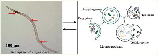
Identification of Autophagy in the Pine Wood Nematode Bursaphelenchus xylophilus and the Molecular Characterization and Functional Analysis of Two Novel Autophagy-Related Genes, BxATG1 and BxATG8

1
Co-Innovation Center for Sustainable Forestry in Southern China, College of Forestry, Nanjing Forestry University, Nanjing 210037, Jiangsu, China
2
Jiangsu Key Laboratory for Prevention and Management of Invasive Species, Nanjing Forestry University, Nanjing 210037, Jiangsu, China
3
Yancheng Institute of Technology, School of Ocean and Biological Engineering, Yancheng 224051, Jiangsu, China
*
Author to whom correspondence should be addressed.
Int. J. Mol. Sci. 2016, 17(3), 279; https://doi.org/10.3390/ijms17030279
Submission received: 9 January 2016 / Revised: 4 February 2016 / Accepted: 14 February 2016 / Published: 3 March 2016
(This article belongs to the Section Biochemistry)

Abstract

:
The pine wood nematode, Bursaphelenchus xylophilus, causes huge economic losses in pine forests, has a complex life cycle, and shows the remarkable ability to survive under unfavorable and changing environmental conditions. This ability may be related to autophagy, which is still poorly understood in B. xylophilus and no autophagy-related genes have been previously characterized. In this study, transmission electron microscopy was used to confirm that autophagy exists in B. xylophilus. The full-length cDNAs of BxATG1 and BxATG8 were first cloned from B. xylophilus, and BxATG1 and BxATG8 were characterized using bioinformatics methods. The expression pattern of the autophagy marker BxATG8 was investigated using in situ hybridization (ISH). BxATG8 was expressed in esophageal gland and hypodermal seam cells. We tested the effects of RNA interference (RNAi) on BxATG1 and BxATG8. The results revealed that BxATG1 and BxATG8 were likely associated with propagation of nematodes on fungal mats. This study confirmed the molecular characterization and functions of BxATG1 and BxATG8 in B. xylophilus and provided fundamental information between autophagy and B. xylophilus.

Graphical Abstract

1. Introduction

Bursaphelenchus xylophilus (Steiner & Buhrer) Nickle is the pine wood nematode that is the causal agent of pine wilt disease (PWD), which results in large economic losses [1]. B. xylophilus is native to North America [2] but has been introduced to, and spread throughout, many parts of the world, including Asia and Europe, including Japan, China, South Korea and Portugal [3,4,5]. It has become a severe threat to pine forests worldwide [4,5,6]. At present, there are many different hypotheses to explain the pathogenesis of PWD, such as the cellulose (which suggests that the destruction of pine cells is triggered by cell wall-degrading enzymes, such as cellulose), phytotoxin and terpenoid hypotheses [7,8,9], but the pathogenic mechanism of B. xylophilus remains unknown.
B. xylophilus is a pathogenic nematode with a complex life cycle and occurs in two phases—dispersal and propagation [10]. Under unfavorable environmental conditions, such as limited food and cooler temperatures, the second-stage propagative juvenile molts into the third-stage dispersal juvenile, then they molt into specialized dispersal-stage dauer juvenile [10,11]. B. xylophilus shows a remarkable adaptability to changing environmental conditions, but the mechanism behind this adaptability is still not well understood. Under conditions of high population density, limited food or increased temperature, Caenorhabditis elegans nematodes can induce the process of autophagy [12,13]. C. elegans is used as a model organism and provides a wealth of information for research on other nematodes. Does the process of autophagy exist in B. xylophilus? Does autophagy assist the nematodes’ responses to various changing environmental conditions and allow them to invade pine trees successfully?
Autophagy exists widely in eukaryotic organisms and is an evolutionarily conserved process [14,15], in which protein and organelles are sequestered within double membrane vesicles that deliver the contents to the lysosome/vacuole for degradation and the recycling of the resultant macromolecules [16]. Autophagy is the major cellular pathway for the degradation of long-lived proteins and cytoplasmic organelles. It involves the rearrangement of subcellular membranes to sequester cargo for delivery to the lysosome where the sequestered material is degraded and recycled [14]. It has a greater variety of physiological and pathophysiological roles than expected, such as starvation adaptation, intracellular clearance, development, anti-aging, degradation of invading bacteria and cell death [15,17,18]. Recently, the role of autophagy was confirmed in pathogens and insect pests, such as Magnaporthe grisea, Tenebrio molitor and Rhipicephalus (Boophilus) microplus [19,20,21,22], and it plays an important role in their growth, development, reproduction and pathogenicity. Whether the autophagy of B. xylophilus is associated with adaptability to changing environmental conditions, vitality, reproduction, invasiveness and pathogenicity is still unknown. Therefore, insights into the characteristics of autophagy and its functions in B. xylophilus may help in better understanding the biological adaptation and pathogenic mechanisms.
An objective of this study is to show that autophagy exists in B. xylophilus using transmission electron microscopy (TEM). TEM is a very reliable approach to analyzing and quantifying autophagic compartments. TEM allows the visualization of every step of the autophagic pathway [23]. The genes responsible for autophagy were first characterized in the yeast Saccharomyces cerevisiae [24]. Out of the many ATG gene nucleotide sequences of eukaryotic organisms, from yeast to mammals [15,21,24], we were particularly interested in ATG1 and ATG8 because the ATG1 product plays an essential role in the regulation of autophagy [19,25], and the ATG8 product performs an important role in the formation of double-membrane autophagosomes, a central step in the intracellular degradation pathway of autophagy, which is routinely used as a marker when studying autophagy [20,26]. Our study sought to clone two novel autophagy-related genes, ATG1 and ATG8, in B. xylophilus, named BxATG1 and BxATG8, respectively. The serine/threonine kinase ATG1 plays an essential role in stimulating autophagy, however, autophagy is a process, and the localization of BxATG8 allows us to track autophagosomes from their initiation in the cytoplasm to their degradation inside the vacuole. Thus, we assessed the functions of autophagy in B. xylophilus using in situ hybridization (ISH) to investigate the localization of BxATG8 expression. RNA interference (RNAi) was used to assess the functions of BxATG1 and BxATG8. The role of the autophagy genes BxATG1 and BxATG8 in development and reproduction through the turnover of organelles and proteins forms an attractive topic for research and is the focus of this paper.

2. Results

2.1. Qualitative Identification of Autophagy in B. xylophilus by Transmission Electron Microscopy (TEM)

TEM was used to identify autophagy in B. xylophilus. Many types of autophagic vacuoles of B. xylophilus are shown in Figure 1. The initial form is an autophagic body delineated by double-membranes (Figure 1A). Autophagosomes, which are characteristic features of the sequestering membrane are liable to be split into myelinated structures (Figure 1B). Autophagosomes that fuse with lysosomes degrade the content resulting in only clumps of the dense material (Figure 1C). The breakdown of the vesicle membrane allows the degradation of its cargo and the eventual recycling of the amino acids (Figure 1D). The TEM observations showed that the process of autophagy exists in B. xylophilus.

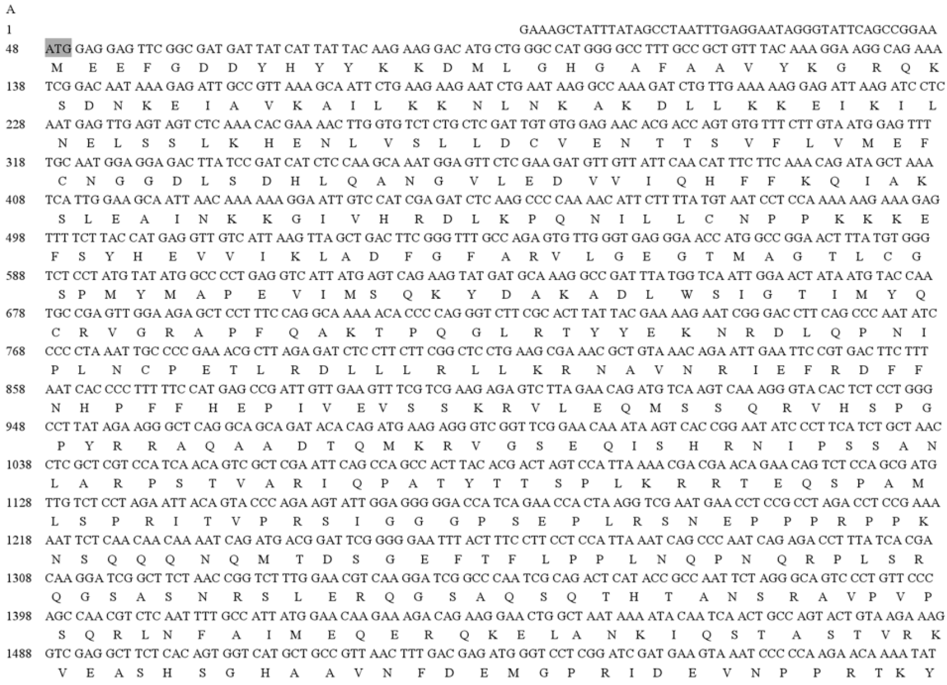
2.2. Autophagy-Related Gene Homologues in B. xylophilus

A homology-based cloning approach was used to obtain partial sequences of the ATG1 and ATG8 homologous sequences from B. xylophilus. The 3′ RACE and 5′ RACE PCR amplifications were used to obtain the full-length cDNA sequences of BxATG1 and BxATG8 from B. xylophilus. The flanking region of the BxATG1 cDNA is 2901 bps, has an open reading frame (ORF) at position 48–2834 and encodes a 928 amino acid polypeptide (Figure 2A). The flanking region of BxATG8 cDNA is 581 bps, has an ORF at position 40–402 and encodes a 120 amino acid polypeptide (Figure 2B).

2.3. In Situ Hybridization (ISH) for the Localization of BxATG8 in B. xylophilus

An ISH method was used to analyze the subcellular localization of the autophagy gene BxATG8, and a digoxigenin (DIG)-labeled probe generated from BxATG8 was specifically hybridized. The hybridization was observed in the oesophageal gland cells, as indicated by the dunk punctate color, and lateral hypodermal seam cells of B. xylophilus, as indicated by the light punctate color (Figure 3A). No hybridization was observed in the control group (Figure 3B). These results indicated that BxATG8 was more highly expressed in oesophageal gland cells, which function to dilute abnormal proteins quickly and to recycle amino acids, thereby assisting B. xylophilus in invading its host. BxATG8 was expressed in lateral hypodermal seam cells, which participate in metabolism and the storage of nutrients. We speculated that the function of autophagy in lateral hypodermal seam cells was regulating cellular metabolism and homeostasis.

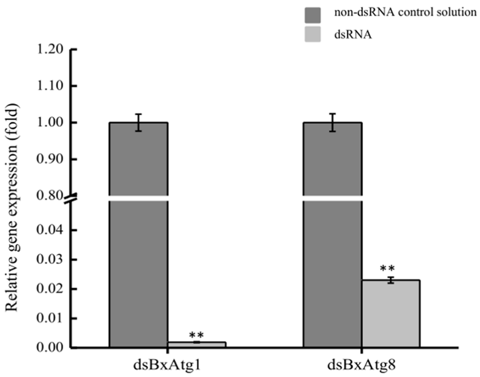
2.4. Detection of RNAi Efficiency

RNAi was used to assess the functions of BxATG1 and BxATG8 in B. xylophilus in this study. Quantitative reverse transcription PCR (qRT-PCR) was performed to determine the effect of RNAi on the BxATG1 and BxATG8 mRNA levels. Soaking nematodes in dsBxATG1 and dsBxATG8 solutions resulted in a marked decrease in BxATG1 and BxATG8 gene expression levels compared with those of nematodes soaked in a non-dsRNA control solution. When the mRNA expression level of the control was considered as 100%, the mean expression level of dsBxATG1- and dsBxATG8-treated samples were 0.19% and 2.30%, respectively (Figure 4). These results suggested that BxATG1 and BxATG8 were silenced by RNAi effectively.

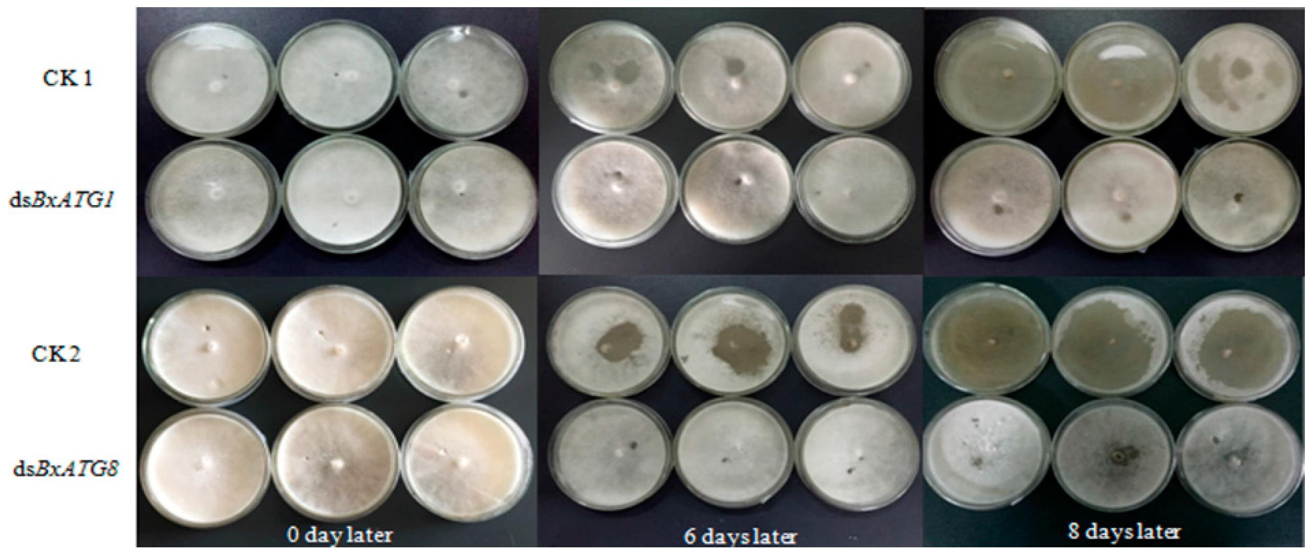
2.5. Effect of RNAi on B. xylophilus Reproduction on Fungal Mats

The effect of RNAi on B. xylophilus reproduction was tested on potato dextrose agar (PDA) plates inoculated with Botrytis cinerea at 25 °C. The nematodes soaked in double-stranded RNA (dsBxATG1 and dsBxATG8) solutions showed significantly reduced reproduction rates compared with the nematodes soaked in non-dsRNA control solutions (CK1 and CK2) and this result was confirmed by the remaining area of B. cinerea (Figure 5). After eight days, the reproduction rates of nematodes in the dsBxATG1 and non-dsRNA control solution (CK1) treatment were 161- and 520-fold (p < 0.01, Student’s t-test) (Figure 6A), and the reproduction rates of nematodes in the dsBxATG8 and non-dsRNA control solution (CK2) treatment were 194- and 529-fold (p < 0.01, Student’s t-test) (Figure 6B). These results indicated that B. xylophilus reproduction was significantly influenced by the RNAi treatment.

3. Discussion

Recently, a number of studies have focused on the functions of autophagy in eukaryotic organisms. The process of autophagy results in the turnover of intracellular proteins for guaranteed rejuvenation, which assists the clearance of misfolded proteins, and degrades organelles and proteins into small polypeptides to help maintain amino acid pools and the energy balance. Autophagy occurs constitutively at low levels even under normal growth conditions [17,27]. In this study, for the first time, the B. xylophilus autophagy was qualitatively identified during starvation, which is the best inducer of autophagy. It indicated that the process of autophagy exists in B. xylophilus as a response to stressful environmental conditions. The full-length cDNAs of autophagy-related genes (BxATG1 and BxATG8) from B. xylophilus, which had never been reported previously, were cloned and analyzed. These findings are relevant given the central roles that their products play in the autophagy process.
ISH enables the investigation of gene expression patterns and gene functions in nematodes [28,29,30]. ATG8/LC3/LGG-1 is routinely used as a marker to study autophagy, and researchers rely heavily on the expression patterns of reporters for ATG8/LC3/LGG-1 [31,32,33]. Thus, ISH was used to locate BxATG8 in B. xylophilus in this study, and the pattern of BxATG8 in B. xylophilus showed that it was expressed in the oesophageal gland and lateral hypodermal seam cells. The oesophageal gland of nematodes secretes a large number of proteins, including glucanases and pectate lyase [34,35]. Both cellulase and pectate lyase proteins are secreted through the nematode stylet into plant tissues and participate in weakening the cell walls, which facilitates the feeding, penetration and migration of nematodes in pine tissues [35]. The roles of lateral hypodermal seam cells in B. xylophilus are in metabolism and storage of nutrients [36]. The role of autophagy is to quickly break down abnormal proteins and to recycle amino acids for combining proteins. The autophagic compartments are a continuous source of small peptides and amino acids used to rebuild cell structures [17]. Therefore, the results suggested that autophagy gene BxATG8 might play an important role in plant–nematode interactions.
Furthermore, autophagy in pathogens, such as Aedes aegypti, Magnaporthe oryzae and Colletotrichum orbiculare, plays an important role in reproductive development, promoting their survival when environmental stress affects and changes their pathology [19,37,38]. Based on our results, we have found that these phenomena also occur in B. xylophilus. This was the first example of autophagy-related gene functions in B. xylophilus. RNAi technology was used to demonstrate the functions of BxATG1 and BxATG8. RNAi was first described by Fire et al. [39]. Later, RNAi was developed as an effective tool in plants and animals to study gene functions and for genetic manipulation [40,41]. Moreover, RNAi has also been used to assess the pathogenic and molecular effects of silenced B. xylophilus genes [42,43,44,45]. Autophagy is believed to be associated with changes in cellular architecture during differentiation and development [14]. As in Caenorhabditis elegans, autophagy functions in the cellular processes that regulate life-span during non-stressed conditions, and UNC-51 and BEC-1 are required for male tail development. C. elegans failed to resume reproduction even under favorable environmental conditions when BEC-1 was silenced [12,46]. Our results showed that the silencing of BxATG1 and BxATG8 reduced its reproductive capability. It demonstrated that BxATG1 and BxATG8 were necessary in the developmental processes of B. xylophilus. However, the potential role of autophagy in B. xylophilus needs to be further investigated.

4. Materials and Methods

4.1. B. xylophilus Growth Conditions and Experimental Organisms

The highly virulent AmA3 strain of B. xylophilus was isolated from wood chips of infested Pinus thunbergii Parl from Maanshan city, China. The virulence of AmA3 strain was evaluated by Xiang et al. [47]. The nematodes were grown in colonies of B. cinerea Pers, cultured on PDA plates for 7 days at 25 °C. Then, they were extracted overnight from PDA plates using the Baermann funnel method [48]. Two-year-old P. thunbergii seedlings were obtained from the greenhouse at Nanjing Forestry University (Nanjing, China).

4.2. TEM as Tool to Study Autophagy in B. xylophilus

The nematodes were subject to long-term starvation for 12, 24 and 36 h in double-distilled water (ddH2O). According to the TEM method [23,49], phosphate buffered saline (PBS) was made up of dibasic sodium phosphate and sodium dihydrogen phosphate (pH = 7.2). Nematodes were washed three times in PBS and placed in a 1.5 mL centrifuge tube with 4% glutaraldehyde fixative and fixed overnight at 4 °C, and then prepared for post-fixation in 2% osmium tetroxide (OsO4). The nematodes were placed in a graded series of acetone for 30 min each: 30%, 50%, 75%, 95%, and 2 × 100%. After adding 100% acetone, the centrifuge tube was capped to prevent moisture from entering. Acetone was mixed at ratios of 3:1, 1:1 and 1:3 with the Epon 812 resin mixture, and then added to the nematodes. Pure Epon 812 resin mixture was added to the nematodes, and one piece of the nematodes was placed into the bottom of each capsule. The capsules were placed into a 75 °C oven overnight to polymerize and were then cut into thin sections of 50–70 nm. Finally, the thin sections were stained and photographed under a TEM (JEM1400, Tokyo, Japan).

4.3. RNA Isolation and cDNA Synthesis of B. xylophilus

The total RNA of collected nematodes (a mixture of adults and juveniles) was extracted using TRIzol reagent (Invitrogen, Waltham, MA, USA). The RNA was quantified using a spectrophotometer and examined by electrophoresis on a 1% agarose gel. cDNA was synthesized from 2 µg of total RNA using the TransScript II One-Step gDNA Removal and cDNA Synthesis SuperMix according to the manufacturer’s instructions (TransGen Biotech, Beijing, China).

4.4. Homology-Based Cloning of Partial BxATG1 and BxATG8 Sequences from B. xylophilus

A homology-based cloning approach was used to clone the full-length cDNAs of two novel autophagy-related genes BxATG1 and BxATG8. Two degenerate primer sets (BxATG1 and BxATG8) were designed based on bioinformatics analyses [50]. The following primers were used: F-BxATG1 and R-BxATG1; and F-BxATG8 and R-BxATG8 (Table 1). The PCR conditions were 94 °C for 3 min followed by 30 cycles, each consisting of denaturation at 94 °C for 30 s, annealing at 55 °C for 30 s, and extension at 72 °C for 1 min. The final extension step was at 72 °C for 10 min.

4.5. Full-Length cDNA Cloning of BxATG1 and BxATG8 from B. xylophilus

The full-length BxATG1 and BxATG8 cDNA were obtained using the 3′-Full RACE CoreSet with PrimeScript™ RTase kit (TaKaRa Biotechnology, Dalian, China) and 5′-Full RACE Kit with TAP (TaKaRa Biotechnology). Gene-specific primers of BxATG1: GSP1-1 (3′-Full RACE first round of PCR) and GSP1-2 (3′-Full RACE second round of PCR), and GSP1-3 (5′-Full RACE first round of PCR), and GSP1-4 (5′-Full RACE second round of PCR) (Table 1). Gene-specific primers of BxATG8: GSP8-1 (3′-Full RACE first round of PCR) and GSP8-2 (3′-Full RACE first round of PCR), and GSP8-3 (5′-Full RACE first round of PCR) and GSP8-4 (5′-Full RACE second round of PCR) (Table 1). There were designed for 3′ and 5′ RACE amplification based on the two partial sequences of BxATG1 and BxATG8, which were obtained from the homology-based cloning results. The cycling profiles used were as follows: a cycle at 94 °C for 3 min, followed by 30 cycles, each consisting of denaturation at 94 °C for 30 s, annealing at 55 °C for 30 s, and extension at 72 °C for 2 min. The final extension step was at 72 °C for 10 min.

4.6. Cloning and Sequencing of BxAtg1 and BxAtg8

The amplified PCR products were confirmed by electrophoresis on 1% agarose gels and purified according to the Gel Extraction Kit (Axygen, Hangzhou, China) instructions. They were then cloned into the pEASY-T1 vector (TransGen Biotech, Beijing, China), which was used to transformed Escherichia coli Trans1-T1 (E. coli) competent cells (TransGen Biotech). The E. coli was then incubated overnight at 37 °C on LB plates containing ampicillin. The positive transformants were analyzed by PCR using primers M13F(-47) and M13R(-48) (Table 1). Once the correct clone was identified, a fresh bacterial suspension was submitted to the Nanjing Genscript sequencing company (Nanjing, China) for sequence analysis. The open reading frames of the cDNA sequences of BxATG1 and BxATG8 were found using the ORF Finder tool (available online: http://www.ncbi.nlm.nih.gov/projects/gorf/).

4.7. ISH

ISH was used to evaluate the functions of the autophagy-related gene BxATG8. For ISH, the DNA fragment used as the probe was amplified from the full-length cDNA clones of BxATG8 with a specific primer pairs, BxATG8-I-F and BxATG8-I-R (Table 1). The DIG-labeled sense random primer and anti-sense cDNA probes were synthesized from BxATG8’ PCR products. The nematodes were pre-treated before the post-hybridization washing step according to the manufacturer’s instructions. Hybridization and detection were performed with the DIG-High Prime DNA Labelling and Detection Starter Kit I (Roche Diagnostics, Mannheim, Germany), and finally examined using a Zeiss Axio Image M2 microscope (Zeiss MicroImaging GmbH, Oberkochen, Germany).

4.8. BxATG1 and BxATG8 Interference Using Double-Stranded RNA

Double-stranded RNA (dsRNA) was synthesized using the MEGscript RNAi Kit (Ambion Inc., Austin, TX, USA) with the primers BxATG1-T7I-F, BxATG1-I-R, BxATG1-I-F, BxATG1-T7I-R, BxATG8-T7I-F, BxATG8-I-R, BxATG8-I-F and BxATG8-T7I-R (Table 1). The RNAi soaking method was performed according to Urwin et al. [51]. Freshly cultured nematodes were soaked in dsRNA solution (800 ng/µL) and incubated at 180 rpm for 48 h at 20 °C. The nematodes soaked in the corresponding non-dsRNA solution were used as controls. Each treatment had three replicates. Samples from each treatment were washed thoroughly with ddH2O several times after soaking and then used for additional experiments.

4.9. Analysis of Reproduction of B. xylophilus after RNAi

The method of picking adult virgin female nematodes was modified by Wang et al. [52]. The eggs collected in the watch glass were covered with 2 mL of distilled water and incubated at 25 °C in the dark. The eggs took 25–32 h to hatch in water. Second-stage juveniles were picked and transferred onto a PDA plate with B. cinerea and cultured at 25 °C for one day. Then, female propagative four stage juveniles were collected under a stereo microscope (Zeiss MicroImaging GmbH) at 1 h intervals. The female and male nematodes were soaked in non-dsRNA solution and dsRNA solution, respectively. Fifteen pairs of female and male nematodes were picked and transferred onto a PDA plate with B. cinerea and cultured at 25 °C for 8 days. Three biological replicates were conducted. Subsequently, the nematodes were extracted from PDA plates using the Baermann funnel method and the nematodes were counted.

4.10. Quantitative Reverse Transcription PCR (qRT-PCR)

qRT-PCR was performed to determine the effect of RNAi on BxATG1 and BxATG8 mRNA levels. qRT-PCR was then carried out using SYBR Green Master Mix (Vazyme, Nanjing, China). The Actin gene of B. xylophilus was used as an internal control, with the primers listed in Table 1. Relative expression levels were determined using the ABI Prism 7500 software (Applied Biosystems, Foster City, CA, USA) and the 2−ΔΔCt method. qRT-PCR was conducted with three biological replicates and three technical replicates.

4.11. Statistical Analysis

All assays were performed in triplication. The results shown are the means and standard deviation (SD) of three independent experiments calculated using Microsoft Excel. The statistical significance was determined using SPSS Statistics 17.0 software (IBM China Company Ltd., Beijing, China) to perform the paired t-tests. Asterisks indicate statistically significant differences (** p < 0.01, Student’s t-test).

5. Conclusions

In summary, this study focused on the autophagy, which was identified by TEM under starvation, in B. xylophilus. Autophagy played a significant role in B. xylophilus’ resistance to an adverse starvation-inducing environment. The molecular characterization and functional analysis by ISH and RNAi of BxATG1 and BxATG8 from B. xylophilus indicated these autophagy genes are associated with development and reproduction. These discoveries regarding the relationship between autophagy and B. xylophilus helped us to understand the biological adaptation mechanism of B. xylophilus under adverse environments, and the functions of autophagy genes (BxATG1 and BxATG8) in the process of PWD. The process of autophagy may serve as a survival mechanism in B. xylophilus and provides fundamental information for facilitating understanding of PWD.

Acknowledgments

This work was financially supported by the National Natural Science Foundation of China (No. 31270683), and the Priority Academic Program Development of Jiangsu Higher Education Institutions (PAPD). We thank De-Wei Li, The Connecticut Agricultural Experiment Station, Windsor, CT, USA for critically reviewing the manuscript.

Author Contributions

Li-Na Deng: Designed study, analyzed data, conducted experiments and prepared manuscript; Jian-Ren Ye and Xiao-Qin Wu: Designed study and acquired data; Xiao-Qin Wu: analyzed data and contributed reagents/materials/analysis tools; and Xiao-Qin Wu and Qi Xue: approved final manuscript.

Conflicts of Interest

The authors declare no conflict of interest.

References

  1. Mamiya, Y. Pathology of the pine wilt disease caused by Bursaphelenchus xylophilus. Annu. Rev. Phytopathol. 1983, 21, 201–220. [Google Scholar] [CrossRef] [PubMed]
  2. Kanzaki, N.; Futai, K. A PCR primer set for determination of phylogenetic relationships of Bursaphelenchus species within the xylophilus group. Nematology 2002, 4, 35–41. [Google Scholar] [CrossRef]
  3. Ceng, H.R.; Lin, M.S.; Ni, W.Q.; Fang, Z.D. First report of pine wilt disease from Pinus thunbergii Parl in Nanjing. Pak. J. Nematol. 1983, 4, 1–5. [Google Scholar]
  4. Mota, M.M.; Braasch, H.; Bravo, M.A.; Burgermeister, W.; Metge, K.; Sousa, E. First report of Bursaphelenchus xylophilus in Portugal and in Europe. Nematology 1999, 1, 727–734. [Google Scholar] [CrossRef]
  5. Yang, B.J.; Wang, Q.L. Distribution of the pinewood nematode in China and susceptibility of some Chinese and exotic pines to the nematode. Can. J. For. Res. 1989, 19, 1527–1530. [Google Scholar]
  6. Braasch, H.; Tomiczek, C.; Metge, K.; Hoyer, U.; Burgermeister, W.; Wulfert, I.; Schonfeld, U. Records of Bursaphelenchus spp. (Nematoda, Parasitaphelenchidae) in coniferous timber imported from the Asian part of Russia. For. Pathol. 2001, 31, 129–140. [Google Scholar] [CrossRef]
  7. Son, J.A.; Hogetsu, T.; Moon, Y.S. The process of epithelial cell death in Pinus thunbergii caused by the pine wood nematode, Bursaphelenchus xylophilus. Nematology 2014, 16, 663–668. [Google Scholar] [CrossRef]
  8. Zhao, B.G.; Wang, H.L.; Han, S.F.; Zheng, M.H. Distribution and pathogenicity of bacteria species carried by Bursaphelenchus xylophilus in China. Nematology 2003, 5, 899–906. [Google Scholar] [CrossRef]
  9. Wang, Z.; Wang, C.Y.; Fang, Z.M.; Zhang, D.L.; Liu, L.; Lee, M.R.; Li, Z.; Li, J.J.; Sung, C.K. Advances in research of pathogenic mechanism of pine wilt disease. Afr. J. Microbiol. Res. 2010, 4, 437–442. [Google Scholar]
  10. Kikuchi, T.; Cotton, J.A.; Dalzell, J.J.; Hasegawa, K.; Kanzaki, N.; McVeigh, P.; Takanashi, T.; Tsai, I.J.; Assefa, S.A.; Cock, P.J.A.; et al. Genomic insights into the origin of parasitism in the emerging plant pathogen Bursaphelenchus xylophilus. PLoS Pathog. 2011, 7, 1059–1074. [Google Scholar] [CrossRef] [PubMed]
  11. Ogura, N.; Nakashima, T. In vitro occurrence of dispersal fourth stage juveniles in Bursaphelenchus xylophilus co-incubated with Monochamus alternatus. Jpn. J. Nematol. 2002, 32, 53–59. [Google Scholar]
  12. Meléndez, A.; Talloczy, Z.; Seaman, M.; Eskelinen, E.L.; Hall, D.H.; Levine, B. Autophagy genes are essential for dauer development and life-span extension in C. elegans. Science 2003, 301, 1387–1391. [Google Scholar] [CrossRef] [PubMed]
  13. Zhang, H.; Chang, J.T.; Guo, B.; Hansen, M.; Jia, K.; Kovács, A.L.; Kumsta, C.; Lapierre, L.R.; Legouis, R.; Lin, L.; et al. Guidelines for monitoring autophagy in Caenorhabditis elegans. Autophagy 2015, 11, 9–27. [Google Scholar] [PubMed]
  14. Levine, B.; Klionsky, D.J. Development by self-digestion: Molecular mechanisms and biological functions of autophagy. Dev. Cell 2004, 6, 463–477. [Google Scholar] [CrossRef]
  15. Meléndez, A.; Levine, B. Autophagy in C. elegans. NCBI [Internet]. Available online: http://www.ncbi.nlm.nih.gov/books/NBK116074/ (accessed on 21 August 2015).
  16. Klionsky, D.J. The molecular machinery of autophagy: Unanswered questions. J. Cell Sci. 2005, 118, 7–18. [Google Scholar] [CrossRef] [PubMed]
  17. Mizushima, N. The pleiotropic role of autophagy: From protein metabolism to bactericide. Cell Death Differ. 2005, 12, 1535–1541. [Google Scholar] [CrossRef] [PubMed]
  18. Lee, H.N.; Jeong, T.J. Overview of autophagy in plant cells. J. Life Sci. 2014, 24, 209–217. [Google Scholar] [CrossRef]
  19. Liu, X.H.; Lu, J.P.; Zhang, L.; Dong, B.; Min, H.; Lin, F.C. Involvement of a Magnaporthe grisea serine/threonine kinase gene, MgATG1, in appressorium turgor and pathogenesis. Eukaryot. Cell 2007, 6, 997–1005. [Google Scholar] [CrossRef] [PubMed]
  20. Tindwa, H.; Jo, Y.H.; Patnaik, B.B.; Lee, Y.S.; Kang, S.S.; Han, Y.S. Molecular cloning and characterization of autophagy-related gene TmATG8 in Listeria-invaded hemocytes of Tenebrio molitor. Dev. Comp. Immunol. 2015, 51, 88–98. [Google Scholar] [CrossRef] [PubMed]
  21. Fernández, J.M.F.; Ortega, A.G.; Cruz, R.R.; Camberos, E.P.; Álvarez, Á.H.; Velázquez, M.M. Molecular cloning and characterization of two novel autophagy-related genes belonging to the ATG8 family from the cattle tick Rhipicephalus (Boophilus) microplus (Acari: Ixodidae). Exp. Appl. Acarol. 2014, 64, 533–542. [Google Scholar] [CrossRef] [PubMed]
  22. Valent, B.; Farrall, L.; Chumley, F.G. Magnaporthe grisea genes for pathogenicity and virulence identified through a series of backcrosses. Genetics 1991, 127, 87–101. [Google Scholar] [PubMed]
  23. Ylä-Anttila, P.; Vihinen, H.; Jokitalo, E.; Eskelinen, E.L. Monitoring autophagy by electron microscopy in Mammalian cells. Methods Enzymol. 2009, 452, 143–164. [Google Scholar] [PubMed]
  24. Spellman, P.T.; Sherlock, G.; Zhang, M.Q.; Iyer, V.R.; Anders, K.; Eisen, M.B.; Brown, P.O.; Botstein, D.; Futcher, B. Comprehensive identification of cell cycle-regulated genes of the yeast Saccharomyces cerevisiae by microarray hybridization. Mol. Biol. Cell 1998, 9, 3273–3297. [Google Scholar] [CrossRef] [PubMed]
  25. Chan, E.Y.; Tooze, S.A. Evolution of Atg1 function and regulation. Autophagy 2009, 5, 758–765. [Google Scholar] [CrossRef] [PubMed]
  26. Shpilka, T.; Weidberg, H.; Pietrokovski, S.; Elazar, Z. Atg8: An autophagy-related ubiquitin-like protein family. Genome Biol. 2011, 12. [Google Scholar] [CrossRef] [PubMed]
  27. Ryter, S.W.; Cloonan, S.M.; Choi, A.M.K. Autophagy: A critical regulator of cellular metabolism and homeostasis. Mol. Cells 2013, 36, 7–16. [Google Scholar] [CrossRef] [PubMed]
  28. Vandekerckhove, T.T.M.; Coomans, A.; Cornelis, K.; Cornelis, K.; Baert, P.; Gillis, M. Use of the Verrucomicrobia-specific probe EUB338-III and fluorescent in situ hybridization for detection of “Candidatus Xiphinematobacter” cells in nematode hosts. Appl. Environ. Microbiol. 2002, 68, 3121–3125. [Google Scholar] [CrossRef] [PubMed]
  29. De Boer, J.M.; Yan, Y.; Smant, G.; Davis, E.L.; Baum, T.J. In-situ hybridization to messenger RNA in Heterodera glycines. J. Nematol. 1998, 30, 309–312. [Google Scholar] [PubMed]
  30. Wang, F.; Wang, Z.Y.; Li, D.L.; Chen, Q.L. Identification and characterization of a Bursaphelenchus xylophilus (Aphelenchida: Aphelenchoididae) thermotolerance-related gene: Bx-hsp90. Int. J. Mol. Sci. 2012, 13, 8819–8833. [Google Scholar] [CrossRef] [PubMed]
  31. Cheong, H.; Klionsky, D.J. Biochemical methods to monitor autophagy-related processes in yeast. Methods Enzymol. 2008, 451, 1–26. [Google Scholar] [PubMed]
  32. Navale, R.; Allanki, A.D.; Sijwali, P.S. Characterization of the autophagy marker protein Atg8 reveals atypical features of autophagy in Plasmodium falciparum. PLoS ONE 2014, 9, e113220. [Google Scholar] [CrossRef] [PubMed]
  33. Mizushima, N.; Yoshimori, T.; Levine, B. Methods in mammalian autophagy research. Cell 2010, 140, 313–326. [Google Scholar] [CrossRef] [PubMed]
  34. De Boer, J.M.; Smant, G.; Goverse, A.; Davis, E.L.; Ovremars, H.A.; Pomp, H.; Gent-Pelzer, M.V.; Zilverentant, J.F.; Stokkermans, J.P.W.G.; Hussey, R.S.; et al. Secretory granule proteins from the subventral esophageal glands of the potato cyst nematode identified by monoclonal antibodies to a protein fraction from second-stage juvenile. Mol. Plant Microbe Interact. 1996, 9, 39–46. [Google Scholar] [CrossRef] [PubMed]
  35. Kikuchi, T.; Shibuya, H.; Aikawa, T.; Jones, J.T. Cloning and characterization of pectate lyases expressed in the esophageal gland of the pine wood nematode Bursaphelenchus xylophilus. Mol. Plant Microbe Interact. 2006, 19, 280–287. [Google Scholar] [CrossRef] [PubMed]
  36. Feng, Z.X. Plant Nematology; Chinese Agricultural Publishing House: Beijing, China, 2001; p. 11. [Google Scholar]
  37. Asakura, M.; Ninomiya, S.; Sugimoto, M.; Oku, M.; Yamashita, S.I.; Okuno, T.; Sakai, Y.; Takano, Y. Atg26-mediated pexophagy is required for host invasion by the plant pathogenic fungus Colletotrichum orbicular. Plant Cell 2009, 21, 1291–1304. [Google Scholar] [CrossRef] [PubMed]
  38. Bryant, B.; Raikhel, A.S. Programmed autophagy in the fat body of Aedes aegypti is required to maintain egg maturation cycles. PLoS ONE 2011, 6, e25502. [Google Scholar] [CrossRef] [PubMed]
  39. Fire, A.; Xu, S.Q.; Montgomery, M.K.; Kostas1, S.A.; Driver, S.E.; Mello, C.C. Potent and specific genetic interference by double-stranded RNA in Caenorhabditis elegans. Nature 1998, 391, 806–811. [Google Scholar] [CrossRef] [PubMed]
  40. Wesley, S.V.; Helliwell, C.A.; Smith, N.A.; Wang, M.B.; Rouse, D.T.; Liu, Q.; Gooding, P.S.; Singh, S.P.; Abbott, D.; Stoutjesdijk, P.A.; et al. Construct design for efficient. effective and high-throughput gene silencing in plants. Plant J. 2001, 27, 581–590. [Google Scholar] [CrossRef] [PubMed]
  41. Chang, C.H.; Wang, H.I.; Lu, H.C.; Chen, C.E.; Chen, H.H.; Yeh, H.H.; Tang, C.Y. An efficient RNA interference screening strategy for gene functional analysis. BMC Genom. 2012, 13. [Google Scholar] [CrossRef] [PubMed]
  42. Ma, H.B.; Lu, Q.; Liang, J.; Zhang, X.Y. Functional analysis of the cellulose gene of the pine wood nematode, Bursaphelenchus xylophilus, using RNA interference. Genet. Mol. Res. 2011, 10, 1931–1941. [Google Scholar] [CrossRef] [PubMed]
  43. Kang, J.S.; Koh, Y.H.; Moon, Y.S.; Lee, S.H. Molecular properties of a venom allergen-like protein suggest a parasitic function in the pinewood nematode Bursaphelenchus xylophilus. Int. J. Parasitol. 2012, 42, 63–70. [Google Scholar] [CrossRef] [PubMed]
  44. Cheng, X.Y.; Dai, S.M.; Xiao, L.; Xie, B.Y. Influence of cellulase gene knockdown by dsRNA interference on the development and reproduction of the pine wood nematode, Bursaphelenchus xylophilus. Nematology 2010, 12, 225–233. [Google Scholar] [CrossRef]
  45. Xu, X.L.; Wu, X.Q.; Ye, J.R.; Huang, L. Molecular characterization and functional analysis of three pathogenesis-related Cytochrome P450 genes from Bursaphelenchus xylophilus (Tylenchida: Aphelenchoidoidea). Int. J. Mol. Sci. 2015, 16, 5216–5234. [Google Scholar] [CrossRef] [PubMed]
  46. Aladzsity, I.; Tóth, M.L.; Sigmond, T.; Szabó, E.; Bicsák, B.; Barna, J.; Regős, Á.; Orosz, L.; Kovács, A.L.; Vellai, T. Autophagy genes unc-51 and bec-1 are required for normal cell size in Caenorhabditis elegan. Genetics 2007, 177, 655–660. [Google Scholar] [CrossRef] [PubMed]
  47. Xiang, Y.; Wu, X.Q.; Zhou, A.D. Bacterial diversity and community structure in the pine wood nematode Bursaphelenchus xylophilus and B. mucronatus with different virulence by high-throughput sequencing of the 16S rDNA. PLoS ONE 2015, 10, e0137386. [Google Scholar] [CrossRef] [PubMed]
  48. Viglierchio, D.R.; Schmitt, R.V. On the methodology of nematode extraction from field samples: Baermann funnel modifications. J. Nematol. 1983, 15, 438–444. [Google Scholar] [PubMed]
  49. Martinet, W.; Timmermans, J.P.; de Meyer, G.R.Y. Methods to assess autophagy in situ—Transmission electron microscopy versus immunohistochemistry. Methods Enzymol. 2014, 543, 89–114. [Google Scholar] [PubMed]
  50. Consensus-degenerate hybrid oligonucleotide primers. Available online: http://blocks.fhcrc.org/codehop.html (accessed on 25 August 2015).
  51. Urwin, P.E.; Lilley, C.J.; Atkinson, H.J. Ingestion of double-stranded RNA by preparasitic juvenile cyst nematodes leads to RNA interference. Mol. Plant Microbe Interact. 2002, 15, 747–752. [Google Scholar] [CrossRef] [PubMed]
  52. Wang, Y.; Yamada, T.; Sakaue, D.; Suzuki, K. Variations in life history parameters and their influence on rate of population increase of different pathogenic isolates of the pine wood nematode, Bursaphelenchus xylophilus. Nematology 2005, 7, 459–467. [Google Scholar] [CrossRef]
Figure 1. Autophagy in the cells of the highly virulent strain AmA3 of Bursaphelenchus xylophilus after starvation was induced for 12 h (A,B); 24 h (C); and 36 h (D), with autophagic bodies (right arrows), autophagosomes (left arrows), autolysosomes (down arrows) and vesicle breakdown (up arrows) Scale bars: (A) 0.2 µm; (B) 0.5 µm; (C) 1 µm; and (D) 2 µm.
Ijms 17 00279 g001
Figure 2. Full-length cDNA sequences and deduced amino acid sequence of BxATG1 (A) and BxATG8 (B) from B. xylophilus. Note: The initiation codons are shown with a dark background, and asterisks indicate the stop codons.
Ijms 17 00279 g002aIjms 17 00279 g002b
Figure 3. Localization of Bx-ATG8 mRNA by in situ hybridization (ISH) using a digoxigenin (DIG)-labeled BxATG8 antisense or sense probe. The hybridization sites are shown in B. xylophilus (A), including the oesophageal gland cells (right arrows), and lateral hypodermal seam cells (left arrows). Control sense probe in B. xylophilus (B). The scale bars = 100 μm.
Ijms 17 00279 g003
Figure 4. Quantitative reverse transcription PCR (qRT-PCR) analysis of the RNAi efficiency in B. xylophilus after treatment with dsBxATG1 and dsBxATG8. B. xylophilus soaked in a non-dsRNA solution was used as the control. The expression level of the control was set as 100%. Data represent mean values ± standard deviation (SD) from three independent experiments. Bars show standard deviations of the mean. Asterisks on top of the bars indicate statistically significant differences (** p < 0.01, Student’s t-test).
Ijms 17 00279 g004
Figure 5. Effect of RNAi on B. xylophilus inoculated onto Botrytis cinerea. The nematodes soaked in dsBxATG1, dsBxATG8 and non-dsRNA control solutions were grown in B. cinerea.
Ijms 17 00279 g005
Figure 6. Effect of RNAi on B. xylophilus reproduction. Reproduction rates of B. xylophilus washed from potato dextrose agar (PDA) plates of B. cinerea with dsBxATG1 and dsBxATG8 and CK1 and CK2, respectively. Data represent mean values ± SD from three independent experiments. Bars show standard deviations of the mean. Asterisks on top of the bars indicate statistically significant differences between the dsBxATG1 (A) and dsBxATG8 (B) nematodes and controls (** p < 0.01, Student’s t-test).
Ijms 17 00279 g006
Table 1. Polymerase chain reaction (PCR) primers used in the study.
Name of PrimersSequence (5′–3′)
F-BxATG1GGGCCGAGCAGCTGGTNGTNTAYGT
R-BxATG1CACAGCTCGATGGCGTGNYKRTACAT
F-BxATG8ACTTTGAGAAGCGTCGTG
R-BxATG8TGTGGAATGACATTGTTGAC
GSP1-1TCTTGTGATGGCTCAACGAC
GSP1-2CTCGTTCTCAAGAGCTGGCT
GSP1-3TGGAGATGGCTGAAGAGTCG
GSP1-4CGTTGAGCCATCACAAGACT
GSP8-1GTCGTGCTGAAGGTGAGAAGAT
GSP8-2TGAAGGTGAGAAGATCCGTCGCAAGT
GSP8-3ATAAAGCTGTCCCATCGTGGTCGT
GSP8-4TCAGAGGGAACCAGATACTT
BxATG1-T7I-FTAATACGACTCACTATAGGGAAGGCAGAAATCGGACA
BxATG1-I-RAATCGGCTCATGGAAAA
BxATG1-I-FAAGGCAGAAATCGGACA
BxATG1-T7I-RTAATACGACTCACTATAGGGAATCGGCTCATGGAAAA
BxATG8-T7I-FTAATACGACTCACTATAGGGAACCCAAGTTTGAGACCT
BxATG8-I-RCGAAAACACTACAATAAGA
BxATG8-I-FAACCCAAGTTTGAGACCT
BxATG8-T7I-RTAATACGACTCACTATAGGGCGAAAACACTACAATAAGA
Actin FGCAACACGGAGTTCGTTGTAGA
Actin RGTATCGTCACCAACTGGGATGA
qBxATG1-FAGAGTGTTGGGTGAGGGA
qBxATG1-RCTCGGCATTGGTACATTATA
qBxATG8-FGTCAACGATGTCATTCCCCA
qBxATG8-RAACTGATCACTCTTCGGCGG
M13F(-47)CGCCAGGGTTTTCCCAGTCACGAC
M13R(-48)AGCGGATAACAATTTCACACAGGA

