Article

Role of the Genes of Type VI Secretion System in Virulence of Rice Bacterial Brown Stripe Pathogen Acidovorax avenae subsp. avenae Strain RS-2

1
State Key Laboratory of Rice Biology, Ministry of Agriculture Key Lab of Molecular Biology of Crop Pathogens and Insects, Institute of Biotechnology, Zhejiang University, Hangzhou 310058, China
2
Department of Plant Pathology, Bangabandhu Sheikh Mujibur Rahman Agricultural University, Gazipur 1706, Bangladesh
3
State Key Laboratory Breeding Base for Zhejiang Sustainable Pest and Disease Control, Institute of Plant Protection and Microbiology, Zhejiang Academy of Agricultural Sciences, Hangzhou 310021, China
*
Authors to whom correspondence should be addressed.
Int. J. Mol. Sci. 2017, 18(10), 2024; https://doi.org/10.3390/ijms18102024
Submission received: 18 August 2017 / Revised: 9 September 2017 / Accepted: 19 September 2017 / Published: 21 September 2017
(This article belongs to the Section Molecular Plant Sciences)

Abstract

:
The Type VI secretion system (T6SS) is a class of macromolecular machine that is required for the virulence of gram-negative bacteria. However, it is still not clear what the role of T6SS in the virulence of rice bacterial brown stripe pathogen Acidovorax avenae subsp. avenae (Aaa) is. The aim of the current study was to investigate the contribution of T6SS in Aaa strain RS2 virulence using insertional deletion mutation and complementation approaches. This strain produced weak virulence but contains a complete T6SS gene cluster based on a genome-wide analysis. Here we compared the virulence-related phenotypes between the wild-type (RS-2) and 25 T6SS mutants, which were constructed using homologous recombination methods. The mutation of 15 T6SS genes significantly reduced bacterial virulence and the secretion of Hcp protein. Additionally, the complemented 7 mutations ΔpppA, ΔclpB, Δhcp, ΔdotU, ΔicmF, ΔimpJ, and ΔimpM caused similar virulence characteristics as RS-2. Moreover, the mutant ΔpppA, ΔclpB, ΔicmF, ΔimpJ and ΔimpM genes caused by a 38.3~56.4% reduction in biofilm formation while the mutants ΔpppA, ΔclpB, ΔicmF and Δhcp resulted in a 37.5~44.6% reduction in motility. All together, these results demonstrate that T6SS play vital roles in the virulence of strain RS-2, which may be partially attributed to the reductions in Hcp secretion, biofilm formation and motility. However, differences in virulence between strain RS-1 and RS-2 suggest that other factors may also be involved in the virulence of Aaa.

Graphical Abstract

1. Introduction

The gram-negative bacteria Acidovorax avenae subsp. avenae (Aaa) causes diseases in a wide range of economically important plants such as rice, corn, oats, sugarcane, millet, and foxtail [1]. Particularly, this well-known seed-borne bacterium [1,2] causes bacterial brown stripe (BBS) in rice, which leads to heavy economic losses. It has been reported in many rice-growing countries such as Asia, Africa, North America and Europe [3,4]. The contaminated seeds represent the most important primary source of inocula [2,5,6] for outbreak of the disease. Recently, this disease has achieved increased attention, especially in China [7,8,9,10]. The economic importance of BBS makes it necessary to know the molecular basis for the infection of A. avenae subsp. avenae [9,11] to rice plants.
The bacterial phytopathogens infections are highly associated with secreted effectors proteins in their host plants. It has been well documented that bacteria use a remarkable array of sophisticated macromolecular nanomachines to deliver extracellular proteins or effectors molecules into the surrounding environment or, in some cases, directly to the target site of the host cell [12,13]. At least six different secretion systems have been found in gram-negative pathogenic bacteria [12,14]. In particular, a novel secretary system T6SS has been described to be largely association with various biological functions such as pathogenicity, biofilm formation, adaptation, modulation of quorum sensing and survival [15,16,17,18]. Furthermore, according to our previous studies that the plant height was closely associated with bacterial virulence while T6SS genes showed differential responses to in vivo infection of Aaa to rice host [11,19]. Interestingly, genome-wide in silico analysis has identified a large number of secretion system-related genes including T6SS (Figure 1) in Aaa strain RS-2. However, the role of each gene of the T6SS gene cluster in Aaa strain RS-2 remains poorly understood.
The aim of this study was to examine the role of T6SS in Aaa strain RS-2 through comparing bacterial pathogenicity to rice, growth measurement, swimming motility, biofilm formation, and the secretion of effector proteins between wild-type and the mutants constructed in this study.

2. Results

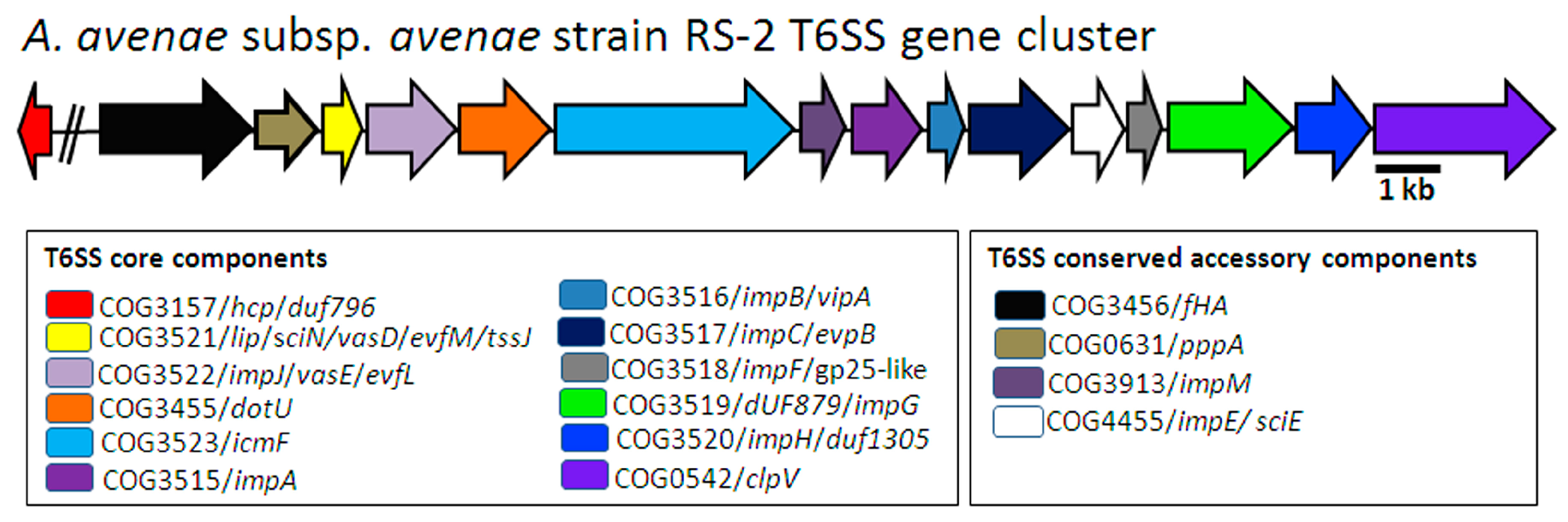
2.1. In Silico Identification of T6SS Genes

Recently, the whole genome of A. avenae subsp. avenae strain RS-1 [7] and RS-2 (unpublished)-isolated from rice-has been sequenced by our laboratory, which is a useful resource for identifying genes involved in some specific biological functions underlying this disease. In this study, we found 25 Type VI secretion system genes in the A. avenae subsp. avenae strain RS-2 genome while the identification of T6SS homologous coding loci were conducted by local BLAST (BLASTN, BLASTX) and results are shown in Supplementary Figure S1. Based on the genome wide analyses of A. avenae subsp. avenae strain RS-1, Acidovorax avenae subsp. avenae ATCC 19860 and A. citrulli AAC00-1, one T6SS gene cluster was also found in Aaa strain RS-2 (Figure 1).
As shown in Figure 1, this cluster contains 16 genes namely hcp, fHA, pppA, lip, impJ, dotU, icmF, impM, impA, impB, impC, impE, impF, dUF879, impH, and clpV, which have been shown to represent core and conserved accessory components in the T6SS of Aaa strain RS-2. Phylogenetic analysis of sequences in this study clearly indicated the presence of T6SS genes in A. avenae subsp. avenae strain RS-2, which were homologs in closely related bacteria such as A. avenae subsp. avenae strain RS-1 and ATCC 19860 as well as A. citrulli AAC00-1 (Supplementary Figure S2). All T6SS genes in the Aaa strain RS-2 genome including their putative functions are presented in the Supplementary Table S1.

2.2. T6SS Gene Mutants and Complementation

To study the involvement of T6SS genes in virulence mechanism, we created the insertional knockout mutants on target genes of T6SS in Aaa strain RS-2 via homologous recombination (primers, constructed strains and plasmids were listed in Supplementary Tables S2 and S3). After successfully transforming the plasmid construct, transformants grown on selective antibiotic were ensured by PCR diagnosis for T6SS mutants (Figure 2) and their complementation as well (Figure 3). Subsequently, the identity of all the obtained strains were verified by blasting their sequence in the NCBI database, and the results showed the highest homology with Aaa (data not shown). The knockout mutants were then examined for phenotypes in several aspects such as pathogenicity, growth, swimming, biofilm formation, and secretion of effector proteins.

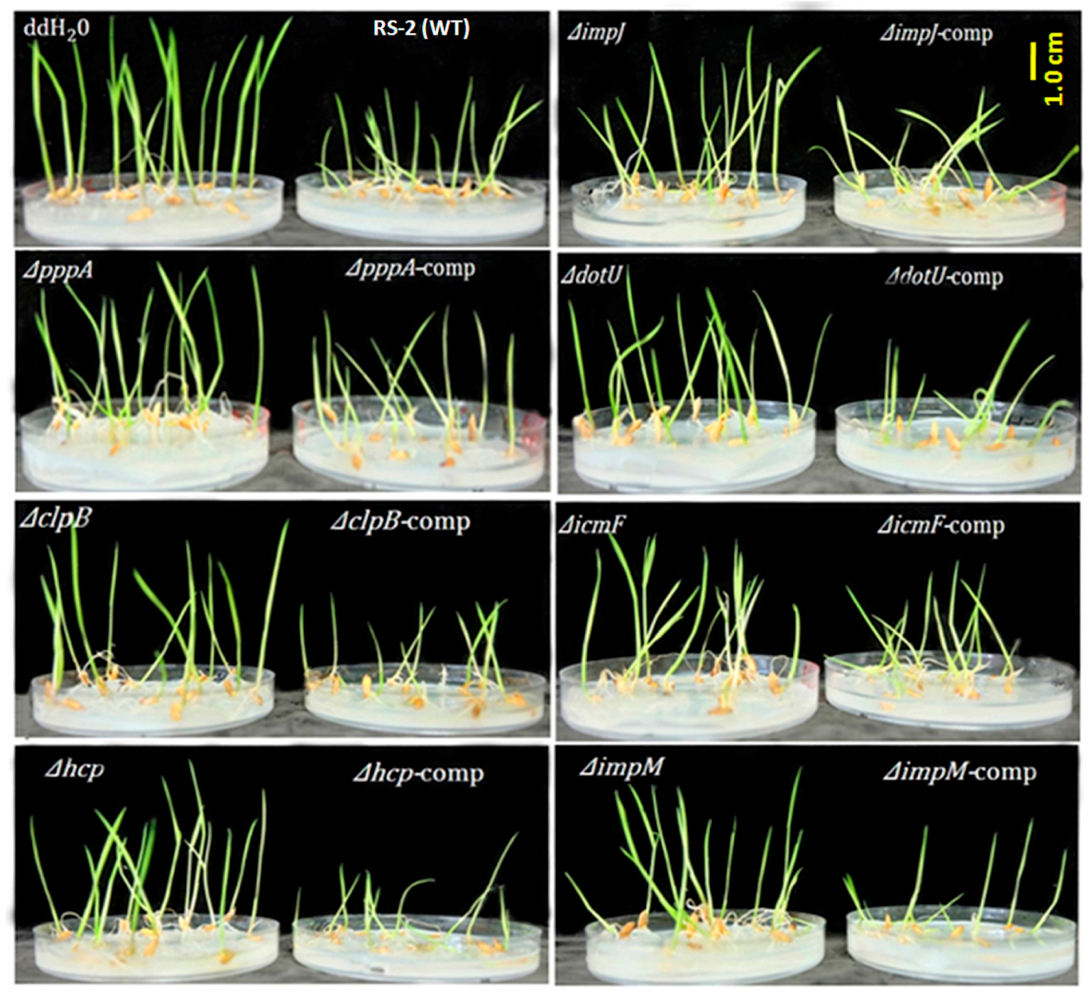
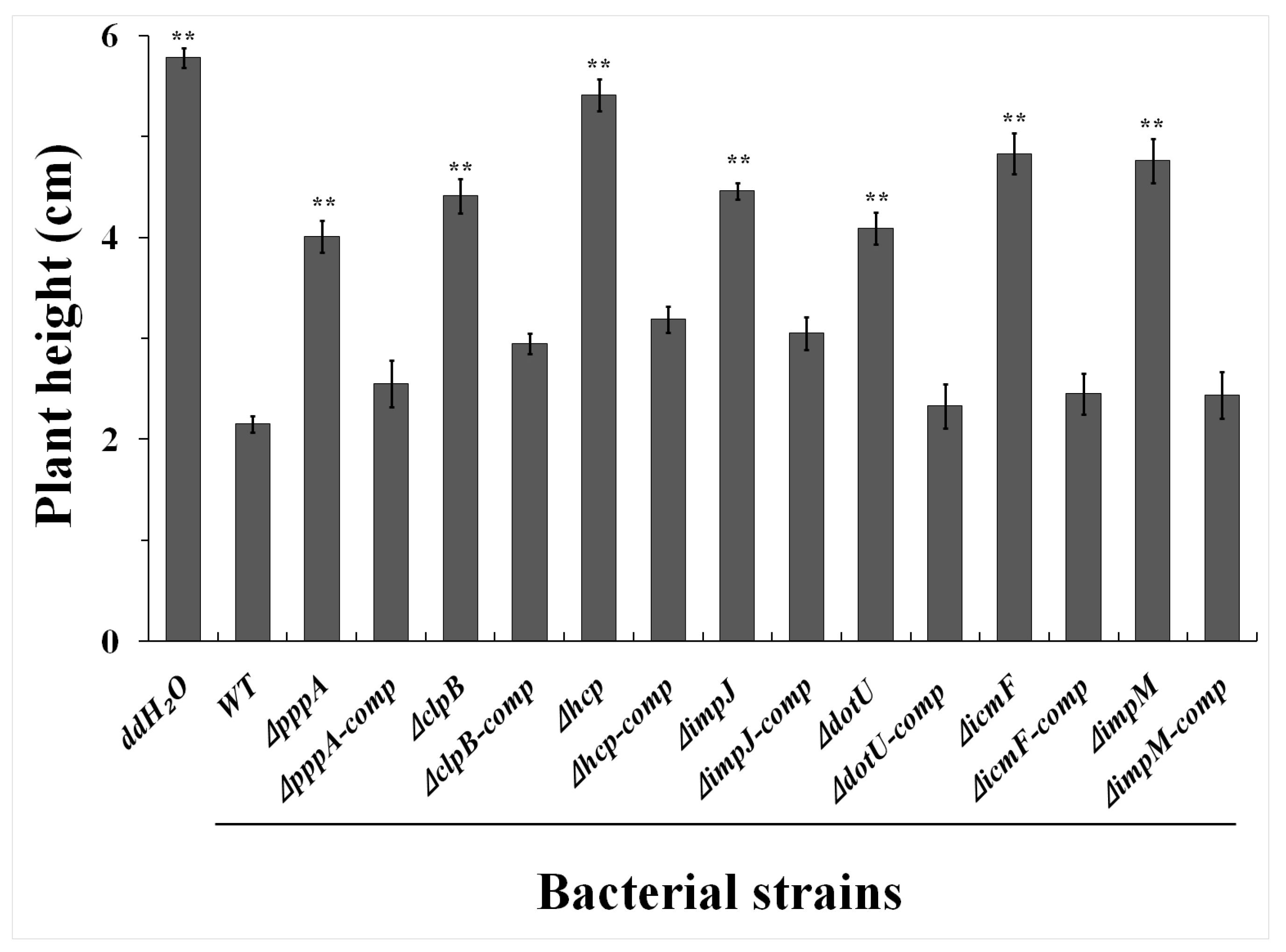
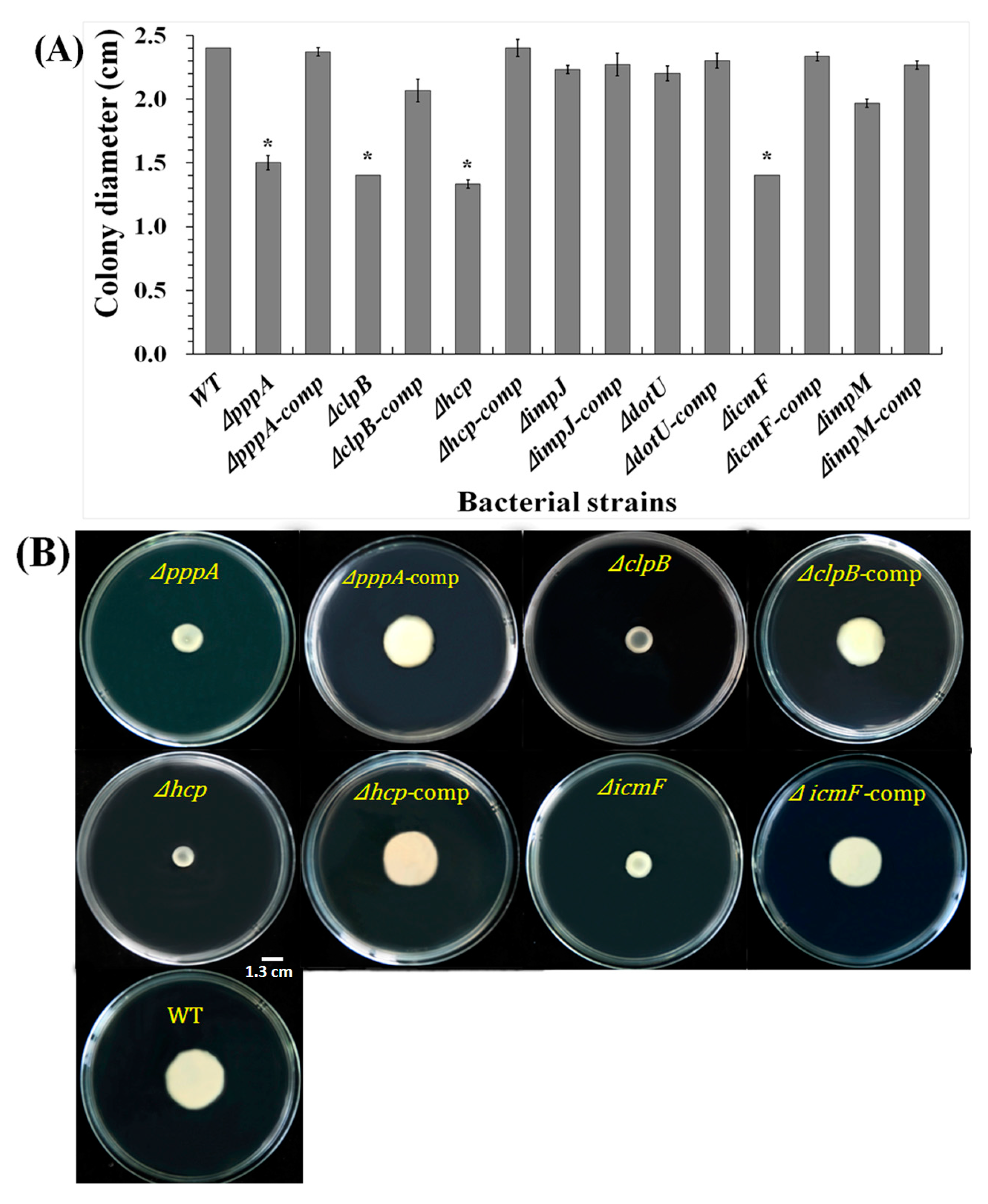
2.3. T6SS Affected Pathogenicity of Aaa Strain RS2 to Rice Seedlings

The effect of T6SS genes on the virulence of the Aaa strain RS-2 was determined by comparing the plant height of rice seedlings between the wild-type and individual T6SS mutants, and results are presented in Figure 4 and Figure 5 and Supplementary Table S4.
As shown in Figure 4 and Figure 5, we observed that rice seedlings treated with wild-type strain RS-2 grows weaker and shorter with an average of only 2.15 cm whereas ddH2O (the negative control) treated seedlings showed the maximum plant height (5.78 cm). Furthermore, results showed that 7 of the mutants-ΔpppA, ΔclpB, Δhcp, ΔimpJ, ΔdotU, ΔicmF and ΔimpM-caused the greatest (p < 0.01) reduction in bacterial virulence compared to Aaa wild-type RS-2 strain, while the corresponding average plant heights were 4.01, 4.41, 5.41, 4.46, 4.09, 4.83, 4.76 cm, respectively.
An association between plant height and bacterial virulence was revealed in the above result. As mentioned above, the wild-type strain RS-2 showed the strongest virulence toward rice seedlings, which reduced the plant height by 62.80% compared with that treated with the ddH2O, whereas the corresponding plant height in mutants ΔpppA, ΔclpB, Δhcp, ΔimpJ, ΔdotU, ΔicmF and ΔimpM decreased by 30.62%, 23.70%, 6.40%, 22.84%, 29.24%, 16.44%, 17.65%, respectively (see Figure 4 and Supplementary Table S4).
Complementation of these mutants restored their virulence (Figure 4 and Figure 5). Besides the above mentioned 7 mutants, the other 8 mutants (ΔvgrG (1–8)) also caused a significant (p < 0.05) reduction in pathogenicity compared with the wild-type strain. Moreover, there was no significant (p > 0.05) difference in the pathogenicity between the 10 mutants (ΔfHA, ΔclpV, ΔdUF879, ΔimpA, ΔimpB, ΔimpC, ΔimpE, ΔimpF, ΔimpH and Δlip) and the wild-type strain RS-2 (Supplementary Table S4).
In conclusion, among the 25 T6SS genes, the mutation of 15 of them had a significant effect on Aaa virulence in respect of rice plant height, and the other 10 genes did not. Based on this information, we can infer that the loss of pathogenicity of rice bacterial brown stripe disease in the mutants was mainly due to the deficiency of different T6SS genes.

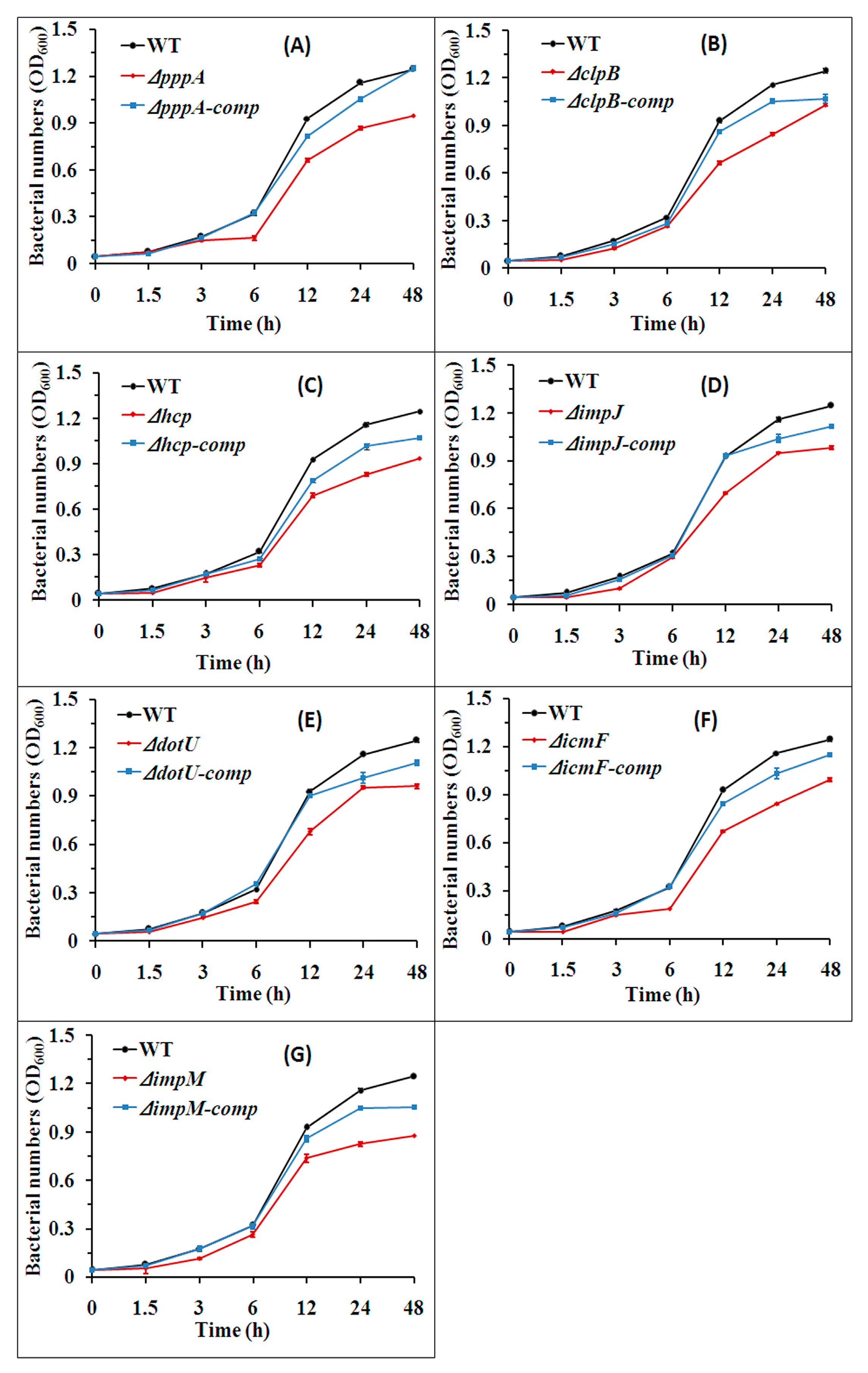
2.4. T6SS Affected the Growth of Aaa Strain RS2

Results from this study indicated that the strain RS-2 wild-type, its mutants and their complemented strains showed the fastest growth rate during incubation for 6~12 h, followed by moderate growth after incubation for 24 h, and then reached maximum growth rate after incubation for 48 h (Figure 6). Indeed, the OD600 of wild-type strain RS-2 was 0.076, 0.175, 0.322, 0.929, 1.158 and 1.245 after incubation for 1.5, 3.0, 6.0, 12.0, 24.0 and 48.0 h, respectively, while the mutation of seven T6SS genes ΔpppA, ΔclpB, Δhcp, ΔimpJ, ΔdotU, ΔicmF, ΔimpM caused 29.03%, 48.38%, 25.81%, 24.73%, 26.88%, 27.96%, 20.43% reduction in bacterial growth after incubation for 12.0 h; 25.00%, 42.24%, 28.45%, 18.10%, 18.10%, 27.59% and 31.03% reduction in bacterial growth after incubation of 24 h; 24.00%, 16.00%, 24.80%, 21.60%, 23.20%, 20.80% and 29.6% reduction in bacterial growth after incubation for 48 h, respectively, compared to the wild-type strain RS-2 (Figure 6). However, there was no significant difference in the growth between the wild-type strain RS-2 and the complemented strains ΔpppA-comp, ΔclpB-comp, Δhcp-comp, ΔimpJ-comp, ΔdotU-comp, ΔicmF-comp, ΔimpM-comp but slight reduction on the OD600 value for complements after culturing for 12 h (Figure 6). In addition, the OD600 values of the other 18 mutant strains were very close to that of the wild-type strain during the whole incubation time, indicating that there were no significant differences between these mutants and the wild-type (data not shown).

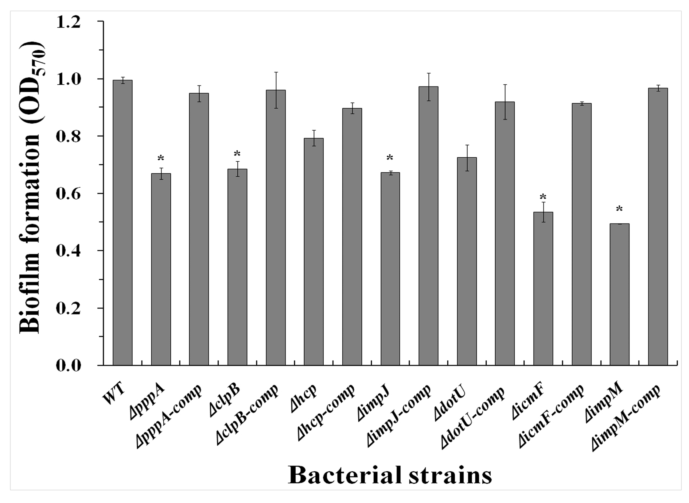
2.5. T6SS Affected the Biofilm Formation of Aaa Strain RS2

After incubation at 30 °C for 48 h without agitation, the quantification of the biofilm confirmed that mutants ΔpppA, ΔclpB, ΔimpJ, ΔicmF, and ΔimpM reduced biofilm adhesion in microtitre plates while the optical density (OD570) value were measured and the results listed in Figure 7 were 0.67, 0.69, 0.79, 0.67, 0.72, 0.53 and 0.49, respectively. The wild-type, strain RS-2 produced a stronger biofilm while the OD570 value for stained biofilm was 0.99, which was significantly (p < 0.05) higher than that of ΔpppA, ΔclpB, ΔimpJ, ΔicmF, and ΔimpM.
However, mutants Δhcp and ΔdotU had statistically similar OD570 value compared with RS-2 wild-type, indicating that hcp and dotU genes did not significantly affect the formation of biofilm. Similar to the wild-type strain, complementation of the mutants ΔpppA, ΔclpB, ΔimpJ, ΔicmF and ΔimpM showed compatible biofilm-forming capability (p > 0.05). Additionally, there were no significant (p > 0.05) differences in biofilm formation between the wild-type strain and the other single T6SS gene mutants of Aaa strain RS-2. Taken together, these results indicate that these T6SS genes might be involved in the biofilm formation of Aaa strainRS-2.

2.6. T6SS Affected the Swimming Motility of Aaa Strain RS2

The effects of T6SS on the motility of the Aaa strain RS2 were determined by measuring the diameter of the area covered by swimming bacteria on LB plates with 0.3% agar after 48 h of incubation and the results are shown in Figure 8.
Results from the swimming assays showed that mutant ΔclpB, Δhcp and ΔicmF had lost motility that was restored upon complementation. The colony diameters of ΔpppA, ΔclpB, Δhcp and ΔicmF were 1.5, 1.4, 1.33, and 1.4 cm, which were decreased by 37.5%, 41.66%, 44.59% and 41.66%, respectively compared with the wild-type strain RS-2, suggesting that these genes might be related to bacterial swimming. However, no significant difference was found between the colony diameter of the wild-type strain and the other mutants (ΔimpJ, ΔdotU and ΔimpM), indicating that the three T6SS genes did not significantly affect the swimming of Aaa strain RS-2.

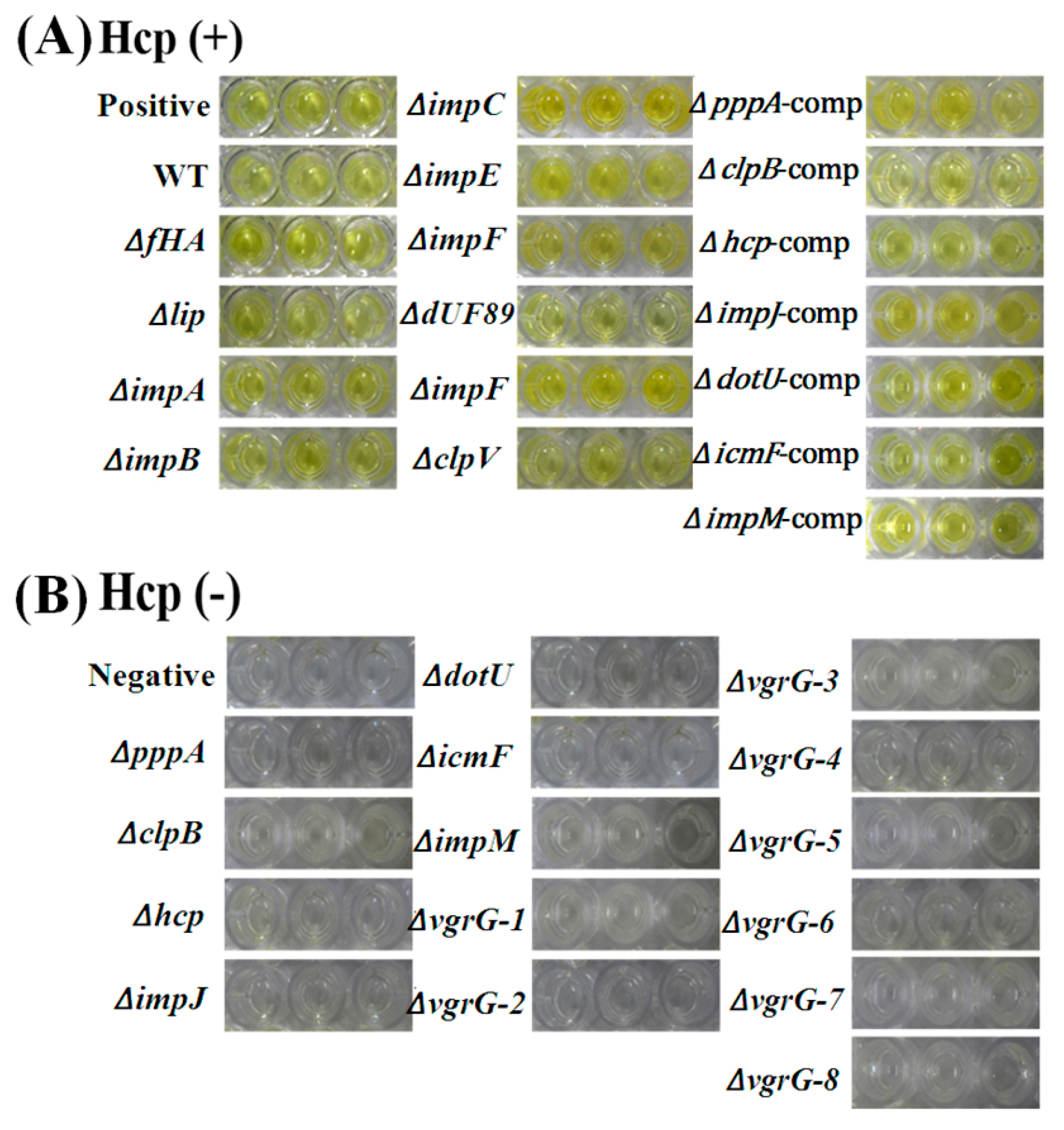
2.7. T6SS Affected Hcp Secretion of Aaa Strain RS2

The effects of T6SS on Hcp effectors protein secretion were examined in the Aaa strain RS-2 based on an ELISA experiment with polyclonal rabbit serum, which was performed using the purified Hcp-His fusion protein at a dilution of 5000. Results are shown in Figure 9. It can be seen that there was a positive (P/N > 2.1) ELISA reaction for the mutants ΔfHA, Δlip, ΔimpA, ΔimpB, ΔimpE, ΔimpF, ΔdUF879, ΔimpH, ΔclpV and the wild-type. Furthermore, the optical density (OD450) for the Hcp secretion of ΔfHA, Δlip, ΔimpA, ΔimpB, ΔimpE, ΔimpF, ΔdUF879, ΔimpH, ΔclpV were 0.361, 0.368, 0.361, 0.359, 0.352, 0.350, 0.355, 0.351, 0.367, 0.364, respectively, while the OD450 of the wild-type strain was 0.413 (Figure 9). Obviously, these T6SS mutants did not reduce significantly the OD450 values (p ≤ 0.5) compared to the wild-type, indicating that mutations of these genes did not affect the secretion of Hcp protein in strain RS-2. In contrast, there was a negative reaction (P/N ≤ 1.5) for the other 15 mutants ΔpppA, ΔclpB, Δhcp, ΔimpJ, ΔdotU, ΔicmF, ΔimpM, ΔvgrG-1, ΔvgrG-2, ΔvgrG-3, ΔvgrG-4, ΔvgrG-5, ΔvgrG-6, ΔvgrG-7 and ΔvgrG-8, while the OD450 values were significantly lower than those of the wild-type, indicating that the 15 genes may significantly affect the secretion of Hcp protein in the RS-2 strain. In addition, there was a positive ELISA reaction for the complementation ΔpppA-comp, ΔclpB-comp, Δhcp-comp, ΔimpJ-comp, ΔdotU-comp, ΔicmF-comp and ΔimpM-comp, while they had similar OD450 values with the wild-type of the Aaa RS-2 strain.

3. Discussion

T6SSs have been shown to be involved in the virulence of a wide range of gram-negative bacteria including plant and animal pathogens such as Edwardsiella tarda, Vibrio cholera, Pseudomonas aeruginosa, Burkholderia mallei, avian pathogenic Escherichia coli, Xanthomonas oryzae, Pectobacterium atrosepticum and Agrobacterium tumefaciens [14,20,21,22,23,24,25]. The T6SS is typically encoded by clusters of contiguous genes and composed of at least 13 conserved proteins along with a variable complement of accessory elements [21,26]. Gram-negative bacteria utilize T6SS as a functional apparatus to expel virulence factors from cytoplasm to the extracellular surroundings and within a eukaryotic target cell for their virulence and survival in hosts [14,20,21,27,28].
In the current study, 25 T6SS genes that are highly homologous to that of the Aaa strain RS-1 were identified in the Aaa strain RS-2 based on genome-wide in silico analysis. Furthermore, the involvement of these T6SS genes in the pathogenicity of Aaa strain RS-2 was justified by the difference in virulence-related phenotypes between the constructed mutants and the wild-type. In agreement with the results of this study, our previous studies not only confirmed the in silico prediction of T6SS component OmpA/MotB and ClpB based on LC-MS/MS analysis of outer membrane proteins in theAaa strain RS-1, but also revealed that T6SS have a strong response to in vivo infection and different stresses based on RNA-Seq analysis [26,29]. These results highlighted that T6SS are highly associated with the virulence of rice bacterial brown stripe pathogen Aaa.
In general, this study found that there were significant differences in virulence among the 25 T6SS genes mutants of the Aaa strain RS-2. This is consistent with the results of our previous studies, which indicated that T6SS genes had differential responses to in vivo infection of the Aaa strain RS-1 to rice host [8,11,29,30]. Furthermore, this study indicated that the virulence of the Aaa strain RS-2 was significantly reduced by the mutation of 15 T6SS genes, but unaffected by the other T6SS genes based on seed transmission assays. This reveals the complexity of the T6SS genes in the virulence of the Aaa strain RS-2, which makes it necessary to examine the function of each T6SS gene in these kind of studies. Indeed, T6SS has been also reported to be involved in a series of cellular activities such as translocation function, stress resistance, extracellular protease production and intracellular communication, which are unrelated to pathogenicity [16,20,21,23]. However, this study showed that 15 T6SS genes of strain RS-2 are highly associated with the virulence of Aaa.
In agreement with the results of this study, Sheng et al. [31] reported that the pppA contributes to virulence actions such as biofilm formation, motility, and cell aggregation. Zhang et al. [19] demonstrated that the deletion of clpB significantly affected bacterial growth, virulence, exopolysaccharide production, biofilm formation of the Aaa strain RS-1. Furthermore, the IcmF-DotU complex consists of the main transmembrane structure in the needle-like T6SS apparatus [15,32]. Thus, the contribution of dotU and icmF to bacterial virulence was mainly due to their regulation in secretion of the putative effector proteins haemolysin co-regulated proteins (Hcp) and valine-glycine repeat (VgrG), which have been shown to play an important role in bacterial virulence [12,14]. In addition, the specific functions of impJ and impM in T6SS is still not known, although impJ has been regarded as a trimeric cytoplasmic protein that interacts with the membrane and phage-like complexes in T6SS [33] and impM is responsible for encoding the member of a Type VI secretion-associated protein that belongs to theBMA_A0400 family in T6SS containing bacteria [14].
Furthermore, results of the ELISA experiment indicated that no Hcp effector protein was detected in the secretory proteins from the mutants of 15 virulence -associated T6SS genes, while their complementation showed a positive reaction similar to the wild-type strain RS-2. This result is consistent with previous studies, which found that T6SS has also been reported to function through the secretion of effector proteins such as Hcp. Indeed, besides its structural function in T6SS, the secretion of Hcp is thought to be an effector protein as the hallmark of a functional T6SS in many bacteria including V. cholerae, A. hydrophila, P. aeruginosa and B. pseudomallei, E. coli, A. tumefaciens [12,27,34,35,36]. Therefore, the results of this study supported the evidence that the role of T6SS in bacterial virulence is highly associated with the secretion of effector Hcpin strain RS-2.
However, there was a difference in pathogenicity among the 15 virulence-associated T6SS genes, while the mutation of the 7 T6SS genes ΔpppA, ΔclpB, Δhcp, ΔimpJ, ΔdotU, ΔicmF, ΔimpM caused a greater reduction in plant height than that of the 8 ΔvgrG (1–8) genes, revealing that the other virulence determinants such as growth, biofilm, swimming other than Hcp effector proteins may also be attributed to the function of T6SS. Indeed, the greater reduction in virulence may be at least partially explained by the result that bacterial growth was impaired by the 7 T6SS genes mutants, but was unaffected by the 8 ΔvgrG (1–8) genes mutants compared with the wild-type. Interestingly, mutation of the 7 T6SS genes showed a differential effect on bacterial swimming and biofilm formation, which has been widely reported to be highly associated with bacterial virulence [11,16,37,38,39]. Furthermore, in agreement with the results of this study, the T6SS gene clusters have been shown to be involved in the biofilm formation of other bacteria such A. citrulli [6], P. fluorescens [40], V. alginolyticus [16] and V. parahaemolyticus [41]. This result reveals the complexity of T6SS in the virulence mechanism of the Aaa strain RS-2.

4. Materials and Methods

4.1. Bacterial Strains, Growth Media and Inoculum Preparation

The bacterial strains and plasmids used in this study are described in Supplementary Table S2. The Aaa wild-type strain RS-2 and mutant strains were cultured in Luria–Bertani (LB) agar or broth medium [42] at 30 °C. Escherichia coli strains were grown in LB agar or broth medium at 37 °C. For inoculation, bacterial strains were grown in LB broth for 48 h, serially diluted into ddH2O, and the final concentration of bacterial suspension was adjusted to an approximate optical density (OD600) of 0.6 (~1 × 10⁸ CFU/mL) with a spectrophotometer (Perkin Elmer Lambda 35 UV/VIS, Waltham, MA, USA). When required, the culture media were supplemented with the following concentrations: ampicillin (Amp), 100 µg mL−1; kanamycin (Km), 50 µg mL−1; rifampicin (Rif), 50 µg mL−1; and Chloramphenicol (Chl), 3.4 µg mL−1.

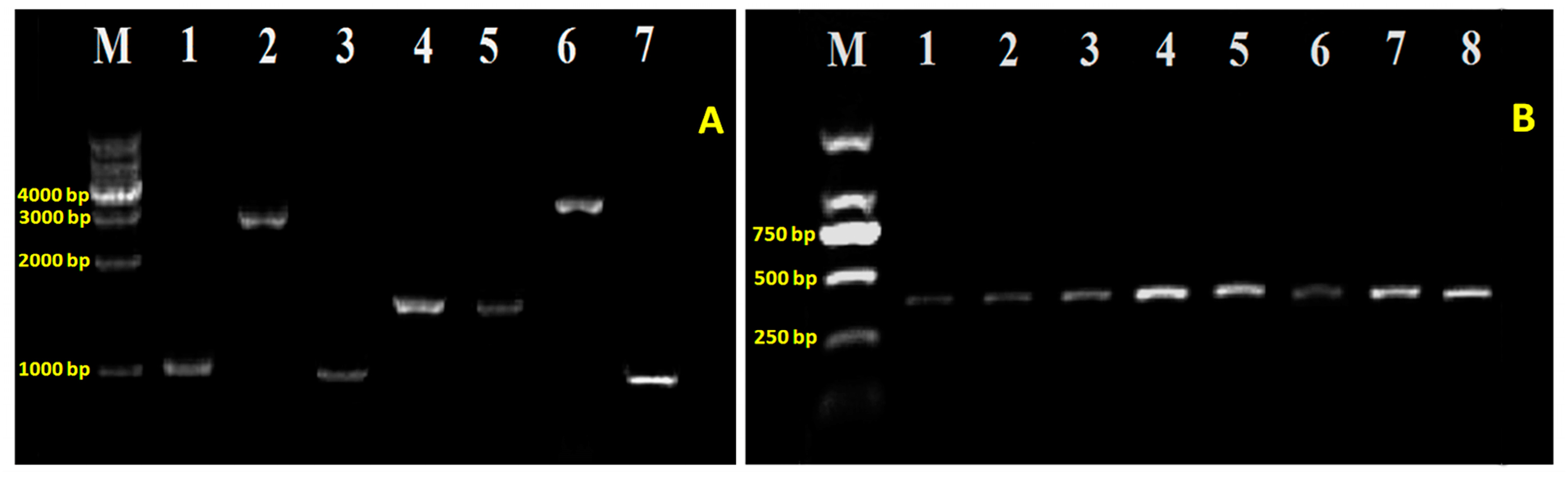
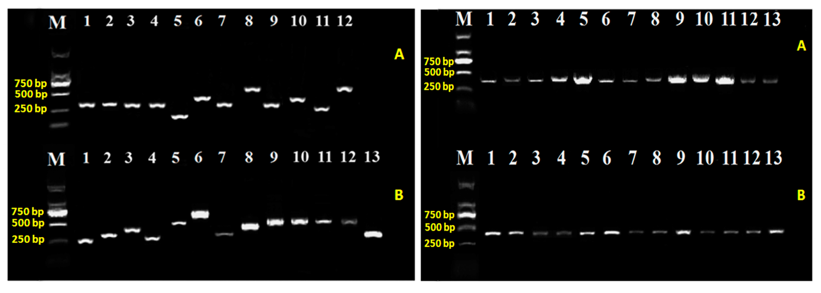
4.2. DNA Extraction and Amplification

Genomic DNA was extracted using the TIANamp bacteria DNA kit (Spin Column) (Tiangen Biotech (Beijing) Co, Ltd., Beijing, China) following the protocol for isolating genomic DNA from bacteria. Bacterial plasmid DNA was isolated using the EZNA® Plasmid DNA Mini Kit I (Omega Bio-tek Inc., Norcross, GA, USA). The concentration and purity of the DNA was measured using the Nano Drop 2000 spectrophotometer (Thermo Fisher Scientific Inc., Waltham, MA, USA). All conventional PCR reactions were carried out in a Bioer XP Thermal Cycler (Hangzhou Bioer Tech. Co., Ltd., Hangzhou, China). Amplification of the DNA was performed in 50 µL total volumes with 2× TSINGKE Master Mix (TsingKe Biological Technology, Beijing, China) while the PCR conditions were 94 °C for 10 min followed by 35 cycles of denaturation at 94 °C for 30 s, annealing at 57~62 °C (specific for each gene) for 30 s, and extension at 72 °C for 1 min/kb.

4.3. In Silico Analysis of Type VI Secretion Loci

A genome wide analysis was performed in this study to reveal the veil of T6SS in the A. avenae subsp. avenae strain RS-2. The components and location of T6SS homologs in Aaa strain RS-2 were determined by BLASTN and TBLASTX searching, in which T6SS information described in [11,30] was used as bait sequences against strain RS-2 genome. In addition, to identify and compare the homology of T6SS genes across A. avenae subsp. avenae strain RS-2, A. avenae subsp. avenae strain RS-1, A. avenae subsp. avenae ATCC 19860 and A.citrulli AAC00-1, a phylogenetic profile—which is matrix of the presence/absence of genes across the above bacteria—was created. A phylogenetic tree was built using the neighbor-joining method in MEGA6 [43] with 1000 bootstraps.

4.4. Generation of T6SS Mutants and Complementation

To examine the role of T6SS in the virulence mechanism of the Aaa strain RS-2, an insertional mutagenesis on target gene was generated by suicide plasmid pJP5603 [44] through homologous recombination on the background of wild-type strain RS-2. In-frame deletion of T6SS genes and their complementation were performed following the procedure of Liu et al. [16]. In brief, the internal DNA fragment of each gene of T6SS was PCR amplified using genomic DNA of wild-type strain RS-2 as template. The PCR product was cloned into pGEM-T Easy vector (Promega Corporation, Madison, WI, USA), verified by sequencing and digested with appropriate restriction enzymes (Supplementary Tables S2 and S3), and then ligated into the suicide vector pJP5603. The resulting plasmid constructs were moved into the wild-type strain RS-2 by biparental mating using E. coli strain S17-1 [45] as a donor. Mutant checking of 25 T6SS genes were confirmed by PCR amplification using primers flanking the genes of interest. For complementation, the whole open reading frame of these genes along with 500 bp upstream of the start codon including its native promoter were amplified by PCR using wild-type strain RS-2 as template and sub-cloned into the expression vector pRADK [46] after sequencing verification. The resulting constructs were introduced into the corresponding mutants by filter mating [47] and the complementation of the corresponding mutants was selected by Chl + Km + Rif resistance. Primers and the restriction enzymes used in making of T6SS mutants and their complementation were listed in Supplementary Table S3.

4.5. Seedling Pathogenicity in Rice

To determine the effect of T6SS on the seedling pathogenicity of bacterial brown stripe of rice, seed transmission assays were carried out as described in Li et al. [9] with some modifications. Briefly, germinated rice seeds (cv. II You 023, n = 100/mutant) were inoculated with gentle agitation using immersion in 10 mL of a cell suspension containing approximately ~1 × 10⁸ CFU/mL (OD600 = 0.6) of Aaa wild-type strain RS-2 and each T6SS mutant for 4 h. Seeds treated with double-distilled water (ddH2O) in the same manner were also used as negative control. After inoculation, bacterial suspensions were discarded and the seeds were then air dried at room temperature for 24 h. The inoculated dried seeds were planted in plate moisture method containing 0.5% agar (15 seeds per plate) and incubated under greenhouse conditions (28 ± 2 °C, 80% humidity) with a 14:10 h light–dark photoperiod. Seedling emergence and plant height was recorded after 5 days of sowing. This experiment was repeated three times with three replications of each treatment.

4.6. Bacterial Growth Assays

Bacterial growth was assessed by inoculating 5.0 µL cell suspensions (overnight broth cultures adjusted to OD600 = 0.6) into 5 mL of LB broth. Bacterial numbers were determined by measuring absorbance at 600 nm using a Thermo Multiskan EX Micro plate Photometer (Perkin Elmer Lambda 35 UV/VIS, Thermo Fisher Scientific Inc.) after incubation at 200 rpm, 30 °C, for 0.0, 1.5, 3.0, 6.0, 12.0, 24.0, and 48.0 h, respectively. Only the LB broth was used as the negative control.

4.7. Biofilm Formation Measurement

Biofilm formation assays were performed on T6SS mutants of the Aaa strain RS-2 in 96-well microtitre plates (Corning-Costar Corp., Corning, NY, USA) using the method of Peeters et al. [48]. Briefly, the overnight cell suspension of the Aaa wild-type strain RS-2 and mutants were re-cultured into a fresh LB broth containing appropriate antibiotics with a 1:100 dilution under shaking to mid exponential growth. Then each well was inoculated with 100 µL of approximately ~1 × 10⁸ CFU/mL (OD600 = 0.6) bacterial suspension and incubated at 30 °C for 48 h of adhesion without agitation while twelve wells filled with sterile ddH2O served as blanks. Culture media were then poured out and each well in the plates was washed three times with sterile ddH2O. Following air-dried for 30 min, each well was stained with 125 µL of 0.1% (w/v) crystal Violet (CV) solution for 45 min at room temperature. The unbound crystal Violet was removed and then washed with ddH2O. To solubilize the crystal Violet stained cells, 150 µL of 33% acetic acid was added into each well. Bacterial biofilm was quantified by measuring their optical density at 590 nm using a Thermo Multiskan EX Micro plate Photometer (Thermo Fisher Scientific Inc.). Twelve replications of each treatment were used for quantitative measurement in the three repeated experiments.

4.8. Motility Assays

Bacteria were cultured overnight in LB broth supplemented with appropriate antibiotic at 30 °C in a shaker, and then centrifuged down, washed and diluted to OD600 = 0.6 in sterile water. The media for motility assays was LB containing 0.3% agar for swimming as described by Liu et al. [49]. Five µL cell suspensions of wild-type strain RS-2 and mutants were spotted on the center of each swimming plate. After 48 h of incubation, the colony diameter was measured. This assay was repeated three times independently with three replications of each treatment.

4.9. Measurement of the Secreted Hcp by ELISA

Enzyme-linked immune sorbent assay (ELISA) was performed for the measurement of the secreted Hcp, which has been found to be associated with the adaptation to various effector and bacterial virulence [26,29,30]. The standard ELISA was conducted in a 96 microtiter plate as described by Slutzki et al. [50]. Briefly, one milliliter of overnight and 30 °C bacterial culture (approximately OD600 = 0.6) was harvested by centrifugation at 4000× g for 10 min and the supernatant was then filtered through 0.22 µm filter. The microtiter plates were coated with 150 µL of filtered antigen diluted 10 times with coating buffer and incubated overnight at 4 °C. The plates were blocked with 175 µL/well of blocking buffer (phosphate-buffered saline (PBS), 10 mM CaCl2, 1% bovine serum albumin (BSA), 0.05% Tween 20) for 1 h at 37 °C and then washed with washing buffer. For the detection of Hcp effector protein, the polyclonal antibody Hrp-conjugated Goat Anti-Rabbit IgG at a 1:5000 dilution from Shanghai Health Company (Shanghai, China) was used. After incubation at 37 °C for periods ranging from 2 to 24 h, the enzymatic reaction (colour development) was recorded and the optical density was measured using a microplate reader (Multiscan microtiter plate reader) set at 450 nm while absorbance (≥0.5) after subtraction of values for negative control samples were considered as positive.

4.10. Statistical Analyses

The software STATGRAPHICS Plus version 4.0 (Copyright manugistics Inc., Rockville, MD, USA) was used to perform the statistical analyses. The levels of significance (p < 0.05) of the main treatments and their interactions were calculated by analysis of variance after testing for normality and variance homogeneity.

5. Conclusions

The current study revealed the diversity of T6SS genes in the pathogenicity of the Aaa strain RS-2 to rice seedlings. Indeed, the virulence was attenuated by the mutation of 15 T6SS genes, but unaffected by the mutation of the other10 T6SS genes. Furthermore, this study found that the mutation of the 15 virulence-associated T6SS genes caused a significant reduction in Hcp secretion, while some of these resulted in a significant reduction in the growth, biofilm formation and swimming of the Aaa strain RS-2. In addition, our preliminary study found that there was a difference in the virulence of strain RS-1 and RS-2 although both of them have the similar T6SS of high homology, indicating that there may be other virulence factors involved in the pathogenicity of Aaa. However, this study clearly highlighted that T6SS played vital roles in the virulence of this rice pathogenic bacteria, which may function by affecting bacterial growth, biofilm formation, swimming ability and the secretion of Hcp effectors.

Supplementary Materials

Supplementary materials can be found at www.mdpi.com/1422-0067/18/10/2024/s1.

Acknowledgments

This work was supported by National Natural Science Foundation of China (31571971, 31371904), Zhejiang Provincial Project (2017C02002), Shanghai Agricultural Basic Research Project (2014:7-3-1), the Zhejiang Provincial Natural Science Foundation of China (R13C140001), the Fundamental Research Funds for the Central Universities (2017QNA6012), the Agricultural Ministry of China (nyhyzx201303015), Dabeinong Funds for Discipline Development and Talent Training in Zhejiang University, Key Subject Construction Program of Zhejiang for Modern Agricultural Biotechnology and Crop Disease Control (2010DS700124-KF1710), China Postdoctoral Science Foundation (2017M612003).

Author Contributions

Conceived and designed the experiments: Bin Li, Guochang Sun. Performed the experiments: Yingzi Yang, Md. Mahidul Islam Masum, Yushi Fang, Yang Zhang. Analyzed the data: Md. Mahidul Islam Masum, Yingzi Yang, Yang Zhang, Bin Li. Contributed reagents/materials/analysis tools: Ogunyemi SolabomiOlaitan, Jie Chen, Yanli Wang, Bin Li, Guochang Sun. Wrote the paper: Md. Mahidul Islam Masum, Bin Li, Wen Qiu.

Conflicts of Interest

The authors declare no conflict of interest.

References

  1. Song, W.Y.; Kim, H.M.; Hwang, C.Y.; Schaad, N.W. Detection of Acidovorax avenae subsp. avenae in rice seeds using BIO-PCR. J. Phytopathol. 2004, 152, 667–676. [Google Scholar] [CrossRef]
  2. Shakya, D.D.; VInther, F.; Mathur, S.B. World wide distribution of a bacterial stripe pathogen of rice identified as Pseudomonas avenae. J. Phytopathol. 1985, 114, 256–259. [Google Scholar] [CrossRef]
  3. Cui, Z.; Ojaghian, M.R.; Tao, Z.; Kakar, K.U.; Zeng, J.; Zhao, W.; Duan, Y.; Vera Cruz, C.M.; Li, B.; Zhu, B.; et al. Multiplex pcr assay for simultaneous detection of six major bacterial pathogens of rice. J. Appl. Microbiol. 2016, 120, 1357–1367. [Google Scholar] [CrossRef] [PubMed]
  4. Wang, Y.; Zhou, Q.; Li, B.; Liu, B.; Wu, G.; Ibrahim, M.; Xie, G.; Li, H.; Sun, G. Differentiation in MALDI-TOF MS and FTIR spectra between two closely related species Acidovoraxoryzae and Acidovorax citrulli. BMC Microbiol. 2012, 12, 182. [Google Scholar] [CrossRef] [PubMed]
  5. Kadota, I.; Ohuchi, A.; Nishiyama, K. Serological properties and specificity of Pseudomonas avenae Manns 1909, the causal agent of bacterial brown stripe of rice. Jpn. J. Phytopathol. 1991, 57, 268–273. [Google Scholar] [CrossRef]
  6. Tian, Y.L.; Zhao, Y.Q.; Wu, X.R.; Liu, F.Q.; Hu, B.S.; Walcott, R.R. The type VI protein secretion system contributes to biofilm formation and seed-to-seedling transmission of Acidovorax citrulli on melon. Mol. Plant Pathol. 2015, 16, 38–47. [Google Scholar] [CrossRef] [PubMed]
  7. Xie, G.-L.; Zhang, G.-Q.; Liu, H.; Lou, M.-M.; Tian, W.-X.; Li, B.; Zhou, X.-P.; Zhu, B.; Jin, G.-L. Genome sequence of the rice-pathogenic bacterium Acidovorax avenae subsp. avenae RS-1. J. Bacteriol. 2011, 193, 5013–5014. [Google Scholar] [CrossRef] [PubMed]
  8. Yang, C.; Li, B.; Ge, M.; Zhou, K.; Wang, Y.; Luo, J.; Ibrahim, M.; Xie, G.; Sun, G. Inhibitory effect and mode of action of chitosan solution against rice bacterial brown stripe pathogen Acidovorax avenae subsp. avenae RS-1. Carbohydr. Res. 2014, 391, 48–54. [Google Scholar] [CrossRef] [PubMed]
  9. Li, B.; Liu, B.; Yu, R.; Tao, Z.; Wang, Y.; Xie, G.; Li, H.; Sun, G. Bacterial brown stripe of rice in soil-less culture system caused by Acidovorax avenae subsp. avenae in China. J. Gen. Plant Pathol. 2011, 77, 64–67. [Google Scholar] [CrossRef]
  10. Li, B.; Wang, L.; Ibrahim, M.; Ge, M.; Wang, Y.; Mannan, S.; Asif, M.; Sun, G. Membrane protein profiling of Acidovorax avenae subsp. avenaeunder various growth conditions. Arch. Microbiol. 2015, 197, 673–682. [Google Scholar] [CrossRef] [PubMed]
  11. Li, B.; Ge, M.; Zhang, Y.; Wang, L.; Ibrahim, M.; Wang, Y.; Sun, G.; Chen, G. New insights into virulence mechanisms of rice pathogen Acidovorax avenae subsp. avenaestrain RS-1 following exposure to β-lactam antibiotics. Sci. Rep. 2016, 6, 22241. [Google Scholar] [CrossRef] [PubMed]
  12. Mougous, J.D.; Cuff, M.E.; Raunser, S.; Shen, A.; Zhou, M.; Gifford, C.A.; Goodman, A.L.; Joachimiak, G.; Ordoñez, C.L.; Lory, S.; et al. A virulence locus of Pseudomonas aeruginosa encodes a protein secretion apparatus. Science 2006, 312, 1526–1530. [Google Scholar] [CrossRef] [PubMed]
  13. Costa, T.R.D.; Felisberto-Rodrigues, C.; Meir, A.; Prevost, M.S.; Redzej, A.; Trokter, M.; Waksman, G. Secretion systems in gram-negative bacteria: Structural and mechanistic insights. Nat. Rev. Microl. 2015, 13, 343–359. [Google Scholar] [CrossRef] [PubMed]
  14. Pukatzki, S.; Ma, A.T.; Sturtevant, D.; Krastins, B.; Sarracino, D.; Nelson, W.C.; Heidelberg, J.F.; Mekalanos, J.J. Identification of a conserved bacterial protein secretion system in Vibrio cholerae using the Dictyostelium host model system. Proc. Natl. Acad. Sci. USA 2006, 103, 1528–1533. [Google Scholar] [CrossRef] [PubMed]
  15. Leiman, P.G.; Basler, M.; Ramagopal, U.A.; Bonanno, J.B.; Sauder, J.M.; Pukatzki, S.; Burley, S.K.; Almo, S.C.; Mekalanos, J.J. Type VI secretion apparatus and phage tail-associated protein complexes share a common evolutionary origin. Proc. Natl. Acad. Sci. USA 2009, 106, 4154–4159. [Google Scholar] [CrossRef] [PubMed]
  16. Liu, H.; Gu, D.; Sheng, L.; Wang, Q.; Zhang, Y. Investigation of the roles of T6SS genes in motility, biofilm formation, and extracellular protease asp production in Vibrio alginolyticus with modified gateway-compatible plasmids. Lett. Appl. Microbiol. 2012, 55, 73–81. [Google Scholar] [CrossRef] [PubMed]
  17. Miyata, S.T.; Kitaoka, M.; Brooks, T.M.; McAuley, S.B.; Pukatzki, S. Vibrio cholerae requires the type VI secretion system virulence factor Vasx to kill Dictyostelium discoideum. Infect. Immun. 2011, 79, 2941–2949. [Google Scholar] [CrossRef] [PubMed]
  18. Weber, B.; Hasic, M.; Chen, C.; Wai, S.N.; Milton, D.L. Type VI secretion modulates quorum sensing and stress response in Vibrio anguillarum. Environ. Microbiol. 2009, 11, 3018–3028. [Google Scholar] [CrossRef] [PubMed]
  19. Zhang, Y.; Zhang, F.; Li, B.; Yang, Y.Z.; Ibrahim, M.; Fang, Y.S.; Qiu, W.; Masum, M.M.I.; Oliva, R. Characterization and functional analysis of clpB gene from Acidovorax avenae subsp. avenae RS-1. Plant Pathol. 2017. [Google Scholar] [CrossRef]
  20. Bingle, L.E.; Bailey, C.M.; Pallen, M.J. Type VI secretion: A beginner’s guide. Curr. Opin. Microbiol. 2008, 11, 3–8. [Google Scholar] [CrossRef] [PubMed]
  21. Boyer, F.; Fichant, G.; Berthod, J.; Vandenbrouck, Y.; Attree, I. Dissecting the bacterial type VI secretion system by a genome wide in silico analysis: What can be learned from available microbial genomic resources? BMC Genom. 2009, 10. [Google Scholar] [CrossRef] [PubMed]
  22. Schell, M.A.; Ulrich, R.L.; Ribot, W.J.; Brueggemann, E.E.; Hines, H.B.; Chen, D.; Lipscomb, L.; Kim, H.S.; Mrázek, J.; Nierman, W.C.; et al. Type VI secretion is a major virulence determinant in Burkholderia mallei. Mol. Microbiol. 2007, 64, 1466–1485. [Google Scholar] [CrossRef] [PubMed]
  23. Shrivastava, S.; Mande, S.S. Identification and functional characterization of gene components of type VI secretion system in bacterial genomes. PLoS ONE 2008, 3, e2955. [Google Scholar] [CrossRef] [PubMed]
  24. Pukatzki, S.; McAuley, S.B.; Miyata, S.T. The type VI secretion system: Translocation of effectors and effector-domains. Curr. Opin. Microbiol. 2009, 12, 11–17. [Google Scholar] [CrossRef] [PubMed]
  25. Zhou, Y.; Tao, J.; Yu, H.; Ni, J.J.; Zeng, L.B.; Teng, Q.H.; Kim, K.S.; Zhao, G.P.; Guo, X.; Yao, Y. Hcp family proteins secreted via the type VI secretion system coordinately regulate Escherichia coli K1 interaction with human brain microvascular endothelial cells. Infect. Immun. 2012, 80, 1243–1251. [Google Scholar] [CrossRef] [PubMed]
  26. Ibrahim, M.; Shi, Y.; Qiu, H.; Li, B.; Jabeen, A.; Li, L.P.; Liu, H.; Kube, M.; Xie, G.; Wang, Y.; et al. Differential expression of in vivoand in vitro protein profile of outer membrane of Acidovorax avenae subsp. avenae. PLoS ONE 2012, 7. [Google Scholar] [CrossRef] [PubMed]
  27. Burtnick, M.N.; Brett, P.J.; Harding, S.V.; Ngugi, S.A.; Ribot, W.J.; Chantratita, N.; Scorpio, A.; Milne, T.S.; Dean, R.E.; Fritz, D.L.; et al. The cluster 1 type VI secretion system is a major virulence determinant in Burkholderia pseudomallei. Infect. Immun. 2011, 79, 1512–1525. [Google Scholar] [CrossRef] [PubMed]
  28. Alteri, C.J.; Mobley, H.L. The versatile type VI secretion system. Microbiol. Spectr. 2016, 4. [Google Scholar] [CrossRef]
  29. Li, B.; Ibrahim, M.; Ge, M.; Cui, Z.; Sun, G.; Xu, F.; Kube, M. Transcriptome analysis of Acidovorax avenae subsp. avenae cultivated in vivo and co-culture with Burkholderia seminalis. Sci. Rep. 2014, 4, 5698. [Google Scholar] [CrossRef] [PubMed]
  30. Cui, Z.; Jin, G.; Li, B.; Kakar, K.; Ojaghian, M.; Wang, Y.; Xie, G.; Sun, G. Gene expression of type VI secretion system associated with environmental survival in Acidovorax avenae subsp. avenaeby principle component analysis. Int. J. Mol. Sci. 2015, 16, 22008–22026. [Google Scholar] [CrossRef] [PubMed]
  31. Sheng, L.; Lv, Y.; Liu, Q.; Wang, Q.; Zhang, Y. Connecting type VI secretion, quorum sensing, and c-di-GMP production in fish pathogen Vibrio alginolyticus through phosphatase Pppa. Vet. Microbiol. 2013, 162, 652–662. [Google Scholar] [CrossRef] [PubMed]
  32. Leung, K.Y.; Siame, B.A.; Snowball, H.; Mok, Y.-K. Type VI secretion regulation: Cross talk and intracellular communication. Curr. Opin. Microbiol. 2011, 14, 9–15. [Google Scholar] [CrossRef] [PubMed]
  33. Zoued, A.; Durand, E.; Bebeacua, C.; Brunet, Y.R.; Douzi, B.; Cambillau, C.; Cascales, E.; Journet, L. Tssk is a trimeric cytoplasmic protein interacting with components of both phage-like and membrane anchoring complexes of the type VI secretion system. J. Biol. Chem. 2013, 288, 27031–27041. [Google Scholar] [CrossRef] [PubMed]
  34. Fu, Y.; Waldor, M.K.; Mekalanos, J.J. Tn-seq analysis of Vibrio cholerae intestinal colonization reveals a role for T6SS-mediated antibacterial activity in the host. Cell Host Microbe 2013, 14, 652–663. [Google Scholar] [CrossRef] [PubMed]
  35. Suarez, G.; Sierra, J.C.; Kirtley, M.L.; Chopra, A.K. Role of Hcp, a type 6 secretion system effector, of Aeromonas hydrophila in modulating activation of host immune cells. Microbiology 2010, 156 Pt 12, 3678–3688. [Google Scholar] [CrossRef] [PubMed]
  36. Wu, H.Y.; Chung, P.C.; Shih, H.W.; Wen, S.R.; Lai, E.M. Secretome analysis uncovers an Hcp-family protein secreted via a type VI secretion system in Agrobacterium tumefaciens. J. Bacteriol. 2008, 190, 2841–2850. [Google Scholar] [CrossRef] [PubMed]
  37. Liu, L.Y.; Hao, S.; Lan, R.T.; Wang, G.X.; Xiao, D.; Sun, H.; Xu, J. The type VI secretion system modulates flagellar gene expression and secretion in Citrobacter freundii and contributes to adhesion and cytotoxicity to host cells. Infect. Immun. 2015, 83, 2596–2604. [Google Scholar] [CrossRef] [PubMed]
  38. Castiblanco, L.F.; Sundin, G.W. New insights on molecular regulation of biofilm formation in plant-associated bacteria. J. Integr. Plant Biol. 2016, 58, 362–372. [Google Scholar] [CrossRef] [PubMed]
  39. Bogino, P.C.; Oliva Mde, L.; Sorroche, F.G.; Giordano, W. The role of bacterial biofilms and surface components in plant-bacterial associations. Int. J. Mol. Sci. 2013, 14, 15838–15859. [Google Scholar] [CrossRef] [PubMed] [Green Version]
  40. Gallique, M.; Decoin, V.; Barbey, C.; Rosay, T.; Feuilloley, M.G.J.; Orange, N.; Merieau, A. Contribution of the Pseudomonas fluorescens MFE01 type VI secretion system to biofilm formation. PLoS ONE 2017, 12. [Google Scholar] [CrossRef] [PubMed]
  41. Mizan, M.F.R.; Jahid, I.K.; Kim, M.; Lee, K.H.; Kim, T.J.; Ha, S.D. Variability in biofilm formation correlates with hydrophobicity and quorum sensing among Vibrio parahaemolyticus isolates from food contact surfaces and the distribution of the genes involved in biofilm formation. Biofouling 2016, 32, 497–509. [Google Scholar] [CrossRef] [PubMed]
  42. Sambrook, J.; Fritsch, E.F.; Maniatis, T. Molecular Cloning:A Laboratory Manual; Cold Spring Harbor Laboratory Press: New York, NY, USA, 1989. [Google Scholar]
  43. Tamura, K.; Stecher, G.; Peterson, D.; Filipski, A.; Kumar, S. MEGA6: Molecular evolutionary genetics analysis version 6.0. Mol. Biol. Evol. 2013, 30, 2725–2729. [Google Scholar] [CrossRef] [PubMed]
  44. Penfold, R.J.; Pemberton, J.M. An Improved Suicide Vector for Construction of Chromosomal Insertion Mutations in Bacteria; Elsevier: Amsterdam, The Netherlands, 1992. [Google Scholar]
  45. Simon, R.; Priefer, U.; Puhler, A. A broad host range mobilization system for in vivogenetic engineering: Transposon mutagenesis in gram negative bacteria. Nat. Biotechnol. 1983, 1, 784–791. [Google Scholar] [CrossRef]
  46. Gao, G.; Lu, H.; Huang, L.; Hua, Y. Construction of DNA damage response gene pprI function-deficient and function-complementary mutants in Deinococcus radiodurans. Chin. Sci. Bull. 2005, 50, 311–316. [Google Scholar] [CrossRef]
  47. Smith, M.D.; Guild, W.R. Improved method for conjugative transfer by filter mating of Streptococcus pneumoniae. J. Bacteriol. 1980, 144, 457–459. [Google Scholar] [PubMed]
  48. Peeters, E.; Nelis, H.J.; Coenye, T. Comparison of multiple methods for quantification of microbial biofilms grown in microtiter plates. J. Microbiol. Methods 2008, 72, 157–165. [Google Scholar] [CrossRef] [PubMed]
  49. Liu, H.; Tian, W.-X.; Ibrahim, M.; Li, B.; Zhang, G.-Q.; Zhu, B.; Xie, G.-L. Chacterization of pilP, a gene required for twitching motility, pathogenicity, and biofilm formation of Acidovoraxavenaesubsp. Avenae RS-1. Eur. J. Plant Pathol. 2012, 134, 551–560. [Google Scholar] [CrossRef]
  50. Slutzki, M.; Barak, Y.; Reshef, D.; Schueler-Furman, O.; Lamed, R.; Bayer, E.A. Indirect elisa-based approach for comparative measurement of high-affinity cohesin-dockerin interactions. J. Mol. Recognit. 2012, 25, 616–622. [Google Scholar] [CrossRef] [PubMed]
Figure 1. Schematic diagram of the genetic organization of Acidovoraxavenae subsp. avenae strain RS-2 T6SS gene cluster. Genes are indicated by arrows and the direction of the arrows represent the direction of transcription of the genes in genome. “//” indicates the presence of other genes but not belongs to T6SS. The database of Clusters of Orthologous Groups of proteins (COGs) was achieved from the National Center of Biotechnology Information (ftp://ftp.ncbi.nih.gov/pub/COG/COG2014/static/lists/listAciave.html).
Ijms 18 02024 g001
Figure 2. Validation of the 25 T6SS using specific primers of each gene (left) and specific primer of Acidovorax avenae subsp. avenae strain RS-2 mutants (right); M: Marker DL2000; (A) 1~13: ΔpppA, ΔclpB, Δhcp, ΔfHA, Δlip, ΔimpJ, ΔdotU, ΔicmF, ΔimpM, ΔimpA, ΔimpB, ΔimpC, the wild-type; (B) 1~13: ΔimpE, ΔimpF, ΔdUF879, ΔimpH, ΔclpV, ΔvgrG-1, ΔvgrG-2, ΔvgrG-3, ΔvgrG-4, ΔvgrG-5, ΔvgrG-6, ΔvgrG-7, ΔvgrG-8.
Ijms 18 02024 g002
Figure 3. Validation for the complementation of T6SS mutants using specific primers of the 7 virulence-associated genes (left) and specific primer of Acidovorax avenae subsp. avenae strain RS-2 (right); (A) M: 1 kb DNA Ladder; 1~7: ΔpppA, ΔicmF, Δhcp, ΔdotU, ΔimpJ, ΔclpB, ΔimpM; (B) M: Marker 2000; 1~7: ΔpppA-comp, ΔicmF-comp, Δhcp-comp, ΔdotU-comp, ΔimpJ-comp, ΔclpB-comp, ΔimpM-comp, the wild-type.
Ijms 18 02024 g003
Figure 4. Seed-transmission assay for the virulence of the wild-type, the T6SS mutants and their corresponding complemented strains of Acidovorax avenae subsp. avenae strain RS-2 to rice seedling. Germinated rice seeds (cv. II You 023, n = 100/mutant) were inoculated with bacterial suspension of ~1×10⁸ colony forming units (CFU)/mL. Fifteen seeds were planted per sterile agar (0.5%) plate and seedlings were evaluated 5 days after planting on plate.
Ijms 18 02024 g004
Figure 5. The effect of T6SS genes of Acidovorax avenae subsp. avenae strain RS-2 on virulence phenotype in respect of plant height for the rice seedling. WT: wild-type strain RS-2; ddH2O: double distilled H2O; ΔpppA/ΔpppA-comp: mutant/complementation of pppA; ΔclpB/ΔclpB-comp: mutant/complementation of clpB; Δhcp/Δhcp-comp: mutant/complementation of hcp; ΔimpJ/ΔimpJ-comp: mutant/complementation of impJ; ΔdotU/ΔdotU-comp: mutant/complementation of dotU; ΔicmF/ΔicmF-comp: mutant/complementation of icmF; ΔimpM/ΔimpM-comp: mutant/complementation of impM. Vertical bars represent standard errors of the means. Asterik symbol above the data bars indicate a significant difference between the wild-type and T6SS mutants, complemented strains or negative control (** p < 0.01).
Ijms 18 02024 g005
Figure 6. Bacterial growth of the wild-type, the T6SS mutants and their corresponding complemented strains of Acidovorax avenae subsp. avenae strain RS-2. (AG) Indicate the comparisons between wild-type and the mutant/complementation of gene pppA, clpB, hcp, impJ dotU, icmF and impM, respectively. Means ± SEM are shown (n = 6). The experiment was repeated three times and here is presented one set of representative data.
Ijms 18 02024 g006
Figure 7. Quantification of biofilm produced by the wild-type, the T6SS mutants and the corresponding complemented strains of Acidovorax avenae subsp. avenae strain RS-2. Means ± SEM are shown (n = 6). Vertical bars represent standard errors of the means. Asterick indicated significant difference between the wild-type strain and the T6SS mutants, or the complemented strains (* 0.01 < p < 0.05).
Ijms 18 02024 g007
Figure 8. The mutation of the T6SS genes pppA, clpB, hcp and icmF inhibited the swimming motility of Acidovorax avenae subsp. avenae strain RS-2. Statistically significant difference (0.01 < * p < 0.05 by student’s t-test) was found between the T6SS mutants and the corresponding complemented strains or the wild-type (A). The swimming motility was determined by measuring the diameters of bacterial colony on the plates from three independent experiments (B).
Ijms 18 02024 g008
Figure 9. ELISA detection for the secretion of Hcp effector protein by the wild-type, the T6SS mutants and the corresponding complemented strains of Acidovorax avenae subsp. avenae strain RS-2.(A) The positive reaction (P/N value ≥ 2.1); (B) The negative reaction (P/N value < 2.1). The purified Hcp-His fusion protein and His protein were used as the positive and the negative control, respectively. The experiment was conducted three times with 3 replicates.
Ijms 18 02024 g009

