Article

Identification and Characterization of Lipopolysaccharide Induced TNFα Factor from Blunt Snout Bream, Megalobrama amblycephala

1
Department of Cell Biology, School of Life Sciences, Central South University, Changsha 410017, China
2
Center of Biological Experiments, School of Life Sciences, Central South University, Changsha 410017, China
3
Guangxi Key Laboratory of Aquatic Genetic Breeding and Healthy Aquaculture, Guangxi Academy of Fishery Sciences, Nanning 530021, China
*
Author to whom correspondence should be addressed.
Int. J. Mol. Sci. 2017, 18(2), 233; https://doi.org/10.3390/ijms18020233
Submission received: 19 October 2016 / Revised: 17 January 2017 / Accepted: 18 January 2017 / Published: 15 February 2017
(This article belongs to the Special Issue Lipopolysaccharides (LPSs))

Abstract

:
Lipopolysaccharide induced TNFα factor (LITAF) is an important transcription factor responsible for regulation of tumor necrosis factor α. In this study, a novel litaf gene (designated as Malitaf) was identified and characterized from blunt snout bream, Megalobrama amblycephala. The full-length cDNA of Malitaf was of 956 bp, encoding a polypeptide of 161 amino acids with high similarity to other known LITAFs. A phylogenetic tree also showed that Malitaf significantly clustered with those of other teleost, indicating that Malitaf was a new member of fish LITAF family. The putative maLITAF protein possessed a highly conserved LITAF domain with two CXXC motifs. The mRNA transcripts of Malitaf were detected in all examined tissues of healthy M. amblycephala, including kidney, head kidney, muscle, liver, spleen, gill, and heart, and with the highest expression in immune organs: spleen and head kidney. The expression level of Malitaf in spleen was rapidly up-regulated and peaked (1.29-fold, p < 0.05) at 2 h after lipopolysaccharide (LPS) stimulation. Followed the stimulation of Malitaf, Matnfα transcriptional level was also transiently induced to a high level (51.74-fold, p < 0.001) at 4 h after LPS stimulation. Taken together, we have identified a putative fish LITAF ortholog, which was a constitutive and inducible immune response gene involved in M. amblycephala innate immunity during the course of a pathogenic infection.

1. Introduction

Blunt snout bream (Megalobrama amblycephala) is one of the major economically important species in freshwater polyculture fish aquaculture in China [1]. It has been widely cultured because of its herbivorous habit, faster growth rate, and delicate flesh quality, as well as increasing demand in China during the last few decades [2,3]. In 2013, its production has reached 0.73 million tons, ranking seventh in Chinese freshwater fish production [4]. Associated with intensive farming, however, diseases caused by infectious bacteria, mainly Aeromonas hydrophila, frequently occur [5]. The infectious disease outbreak with quick spreading has led to serious economic losses in M. amblycephala culture industry. Innate immunity plays crucial roles in defense against bacterial infections in fish [6]. The innate immune response to a bacterial pathogen is characterized by the immediate release of pro-inflammatory cytokines, which act as key mediators of the immune system to eliminate the pathogen [7,8]. Finding more molecular components involved in M. amblycephala’s innate immunity, therefore, will facilitate our understandings in the largely unveiled complex immunity in fish.
Among the pro-inflammatory cytokines, tumor necrosis factor α (TNFα) has been confirmed to significantly trigger host immunity, increase phagocytic activity, and provoke the induction of inflammatory cytokines [9,10]. TNFα is one of the most well-known pleiotropic cytokines and is secreted by various cell types and can be regulated by different transcription factors, such as nuclear factor κB (NF-κB) [11], nuclear factor of activated T-cells (NF-AT) [12], activator protein 1 (AP-1) [13], and lipopolysaccharide induced TNFα factor (LITAF) [14]. Lipopolysaccharide induced TNFα factor (LITAF) is an important transcription factor mediating transcription of various inflammatory cytokines, especially TNFα [15]. It has been demonstrated that LITAF can directly interact with the signal transducer and activator of transcription (STAT) 6B and translocates into the nucleus where it binds to the promoter regions of TNFα and other cytokines to modulate their transcription [16]. LITAF was initially identified and characterized in the human macrophage cell line, THP-1 [17]. Since then, a large amount of LITAF homologues have been obtained in several aquatic animals including mollusk [18,19,20,21,22,23], arthropod [24,25], sea cucumber (Apostichopus japonicus) [26], and amphioxus (Branchiostoma belcheri) [27], suggesting a conserved function in innate immunity. Although the litaf gene has been characterized in several fish species, the knowledge of the LITAF orthologs in most teleosts is still limited [28,29,30,31].
Therefore, in the present study, we identified a novel litaf homolog cDNA (designated as Malitaf) in M. amblycephala, analyzed its phylogenetic relationship, and characterized its expression pattern in response to LPS stimulation. Considering that litaf is a vital regulator for tnfα expression, we therefore subsequently investigated the expression profile of tnfα (Matnfα) in M. amblycephala. To our knowledge, this is the first study in the LPS-induced response of Malitaf. The achieved results will provide a better understanding of the immune defense mechanisms and further improve the healthy management efficiency in this species.

2. Results

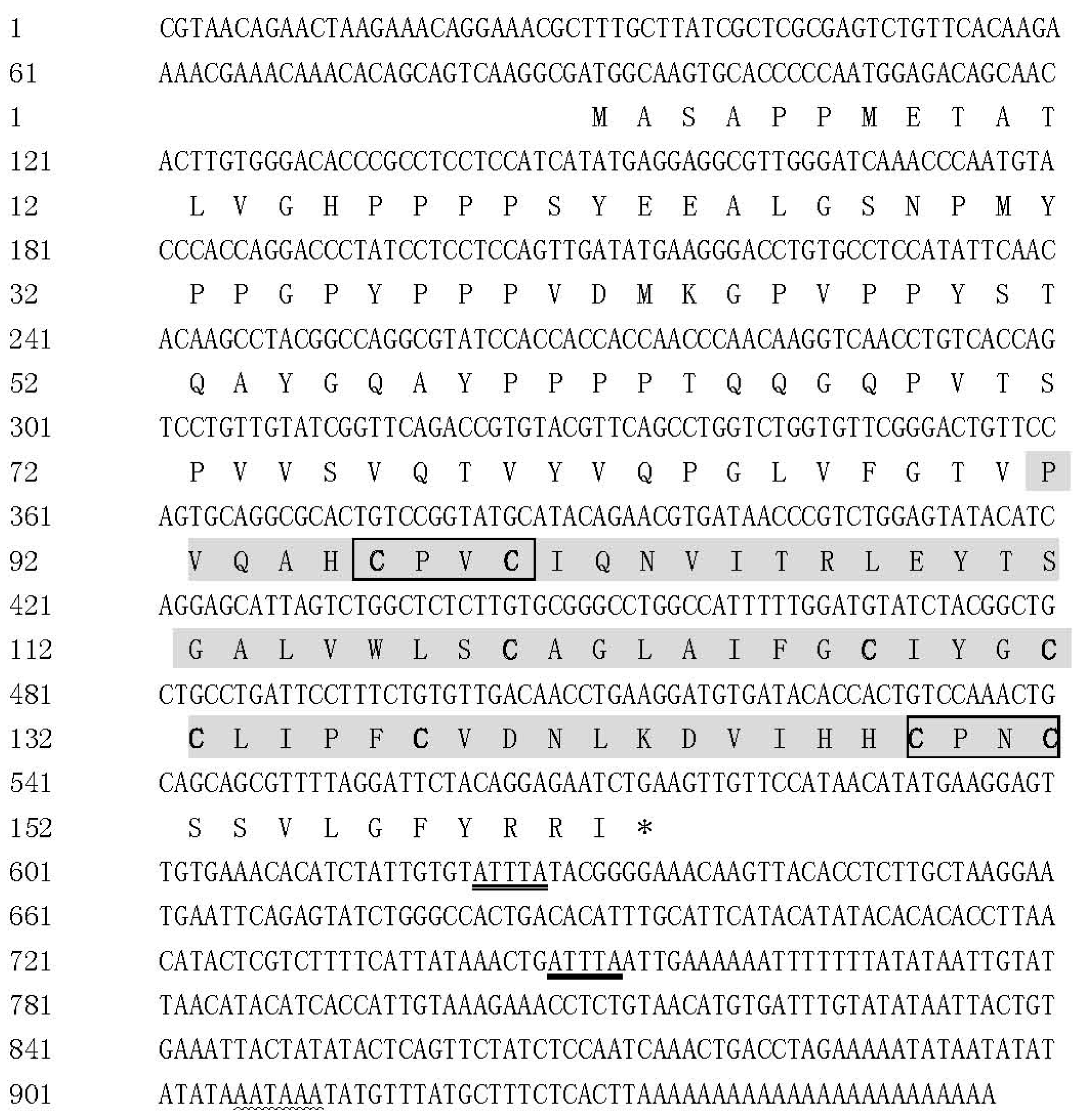
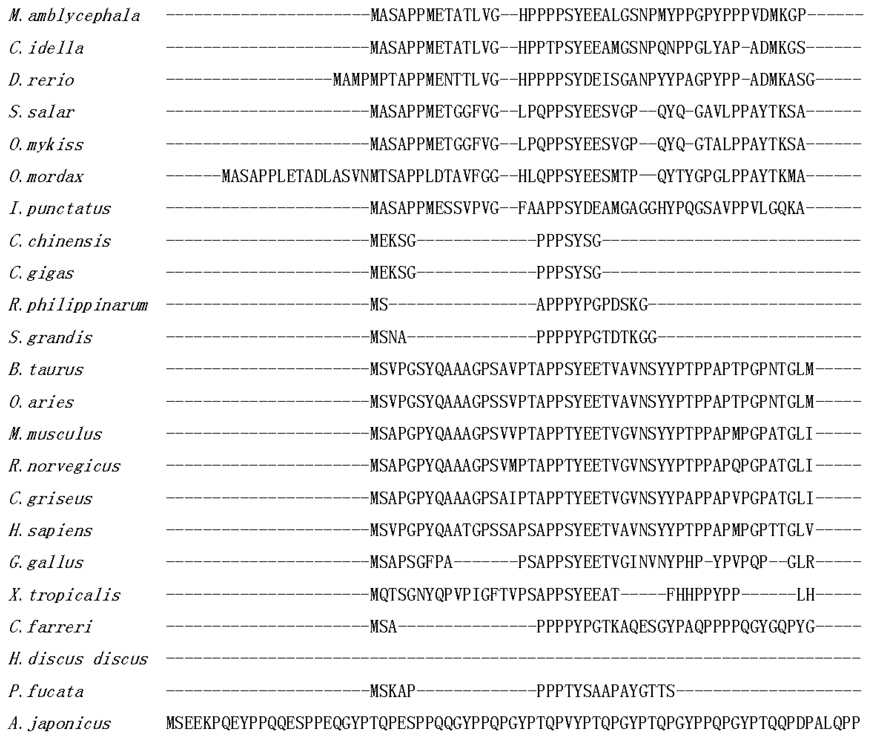
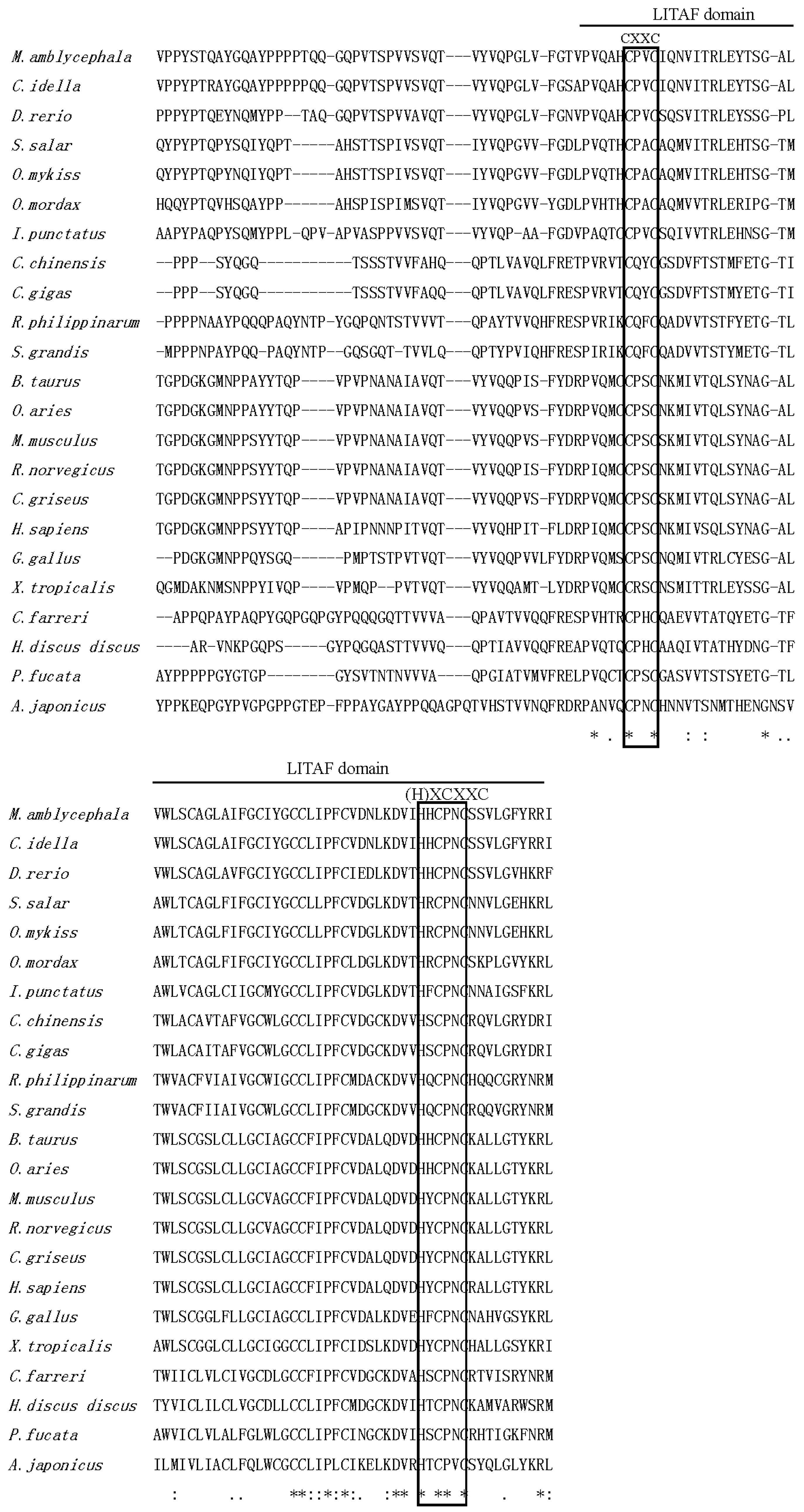
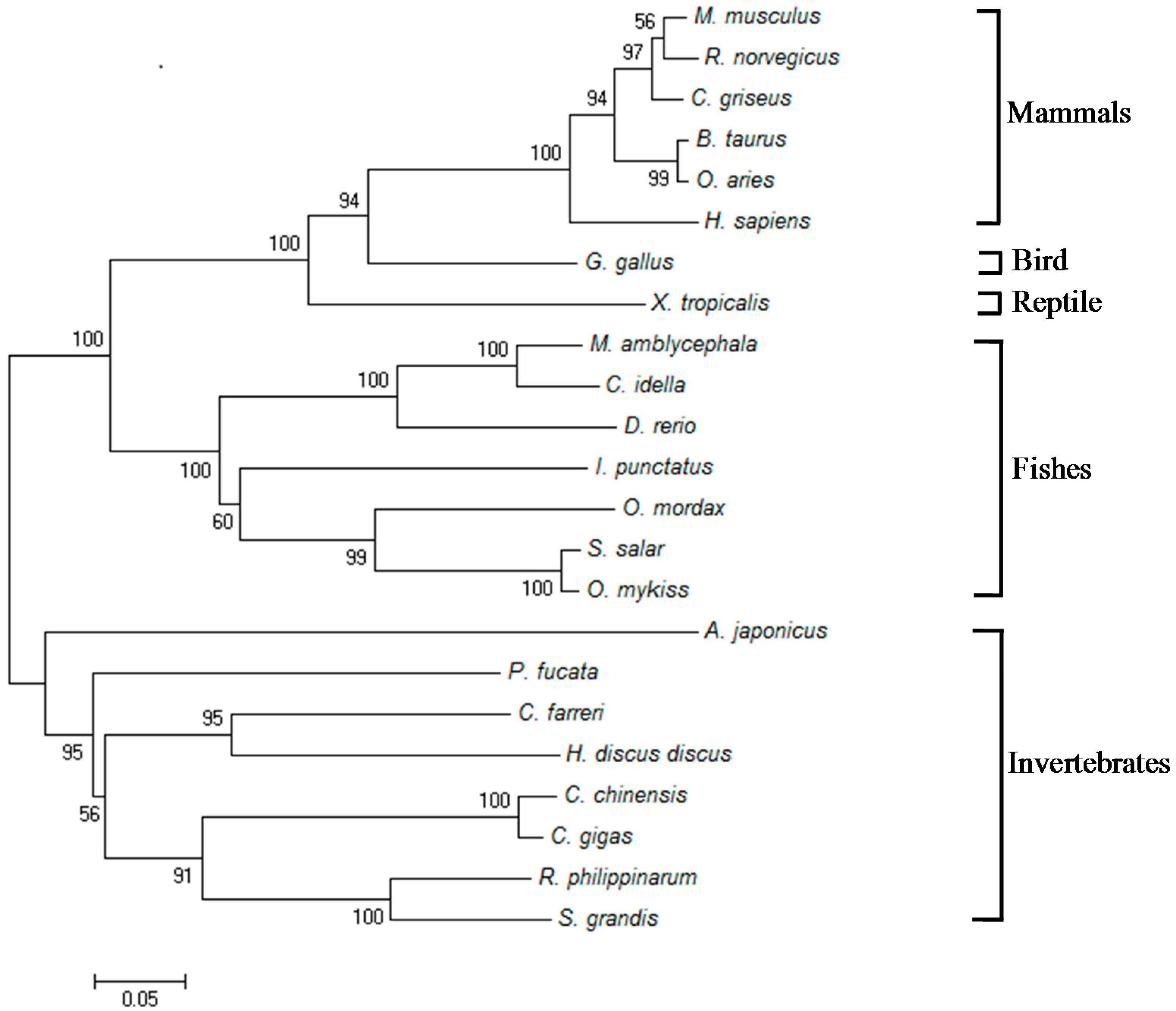
2.1. Isolation and Characterization of Malitaf

The complete Malitaf cDNA sequence was 956 bp, which was composed of an 88-bp length 5′-untranslated region (5′-UTR), a 486-bp open reading frame encoding a protein comprising 161 amino acids, and a 358-bp 3′-UTR followed by a poly (A) tail (Figure 1). One putative polyadenylation signal (AATAAA) was recognized at the nucleotide position 906, which was 21 nucleotides upstream of the poly (A) tail. Furthermore, there were two cytokine RNA instability motifs (ATTTA) at the 3′-UTR of Malitaf, which were also presented in the litaf gene of Paralichthys olivaceus [28]. This cDNA sequence has been deposited in the GenBank database under accession number KX421367.
The deduced protein from Malitaf gene (MaLITAF) possessed with an estimated molecular mass of 17.2 kDa and an isoelectric point of 6.00. The protein analysis by Basic Local Alignment Search Tool (BLAST) showed that MaLITAF protein shared the highest identity (91.3%) with that of grass carp (Ctenopharyngodon idellus). Multiple alignment of amino acid sequences of LITAFs from different species revealed that the C-terminal region of the MaLITAF showed much higher homology than that of other regions (Figure 2). Furthermore, similar to other LITAF homologues, the maLITAF protein possessed the typical LITAF domain (91–160 aa) that contained an N-terminal CXXC “knuckle” followed by a long hydrophobic region, and a C-terminal (H)XCXXC knuckle (Figure 2). The CXXC and HXCXXC motifs are highly conserved among invertebrates, mammals, and fish.
A phylogenetic tree was constructed by the neighbor-joining method based on entire amino acid sequences of LITAFs from M. amblycephala and other species. As shown in Figure 3, all LITAFs were split into five categories, including mammalian, avian, amphibian, teleost, and invertebrate LITAFs. Obviously, Malitaf was located into the fish LITAF group which was distinct from the mammalian cluster. As expected, Malitaf showed the closest relationship to C. idellus. The results indicated that Malitaf represents a new member of fish LITAF family.

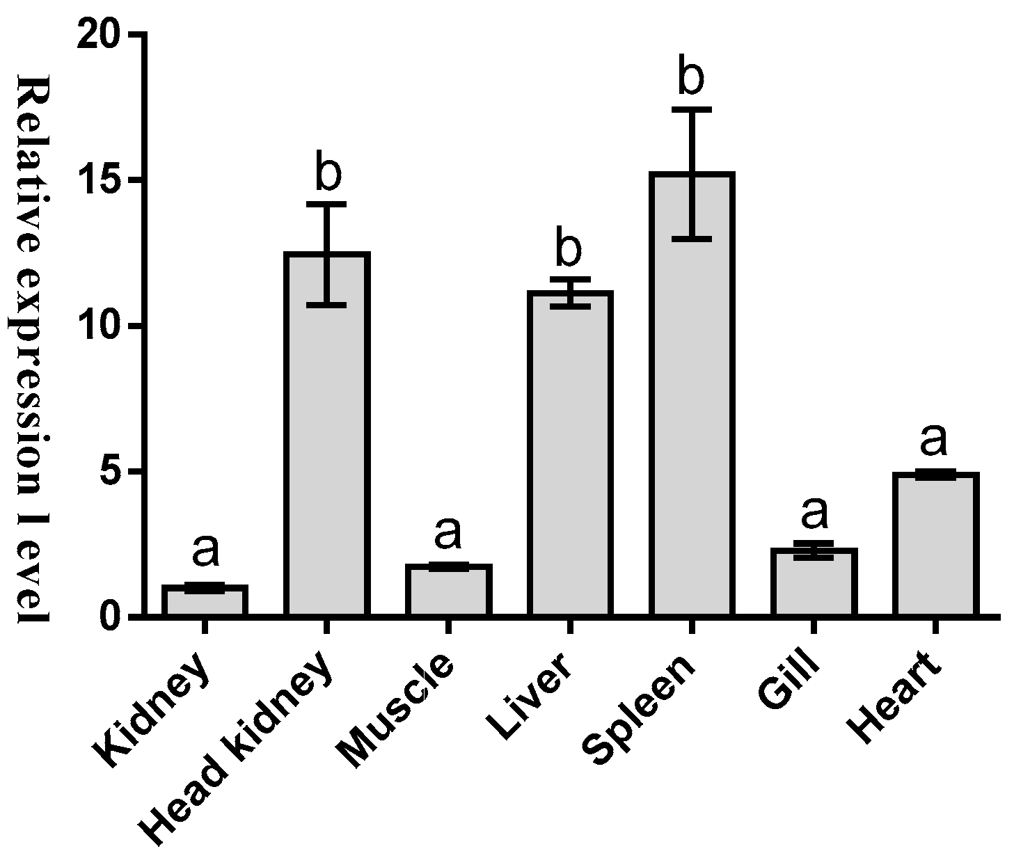
2.2. Tissues Distribution of Malitaf

To further understand the potential function of this new gene, the presence of Malitaf mRNA transcript in different tissues from healthy M. amblycephala was examined by quantitative real-time PCR (qRT-PCR) analysis, which can accurately quantify transcripts at a low copy number [32]. Expression was normalized to the tissue with the lowest observed mRNA level-kidney (set as 1). As shown in Figure 4, the mRNA transcript of Malitaf was ubiquitously detected in a wide range of tissues examined from healthy fish. However, the relative gene expression of Malitaf to kidney was the highest in spleen (15.21-fold, p < 0.001), which was also observed in spleen of rock bream (Oplegnathus fasciatus) [30], followed by head kidney (12.45-fold). The transcriptional level of Malitaf decreased gradually in liver (11.12-fold), heart (4.9-fold), gill (2.29-fold), and muscle (1.75-fold).

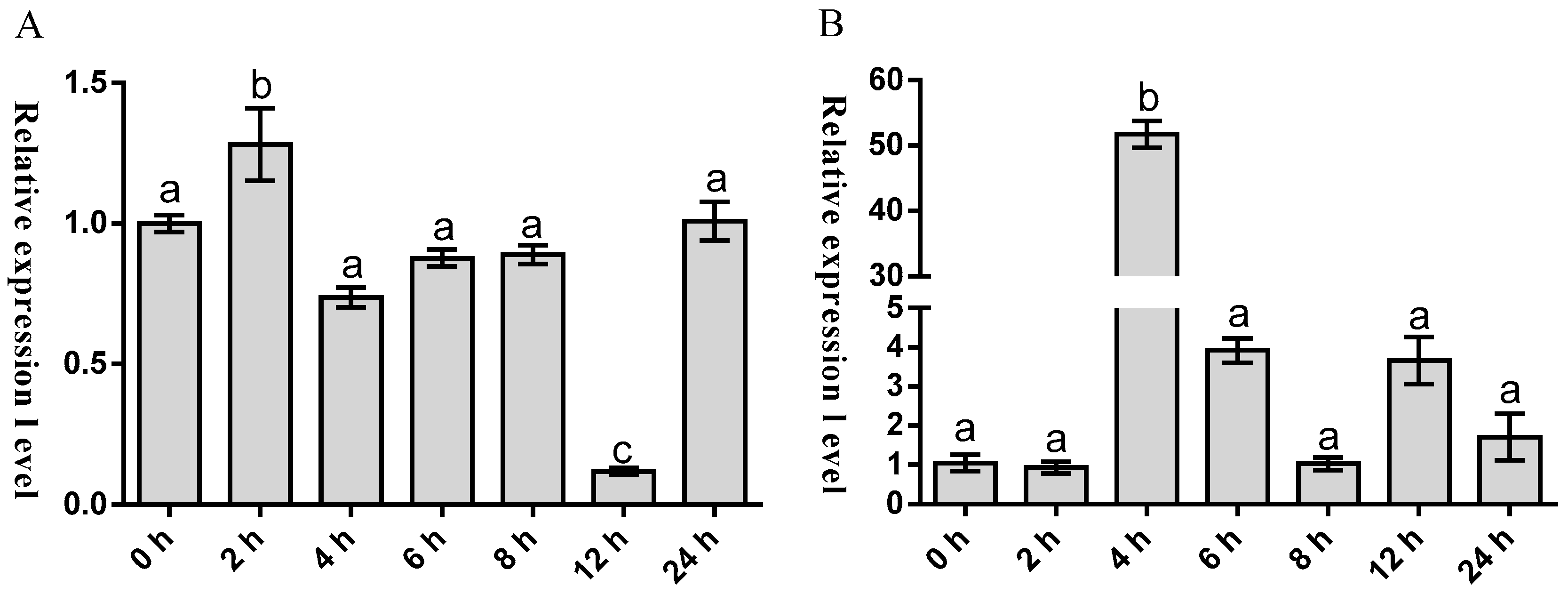
2.3. Temporal Expression of Malitaf and Matnfα after LPS Stimulation

To preliminarily unravel the potential role of Malitaf in innate immunity, we characterized the temporal expression pattern of Malitaf in spleen in response to stimulation with LPS. As shown in Figure 5A, the mRNA expression of Malitaf was rapidly up-regulated after LPS stimulation. The expression reached a peak level at 2 h post-stimulation, which was about 1.29-fold higher than control group (p < 0.05). Subsequently, it was found that its expression decreased gradually to the control level as time elapsed. However, a significant decrease of Malitaf expression was observed at 12 h after LPS stimulation. Considering that litaf is an important regulator for tnfα expression, we therefore tested the expression profile of Matnfα following the elevated expression of Malitaf under LPS stimulations at different time points. As shown in Figure 5B, the transcription of Matnf maintained the control level at 2 h post LPS stimulation. However, a sharp increase of Matnfα mRNA to peak level was detected at 4 h post LPS stimulation (51.74-fold, p < 0.001), and then dropped to the original level at 8 h post LPS stimulation. Interestingly, the transcription of Matnfα surged again to a higher level at 12 and 24 h post LPS stimulation (3.66- and 1.71-fold, respectively) compared with that of the control group.

3. Discussion

Cytokines are the key regulators of innate immunity against pathogens [33]. Until now, litaf gene had been cloned and characterized from many organisms including both vertebrate and invertebrate animals, which suggested a conserved function in innate immunity. In mammals, the litaf has been reported as an important transcript factor for the regulation of TNFα and transcription of various inflammatory cytokines in mammals [34]. However, the information on the systemic reaction of fish litafs during bacterial infection was rather rare. Therefore, we cloned the novel litaf gene from blunt snout bream and examined their expression profiles to understand its potential role in innate immunity.
The LITAF domain, which is a key feature for the LITAF family, exists in viruses, fungi, plants, and Metazoa, [35]. In the present study, the amino acid alignment indicated a highly conserved LITAF domain with N-terminal CXXC and C-terminal HXCXXC knuckles that formed a compact Zn2+-binding structure. These characteristics are a key feature of intracellular Zn2+-binding domains that the N-terminal region binds to the intracellular molecule and the hydrophobic region does not span the membrane [36]. These observations indicated that the LITAF domain of Malitaf appears to be capable of responding similarly to the mammalian LITAF. In a phylogenetic tree of selected vertebrate and invertebrate LITAF amino acid sequences, the MaLITAF was obviously separated from the mammalian cluster and formed one distinct cluster with other teleost LITAFs, suggesting that it is a fish-specific LITAF.
We performed qRT-PCR to monitor the transcriptional level of Malitaf in various tissues from the healthy fish. The results showed the constitutive distribution of Malitaf, indicating that its important role in immune defense against invaders and its immune responses could be occurring in the whole fish body. In teleost fish, spleen and head kidney could be considered as the important central immune organs synthesizing proteins involved in fish immune defense [37]. Therefore, the higher expression level of Malitaf in spleen and head kidney also particularly suggested its crucial role in immune defense against potential pathogens. In other teleost fish, the constitutive expression of litaf mRNA has been reported in a wide range of tissues, but with varying expression levels. For example, the relative gene expression of litaf1 of rock bream was the highest in spleen. In Japanese flounder, the litaf mRNA was detected in all examined tissues with the greatest amount in gill, followed by blood, skin, gonad, hepatopancreas, head kidney, heart, brain, trunk kidney, and spleen in a descending order [28]. The grouper litaf gene was also widely expressed in different tissues analyzed, including liver, spleen, kidney, head kidney, intestine, skin, gill, brain, muscle, heart, and stomach [29]. However, the relatively low expression levels were detected in muscle and liver. In grass carp, the litaf gene was found to express in various tissues but with a high expression level in gill [31]. The inconsistent constitutive expression of litaf gene among different fish species may be a reflection of physiological differences among these species, or even environmental influences. Furthermore, the differences in litaf gene expression are thought to be possibly due to different expressing cells and functions of litaf gene. Certainly, further studies on functional differentiation of litaf gene between fish and other species may yield more novel information on the immune regulatory response of fish.
The Malitaf expression was significantly induced in spleen by LPS, a compound that mimics a Gram-negative bacteria infection, which was similar to the previous reports on human THP-1 cells [17], mouse [38], chicken macrophages [39], Pacific oyster [22], and scallop hemocytes [23]. The results strongly suggested its significantly responsive to LPS and involvement in innate immune response against Gram-negative bacteria. After 12 h of LPS stimulation, the Malitaf mRNA has significantly decreased, which might possibly prevent the excess production of cytokines, and then the innate immune response to pathogens could be efficiently controlled [33]. The expression pattern of Malitaf appeared to be similar to Japanese flounder litaf with respect to the expression profile in response to LPS stimulation [28]. TNFα effects can be both beneficial and detrimental to the host [40]. TNFα must be tightly regulated because over-production can be lethal to the host as in septic shock syndrome. The mouse litaf gene induces activation of tnfα gene expression by itself [38]. Inhibiting human litaf expression in a human monocytic cell line leads to a reduction in the tnfα transcript [15]. Therefore, in the present study, we tested the expression profile of Matnfα following the elevated expression of Malitaf under LPS stimulation. The Matnfα was also induced and rapidly peaked at 4 h, not at 2 h, post LPS administration indicating that Malitaf may significantly contribute to the up-regulated Matnfα expression in the spleen. In addition, the associated up-regulation of Malitaf and Matnfα in spleen during the early phase of LPS stimulation process indicated that Malitaf may be an essential regulator involved in M. amblycephala innate immune response, probably through regulation of Matnfα expression.

4. Materials and Methods

4.1. Fish, LPS Stimulation, and RNA Isolation

Adult M. amblycephala (weight: 405 ± 12.4 g) were obtained from a fish farm (Changsha, China). Fish were maintained with a flow-through water supply at room temperature. After acclimating for one week, the normal fish were used for the stimulation experiments. LPS isolated from Escherichia coli (L2880, Sigma, St. Louis, MO, USA) was suspended into sterilized phosphate buffered saline (PBS), and then was intraperitoneally injected into fish at a dose of 0.1 mg/100 g fish. Afterwards, fish were anesthetized with MS-222 (3-aminobenzoic acid ethyl ester; Sigma). Various tissues from three healthy individuals, including kidney, head kidney, muscle, liver, spleen, gill, and heart, were collected. Similarly, the spleen from three individuals was collected at different time points (0, 2, 4, 6, 8, 12, and 24 h) after stimulation. Total RNA from above tissues was extracted using TRIzol reagent (Invitrogen, Carlsbad, CA, USA), and quantified based on the absorbance at 260 nm. The integrity of RNA was checked by agarose gel electrophoresis. The animal experiments were approved by the Ethics Committee of School of Life Sciences of Central South University with the following reference number (SLSEC0028) in 10 March 2015. The tissues collected at 0 h were from fish injected with the same volume of PBS.

4.2. Cloning and Characterization of Malitaf

To obtain the partial cDNA of Malitaf, the degenerate primers were designed based on an alignment of its counterparts from other teleost fish (Table 1). Gene-specific primers were used to amplify each end according to the manufacturer’s instructions. All resulting PCR products were purified and then cloned into the pMD18-T vector (TaKaRa, Dalian, China) and sequenced bi-directionally at Sangon (Shanghai, China). The open reading frame (ORF) of Malitaf cDNA was detected using the ORF finder (available on: http://www.ncbi.nlm.nih.gov/project/gorf). Protein domains were predicted by the Simple Modular Architecture Research Tool (SMART) [41]. Multiple sequence alignments were created using the Clustal W program [42]. Phylogenetic and molecular evolutionary analyses were constructed by the Neighbor-Joining method in Molecular Evolutionary Genetics Analysis (MEGA) software (version 7.01, Tokyo Metropolitan University, Tokyo, Japan), and support for each node was bootstrapped with 1000 replicates [43].

4.3. Spatial and Temporal Expression Analysis of Malitaf and Matnfα

The quantitative real-time PCR (qRT-PCR) was performed to investigate Malitaf mRNA expression levels in different tissues of healthy M. amblycephala. In addition, the mRNA expression pattern of Malitaf and Matnfα was determined in spleen after LPS stimulation by qRT-PCR. The β-actin and 18S rRNA was selected as an internal control to verify the successful reverse transcription and to calibrate the cDNA template in spatial and temporal expression analysis, respectively. The qRT-PCR was implemented using an ABI 7500 Real-time PCR system (Applied Biosystems, Foster, CA, USA) in a total volume of 20 µL, including 10 µL SYBR® Premix Ex Taq™ II (2×) (TaKaRa, Dalian, China), 0.4 µL ROX Reference Dye II (50×), 0.4 µL of each primer (10 µmol∙L−1), 2 µL 1:5 diluted cDNA, and 6.8 µL of PCR-grade water. The thermal profile was 95 °C for 30 s followed by 40 cycles of 95 °C for 5 s, 60 °C for 34 s, and 72 °C for 30 s. Melting curve analysis of the amplified products was performed at the end of each PCR to confirm that a single PCR product was generated. The 2−∆∆Ct method was used to analyze the expression levels of Malitaf and Matnfα genes [32]. The data obtained from three independent biological replicates were subjected to statistical analysis and the values represented the n-fold difference relative to the references (kidney and 0 h).

4.4. Statistical Analysis

All data of qRT-PCR were presented as means ± SD and checked for homogeneity of variances and normality. Statistical analysis was performed using GraphPad Prism 5.0 (GraphPad Software Inc., San Diego, CA, USA). Significant differences among samples were determined by one-way analysis of variance (one-way ANOVA) followed by a Tukey’s multiple comparison test. Differences were considered significant at p < 0.05 and extremely significant at p < 0.01.

5. Conclusions

We identified and characterized a new member of LITAF family, Malitaf, in M. amblycephala. We confirmed that Malitaf mRNA constitutively expresses in all examined tissues and displays a higher expression level in spleen and head kidney than in other tissues. We also demonstrated that Malitaf expression could be induced by bacterial endotoxin LPS stimulation. Furthermore, we showed that the expression of Matnfα, a pleiotropic cytokine regulated by Malitaf, was up-regulated associated with the enhanced expression of Malitaf in vivo. Our results significantly suggested that Malitaf may play a key role in blunt snout bream innate immunity. Further studies on the functions of Malitaf will contribute to a better understanding of the fish immune system and may help elucidate fish immunoregulatory pathways.

Acknowledgments

This work was supported by the National Natural Science Foundation of China (31201984 and 81500231), the Natural Science Foundation of Hunan Province, China (14JJ3047), the Fundamental Research Funds for the Central Universities (2011QNZT129), the Student Innovation Program for Central South University (Grant no. CX2015508), and the Student Free Exploration Program for Central South University (Grant no. ZY2015785).

Author Contributions

Yina Lv and Yuhong Jiang conducted the gene clone and wrote the manuscript; Leilei Tang conducted the expression analysis of Malitaf and Matnfα; Jun Xiao, Yi Zhou, and Huan Zhong analyzed the data; Jinpeng Yan and Xinying Xiang designed the experiments and modified the manuscript; All authors reviewed and approved the final manuscript.

Conflicts of Interest

The authors declare no conflict of interest.

References

  1. Li, S.F.; Cai, W.Q.; Zhou, B.Y. Variation in morphology and biochemical genetic markers among populations of blunt snout bream (Megalobrama amblycephala). Aquaculture 1993, 111, 117–127. [Google Scholar] [CrossRef]
  2. Tang, L.L.; Liang, Y.H.; Jiang, Y.H.; Liu, S.J.; Zhang, F.Y.; He, X.; Wang, T.Y.; Zhou, Y.; Zhong, H.; Yan, J.P. Identification and expression analysis on bactericidal permeability-increasing protein/lipopolysaccharide-binding protein of blunt snout bream, Megalobrama amblycephala. Fish Shellfish Immunol. 2015, 45, 630–640. [Google Scholar] [CrossRef] [PubMed]
  3. Jiang, Y.H.; Tang, L.L.; Zhang, F.Y.; Jiang, H.Y.; Li, X.W.; Yang, L.Y.; Zhang, L.; Mao, J.R.; Yang, J.P. Identification and characterization of immune-related microRNAs in blunt snout bream, Megalobrama amblycephala. Fish Shellfish Immunol. 2016, 49, 470–492. [Google Scholar]
  4. Bureau, C.F. China Fisheries Yearbook; Chinese Agriculture Press: Beijing, China, 2013. [Google Scholar]
  5. Nielsen, M.E.; Hoi, L.; Schmidt, A.S.; Qian, D.; Shimada, T.; Shen, J.Y.; Larsen, J.L. Is Aeromonas hydrophila the dominant motile Aeromonas species that causes disease outbreaks in aquaculture production in the Zhejiang Province of China? Dis. Aquat. Organ. 2001, 46, 23–29. [Google Scholar] [CrossRef] [PubMed]
  6. Ellis, A.E. Innate host defense mechanisms of fish against viruses and bacteria. Dev. Comp. Immunol. 2001, 25, 827–839. [Google Scholar] [CrossRef]
  7. Zhu, L.Y.; Nie, L.; Zhu, G.; Xiang, L.X.; Shao, J.Z. Advances in research of fish immune-relevant genes: A comparative overview of innate and adaptive immunity in teleosts. Dev. Comp. Immunol. 2013, 39, 39–62. [Google Scholar] [CrossRef] [PubMed]
  8. Tang, L.L.; Xiang, X.Y.; Jiang, Y.H.; Lv, Y.N.; Zhou, Y.; Zhong, H.; Xiao, J.; Zhang, F.Y.; Jiang, H.Y.; Yan, J.P. Identification and characterization of a novel Toll-like receptor 4 homologue in blunt snout bream, Megalobrama amblycephala. Fish Shellfish Immunol. 2016, 57, 25–34. [Google Scholar] [CrossRef] [PubMed]
  9. Grayfer, L.; Walsh, J.G.; Belosevic, M. Characterization and functional analysis of goldfish (Carassius auratus L.) tumor necrosis factor-α. Dev. Comp. Immunol. 2008, 32, 532–543. [Google Scholar] [CrossRef] [PubMed]
  10. Li, M.F.; Zhang, J. CsTNF1, a teleost tumor necrosis factor that promotes antibacterial and antiviral immune defense in a manner that depends on the conserved receptor binding site. Dev. Comp. Immunol. 2016, 55, 65–75. [Google Scholar] [CrossRef] [PubMed]
  11. Collart, M.A.; Baeuerle, P.; Vassalli, P. Regulation of tumor necrosis factor α transcription in macrophages: Involvement of four κB-like motifs and of constitutive and inducible forms of NF-κB. Mol. Cell Biol. 1990, 10, 1498–1506. [Google Scholar] [CrossRef] [PubMed]
  12. Lee, M.H.; Park, J.; Chung, S.W.; Kang, B.Y.; Kim, S.H.; Kim, T.S. Enhancement of interleukin-4 production in activated CD4+ T cells by diphthalate plasticizers via increased NF-AT binding activity. Int. Arch. Allergy Immunol. 2004, 134, 213–222. [Google Scholar] [CrossRef] [PubMed]
  13. Vitiello, M.; D’Isanto, M.; Galdiero, M.; Raieta, K.; Tortora, A.; Rotondo, P.; Peluso, L.; Galdiero, M. Interleukin-8 production by THP-1 cells stimulated by Salmonella enterica serovar Typhimurium porins is mediated by AP-1, NF-κB and MAPK pathways. Cytokine 2004, 27, 15–24. [Google Scholar] [CrossRef]
  14. Stucchi, A.; Reed, K.; O’Brien, M.; Cerda, S.; Andrews, C.; Gower, A.; Bushell, K.; Amar, S.; Leeman, S.; Becker, J. A new transcription factor that regulates TNF-α gene expression, LITAF, is increased in intestinal tissues from patients with CD and UC. Inflamm. Bowel Dis. 2006, 12, 581–587. [Google Scholar] [CrossRef] [PubMed]
  15. Tang, X.; Metzger, D.; Leeman, S.; Amar, S. LPS-induced TNF-α factor (LITAF)-deficient mice express reduced LPS-induced cytokine: Evidence for LITAF-dependent LPS signaling pathways. Proc. Natl. Acad. Sci. USA 2006, 103, 13777–13782. [Google Scholar] [CrossRef] [PubMed]
  16. Tang, X.; Marciano, D.L.; Leeman, S.E.; Amar, S. LPS induces the interaction of a transcription factor, LPS-induced TNF-α factor, and STAT6(B) with effects on multiple cytokines. Proc. Natl. Acad. Sci. USA 2005, 102, 5132–5137. [Google Scholar] [CrossRef] [PubMed]
  17. Myokai, F.; Takashiba, S.; Lebo, R.; Amar, S. A novel lipopolysaccharide-induced transcription factor regulating tumor necrosis factor α gene expression: Molecular cloning, sequencing, characterization, and chromosomal assignment. Proc. Natl. Acad. Sci. USA 1999, 96, 4518–4523. [Google Scholar] [CrossRef]
  18. De Zoysa, M.; Nikapitiya, C.; Oh, C.; Whang, I.; Lee, J.S.; Jung, S.J.; Choi, C.Y.; Lee, J. Molecular evidence for the existence of lipopolysaccharide-induced TNF-α factor (LITAF) and Rel/NF-κB pathways in disk abalone (Haliotis discus discus). Fish Shellfish Immunol. 2010, 28, 754–763. [Google Scholar] [CrossRef]
  19. Li, H.J.; Yang, Q.; Gao, X.G.; Su, H.; Wang, J.; He, C.B. Identification and expression of a putative LPS-induced TNF-α factor from Asiatic hard clam Meretrix meretrix. Mol. Biol. Rep. 2012, 39, 865–871. [Google Scholar] [CrossRef] [PubMed]
  20. Zhang, D.; Jiang, J.; Jiang, S.; Ma, J.; Su, T.; Qiu, L.; Zhu, C.; Xu, X. Molecular characterization and expression analysis of a putative LPS-induced TNF-α factor (LITAF) from pearl oyster Pinctada fucata. Fish Shellfish Immunol. 2009, 27, 391–396. [Google Scholar] [CrossRef] [PubMed]
  21. Yang, D.; Wei, X.; Yang, J.; Yang, J.; Xu, J.; Fang, J.; Wang, S.; Liu, X. Identification of a LPS-induced TNF-α factor (LITAF) from mollusk Solen grandis and its expression pattern towards PAMPs stimulation. Fish Shellfish Immunol. 2013, 35, 1325–1328. [Google Scholar] [CrossRef] [PubMed]
  22. Yu, F.; Zhang, Y.; Yu, Z. Characteristics and expression patterns of the lipopolysaccharide-induced TNF-α factor (LITAF) gene family in the Pacific oyster, Crassostrea gigas. Fish Shellfish Immunol. 2012, 33, 899–908. [Google Scholar] [CrossRef] [PubMed]
  23. Yu, Y.; Qiu, L.; Song, L.; Zhao, J.; Ni, D.; Zhang, Y.; Xu, W. Molecular cloning and characterization of a putative lipopolysaccharide-induced TNF-α factor (LITAF) gene homologue from Zhikong scallop Chlamys farreri. Fish Shellfish Immunol. 2007, 23, 419–429. [Google Scholar] [CrossRef] [PubMed]
  24. Li, S.; Jia, Z.; Li, X.; Geng, X.; Sun, J. Identification and expression analysis of lipopolysaccharide-induced TNF-α factor gene in Chinese mitten crab Eriocheir sinensis. Fish Shellfish Immunol. 2014, 38, 190–195. [Google Scholar] [CrossRef] [PubMed]
  25. Wang, P.H.; Wan, D.H.; Pang, L.R.; Gu, Z.H.; Qiu, W.; Weng, S.P.; Yu, X.Q.; He, J.G. Molecular cloning, characterization and expression analysis of the tumor necrosis factor (TNF) superfamily gene, TNF receptor superfamily gene and lipopolysaccharide-induced TNF-α factor (LITAF) gene from Litopenaeus vannamei. Dev. Comp. Immunol. 2012, 36, 39–50. [Google Scholar] [CrossRef] [PubMed]
  26. Zhang, X.; Zhang, P.; Li, C.; Li, Y.; Jin, C.; Zhang, W. Characterization of two regulators of the TNF-α signaling pathway in Apostichopus japonicus: LPS-induced TNF-α factor and baculoviral inhibitor of apoptosis repeat-containing 2. Dev. Comp. Immunol. 2015, 48, 138–142. [Google Scholar] [CrossRef] [PubMed]
  27. Jin, P.; Hu, J.; Qian, J.; Chen, L.; Xu, X.; Ma, F. Identification and characterization of a putative lipopolysaccharide-induced TNF-α factor (LITAF) gene from Amphioxus (Branchiostoma belcheri): An insight into the innate immunity of Amphioxus and the evolution of LITAF. Fish Shellfish Immunol. 2012, 32, 1223–1228. [Google Scholar] [CrossRef] [PubMed]
  28. Li, S.; Li, X.; Gen, X.; Chen, Y.; Wei, J.; Sun, J. Identification and characterization of lipopolysaccharide-induced TNF-α factor gene from Japanese flounder Paralichthys olivaceus. Vet. Immunol. Immunopathol. 2014, 157, 182–189. [Google Scholar] [CrossRef] [PubMed]
  29. Cai, J.; Huang, Y.; Wei, S.; Ouyang, Z.; Huang, X.; Qin, Q. Characterization of LPS-induced TNF-α factor (LITAF) from orange-spotted grouper, Epinephelus coioides. Fish Shellfish Immunol. 2013, 35, 1858–1866. [Google Scholar] [CrossRef] [PubMed]
  30. Hwang, S.D.; Sang, H.S.; Kwon, M.G.; Chae, Y.S.; Shim, W.J.; Jung, J.H.; Kim, J.W.; Park, C.I. Molecular cloning and expression analysis of two lipopolysaccharide-induced TNF-α factors (LITAFs) from rock bream, Oplegnathus fasciatus. Fish Shellfish Immunol. 2014, 36, 467–474. [Google Scholar] [CrossRef] [PubMed]
  31. Wang, H.; Shen, X.; Xu, D.; Lu, L. Lipopolysaccharide-induced TNF-α factor in grass carp (Ctenopharyngodon idella): Evidence for its involvement in antiviral innate immunity. Fish Shellfish Immunol. 2013, 34, 538–545. [Google Scholar] [CrossRef] [PubMed]
  32. Livak, K.J.; Schmittgen, T.D. Analysis of relative gene expression data using real-time quantitative PCR and the 2−ΔΔCt method. Methods 2001, 25, 402–408. [Google Scholar] [CrossRef] [PubMed]
  33. Bonecchi, R.; Garlanda, C.; Mantovani, A.; Riva, F. Cytokine decoy and scavenger receptors as key regulators of immunity and inflammation. Cytokine 2016, 87, 37–45. [Google Scholar] [CrossRef] [PubMed]
  34. Merrill, J.C.; You, J.; Constable, C.; Leeman, S.E.; Amar, S. Whole-body deletion of LPS-induced TNF-α factor (LITAF) markedly improves experimental endotoxic shock and inflammatory arthritis. Proc. Natl. Acad. Sci. USA 2011, 108, 21247–21252. [Google Scholar] [CrossRef] [PubMed]
  35. Moriwaki, Y.; Begum, N.A.; Kobayashi, M.; Matsumoto, M.; Toyoshima, K.; Seya, T. Mycobacterium bovis Bacillus Calmette-Guerin and its cell wall complex induce a novel lysosomal membrane protein, SIMPLE, that bridges the missing link between lipopolysaccharide and p53-inducible gene, LITAF(PIG7), and estrogen-inducible gene, EET-1. J. Biol. Chem. 2001, 276, 23065–23076. [Google Scholar] [CrossRef] [PubMed]
  36. Ponting, C.P.; Mott, R.; Bork, P.; Copley, R.R. Novel protein domains and repeats in Drosophila melanogaster: Insights into structure, function, and evolution. Genome Res. 2001, 11, 1996–2008. [Google Scholar] [CrossRef] [PubMed]
  37. Bromage, E.S.; Kaattari, I.M.; Zwollo, P.; Kaattari, S.L. Plasmablast and plasma cell production and distribution in trout immune tissues. J. Immunol. 2004, 173, 7317–7323. [Google Scholar] [CrossRef] [PubMed]
  38. Bolcatobellemin, A.L.; Mattei, M.G.; Fenton, M.; Amar, S. Molecular cloning and characterization of mouse LITAF cDNA: Role in the regulation of tumor necrosis factor-α (TNF-α) gene expression. J. Endotoxin Res. 2004, 10, 15–23. [Google Scholar]
  39. Hong, Y.H.; Lillehoj, H.S.; Lee, S.H.; Park, D.W.; Lillehoj, E.P. Molecular cloning and characterization of chicken lipopolysaccharide-induced TNF-α factor (LITAF). Dev. Comp. Immunol. 2006, 30, 919–929. [Google Scholar] [CrossRef] [PubMed]
  40. Beutler, B.; Cerami, A. Cachectin and tumour necrosis factor as two sides of the same biological coin. Nature 1986, 320, 584–588. [Google Scholar] [CrossRef] [PubMed]
  41. Letunic, I.; Copley, R.R.; Pils, B.; Pinkert, S.; Schultz, J.; Bork, P. SMART 5: Domains in the context of genomes and networks. Nucleic Acids Res. 2006, 34, D257–D260. [Google Scholar] [CrossRef] [PubMed]
  42. Thompson, J.D.; Higgins, D.G.; Gibson, T.J. CLUSTAL W: Improving the sensitivity of progressive multiple sequence alignment through sequence weighting, position-specific gap penalties and weight matrix choice. Nucleic Acids Res. 1994, 22, 4673–4680. [Google Scholar] [CrossRef] [PubMed]
  43. Kumar, S.; Stecher, G.; Tamura, K. MEGA7: Molecular evolutionary genetics analysis version 7.0 for bigger datasets. Mol. Biol. Evol. 2016, 33, 1870–1874. [Google Scholar] [CrossRef] [PubMed]
Figure 1. The nucleotide and deduced amino acid sequences of Malitaf. The predicted lipopolysaccharide induced TNFα factor (LITAF) domain is shaded. The two CXXC motifs are in shaded boxes, and the cysteine residues are indicated in shaded bold face. The ATTTA and AATAAA are double and wavy underlined, respectively. “*” shows stop codon.
Ijms 18 00233 g001
Figure 2. Multiple sequence alignment of LITAFs. The identical, highly conserved, and less conserved amino acid residues are indicated by “*”, “:”, and “.”, respectively. The gaps in the alignment are indicated by “-”. The LITAF domain was labeled above the sequences, and two motifs were indicated with rectangles.
Ijms 18 00233 g002aIjms 18 00233 g002b
Figure 3. The neighbor-joining phylogenetic tree of MaLITAF protein and other known homologues from GenBank registered. The numbers at the nodes represent bootstrap values for 1000 replications and the bar (0.1) indicates genetic distance. The sequences of LITAFs from other species were downloaded from GenBank: C. idella (ACT68335), Danio rerio (NP_001002184), Ictalurus punctatus (NP_001187935), Oncorhynchus mykiss (NP_001158593), Osmerus mordax (ACO09164), Salmo salar (ACI67257), Gallus gallus (NP_989598), Xenopus tropicalis (NP_988970), Mus musculus (NP_064364), Homo sapiens (NP_004853), Bos taurus (NP_001039717), Cricetulus griseus (XP_003496739), Ovis aries (XP_004020821), Rattus norvegicus (NP_001099205), Solen grandis (AEW43450), Ruditapes philippinarum (ADX31291), Crassostrea gigas (ABO70331), Cipangopaludina chinensis (AEX08893), Haliotis discus discus (ADI72430), Chlamys farreri (ABI79459), Pinctada fucata (ACN70008), and A. japonicus (AIB51692).
Ijms 18 00233 g003
Figure 4. The expression of Malitaf mRNA in different tissues from healthy M. amblycephala. Significant pairwise expression-level differences between tissues are indicated by different letters above the bars.
Ijms 18 00233 g004
Figure 5. Temporal expression analysis of Malitaf (A); and Matnfα (B) mRNA in spleen from M. amblycephala after LPS stimulation. Significant pairwise expression-level differences among different time-points are indicated by different letters above the bars.
Ijms 18 00233 g005
Table 1. Sequences of primers used in this study.
PrimerSequence(5′–3′)Comment
LITAF-FAARCGCTTTGYTTRTCGCTCGene cloning
LITAF-RAGGTGYAAMTTGTTTCCCCG
3′-Adaptor primerGCTGTCAACGATACGCTACGTAACGGCATGACAGTG(T)183′RACE
3′-PrimerGCTGTCAACGATACGCTACGTAACG
3′-Nested primerCGCTACGTAACGGCATGACAGTG
LITAF-3′-GSPACAGCAACACTTGTGGGACACCCGCCT
LITAF-3′-NGSPAGCCTGGTCTGGTGTTCGGGACTGTT
AAPGGCCACGCGTCGACTAGTACGGGIIGGGIIGGGIIG5′RACE
AUAPGGCCACGCGTCGACTAGTAC
LITAF-5′-GSPCTGTGCCTCCATATTCAACACAAG
LITAF-5′-NGSPGTTGGGATCAAACCCAATGTACC
LITAF-qFCACCAGTCCTGTTGTATCGGReal-time PCR
LITAF-qRCGCACAAGAGAGCCAGACTA
TNFα-qFCTGCTGTCTGCTTCACGCTC
TNFα-qRTAAATGGATGGCTGCCTTGG
β-actin-qFCGGACAGGTCATCACCATTG
β-actin-qRCGCAAGACTCCATACCCAAGA
18S rRNA-qFCAAGACGGACGAGAGCGAAA
18S rRNA-qRGCGGGTTGGCATAGTTTACG

