Review

Regulation of Three Virulence Strategies of Mycobacterium tuberculosis: A Success Story

by
Niels A. Zondervan
1,
Jesse C. J. Van Dam
1,
Peter J. Schaap
1,
Vitor A. P. Martins dos Santos
1,2 and
Maria Suarez-Diez
1,*
1
Laboratory of Systems and Synthetic Biology, Wageningen University & Research, Stippeneng 4, 6708 WE Wageningen, The Netherlands
2
LifeGlimmer GmbH, Markelstrasse 38, 12163 Berlin, Germany
*
Author to whom correspondence should be addressed.
Int. J. Mol. Sci. 2018, 19(2), 347; https://doi.org/10.3390/ijms19020347
Submission received: 28 December 2017 / Revised: 19 January 2018 / Accepted: 21 January 2018 / Published: 24 January 2018
(This article belongs to the Special Issue Molecular Mechanism of Infectious Disease)

Abstract

:
Tuberculosis remains one of the deadliest diseases. Emergence of drug-resistant and multidrug-resistant M. tuberculosis strains makes treating tuberculosis increasingly challenging. In order to develop novel intervention strategies, detailed understanding of the molecular mechanisms behind the success of this pathogen is required. Here, we review recent literature to provide a systems level overview of the molecular and cellular components involved in divalent metal homeostasis and their role in regulating the three main virulence strategies of M. tuberculosis: immune modulation, dormancy and phagosomal rupture. We provide a visual and modular overview of these components and their regulation. Our analysis identified a single regulatory cascade for these three virulence strategies that respond to limited availability of divalent metals in the phagosome.

1. Introduction

Mycobacterium tuberculosis (Mtb) is the most successful known intracellular pathogen infecting roughly one third of the world population and killing about 1.3 million people in 2017 alone [1]. Treating Mtb infection is increasingly difficult due to increasing number of drug-resistant, multidrug-resistant and extensively drug-resistant strains [1]. In order to come up with new drug targets and treatment strategies, there is an urgent need to understand the molecular mechanisms supporting the success of this versatile pathogen. Here, we will review the regulation of three important survival strategies of Mtb: immune modulation, dormancy and phagosomal rupture [2,3,4].
Firstly, Mtb is a master in immune modulation. Its ability to interfere with host cell signalling pathways allows it to carefully balance production of cytokines involved in activation of the pro-inflammatory and anti-inflammatory response [5,6]. By balancing the pro- and anti-inflammatory immune response, Mtb delays phagosome maturation, harvests essential nutrients and stimulates the formation of granulomas. At early infection states, these granulomas are initially dominated by alveolar macrophages and shield the bacteria from more effective immune cells [7].
Secondly, when residing in the hypoxic granuloma, Mtb enters a metabolically near inactive and non-replicating dormant state in which it is immune to most types of drugs [8]. Mtb manipulates the macrophages to accumulate lipids, providing it with the nutrients required to sustain dormancy for multiple decades [7,9,10,11,12].
Thirdly, Mtb has a highly regulated pore formation system that it uses to rupture the phagosome and gain cytosolic access, resulting into necrosis of the host cell and dissemination of the bacilli [13,14].
The fine-tuned regulation of these three virulence strategies is what makes Mtb such a successful pathogen. A large body of literature exists on these virulence strategies and on their molecular components. However, there have been few attempts to provide a systems wide overview of these three virulence strategies, their molecular components and their regulation. Divalent metals play an important role in the regulation of some key aspects of these strategies [15,16,17]. Here, we will present an overview of their involvement in this regulatory process. Detailed inspection of available knowledge pinpoints a single regulatory cascade as a main control hub for these three virulence strategies, representing their interconnectivity as subsequent stages encountered in pathogen host interaction. A modular overview of the molecular components involved in divalent metal homeostasis and their components involved in these three virulence strategies can be found in Figure S1 and Table S1. In the following, we will discuss these components and the environmental cues that control them and we will highlight the role of divalent metals in the phagosome.

2. Divalent Metals at the Interface of M. tuberculosis Host Interaction

Divalent metals such as iron, zinc and manganese are required for proliferation and survival of all living organisms. Divalent metals appear, in all living beings, nearly exclusively as constituents of proteins and act as cofactors in many essential enzymes and environmental sensors [18]. Iron is the most commonly used divalent metal cofactor [18]. Iron containing enzymes are involved, among other processes, in electron transfer, maintaining redox balance and detoxification [19]. Manganese has the strongest affinity for ATP and is the preferred cofactor in cAMP production [20,21]. Zinc is used as cofactor by numerous enzymes and DNA binding proteins and additionally functions to scaffold additional proteins [22].
To prevent growth of bacteria, the host uses high affinity iron binding proteins such as lactoferrin, ferritin and transferrin to keep concentration of free iron in the blood low, in the so-called iron sparing response [16,23]. These proteins also bind other divalent metals such as manganese, albeit with lower specificity than iron. Similarly, calprotectin functions as high affinity calcium binding protein but also binds manganese, zinc and iron in the blood [24]. During infection, macrophages withdraw approximately 30% of the total circulating iron from the blood stream making macrophages environments rich in divalent metals [25]. Some intracellular pathogens use this defence mechanism to their advantage by stimulating phagocytosis by macrophages to get access to divalent metals and other nutrients. During initial infection, Mtb predominantly encounters resident, replicative alveolar macrophages populating the lungs which are rich in divalent metals while having reduced bactericidal abilities compared to other macrophages [12,25].
Upon ingestion by a macrophage, Mtb is engulfed in a special compartment called the phagosome, in a process known as phagocytosis. The phagosome then fuses with vesicles containing enzymes and other proteins that facilitate bacterial digestion. Phagocytosis is a rapid process and leads to phagosomal-endosomal fusion in approximately 3–4 min, acidification of the phagosome within 23–32 min and fusion with lysosome in 74–120 min, based on experiments with epithelial macrophages [26]. However, Mtb blocks phagosome maturation in an early phase leading to fusion with early endosomes and a pH of approximately 5.5 [27].
The macrophage continuously exports divalent metals out of the phagosome via Nramp1 and Nramp2 in a pH dependent manner. Many cell types express Nramp2 while only macrophages express Nramp1. Nramp1 is mechanistically similar to Nramp2 but has a much higher specificity for manganese (Mn) compared to Nramp2 [17,27,28]. Mn is required as cofactor for the bacteria to break down oxidative compounds produced in the phagosome such as H2O2 [16,20,29]. Thus, restricting Mn availability in the phagosome by recruitment of Nramp1 is an essential defence against intracellular pathogens. Nramp2 functions optimally around pH 6, a condition found in the early phagosome while Nramp1 has an optimal activity at a pH of 4.5 Nramp1 is attached to the membrane of maturing phagosomes and is associated with increased recruitment of endosomes and/or lysosomes containing vacuolar V-H+-ATPase, resulting in acidification of the phagosome from pH 6.5 to 5.5 [27,30]. Nramp2 is regulated separately from Nramp1 and co-localizes with transferrin receptors to early endosomes as well as with V-H+-ATPase. V-H+-ATPase provides the electro-genic force needed for Nramp1 and Nramp2 to operate [31,32]. Metal availability in the phagosome is tightly regulated by the host through the combined action of Nramp1 and Nramp2. Therefore, blocking phagosome maturation is an effective strategy to create an environment in which Mtb can outcompete divalent metal export from the phagosome. Mtb uses special high affinity siderophores (mycobactin) to gain access to divalent metals from both extracellular transferrin and the intracellular iron pool [25].
Within Mtb iron, zinc and manganese homeostasis are regulated by IdeR, Zur (previously known as FurB) and MntR respectively [19,22,33]. Ligation of Fe2+ to IdeR and Zn2+ to Zur stabilizes the formation of dimers that have strong affinity to binding sites involved in suppressing the genes in their respective regulons [15,19,34]. MntR in Bacillus subtilis contains two manganese binding sites as well as a dimerization site similar to IdeR and Zur [35]. There is a significant overlap between IdeR, Zur and MntR regulated genes, see Figure 1. An overview of the regulation of molecular components by divalent metal regulators, IdeR, Zur and MntR can be found in Figure S1 and Table S1. Each of these three regulators suppresses the main operon of genes coding for the ESX-3 secretion system and associated PE, PPE and Esx proteins homologues of ESAT-6 and CFP-10 (EsxA and EsxB) [33]. We will further discuss the ESX-3 transport system in a section below. In the following sections, we will discuss main characteristics of genes regulated by Fe, Zn and Mn respectively.

2.1. Iron Homeostasis and Redox Sensing

Mtb produces high affinity hydrophilic and lipophilic siderophores termed carboxy-mycobactin and mycobactin, respectively. Mycobactin can bypass the phagosome membrane to scavenge iron from the extracellular iron storage protein transferrin [25,36,37,38]. In addition, Mtb actively synthesizes deoxy-mycobactin during iron starvation [39].
Mtb combines the expression of a dedicated iron acquisition machinery with cellular components involved in immune modulation. By limiting acidification of the phagosome, Mtb maintains favourable conditions in which it can outperform active export of divalent metals by the macrophages transporter Nramp1. Mtb’s success in acquiring iron is illustrated by a 20-fold increase of iron concentrations in the phagosome between 1 and 24 h of macrophage infection [40]. However, high iron concentrations renders Mtb much more vulnerable to the formation of oxygen and nitrogen radicals upon phagosome maturation, as iron functions as a catalyst in the formation of radicals via the Fenton reaction [41]. Tight regulation of iron homeostasis is, therefore, essential, making IdeR an interesting drug target [42]. Mtb has adapted to deal with oxidative stress outside of the cell but is relatively vulnerable to endogenously generated oxidative stress in comparison to M. smegmatis [41]. Due to this vulnerability, vitamin-C is an effective drug to combat Mtb in the early stage of infection by inducing the Fenton reaction in iron rich phagosomes [43]. The oxidative conditions encountered in the phagosome leads to oxidation of the intracellular iron pool. Oxidation of the iron pool de-represses IdeR regulated genes among which some are involved in virulence. Upregulating expression of virulence genes in low iron and oxidative conditions is a common response in intracellular pathogens and has been observed in Shigella dysenteriae, Corynebacterium diphtheniae, Yersinia pestis and Yersinia pseudotuberculosis, as well as in Mtb [44,45].
The iron pool within Mtb and the phagosome functions as redox sensor to the oxidative conditions encountered in the early phagosome. In oxidative conditions, ferrous iron (Fe2+) is oxidized to ferric iron (Fe3+) [46]. Ferric iron does not bind to IdeR, leading to upregulation of IdeR suppressed genes in oxidative conditions [42]. Genes suppressed by IdeR code for proteins involved in siderophore synthesis (mbtA-G), secretion (mmpL4/5, mmpS4/5) and uptake (irtAB) as well as 11 genes coding for the ESX-3 secretion system, among others [47,48,49]. Even though IdeR mainly functions as iron dependent repressor, IdeR also induces transcription of four genes. Among the induced genes, bfrB and, to a lesser extent bfrA, code for mycobacterial ferritin-like iron storage proteins, which prevent overload of iron within Mtb [19,50]. Analysis of the promoter region of bfrB revealed it contains two tandem IdeR binding sites involved in alleviating repression by Lsr2. Lsr2 is a histone like regulator that binds AT-rich regions virulence islands, including those coding for ESX-1, espACD and PDIM coding genes, acting as a global regulator to aid in the adaptation to extremes in oxygen availability [50,51,52,53,54,55]. Combined regulation of bfrB by Lsr2 and IdeR, suggests iron storage by BfrB is suppressed by Lsr2 during infection under changing oxygen conditions unless IdeR detects availability of intracellular ferrous iron which indicates a lack of oxidative conditions. Under low iron conditions, BfrA is required to mobilize stored iron. On the other hand, on high iron conditions, BfrB is needed for iron storage [56]. BrfB was shown to be required for the long term persistence of Mtb in iron-starved granulomas [23].
Iron homeostasis is an essential process for bacterial survival, therefore its cellular components are interesting drug targets. This was shown in a knockout study of the mmpS4/5 siderophore secretion, which resulted in limited intracellular availability of iron as well as intracellular accumulation of siderophores toxic to Mtb [57]. Another interesting drug target is HupB, a nucleoid-associated protein that protects Mtb against reactive oxygen species, regulates siderophore synthesis and was proposed to facilitate transfer of iron from ferri-carboxymycobactin to mycobactin [58,59]. HupB stimulates transcription of its own operon in the absence of IdeR-Fe2+ [59].
IdeR also regulates genes involved in response to oxidative and acidic stress, among which the two-component system PhoPR. Two-component systems contain a histidine kinase sensor that senses specific environmental stimulus and a response regulator that gets phosphorylated by the sensor upon specific environmental stimuli. Many two-component regulators, among which PhoPR, also regulate their own operon [60]. Presence of multiple binding sites allows both positive and negative regulation depending on the concentration and phosphorylation state of the response regulator, as is the case for PhoPR [61,62]. PhoPR is the main regulator of the oxidative and acidic stress response but also it is the initial step in a regulatory cascade controlling pore formation and phagosomal rupture. Six putative IdeR binding sites upstream of the phoP-phoR operon were located, of which five were observed to bind IdeR in the presence of iron [63]. This points to a possible link between iron homeostasis and PhoPR regulation of the oxidative stress response and virulence genes.
Nevertheless, the exact role of IdeR in upstream binding of PhoPR remains to be determined.
Oxidation of the iron pool is also sensed by proteins containing iron-sulphur clusters such as the enzyme aconitase (Acn) and the regulators FurA and WhiB1-7. Acn catalyses the isomerization of citrate to isocitrate via cis-aconitate in normal conditions. However, in low iron or oxidative conditions it binds to and suppresses translation of IdeR-mRNA while increasing translation of TrxC-mRNA [64]. The function of Acn as redox sensitive translational regulator is conserved in many organisms [46,65].
FurA (ferric uptake regulator A) regulates the oxidative stress response by modulating expression of the operon coding for FurA and the KatG catalase [66]. KatG is essential for the breakdown of H2O2 radicals formed upon phagosome endosome fusion and activates the anti-cell-wall drug isoniazid. Recently, transcriptional activation of furA-katG was found to be regulated by RbpA, which is induced by H2O2 in a SigE dependent manner [67].
A third iron sensitive regulator is WhiB7. WhiB proteins are iron-sulphur cluster-containing redox-sensing transcription factors. WhiB7 expression is auto-regulated by binding to its own promoter in response to antibiotics or redox stress [68]. An 80-fold upregulation of WhiB7 was observed upon treatment with antibiotics that bind to the 30S ribosomal subunit such as kanamycin and streptomycin [68]. WhiB7 is upregulated by iron starvation and was shown to induce transcription of eis and tap [69], two antibiotic resistance genes. Upregulation of eis increases secretion of IL-10 and slightly represses production of TNF-α by the host. IL-10 and TNF-α are involved in the anti-inflammatory and pro-inflammatory responses respectively [70].
In summary, oxidation of the iron pool is an important environmental cue to activate molecular components involved in iron sequestering, immune modulation and virulence. IdeR, FurA, Acn, WhiB7, Lsr2 and SigE are all involved in the response to the oxidative conditions encountered in the phagosome and subsequent adaption through expression of a vast repertoire of molecules involved in iron homeostasis as well as genes involved in modulation of the immune response.

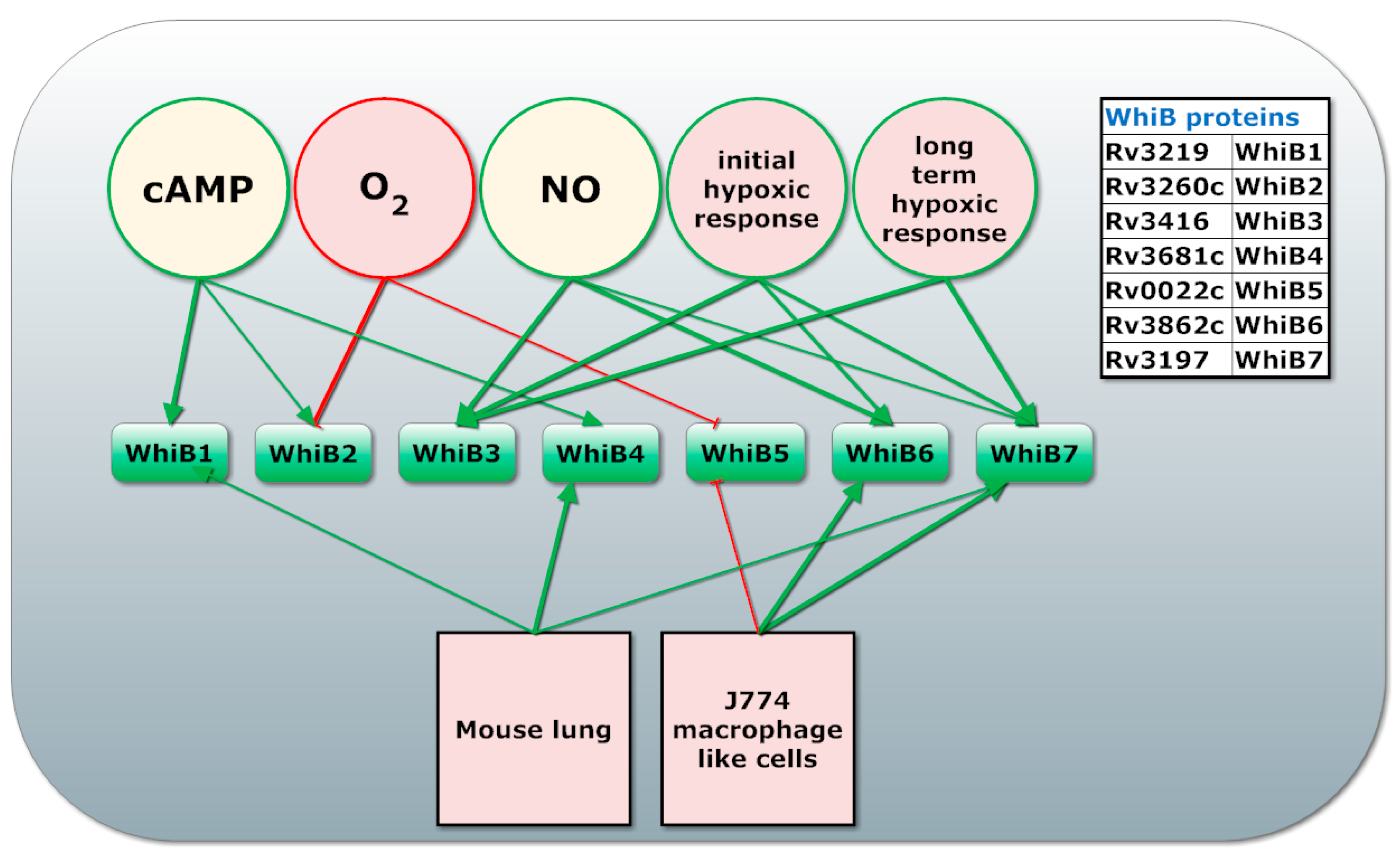
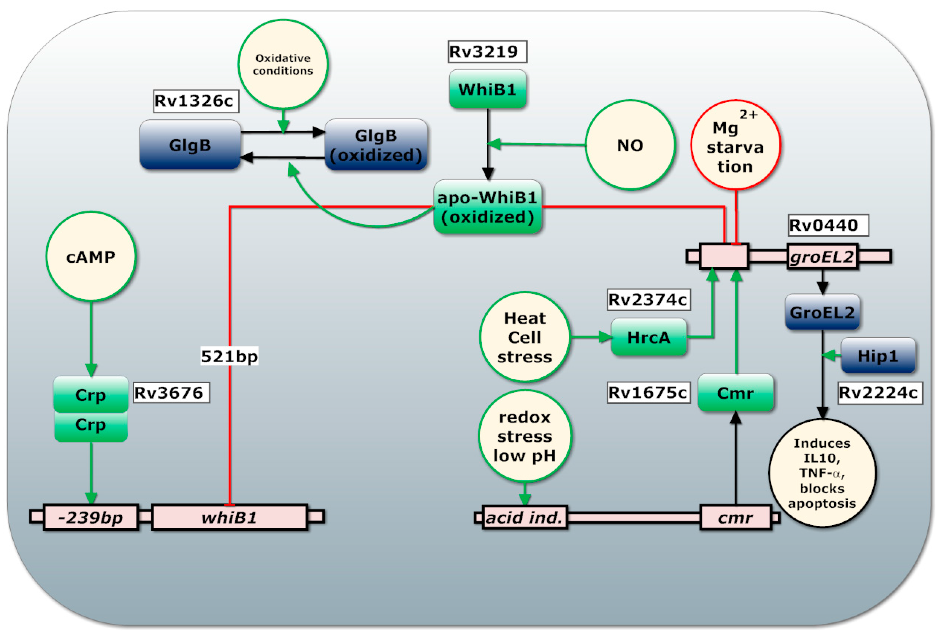
2.2. Manganese Homeostasis and cAMP Production

Manganese is one of the most abundant metal elements in nature [71]. Mn is involved in enzymes of diverse functionality such as photosynthesis and detoxification: Mn is used as cofactor for both synthesis and degradation of H2O2, superoxide and radicals [16]. The oxidative burst is a very effective bactericidal process to defend against intracellular pathogens such as Mtb and Y. Pestis [54,72,73]. As previously stated MntR is a regulator of Mn homoeostasis, however MntR is dispensable for Mtb growth in human and/or mice macrophages due to the limited availability of Mn in the phagosome. Manganese transport on the other hand is required for virulence and to break down oxygen radicals [33]. Mtb contains two superoxide dismutases, SodA and SodC. SodA uses manganese as preferred cofactor and requires CtpC for metalation and export to the phagosome. Interestingly, ctpC transcription is induced in the presence of PhoP, while sodA is predicted to contain upstream cAMP-CRP binding sites implicating it in its regulation [60,74]. CRP is a cAMP dependent regulatory protein.
Another role of Mn we would like to discuss here is the Mn dependent activation of cAMP production in the early phagosome which was first proposed by S. Reddy et al. in 2001 [21]. S. Reddy and co-workers studied the kinetics of membranes containing Mtb adenylyl cyclase CyA (Rv1625c). Their study revealed that the Michaelis-Menten constant (Km) for Mn-ATP is 70-fold lower than for Mg-ATP. This results in a 47-fold activation by 1 mM Mn-ATP compared to 1 mM of Mg-ATP at physiological conditions [21]. Mn is also essential for the CRP regulated, virulence associated type III phosphodiesterase Rv0805 [75,76].
During infection, intracellular cAMP concentration increases ~50 fold and this is associated with a decrease in pH from 6.7 to 5.5 [77]. Among the 15 Adenylate Cyclases (AC) present in Mtb H37Rv, CyA has the highest measured cAMP production while AC (Rv1264) functions optimally at pH 6, which is typically found in early phagosomes [77,78]. Mtb was shown to secrete cAMP in a burst into the macrophage cytosol, resulting in a 10-fold increase in the host’s TNF-α concentration, an important inducer of granuloma formation [79]. Rv0386 is needed for this cAMP burst [79].
The MntR regulon contains mntH (Rv0924c), coding for Mramp, an Nramp homolog that imports manganese (Mn) in a pH dependent manner; mntABCD (Rv1283c-Rv1280c) coding for an ATP dependent manganese transporter and Rv2477c coding for a manganese dependent ATPase which optimally functions at pH 5.2 [80]. Interestingly, Rv2477c was postulated to be involved in resistance to tetracyclines and macrolides [80]. Additionally, MntR and Zur regulate Rv2059-Rv2060 coding for two components of an incomplete ABC transporter of unknown function. Therefore, it is more likely that this transporter is involved in transporting other divalent cations like Co2+, Cu2+ or Ca2+ to substitute Mn and Zn in some conditions. A second possibility is that this operon codes for a divalent cation exporter to counter the side effect of unwanted uptake of divalent cations such as Cu2+ by the high expression of manganese and zinc transporters [33]. Manganese uptake plays an important role in virulence of many bacteria. For instance, supplementing Salmonella typhimurium with manganese prior to infecting macrophages, decreased its lethal dose 50-fold [81]. Similarly, manganese acquisition in the gut was shown to allow S. typhimurium and Salmonella enterica to evade neutrophil killing by calprotectin and reactive oxygen species, while patients with mutations in manganese transporter Nramp1 were shown to be much more susceptible to pathogens such as Mtb [20,27,54,72,82,83].
MntR regulates WhiB6 which regulates espACD and some DevR (previously known as DosR) regulated genes [84]. DevR is the main regulator of dormancy and espACD is involved in pore formation [85] and will be discussed below. The WhiB6 iron sulphur cluster is necessary for the negative control of the DevR regulon and positive control of the ESX-1 secretion system, whereas apo-WhiB6 induces the DevR regulon and suppresses ESX-1 expression in M. marinum [85]. A model was proposed where holo-WhiB6 positively regulate ESX-1 operon while upon reaction with reactive oxygen species and NO, apo-WhiB6 and WhiB6-DNIC are formed respectively. Both apo-WhiB6 and WhiB6-DNIC activate DevR regulated genes to shift metabolism and maintain energy and redox homeostasis [85].
MntR interacts with the toxin-antitoxin system RelJ and RelK in which MntR functions as antitoxin [86,87]. Additionally, VapBC26 and VapB30 toxin-antitoxin system both requires Mg or Mn for their ribonuclease activity, which inhibits growth [88,89]. These results indicate Mn might function as environmental cue in the regulation of growth.

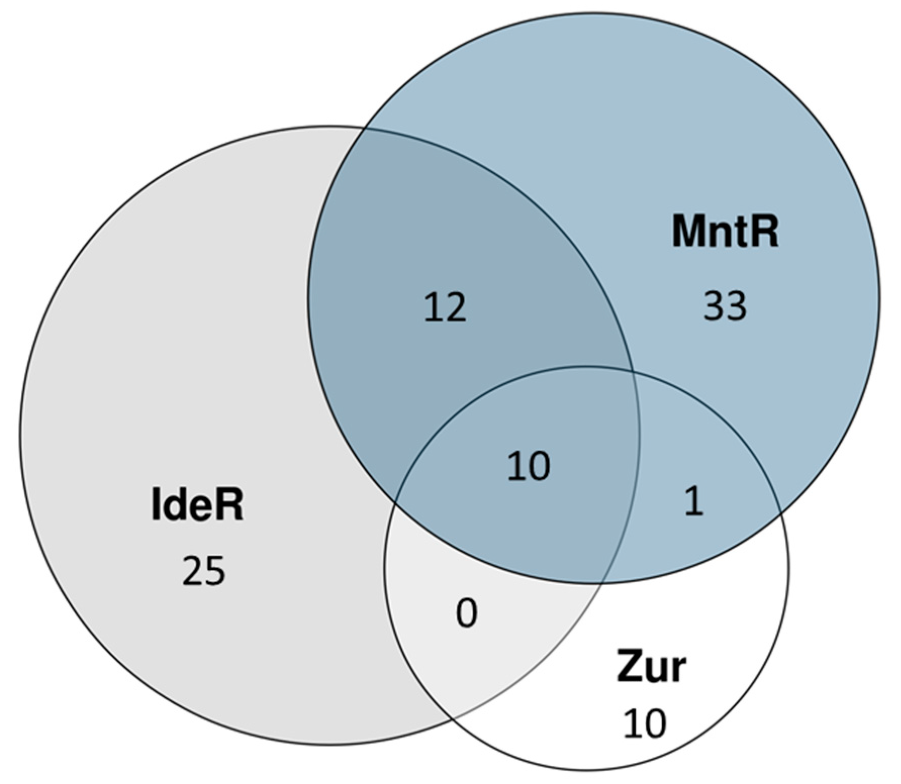
2.3. Zinc Homeostasis

The third and final divalent cation we would like to discuss is zinc, the only redox stable divalent metal of the three. As previously stated, zinc homeostasis is regulated by Zur (FurB), a Zn2+ dependent repressor. Zur knockout studies identified 32 genes that are upregulated in the zur knockout mutant of which 24 belong to eight transcriptional units that were shown to be directly regulated by Zur [22]. Zur expression levels are regulated by SmtB encoded by an upstream gene, which is co-operonic with zur. SmtB functions as a repressor which is deactivated upon binding to Zn2+ [22].
There are three possible zinc uptake systems regulated by Zur. Firstly, Zur regulates the sitABC like genes (Rv2059-2060), which are also regulated by MntR that were previously discussed. This suggest that this transporter might function as Zn importer [20,90,91]. Secondly, Zur regulates Rv0106 coding for a protein similar to the B. subtilis putative zinc low-affinity transporter YciCas [90]. Thirdly, EsxG-EsxH proteins were shown to be able to bind zinc, which might implicate them in zinc transport [92].
Other interesting targets of Zur are five genes coding for ribosomal proteins that can function in the absence of zinc, in contrast to their zinc dependent counterparts which normally bind to the 30S ribosomal subunits [22,93]. Although Zur was found to be able to positively regulate some genes in other pathogenic bacteria via repression of non-coding small RNAs, no such regulation was found in a zur knockout Mtb mutant [15].

2.4. ESX-3 Secretion System

The ESX-3 secretion system is the only one of the five ESX systems that is essential for in vitro growth of Mtb [94,95]. ESX-3 is involved in divalent metal homeostasis and immune modulation. ESX-3 is involved in divalent metal homeostasis and immune modulation. ESX systems secret extracellular proteins [96,97].
Regulatory binding site for all three divalent metal regulators IdeR, Zur and MntR can be found in the ESX-3 core operon promoter [48,92], as summarized in Table 1. The triple control of ESX-3 might allow Mtb to switch partly to other divalent metals in the absence of one of these three. This hypothesis is supported by the observation that siderophore knockout mutants low in iron contain much higher zinc concentrations [32]. However, many ESX-3 associated genes are regulated by only one or two of these regulators, indicating dedicated roles in homeostasis of specific metals [98].
All three divalent metal regulators regulate EsxG and EsxH which play an essential role in secretion of PE and PPE proteins [98]. PE and PPE proteins comprise nearly 10% of the coding potential of the Mtb genome and, for many of them, immune modulating properties have been reported [100]. A large number of studies exist on the immune modulating properties of ESX-3 secreted PE and PPE proteins [95,98,100,101,102,103,104,105]. The ESX-3 secreted protein pair EsxG-EsxH, targets the endosomal sorting complex to impair fusion of the phagosome with the lysosomes, while increasing association with the endocytic pathway leading to fusion with transferrin containing vesicles [92,95,97]. PE5-PPE4 were found to be critical for the siderophore-mediated iron-acquisition functions of ESX-3 [98]. PPE38 inhibits macrophage MHC Class I expression, dampens CD8+ T-Cell responses and was shown to be required for virulence of M. marinum [104,105]. PPE37 was found to reduce the production of pro-inflammatory factors TNF-α and IL-6 [102]. PE_PGRS61 binds TLR2 in a Ca2+ dependent manner, leading to increased IL-10 production. Finally, PE5 and PE15 trigger activation of the host MAP kinases required for IL-10 production [100,103]. IL-10 is an important anti-inflammatory cytokine. IL-10 reduces the expression of iNOS, limiting production of nitric oxide (NO) in the phagosome [95,100]. Enhanced IL-10 expression plays an important role in inhibiting early protective immunity and blocking phagosome activation [106,107]. In addition, a direct role for IL-10 in Mtb reactivation has been observed [106]. Interestingly, IL-10 also modulates lipid metabolism by enhancing uptake and efflux of cholesterol in macrophages [106,107,108]. Mtb is known to induce foamy macrophages using immune modulating proteins as well as secreted lipids. This leads to deregulation of the macrophages lipid metabolism via the macrophages’ lipid-sensing nuclear receptors PPARγ and TR4 [12,107]. One study reported observing Mtb to exploit host vesicle trafficking and lipid storage by recruitment of iron bound mycobactin to lipid droplets which move to the phagosome and discharge their content [36]. Another study found that Mtb uses membrane vesicles containing immune modulating molecules as well as mycobactin to interact with the macrophage during infection [109]. Further research is needed to investigate the proposed synergy between modulation of host vesicle trafficking, lipid acquisition and iron acquisition.

3. Three Main Virulence Strategies of Mtb

The three virulence strategies discussed in this review, namely immune modulation, dormancy and phagosomal rupture, represent subsequent stages in Mtb-host interaction. These strategies extend and complement each other, which is reflected in their regulation. While many pathogens directly express components involved in phagosomal rupture, Mtb keeps a low profile and activates key virulence strategies, such as phagosomal rupture, only when immune modulation fails and the phagosome becomes inhospitable. However, immune modulation also complements phagosomal rupture and dormancy, since immune modulation leads to conditions, such as granuloma formation and cholesterol accumulation, which are needed to prepare Mtb for dormancy and phagosomal rupture.

3.1. Immune Modulation

Mtb uses a number of virulence proteins, complex lipids and secreted metabolites, to modulate the immune response and arrest phagosome maturation to prevent fusion with late endosomes and lysosomes [2,77,97,110,111,112,113]. In case of successful immune modulation, phagosome maturation is halted resulting in a pH of approximately 5.5 [27,30]. The macrophage controls intracellular trafficking, including phagosome maturation, through 42 distinct Rab GTPases. Rab5 is associated with phagosomes immediately after phagocytosis and normally diffuses quickly, allowing Rab7 to associate to the phagosome, which allows fusion of the phagosome with lysosomes. Studies with M. bovis have shown that Mycobacteria halts phagosome maturation, by blocking vesicle fusion between stages controlled by Rab5 and Rab7, with no Rab7 being accumulated in macrophages even after 7 days [111]. Similarly, for Mtb Rab7 was shown to be recruited by the phagosome but its premature release prevents fusion of the phagosome with late endosomes [110,114].
In addition to the earlier discussed ESX-3 secreted proteins, several other proteins and molecules are involved in blocking phagosome maturation. Secreted tyrosine phosphatase (PtpA) is involved in the exclusion of the vacuolar V-ATPase, thereby preventing acidification and fusion with lysosomes [112,115]. cAMP secreted by Mtb blocks phagosome lysosome fusion by inhibiting actin assembly [113]. Additionally, a number of virulence lipids interfere with the phagosome’s Golgi trafficking, needed for maturation of the phagosome [114,116]. Among these virulence lipids are monomycolate, dimycolate, sulpholipid-1, diacyl trehalose, polyacyl trehalose as well as phthiocerol dimycocerosate (PDIM). Of these lipids, PDIM was shown to play a role in phagosomal rupture and will be discussed in the section below.
Mtb is very successful in balancing the expression of molecular systems involved in activating the pro- and anti- inflammatory responses of the host to direct the immune response to favourable conditions for its survival. Mtb achieves this balance through multitude sensors and that integrate many environmental cues. One important family of regulators involved in sensing internal conditions are the iron-sulphur cluster containing WhiB family of regulators, already mentioned in the section on iron homeostasis. Different WhiB regulators have different redox potential and sensitivity to oxidative agents such as O2 and NO and for some, thioredoxin like protein disulphide reductase activity has been reported [68,117,118,119]. Many whiB genes are regulated by cAMP-CRP [68], as summarized in Figure 2.
WhiB1 is an essential regulator that senses NO, is regulated by cAMP-CRP and is associated with resuscitation [119,120]. WhiB4 is associated to the oxidative stress response while WhiB5 is required for resuscitation [121,122]. DNA binding has only been experimentally proven for WhiB1, WhiB2, WhiB3, WhiB6 and WhiB7 [68,85]. Interestingly, WhiB1-3 are induced during infection and, upon nutrient limitation, by exogenous cAMP. This indicates they are involved in sensing the redox state of Mtb [123]. For WhiB1-3 it was shown that their DNA binding ability is enabled by NO by bringing their iron-sulphur cluster in their nitrosylated or apo-form [68,124]. whiB2 and whiB3 are down regulated in presence of O2 while whiB3, whiB6 and whiB7 are upregulated in the early or late hypoxic response. Of the whiB genes, whiB7 is most upregulated in the macrophage with a 13 fold induction while being 80 fold induced by antibiotics that bind the 30S ribosomal unit [118]. WhiB3 senses NO and O2 via its iron-sulphur cluster [73] and regulates genes involved in assimilation of propionate, a by-product of cholesterol degradation, into virulence lipids [125,126,127,128]. Virulence lipids regulated by WhiB3 include sulfolipids, diacyltrehaloses and polyacyltrehaloses, which results in both higher pro- and anti-inflammatory cytokine levels and function as redox sync [126,129]. WhiB3, PhoP and Lsr2 bind to and regulate the whiB3 operon. MprAB might induce whiB3 through upregulation of Rv0081, which was predicted to induce the whiB3 operon [129]. In addition, WhiB3 together with DevSTR regulates expression of tgs1 which is needed for the production of triacylglycerol, a storage lipid without which Mtb cannot resuscitate from dormancy [9,73,130]. WhiB1 is associated with resuscitation as it induces transcription of whib1, rpfA, ahpC and groEL2 in the absence of NO upon upregulation of WhiB1 by cAMP-CRP [119]. Interestingly, WhiB1 also interacts with GlgB, which is essential for optimal growth of Mtb, by reducing intramolecular disulphide bonds [68,119,122].
For a full review of WhiB proteins we refer to the excellent paper by Larsson et al. [118]. For a review of the function of WhiB like proteins and a network view of WhiB1-3 regulated genes and their connection to other virulence factors such as cAMP and CRP we refer to the review by Fei Zheng et al. [68]. An overview of WhiB regulators and the environmental cues they respond to can be found in Figure 2.
Two highly regulated virulence systems are EspACD, involved in phagosomal rupture and GroEL2, an abundant chaperonin involved in blocking apoptosis. Regulation of GroEL2 is summarized in Figure 3. GroEL2 is a highly antigenic gene and is associated with increased release of IL-10 and TNF-α which is also associated with cAMP secretion into the cytoplasm of the macrophage [77,79,113,124,131]. GroEL2 forms a dimer and is normally associated to the cell wall. However, Hip1 cleaves cell wall associated GroEL2 to form monomers that are able to cross the phagosome membrane and inhibit apoptosis by interacting with mitochondrial mortalin [132,133]. In this way, Hip1 modulates the macrophage responses by limiting macrophage activation and dampening the activation of TLR2-dependent pro-inflammatory responses [133]. Interestingly, Hip1 has also been reported to function as lipase, making the proteolytic function of Hip1 somewhat disputed [134]. Mtb inhibits apoptosis of the macrophage through aggregation of mitochondria around the phagosome and increased activation of mitochondria resulting in limited cytochrome C release, an important inducer of apoptosis [135].
CMR and HrcA positively regulate groEL2 expression upon acidic and anaerobic stress [124,136]. CRP induces whiB1 expression in presence of cAMP while WhiB1 represses its own operon as well as GroEL2 in the presence of NO [124,137]. GroEL2 is therefore only expressed in the presence of cAMP or pH and redox responsive transcription factor CMR or heat stress, while NO is absent (Figure 3). GroEL2 expression is induced 24 h post infection but not at 2 h after infection while other CMR regulated genes, like Rv1265 and PE_PGRS6, are induced at 2 h post-infection [138].

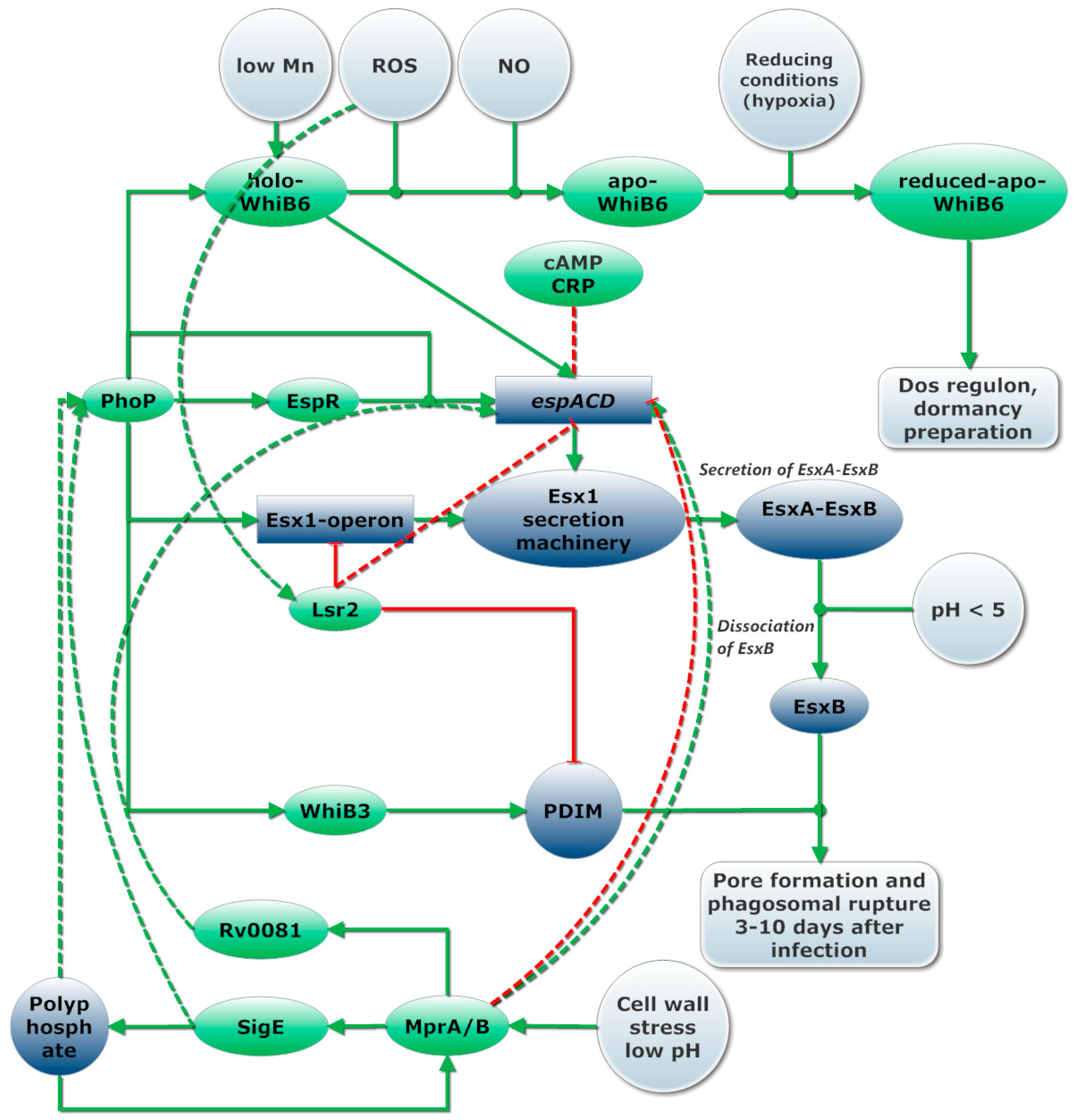
3.2. Phagosomal Rupture and Pore Formation

The second main virulence strategy deployed by Mtb is phagosomal rupture. A model of regulation of pore formation can be found in Figure 4.
ESX-1 and ESX-1 secreted proteins EsxA (ESAT-6) and EsxB (CFP-10) have been implicated in phagosomal rupture of many Mycobateria such as M. marinum, M. kansii and Mtb [139,140,141,142]. The virulence lipid phthiocerol dimycocerosates (PDIM) and EsxA from Mtb were shown to interact with the host cell membrane and in concert, induce phagosome membrane damage and rupture in infected macrophages [142,143]. A recent study reported that many claims about pore formation at neutral pH are due to contamination with detergent from the washing step [4]. The same study found membrane-lysing capabilities for EsxA only to occur below pH 5, to be contact dependent and accompanied by gross membrane disruptions rather than discrete pores. For the sake of simplicity, we refer here to the process of cytosolic access as phagosomal rupture although more research is needed to find out if cytosolic access is only achieved through lesions or also through formation of pores. Additionally there are reports of Mtb and other Mycobacteria to escape the phagosome [144]. However, the data generate by electron microscopy—the only direct approach—remains controversial.
The ESX-1 secretion system is involved in secretion of virulence proteins among which those shown to be involved in pore formation and phagosomal rupture EsxA (ESAT-6) and EsxB (CFP-10), secretion associated proteins EspA-D, EspF and secreted immune modulating PE and PPE proteins [96,145,146,147]. Although EsxB is the main pore forming protein, other ESX-1 secreted genes are required for EsxB secretion and proper functioning of the ESX-1 secretion machinery. EspD stabilizes the extracellular levels of EspA and EspC and it is required for EsxA secretion but does not require ESX-1 for its own secretion [148]. Secretion of EspA, EspC, EsxA is codependent on each other, suggesting they might be secreted as a multimeric complex or that they are part of the secretion machinery itself [149,150]. This theory is supported by a study showing that EspA forms dimers by disulphide bond formation after secretion; disruption of this disulphide bond affects cell wall stability as well as the functioning of the whole ESX-1 secretion system [151]. Recently, an EspC-multimeric complex was observed to form filamentous structure that could represent a secretion needle [152]. Inactivation of MyCP1 protease causes hyper-activation of ESX-1 while protease inhibition leads to attenuated virulence during chronic infection [153,154]. A balanced activation and deactivation of ESX-1 through MycP1 proteolysis of EspB is required during chronic infection. MyCP1 and MyCP5 are required for stability of the ESX-1 and ESX-5 secretion complex respectively [155]. Without ESX-1, Mtb is unable to disrupt the phagosome membrane and make contact with the cytosol, leading to highly diminished pathogenicity [145].
ESX-1 and secreted factors EsxA and EsxB are regulated by the two-component systems PhoPR, previously mentioned. The importance of PhoP for virulence was confirmed in knockout studies that showed phoP knockout mutants to be attenuated in mouse bone marrow derived macrophages, lungs, livers and spleen [156]. A single point mutation in phoP in Mtb H37Ra decreases the DNA affinity of PhoP and strongly contributes to the reduced virulence of this strain [157]. PhoPR regulated genes are upregulated in acidic and oxidative conditions encountered during the first two days of infection [40]. Recent studies show that PhoP interacts with SigE, which is upregulated in acidic pH and upon cell stress during the first three days of infection [40,158]. Additionally, polyphosphate was needed for normal transcription of phoP as well as for transcriptional regulation of sigE by MprAB, although these results could not be reproduced [159,160]. PhoP/R influences transcription of some 80 (according to some sources up to 150 [161]) genes directly as well as the transcription of a large number of genes indirectly via upregulation of WhiB6, EspR, DevS/R and WhiB3 [60,129].
EspR is a transcriptional regulator upregulated by PhoP. EspR induces transcription of the espACD (Rv3612-16c) operon which is essential for phagosomal rupture and potential escape from the phago(-lyso)some [148,151,162]. PhoP, therefore, controls, directly (espB/E-L) or indirectly (espA/C/D), the 13 Esp proteins secreted by ESX-1 [162,163,164]. Recently it was found that holo-WhiB6 increases transcription of its own operon, the ESX-1 regulon and suppressed the DevR regulon, while apo-WhiB6 formed in anaerobic conditions and by prolonged exposure to NO, suppresses the ESX-1 regulon and induces the DevR dormancy regulon [85]. Interestingly, gene expression of EsxB by WhiB6 was highly induced after 30-min of NO exposure, decreased at 60 min and is highly reduced after 3 h of exposure to NO, indicating a short but intense activation of espACD by holo-WhiB6. Additionally binding sites for WhiB6 and Rv0081, a transcriptional factor regulated by MprAB, were predicted upstream of espACD [84]. These results suggest WhiB6, which is induced by PhoPR and MntR, plays an essential role in the regulation of phagosomal rupture and dormancy.
Induction of transcription of espACD by EspR requires the presence of PhoP [162]. In addition, MprAB, Lsr2 and CRP bind to the promotor region of espACD operon. Lsr2 represses transcription of both the espACD and the ESX-1 operon [84], while CRP binding inhibits expression of espACD [165]. Lsr2 binds to AT rich regions in the DNA, mostly virulence genes and is required for adaptation to extreme oxygen conditions [53,54]. We hypothesize it is likely that Lsr2 represses the operon containing ESX-1 genes and espACD in oxidative conditions. This could serve to avoid further aggravation of the immune response. MprAB functions as a repressor of the espACD operon in cellular stress conditions, however MprA/B is also required for full expression of espACD. It is plausible to assume both positive and negative regulation by MprAB occurs based on the presence of multiple binding sites for MprA and two transcriptional start in the espACD operon [84].
Like the post-translational activation of GroEL2 by HiP1, membrane lysing capability of EsxA is activated only upon dissociation of EsxA from EsxB in acidic environment (pH 4–5) encountered when the phagosome matures. Acetylation of proteins in Mtb is cAMP dependent [141]. Acetylation improves dissociation of EsxA from EsxB at higher pH, a model where acetylation leads to reduced virulence was proposed [166]. Taken together, these studies indicate pore formation is strictly regulated, most likely only occurs when cAMP is depleted (no cAMP-CRP), might be inhibited by sudden changes in oxidative conditions (Lsr2), the phagosome acidifies and become hypoxic (PhoPR) and pore formation is transiently induced by WhiB6 upon NO sensing [85]. MprAB further modifies activation of espACD, most likely both positively upon initial cell damage and negatively after prolonged cell stress and accumulation of polyphosphate, as indicated in Figure 4.
It should be mentioned that in addition to their role as regulators, Lsr2, CRP and EspR have also been characterized as nucleoid-associated proteins and as such might serve additional functions such as structuring the organization of the chromosome and, as has been shown for the ESX-1 and espACD operon, protecting DNA region from oxygen radicals [53,165,167].

3.3. Dormancy and Modulation of Granuloma Formation

The third virulence strategy deployed by Mtb is onset of dormancy. Dormancy is a non-replicating and metabolically near inactive state at which Mtb is immune to most drugs and can survive for decades [3,9]. Dormancy occurs upon formation of mostly hypoxic granulomas [168]. Immune modulation that stimulates granuloma formation will therefore be discussed as a part of the dormancy virulence strategy.
When Mtb runs out of cAMP to secrete thereby suppressing phagosome lysosome fusion, the macrophages phagosome will fuse with late endosomes and lysosomes. As a result, the phagosome becomes increasingly hostile with lower pH, production of oxygen radicals and NO and fusion with vesicles containing lysozymes. In contrast, conditions encountered in granulomas are slightly more favourable for Mtb. Granulomas have reduced capacity to form oxidative radicals [11].
Mtb stimulates TNF-α production which leads to granuloma formation among others through secretion of cAMP into the cytosol [70,106,169]. A number of studies indicate that granuloma may be dispensable for preventing bacterial dissemination and may actually contribute to Mtb persistence and shield Mtb from more successful immune cells [7,10,11]. According to some models, Mtb containing granuloma’s contain two types of macrophages: classically activated and alternatively activated [7]. Mtb shifts the macrophage population within the granuloma from being classically activated to alternatively activated macrophage which produce more anti-inflammatory cytokines (TGF-β, IL-10) and arginase. These diminish the amount of arginine available to iNOS, which results in reduced NO production [7,11,170]. A balance of pro-inflammatory and anti-inflammatory response via stimulation of TNF-α and IFN-γ production is needed for granuloma formation while IL-10 is the main negative regulator for this response, inhibiting formation of dense and hypoxic mature fibrotic granuloma’s [7,106]. Moreover, parameter sensitivity analysis for a granuloma model, showed IL-10 had the strongest influence on myofibroblast numbers at 300 days post infection and indicated IL-10 to play a major role in preventing differentiation of immune cells needed to develop protective immunity [7,106].
A number of regulators allow Mtb to sense and adapt to hypoxia and maturation of the phagosome. The most important of these regulators is the two-component regulator DevRST which regulate genes coding for proteins that help Mtb prepare for dormancy and subsequent resuscitation [171,172,173]. A visual representation of DevRST response to environmental cues is present as part of Supplementary File 1. Both DevS and DevT can activate the DevR regulon through phosphorylation of DevR, which autoregulates its own operon through cooperative binding to two binding sites [172,173,174,175]. DevT provides initial activation of the DevR regulon through phosphorylation of DevR and has the strongest sensitivity to CO and a weaker binding to NO and O2 compared to DevS. DevS is sufficient for DevR activation after 5 days of infection [176,177]. DevS phosphorylates DevR even in the presence of small concentrations of NO, negatively regulates the DevR regulon through phosphatase activity in the presence of O2 while positively regulating the DevR regulon in reducing conditions [176,178,179].
Interestingly, even under non-inducing conditions and as such no phosphorylation of DevR, the DevR regulon is activated upon high enough concentrations of DevR, providing a possible explanation for enduring induction of the DevR regulon which might occur after prolonged autoactivation of its own regulon [175]. Among DevR regulated genes there are a few types of regulation. While some genes are strongly upregulated within a few hours of infection others are only mildly induced after 12–24 h in hypoxic and high NO conditions [174]. DevR and other two-component regulators can fine tune expression of genes through the presence of multiple binding sites and through phosphorylation which stimulates cooperative binding [173].
CO is released by the enzymatic activity of heme oxygenase-1 (HO-1) in lungs infected by Mtb [180,181]. CO is an important dormancy inducer. Interestingly, Mtb has a unique heme scavenging and degrading systems that does not produce CO allowing Mtb to degrade heme without inducing the immune response or its own dormancy regulon.
Interestingly, there is evidence for two DevR regulated proteins to be involved in stabilizing the 30S ribosomal units under hypoxic conditions, while slowing down translation and protein synthesis in the process [168,182]. Mtb uses lipids such as cholesterol as primary nutrient in this phase of infection via genes regulated by KstR and IdeR [127,129], while increasing production of triacylglyceride (TAG) via tgs1 which is under control of DevR and Whib3 [73].
Protein-protein interaction was observed between DevT and NarL, a lone two-component response regulator involved in nitrate and nitrite respiration in Escherechia coli [183,184,185]. Although the genes regulated by NarL in Mtb are unknown, we argue it is plausible that NarL is involved in regulation of nirB, narU, narX, narU, nuoB that are currently thought to be part of the DevR regulon.
NO is produced in the maturing phagosome and is an important dormancy cue sensed by DevT and DevS. Mtb expresses two truncated heme proteins, GlbN and GlbO, that help it detoxify from nitrate containing oxygen radicals such as NO while residing in the macrophage [186,187,188,189].
Interestingly, GlbN is co-transcribed with lpRl coding for Lipoprotein LprI, which Acts as a lysozyme inhibitor [190]. The GlbN-lpR1 Activated isoniazid inhibits truncated haemoglobin N that protects against reactive nitrogen and oxygen species as well as AcpM, which is required for mycolic-acid production [15,191,192,193]. NO was found to help Mtb to survive in hypoxic and acidic conditions through anaerobic respiration [185,194]. In addition, nitrate respiration plays an important role in dormancy and protection against hypoxic and acidic stress [194,195].
Although DevRST and WhiB3 are involved in the preparation for dormancy, the enduring hypoxic response measured in a devR knockout mutant showed 230 genes to be differentially expressed with roughly half of them upregulated in in the first day of hypoxia and the other half only upregulated at 4 and 7 days of hypoxia [196]. These results indicate many genes involved in the enduring hypoxia response are not regulated by DevR. Resuscitation from dormancy is more elusive and less studied than dormancy. Resuscitation involves ClgR and both SigH and SigE are upregulated upon reaeration [197]. Also cAMP-CRP plays a role in resuscitation as it upregulates rpfA one of the five resuscitation promoting factors [137,198,199].

4. Success through Tight Regulation of Virulence Strategies

Mtb anticipates changes in the interaction with the host by upregulating both internal and external sensors and regulators involved in sensing progression of the immune response. This allows the bacteria to adjust more quickly to progression of the immune response. External sensors involved in survival in the macrophage consists mostly of two-component regulators [161] (such as DevRST, PhoPR, MprAB, SenX3-RegX3, NarL) while for internal sensors, WhiB family proteins and regulators such as CRP and CMR are used. These sensors and regulators appear interconnected, thus forming a single regulatory cascade that controls the three virulence strategies, as represented in Figure 5. This regulatory cascade integrates many internal (cAMP, Mn, Mg, oxidative conditions and presence of NO) and external environmental cues (phagosome pH or cell wall damage) for fine-tuned regulation of key virulence systems. Examples of such virulence systems downstream this cascade are GroEL2, ESX-1, EsxAB and EspACD. Pore formation by EsxA depends on the regulation of ESX-1 by PhoP, Lsr2 and WhiB6 and on regulation of EspACD by Lsr2, EspR, PhoPR, MprAB, WhiB6 and Rv0081. Post translationally, pore formation by EsxA is regulated by proteolytic activity of MycP1, acetylation of EsxA and dissociation of EsxA-EsxB upon acidification of the phagosome [13,53,54,84,85,139,141,165,166,167]. Similarly, GroEL2 is regulated by CRP, WhiB1, HrCA and Mg2+ starvation and post-translationally regulated by proteolytic cleavage by Hip1 [124,132,133,136,137,138].
There is a great amount of overlap in this cascade, so that multiple environmental signals are considered in the regulation of these genes, as indicated in Figure 5. For example, some PhoPR regulated genes are predicted to have cAMP-CRP binding sites [200]. These genes are upregulated upon oxidative stress and low pH but suppressed in the presence of cAMP-CRP, as is the case for espACD [201]. Some PhoPR regulated genes are also regulated by DevRST, WhiB3 and by MprAB. An even larger overlap exists in genes regulated by DevRST and MprAB, indicating integration of CO, NO, hypoxia and cell stress in the regulation of these genes [202,203,204]. We argue that based on the overlapping regulation of the three virulence strategies, these strategies extend and overlap each other. The order of activation of these strategies is likely to vary depending on the dynamics between Mtb and the host. Timing of specific virulence strategies also vary for different Mtb strains [144]. Some strains gain cytosolic access within hours of phagocytosis while others require 3–10 days [13,144].
Pore or lesion formation is linked to immune modulation. Cytosolic access is need for secretion of cAMP and other immune modulating factors, such as GroEL2, into the macrophage cytosol [144]. There are still many unanswered questions regarding the exact role and regulation of GroEL2. Firstly, it is unknown at which conditions proteolysis of GroEL2 by Hip1 (Rv2224c) occurs. Secondly, Hip1 was reported to mainly function as lipase in one study [134], further research is needed to confirm whether GroEL2 is a direct substrate of Hip1. Strict regulation of GroEL2 suggests it to have an important role in virulence.
Interestingly, there are many parallels in regulation of virulence systems between Mtb and other pathogens. Understanding Mtb as one of the most successful intracellular pathogens can therefore provide insight in common strategies deployed by intracellular pathogens. For instance, positive regulation of virulence genes by PhoPR and suppression by cAMP-CRP appears to occur in more pathogens. In Y. pestis, PhoP directly binds to and transcriptionally activates crp and cyA leading to merging of the PhoPQ and CRP-cAMP regulon [205]. Similarly, a major virulence island is positively regulated by PhoP while being suppressed by cAMP-CRP in S. typhimurium [206]. In Mtb, PhoPR regulates pro-inflammatory virulence genes such as the ESX-1 operon as well as genes involved in protecting against oxidative stress, when cAMP is depleted. cAMP does not only suppress phagosome maturation but also acts as an internal sensor of phagosome maturation, through pH dependent secretion of cAMP.
Some aspects in the regulation of PhoPR and cAMP in Mtb require more research. Firstly, the function of multiple IdeR binding sites upstream of the phoPR suggests complex regulation of the phoPR operon by IdeR and thus by iron bioavailability. Secondly, the exact cue for activation of PhoP remains unknown. Upregulation of phoPR in acidic conditions has been observed as well as under Mg2+ starvation, however this later observation could not be reproduced [125]. Transcriptional analysis of Mtb showed many genes in the PhoPR regulon to be upregulated during the first hours of infection (20 min to 2 h) while the phagosome acidified from pH of 6.5 to pH 5.5 [66]. PhoPR stimulates expression of aprABC, an Mtb specific pH sensing locus involved in the regulation of among others a number of PhoP regulated genes [125]. These results indicated PhoPR directly or indirectly senses pH. Recently, it was discovered that PhoP interacts with acid inducible extracytoplasmic sigma factor SigE, providing a possible explanation for activation of the PhoP regulon at low pH [158]. Extracytoplasmic sigma factors provide a means of regulating gene expression in response to various extracellular changes, hence their name.
Secondly, we argue entrance of Mtb in the early phagosome is likely to lead to higher abundance of Mn. Pathogenic Mycobacteria species such as Mtb and M. avium, have high manganese concentrations at 1 and at 24 h after infection compared to non- pathogenic M. smegmatis [32]. Mn availability might also be affected by Mramp, a pH dependent Mn H+ symporter with maximal activity between pH 5.5 and 6.5 matching the conditions found in the early phagosome. Mn is an important cofactor for cAMP synthesis and it is likely to increase cAMP production in the early phagosome. cAMP-CRP and PhoPR co-regulate virulence genes directly or via regulators such as WhiB6, which is linked to Mn deficiency. Based on the strong affinity of PhoP for Mn we hypothesize Mn might play a role in both cAMP and PhoPR regulation [20,83]. Depletion of Mn and secretion of cAMP might lead to de-repression of cAMP-CRP suppressed genes such as espACD as well as activation of these genes through PhoPR.
Thirdly, polyphosphate is needed for optimal PhoP activation [159]. Polyphosphates are potent inhibitors of type III adenylyl cyclases in M. bovis which agrees with the opposing roles of cAMP-CRP and PhoPR in respectively inducing genes involved in the anti- and pro-inflammatory response in Mtb and other pathogens. Polyphosphate is implicated in the activation of PhoP and is part of one of two positive feedback loops in the regulation of mprAB and sigE [158,159,160]. Polyphosphates kinase production is conserved in all bacteria and is associated to induction of dormancy and activation of virulence genes in many pathogens [207]. Knockout polyphosphate kinases ppk1 mutants, have reduced biofilm formation, are more susceptible to drugs and are impaired in growth in guinea pigs [159,208]. Interestingly, SigE is involved in regulation of polyphosphate. MprAB and SigX3-RegX3, induce transcription of sigE upon cell wall stress or phosphate starvation, while anti sigma factor RseA binds to and neutralizes SigE in reducing conditions [209,210]. RseA is degraded by ClpC1P2-dependent proteolytic activity depending on its phosphorylation by the eukaryotic-like Ser/Thr protein kinase PknB [210]. SigE, polyphosphate and MprAB are involved in a double positive feedback loops through polyphosphate and ClpC1P2 of which a visual model is provided by Manganelli et al. [210]. Polyphosphate functions as phosphate donor for MprAB under low ATP condition. Additionally, SigE regulates the transcription of the furA-katG operon in response to oxidative stress in Mycobacteria [67]. SigE knockout strains are strongly attenuated and a recent study shows a sigE knockout strain provide an even more effective live vaccine than BCG [211]. Taken together, these studies indicate SigE plays an important role in adapting to low pH, cell wall and oxidative stress through upregulation furA-katG, activation of some PhoPR induced genes, MprAB and inhibition of cAMP-CRP through polyphosphate production. The interplay of SigE, polyphosphate and the hypothesized role of Mn in PhoPR and cAMP regulation should be further investigated.
Another aspect we want to address is the link between IdeR, cAMP, cholesterol degradation and phagosomal rupture. IdeR, KstR and KstR2 co-regulate the cholesterol degradation pathway in M. bovis [127]. We suggest a similar synergy between IdeR regulation and cholesterol degradation in Mtb. Transcription of cholesterol degradation genes in Mtb is dependent on the presence of CyA [212]. Regulation of cholesterol degradation by IdeR and cAMP would suggest access to cholesterol is associated to the initial stage of Mtb host interaction when the iron pool is oxidized and cAMP is produced to avoid phagosome maturation. Interestingly, EsxA and other pore forming toxins specifically inserts themselves into phosphor lipid (phosphatidylcholine) and cholesterol-containing liposomes [166,213]. Giant foamy macrophages rich in cholesterol are at the centre of Mtb containing granuloma’s that turn necrotic [7,11,12,107,213]. Accumulation of cholesterol was shown to be essential for uptake of Mtb by the macrophage [214]. Additionally, cholesterol was shown to increase association of TACO, a coat protein that prevents degradation of Mycobacteria upon fusion with lysosomes [214]. We argue that accumulation of cholesterol in macrophages not only increases Mtb survival in the phagosome by serving as carbon source but also might assists in phagosomal rupture and possibly in escape from the phagosome.
In summary, in this review we provide an overview for understanding divalent metal homeostasis and their role in regulating three essential virulence strategies of Mtb: immune modulation, dormancy and phagosomal rupture. Sensors of environmental and internal cues, including divalent metal availability, form a single regulatory cascade that controls these three virulence strategies. The role of polyphosphate, cAMP and manganese in this cascade requires further investigation.

Supplementary Materials

The following are available online at https://www.mdpi.com/1422-0067/19/2/347/s1.

Acknowledgments

This work has been supported by European Union through the SysteMTb project (HEALTH-F4-2010-241587), the Horizon 2020 research and innovation programme under grant agreement No. 634942 (MycoSynVac) and the FP7 programme under grant agreement No. 305340 (INFECT).

Conflicts of Interest

The authors declare no conflict of interest.

Abbreviations

ACAdenylate cyclase
cAMPCyclic adenosine monophosphate
CMRCyclic-AMP and redox responsive transcription factor
CRPCyclic-AMP dependent regulatory protein
DAGDiacylglycerol
DevRSTDevRST is a two component regulator and sensor, which regulate genes coding for proteins that help Mtb prepare for dormancy and subsequent resuscitation
EspRA virulence associated transcriptional regulator upregulated by PhoP
IdeRIron-dependent regulator
Lsr2A histone like regulator that binds AT-rich regions virulence islands, acting as a global regulator to aid in the adaptation to extremes in oxygen availability
MntRManganese-dependent transcriptional repressor
MprABA two component sensor and regulator that responds to cell envelop stress
MtbMycobacterium tuberculosis
PDIMPhthiocerol dimycocerosates
SigEExtracytoplasmic alternative Sigma factor E, involved in response to low pH and cell stress
ZurZinc uptake regulator

References

  1. World Health Organization (WHO). Global Tuberculosis Report 2017; World Health Organization: Geneva, Switzerland, 2017. [Google Scholar]
  2. Meena, L.S. Rajni Survival mechanisms of pathogenic Mycobacterium tuberculosis H37Rv. FEBS J. 2010, 277, 2416–2427. [Google Scholar] [CrossRef] [PubMed]
  3. Gengenbacher, M.; Kaufmann, S.H.E. Mycobacterium tuberculosis: Success through dormancy. FEMS Microbiol. Rev. 2012, 36, 514–532. [Google Scholar] [CrossRef] [PubMed]
  4. Conrad, W.H.; Osman, M.M.; Shanahan, J.K.; Chu, F.; Takaki, K.K.; Cameron, J.; Hopkinson-Woolley, D.; Brosch, R.; Ramakrishnan, L. Mycobacterial ESX-1 secretion system mediates host cell lysis through bacterium contact-dependent gross membrane disruptions. Proc. Natl. Acad. Sci. USA 2017, 114, 1371–1376. [Google Scholar] [CrossRef] [PubMed]
  5. Dietzold, J.; Gopalakrishnan, A.; Salgame, P. Duality of lipid mediators in host response against Mycobacterium tuberculosis: Good cop, bad cop. F1000Prime Rep. 2015, 7, 1–8. [Google Scholar] [CrossRef] [PubMed]
  6. Guirado, E.; Schlesinger, L.S. Modeling the Mycobacterium tuberculosis granuloma—The critical battlefield in host immunity and disease. Front. Immunol. 2013, 4, 1–7. [Google Scholar] [CrossRef] [PubMed]
  7. Silva Miranda, M.; Breiman, A.; Allain, S.; Deknuydt, F.; Altare, F. The tuberculous granuloma: An unsuccessful host defence mechanism providing a safety shelter for the bacteria? Clin. Dev. Immunol. 2012, 2012. [Google Scholar] [CrossRef] [PubMed]
  8. Gomez, J.E.; McKinney, J.D.M. Tuberculosis persistence, latency, and drug tolerance. Tuberculosis 2004, 84, 29–44. [Google Scholar] [CrossRef] [PubMed]
  9. Kapoor, N.; Pawar, S.; Sirakova, T.D.; Deb, C.; Warren, W.L.; Kolattukudy, P.E. Human granuloma in vitro model, for TB dormancy and resuscitation. PLoS ONE 2013, 8, e53657. [Google Scholar] [CrossRef] [PubMed]
  10. Paige, C.; Bishai, W.R. Penitentiary or penthouse condo: The tuberculous granuloma from the microbe’s point of view. Cell. Microbiol. 2010, 12, 301–309. [Google Scholar] [CrossRef] [PubMed]
  11. Shaler, C.R.; Horvath, C.N.; Jeyanathan, M.; Xing, Z. Within the Enemy’s Camp: Contribution of the granuloma to the dissemination, persistence and transmission of Mycobacterium tuberculosis. Front. Immunol. 2013, 4. [Google Scholar] [CrossRef] [PubMed]
  12. Russell, D.; Cardona, P.; Kim, M. Foamy macrophages and the progression of the human tuberculosis granuloma. Nat. Immunol. 2009, 10, 943–948. [Google Scholar] [CrossRef] [PubMed]
  13. Simeone, R.; Bobard, A.; Lippmann, J.; Bitter, W.; Majlessi, L.; Brosch, R.; Enninga, J. Phagosomal rupture by Mycobacterium tuberculosis results in toxicity and host cell death. PLoS Pathog. 2012, 8, e1002507. [Google Scholar] [CrossRef] [PubMed]
  14. Sani, M.; Houben, E.N.G.; Geurtsen, J.; Pierson, J.; de Punder, K.; van Zon, M.; Wever, B.; Piersma, S.R.; Jiménez, C.R.; Daffé, M.; et al. Direct visualization by Cryo-EM of the mycobacterial capsular layer: A labile structure containing ESX-1-secreted proteins. PLoS Pathog. 2010, 6, e1000794. [Google Scholar] [CrossRef] [PubMed] [Green Version]
  15. Lucarelli, D.; Vasil, M.L.; Meyer-Klaucke, W.; Pohl, E. The metal-dependent regulators FurA and FurB from Mycobacterium tuberculosis. Int. J. Mol. Sci. 2008, 9, 1548–1560. [Google Scholar] [CrossRef] [PubMed] [Green Version]
  16. Juttukonda, L.J.; Skaar, E.P. Manganese homeostasis and utilization in pathogenic bacteria. Mol. Microbiol. 2015, 97, 216–228. [Google Scholar] [CrossRef] [PubMed]
  17. Forbes, J.R.; Gros, P. Iron, manganese, and cobalt transport by Nramp1 (Slc11a1) and Nramp2 (Slc11a2) expressed at the plasma membrane. Blood 2003, 102, 1884–1892. [Google Scholar] [CrossRef] [PubMed]
  18. Indriate, M.; Skaar, E.P. Nutritional immunity: Transition metals at the pathogen-host interface. Nat. Rev. Microbiol. 2013, 10, 646–656. [Google Scholar] [CrossRef]
  19. Pandey, R.; Rodriguez, G.M. IdeR is required for iron homeostasis and virulence in Mycobacterium tuberculosis. Mol. Microbiol. 2014, 91, 98–109. [Google Scholar] [CrossRef] [PubMed]
  20. Papp-Wallace, K.M.; Maguire, M.E. Manganese transport and the role of manganese in virulence. Annu. Rev. Microbiol. 2006, 60, 187–209. [Google Scholar] [CrossRef] [PubMed]
  21. Reddy, S.K.; Kamireddi, M.; Dhanireddy, K.; Young, L.; Davis, A.; Reddy, P.T. Eukaryotic-like adenylyl cyclases in Mycobacterium tuberculosis H37Rv: Cloning and characterization. J. Biol. Chem. 2001, 276, 35141–35149. [Google Scholar] [CrossRef] [PubMed]
  22. Maciag, A.; Dainese, E.; Rodriguez, G.M.; Milano, A.; Provvedi, R.; Pasca, M.R.; Smith, I.; Palù, G.; Riccardi, G.; Manganelli, R. Global analysis of the Mycobacterium tuberculosis Zur (FurB) regulon. J. Bacteriol. 2007, 189, 730–740. [Google Scholar] [CrossRef] [PubMed]
  23. Kurthkoti, K.; Amin, H.; Marakalala, M.J.; Ghanny, S.; Subbian, S.; Sakatos, A.; Livny, J.; Fortune, S.M.; Berney, M.; Rodriguez, G.M. The capacity of Mycobacterium tuberculosis to survive iron starvation might enable it to persist in iron-deprived microenvironments of human granulomas. mBio 2017, 8, e01092-17. [Google Scholar] [CrossRef] [PubMed]
  24. Lin, H.; Andersen, G.R.; Yatime, L. Crystal structure of human S100A8 in complex with zinc and calcium. BMC Struct. Biol. 2016, 16, 8. [Google Scholar] [CrossRef] [PubMed]
  25. Olakanmi, O.; Schlesinger, L.S.; Ahmed, A.; Britigan, B.E. Intraphagosomal Mycobacterium tuberculosis acquires iron from both extracellular transferrin and intracellular iron pools. Impact of interferon-gamma and hemochromatosis. J. Biol. Chem. 2002, 277, 49727–49734. [Google Scholar] [CrossRef] [PubMed]
  26. Blanchette, C.D.; Woo, Y.-H.; Thomas, C.; Shen, N.; Sulchek, T.A.; Hiddessen, A.L. Decoupling internalization, acidification and phagosomal-endosomal/lysosomal fusion during phagocytosis of InlA coated beads in epithelial cells. PLoS ONE 2009, 4, e6056. [Google Scholar] [CrossRef] [PubMed]
  27. Forbes, J.R.; Gros, P. Divalent-metal transport by NRAMP proteins at the interface of host-pathogen interactions. Trends Microbiol. 2001, 9, 397–403. [Google Scholar] [CrossRef]
  28. Olakanmi, O.; Schlesinger, L.S.; Ahmed, A.; Britigan, B.E. The nature of extracellular iron influences iron acquisition by Mycobacterium tuberculosis residing within human macrophages. Infect. Immun. 2004, 72, 2022–2028. [Google Scholar] [CrossRef] [PubMed]
  29. Jabado, N.; Jankowski, A.; Dougaparsad, S.; Picard, V.; Grinstein, S.; Gros, P. Natural resistance to intracellular infections: Natural resistance-associated macrophage protein 1 (Nramp1) functions as a pH-dependent manganese transporter at the phagosomal membrane. J. Exp. Med. 2000, 192, 1237–1248. [Google Scholar] [CrossRef] [PubMed]
  30. Supek, F.; Supekova, L.; Nelson, H.; Nelson, N. A yeast manganese transporter related to the macrophage protein involved in conferring resistance to mycobacteria. Proc. Natl. Acad. Sci. USA 1996, 93, 5105–5110. [Google Scholar] [CrossRef] [PubMed]
  31. Peracino, B.; Buracco, S.; Bozzaro, S. The Nramp (Slc11) proteins regulate development, resistance to pathogenic bacteria and iron homeostasis in Dictyostelium discoideum. J. Cell Sci. 2013, 126, 301–311. [Google Scholar] [CrossRef] [PubMed]
  32. Wagner, D.; Maser, J.; Lai, B.; Cai, Z.; Barry, C.E., III; Höner zu Bentrup, K.; Russell, D.G.; Bermudez, L.; Iii, C.E.B. Elemental analysis of Mycobacterium avium-, Mycobacterium tuberculosis-, and Mycobacterium smegmatis-containing phagosomes indicates pathogen-induced microenvironments within the host cell’s endosomal system. J. Immunol. 2013, 174, 1491–1500. [Google Scholar]
  33. Pandey, R.; Russo, R.; Ghanny, S.; Huang, X.; Helmann, J.; Rodriguez, G.M. MntR(Rv2788): A transcriptional regulator that controls manganese homeostasis in Mycobacterium tuberculosis. Mol. Microbiol. 2015, 98, 1168–1183. [Google Scholar] [CrossRef] [PubMed]
  34. Pohl, E.; Holmes, R.K.; Hol, W.G. Crystal structure of the iron-dependent regulator (IdeR) from Mycobacterium tuberculosis shows both metal binding sites fully occupied. J. Mol. Biol. 1999, 285, 1145–1156. [Google Scholar] [CrossRef] [PubMed]
  35. Dewitt, M.A.; Kliegman, J.I.; Helmann, J.D.; Brennan, R.G.; David, L.; Glasfeld, A. The conformations of the manganese transport regulator of Bacillus subtilis in its metal-free state. J. Mol. Biol. 2007, 365, 1257–1265. [Google Scholar] [CrossRef] [PubMed]
  36. Luo, M.; Fadeev, E.A.; Groves, J.T. Mycobactin-mediated iron acquisition within macrophages. Nat. Chem. Biol. 2005, 1, 149–153. [Google Scholar] [CrossRef] [PubMed]
  37. McMahon, M.D.; Rush, J.S.; Thomas, M.G. Analyses of MbtB, MbtE, and MbtF suggest revisions to the Mycobactin biosynthesis pathway in Mycobacterium tuberculosis. J. Bacteriol. 2012, 194, 2809–2818. [Google Scholar] [CrossRef] [PubMed]
  38. Boradia, V.M.; Malhotra, H.; Thakkar, J.S.; Tillu, V.A.; Vuppala, B.; Patil, P.; Sheokand, N.; Sharma, P.; Chauhan, A.S.; Raje, M.; et al. Mycobacterium tuberculosis acquires iron by cell-surface sequestration and internalization of human holo-transferrin. Nat. Commun. 2014, 5, 1–13. [Google Scholar] [CrossRef] [PubMed] [Green Version]
  39. Madigan, C.A.; Cheng, T.-Y.; Layre, E.; Young, D.C.; McConnell, M.J.; Debono, C.A.; Murry, J.P.; Wei, J.-R.; Barry, C.E.; Rodriguez, G.M.; et al. Lipidomic discovery of deoxysiderophores reveals a revised mycobactin biosynthesis pathway in Mycobacterium tuberculosis. Proc. Natl. Acad. Sci. USA 2012, 109, 1257–1262. [Google Scholar] [CrossRef] [PubMed]
  40. Rohde, K.H.; Veiga, D.F.T.; Caldwell, S.; Balázsi, G.; Russell, D.G. Linking the transcriptional profiles and the physiological states of Mycobacterium tuberculosis during an extended intracellular infection. PLoS Pathog. 2012, 8. [Google Scholar] [CrossRef] [PubMed]
  41. Tyagi, P.; Dharmaraja, A.T.; Bhaskar, A.; Chakrapani, H.; Singh, A. Mycobacterium tuberculosis has diminished capacity to counteract redox stress induced by elevated levels of endogenous superoxide. Free Radic. Biol. Med. 2015, 84, 344–354. [Google Scholar] [CrossRef] [PubMed]
  42. Rodriguez, G.M.; Voskuil, M.I.; Gold, B.; Schoolnik, G.K.; Smith, I. ideR, an essential gene in Mycobacterium tuberculosis: Role of IdeR in iron-dependent gene expression, iron metabolism, and oxidative stress response. Infect. Immun. 2002, 70, 3371–3381. [Google Scholar] [CrossRef] [PubMed]
  43. Vilchèze, C.; Hartman, T.; Weinrick, B.; William, R.J., Jr. Mycobacterium tuberculosis is extraordinarily sensitive to killing by a vitamin C-induced Fenton reaction. Nat. Commun. 2013, 4. [Google Scholar] [CrossRef] [PubMed]
  44. Litwin, C.M.; Calderwood, S.B. Role of iron in regulation of virulence genes. Clin. Microbiol. Rev. 1993, 6, 137–149. [Google Scholar] [CrossRef] [PubMed]
  45. Schaible, U.E.; Kaufmann, S.H.E. Iron and microbial infection. Nat. Rev. Microbiol. 2004, 2, 946–953. [Google Scholar] [CrossRef] [PubMed]
  46. Outten, F.W.; Theil, E.C. Iron-based redox switches in biology. Antioxid. Redox Signal. 2009, 11, 1029–1046. [Google Scholar] [CrossRef] [PubMed]
  47. Siegrist, M.S.; Unnikrishnan, M.; Mcconnell, M.J.; Borowsky, M.; Cheng, T.; Siddiqi, N.; Fortune, S.M.; Moody, D.B.; Rubin, E.J. Mycobacterial Esx-3 is required for mycobactin-mediated iron acquisition. Proc. Natl. Acad. Sci. USA 2009, 106, 18792–18797. [Google Scholar] [CrossRef] [PubMed]
  48. Serafini, A.; Boldrin, F.; Palù, G.; Manganelli, R.; Palu, G. Characterization of a Mycobacterium tuberculosis ESX-3 conditional mutant: Essentiality and rescue by iron and zinc. J. Bacteriol. 2009, 191, 6340–6344. [Google Scholar] [CrossRef] [PubMed]
  49. Farhana, A.; Kumar, S.; Rathore, S.S.; Ghosh, P.C.; Ehtesham, N.Z.; Tyagi, A.K.; Hasnain, S.E. Mechanistic insights into a novel exporter-importer system of Mycobacterium tuberculosis unravel its role in trafficking of iron. PLoS ONE 2008, 3, e2087. [Google Scholar] [CrossRef] [PubMed]
  50. Gold, B.; Rodriguez, G.M.; Marras, S.A.; Pentecost, M.; Smith, I. The Mycobacterium tuberculosis IdeR is a dual functional regulator that controls transcription of genes involved in iron acquisition, iron storage and survival in macrophages. Mol. Microbiol. 2001, 42, 851–865. [Google Scholar] [CrossRef] [PubMed]
  51. Rodriguez, G.M.; Smith, I. Mechanisms of iron regulation in mycobacteria: Role in physiology and virulence. Mol. Microbiol. 2003, 47, 1485–1494. [Google Scholar] [CrossRef] [PubMed]
  52. Oldridge, D.A.; Wood, A.C.; Weichert-leahey, N.; Crimmins, I.; Winter, C.; Mcdaniel, L.D.; Diamond, M.; Hart, L.S.; Durbin, A.D.; Abraham, B.J.; et al. The mycobacterial iron dependent regulator IdeR induces ferritin (bfrB) by alleviating Lsr2 repression. Mol. Microbiol. 2016, 528, 418–421. [Google Scholar] [CrossRef]
  53. Fu, G.; Lees, R.S.; Aw, D.; Jin, L.; Gray, P.; Berendonk, T.U.; White-cooper, H.; Scaife, S.; Phuc, H.K.; Jasinskiene, N.; et al. Lsr2 is a nucleoid-associated protein that targets AT-rich sequences and virulence genes in Mycobacterium tuberculosis. Proc. Natl. Acad. Sci. USA 2010, 107, 18741. [Google Scholar] [CrossRef]
  54. Colangeli, R.; Haq, A.; Arcus, V.L.; Summers, E.; Magliozzo, R.S.; McBride, A.; Mitra, A.K.; Radjainia, M.; Khajo, A.; Jacobs, W.R.; et al. The multifunctional histone-like protein Lsr2 protects mycobacteria against reactive oxygen intermediates. Proc. Natl. Acad. Sci. USA 2009, 106, 4414–4418. [Google Scholar] [CrossRef] [PubMed]
  55. Colangeli, R.; Helb, D.; Vilchèze, C.; Hazbón, M.H.; Lee, C.G.; Safi, H.; Sayers, B.; Sardone, I.; Jones, M.B.; Fleischmann, R.D.; et al. Transcriptional regulation of multi-drug tolerance and antibiotic-induced responses by the histone-like protein Lsr2 in M. tuberculosis. PLoS Pathog. 2007, 3, 780–793. [Google Scholar] [CrossRef] [PubMed]
  56. Khare, G.; Nangpal, P.; Tyagi, A.K. Differential roles of iron storage proteins in maintaining the iron homeostasis in Mycobacterium tuberculosis. PLoS ONE 2017, 12, 1–18. [Google Scholar] [CrossRef] [PubMed]
  57. Jones, C.M.; Wells, R.M.; Madduri, A.V.R.; Renfrow, M.B.; Ratledge, C.; Moody, D.B.; Niederweis, M. Self-poisoning of Mycobacterium tuberculosis by interrupting siderophore recycling. Proc. Natl. Acad. Sci. USA 2014, 111, 1945–1950. [Google Scholar] [CrossRef] [PubMed]
  58. Pandey, S.D.; Choudhury, M.; Yousuf, S.; Wheeler, P.R.; Gordon, S.V.; Ranjan, A.; Sritharan, M. Iron-regulated protein HupB of Mycobacterium tuberculosis positively regulates siderophore biosynthesis and is essential for growth in macrophages. J. Bacteriol. 2014, 196, 1853–1865. [Google Scholar] [CrossRef] [PubMed]
  59. Sritharan, M. Iron homeostasis in Mycobacterium tuberculosis: Mechanistic insights into siderophore-mediated iron uptake. Bacteriology 2016, 198, 2399–2409. [Google Scholar] [CrossRef] [PubMed]
  60. Gonzalo-Asensio, J.; Mostowy, S.; Harders-Westerveen, J.; Huygen, K.; Hernández-Pando, R.; Thole, J.; Behr, M.; Gicquel, B.; Martín, C. PhoP: A missing piece in the intricate puzzle of Mycobacterium tuberculosis virulence. PLoS ONE 2008, 3, e3496. [Google Scholar] [CrossRef] [PubMed]
  61. Gupta, S.; Sinha, A.; Sarkar, D. Transcriptional autoregulation by Mycobacterium tuberculosis PhoP involves recognition of novel direct repeat sequences in the regulatory region of the promoter. FEBS Lett. 2006, 580, 5328–5338. [Google Scholar] [CrossRef] [PubMed]
  62. Gonzalo-Asensio, J.; Soto, C.Y.; Arbués, A.; Sancho, J.; del Carmen Menéndez, M.; García, M.J.; Gicquel, B.; Martín, C. The Mycobacterium tuberculosis phoPR operon is positively autoregulated in the virulent strain H37Rv. J. Bacteriol. 2008, 190, 7068–7078. [Google Scholar] [CrossRef] [PubMed]
  63. Manabe, Y.C.; Saviola, B.J.; Sun, L.; Murphy, J.R.; Bishai, W.R. Attenuation of virulence in Mycobacterium tuberculosis expressing a constitutively active iron repressor. Proc. Natl. Acad. Sci. USA 1999, 96, 12844–12848. [Google Scholar] [CrossRef] [PubMed]
  64. Banerjee, S.; Nandyala, A.K.; Raviprasad, P.; Ahmed, N.; Hasnain, S.E. Iron-dependent RNA-binding activity of Mycobacterium tuberculosis aconitase. J. Bacteriol. 2007, 189, 4046–4052. [Google Scholar] [CrossRef] [PubMed]
  65. Pechter, K.B.; Meyer, F.M.; Serio, A.W.; Stülke, J.; Sonenshein, A.L. Two roles for aconitase in the regulation of tricarboxylic acid branch gene expression in Bacillus subtilis. J. Bacteriol. 2013, 195, 1525–1537. [Google Scholar] [CrossRef] [PubMed]
  66. Rohde, K.H.; Abramovitch, R.B.; Russell, D.G. Mycobacterium tuberculosis invasion of macrophages: Linking bacterial gene expression to environmental cues. Cell Host Microbe 2007, 2, 352–364. [Google Scholar] [CrossRef] [PubMed]
  67. Hu, Y.; Wang, Z.; Feng, L.; Chen, Z.; Mao, C.; Zhu, Y.; Chen, S. σE-dependent activation of RbpA controls transcription of the furA-katG operon in response to oxidative stress in mycobacteria. Mol. Microbiol. 2016, 102, 107–120. [Google Scholar] [CrossRef] [PubMed]
  68. Zheng, F.; Long, Q.; Xie, J. The function and regulatory network of WhiB and WhiB-like protein from comparative genomics and systems biology perspectives. Cell Biochem. Biophys. 2012, 63, 103–108. [Google Scholar] [CrossRef] [PubMed]
  69. Reeves, A.Z.; Campbell, P.J.; Sultana, R.; Malik, S.; Murray, M.; Plikaytis, B.B.; Shinnick, T.M.; Posey, J.E. Aminoglycoside cross-resistance in Mycobacterium tuberculosis due to mutations in the 5′ untranslated region of whiB7. Antimicrob. Agents Chemother. 2013, 57, 1857–1865. [Google Scholar] [CrossRef] [PubMed]
  70. Samuel, L.P.; Song, C.-H.; Wei, J.; Roberts, E.A.; Dahl, J.L.; Barry, C.E.; Jo, E.-K.; Friedman, R.L. Expression, production and release of the Eis protein by Mycobacterium tuberculosis during infection of macrophages and its effect on cytokine secretion. Microbiology 2007, 153, 529–540. [Google Scholar] [CrossRef] [PubMed]
  71. Farina, M.; Avila, D.S.; Da Rocha, J.B.T.; Aschner, M. Metals, oxidative stress and neurodegeneration: A focus on iron, manganese and mercury. Neurochem. Int. 2013, 62, 575–594. [Google Scholar] [CrossRef] [PubMed]
  72. Champion, O.L.; Karlyshev, A.; Cooper, I.A.M.; Ford, D.C.; Wren, B.W.; Duffield, M.; Oyston, P.C.F.; Titball, R.W. Yersinia pseudotuberculosis mntH functions in intracellular manganese accumulation, which is essential for virulence and survival in cells expressing functional Nramp1. Microbiology 2011, 157, 1115–1122. [Google Scholar] [CrossRef] [PubMed]
  73. Kumar, A.; Farhana, A.; Guidry, L.; Saini, V.; Hondalus, M.; Steyn, A.J.C. Redox homeostasis in mycobacteria: The key to tuberculosis control? Expert Rev. Mol. Med. 2011, 13, e39. [Google Scholar] [CrossRef] [PubMed]
  74. Akhter, Y.; Yellaboina, S.; Farhana, A.; Ranjan, A.; Ahmed, N.; Hasnain, S.E. Genome scale portrait of cAMP-receptor protein (CRP) regulons in mycobacteria points to their role in pathogenesis. Gene 2008, 407, 148–158. [Google Scholar] [CrossRef] [PubMed]
  75. Matange, N. Revisiting bacterial cyclic nucleotide phosphodiesterases: Cyclic AMP hydrolysis and beyond. FEMS Microbiol. Lett. 2015, 362, fnv183. [Google Scholar] [CrossRef] [PubMed]
  76. Dass, B.K.M.; Sharma, R.; Shenoy, A.R.; Mattoo, R.; Visweswariah, S.S. Cyclic AMP in mycobacteria: Characterization and functional role of the Rv1647 ortholog in Mycobacterium smegmatis. J. Bacteriol. 2008, 190, 3824–3834. [Google Scholar] [CrossRef] [PubMed]
  77. Bai, G.; Knapp, G.S.; McDonough, K.A. Cyclic AMP signalling in mycobacteria: Redirecting the conversation with a common currency. Cell. Microbiol. 2011, 13, 349–358. [Google Scholar] [CrossRef] [PubMed]
  78. Dittrich, D.; Keller, C.; Ehlers, S.; Schultz, J.E.; Sander, P. Characterization of a Mycobacterium tuberculosis mutant deficient in pH-sensing adenylate cyclase Rv1264. Int. J. Med. Microbiol. 2006, 296, 563–566. [Google Scholar] [CrossRef] [PubMed]
  79. Agarwal, N.; Lamichhane, G.; Gupta, R.; Nolan, S.; Bishai, W.R. Cyclic AMP intoxication of macrophages by a Mycobacterium tuberculosis adenylate cyclase. Nature 2009, 460, 98–102. [Google Scholar] [CrossRef] [PubMed]
  80. Daniel, J.; Abraham, L.; Martin, A.; Pablo, X.; Reyes, S. Rv2477c is an antibiotic-sensitive manganese-dependent ABC-F ATPase in Mycobacterium tuberculosis. Biochem. Biophys. Res. Commun. 2017, 1–6. [Google Scholar] [CrossRef] [PubMed]
  81. Rishi, P.; Jindal, N.; Bharrhan, S.; Tiwari, R.P. Salmonella-macrophage interactions upon manganese supplementation. Biol. Trace Elem. Res. 2010, 133, 110–119. [Google Scholar] [CrossRef] [PubMed]
  82. Diaz-ochoa, V.E.; Lam, D.; Lee, C.S.; Chazin, W.J.; Skaar, E.P.; Raffatellu, M.; Behnsen, J.; Liu, J.Z.; Chim, N. Salmonella mitigates oxidative stress and thrives in the inflamed gut by evading calprotectin-mediated article salmonella mitigates oxidative stress and thrives in the inflamed gut by evading calprotectin-mediated manganese sequestration. Cell Host Microbe 2016, 19, 814–825. [Google Scholar] [CrossRef] [PubMed]
  83. Agranoff, D.; Monahan, I.M.; Mangan, J.A.; Butcher, P.D.; Krishna, S. Mycobacterium tuberculosis expresses a novel pH-dependent divalent cation transporter belonging to the Nramp family. J. Exp. Med. 1999, 190, 717–724. [Google Scholar] [CrossRef] [PubMed]
  84. Pang, X.; Samten, B.; Cao, G.; Wang, X.; Tvinnereim, A.R.; Chen, X.-L.; Howard, S.T. MprAB regulates the espA operon in Mycobacterium tuberculosis and modulates ESX-1 function and host cytokine response. J. Bacteriol. 2013, 195, 66–75. [Google Scholar] [CrossRef] [PubMed]
  85. Chen, Z.; Hu, Y.; Cumming, B.M.; Lu, P.; Feng, L.; Deng, J.; Steyn, A.J.C.; Chen, S. Mycobacterial WhiB6 differentially regulates ESX-1 and the dos regulon to modulate granuloma formation and virulence in zebrafish. Cell Rep. 2016, 16, 2512–2524. [Google Scholar] [CrossRef] [PubMed]
  86. Korch, S.B.; Contreras, H.; Clark-curtiss, J.E. Three Mycobacterium tuberculosis Rel Toxin-antitoxin modules inhibit mycobacterial growth and are expressed in infected human macrophages. J. Bacteriol. 2009, 191, 1618–1630. [Google Scholar] [CrossRef] [PubMed]
  87. Yang, M.; Gao, C.H.; Hu, J.; Dong, C.; He, Z.G. Characterization of the interaction between a SirR family transcriptional factor of Mycobacterium tuberculosis, encoded by Rv2788, and a pair of toxin-antitoxin proteins RelJ/K, encoded by Rv3357 and Rv3358. FEBS J. 2014, 281, 2726–2737. [Google Scholar] [CrossRef] [PubMed]
  88. Kang, S.-M.; Kim, D.-H.; Lee, K.-Y.; Park, S.J.; Yoon, H.-J.; Lee, S.J.; Im, H.; Lee, B.-J. Functional details of the Mycobacterium tuberculosis VapBC26 toxin-antitoxin system based on a structural study: Insights into unique binding and antibiotic peptides. Nucleic Acids Res. 2017, 45, 8564–8580. [Google Scholar] [CrossRef] [PubMed]
  89. Lee, I.; Lee, S.J.; Chae, S.; Lee, K.; Kim, J.; Lee, B. Structural and functional studies of the Mycobacterium tuberculosis VapBC30 toxin-antitoxin system: Implications for the design of novel antimicrobial peptides. Nucleic Acids Res. 2015, 43, 7624–7637. [Google Scholar] [CrossRef] [PubMed]
  90. Serafini, A.; Pisu, D.; Palù, G.; Rodriguez, G.M.; Manganelli, R. The ESX-3 secretion system is necessary for iron and zinc homeostasis in Mycobacterium tuberculosis. PLoS ONE 2013, 8, 1–15. [Google Scholar] [CrossRef] [PubMed]
  91. Bretl, D.J.; Bigley, T.M.; Terhune, S.S.; Zahrt, T.C. The MprB extracytoplasmic domain negatively regulates activation of the Mycobacterium tuberculosis MprAB two-component system. J. Bacteriol. 2014, 196, 391–406. [Google Scholar] [CrossRef] [PubMed]
  92. Ilghari, D.; Lightbody, K.L.; Veverka, V.; Waters, L.C.; Muskett, F.W.; Renshaw, P.S.; Carr, M.D.; Muskett, W. Solution structure of the Mycobacterium tuberculosis EsxG·EsxH complex: Functional implications and comparisons with other M. tuberculosis Esx family complexes. J. Biol. Chem. 2011, 286, 29993–30002. [Google Scholar] [CrossRef] [PubMed]
  93. Gabriel, S.E.; Helmann, J.D. Contributions of zur-controlled ribosomal proteins to growth under zinc starvation conditions. J. Bacteriol. 2009, 191, 6116–6122. [Google Scholar] [CrossRef] [PubMed]
  94. Houben, E.N.G.; Korotkov, K.V.; Bitter, W. Take five—Type VII secretion systems of mycobacteria. Biochim. Biophys. Acta 2013. [Google Scholar] [CrossRef] [PubMed]
  95. Newton-Foot, M. The Mycobacterium tuberculosis ESX-3 Secretion System Interactome. Master’s Thesis, University of Stellenbsoch, Stellenbsoch, South Africa, 2010. [Google Scholar]
  96. Simeone, R.; Bottai, D.; Brosch, R. ESX/type VII secretion systems and their role in host-pathogen interaction. Curr. Opin. Microbiol. 2009, 12, 4–10. [Google Scholar] [CrossRef] [PubMed]
  97. Mehra, A.; Zahra, A.; Thompson, V.; Sirisaengtaksin, N.; Wells, A.; Porto, M.; Köster, S.; Penberthy, K.; Kubota, Y.; Dricot, A.; et al. Mycobacterium tuberculosis Type VII Secreted Effector EsxH Targets Host ESCRT to Impair Trafficking. PLoS Pathog. 2013, 9. [Google Scholar] [CrossRef] [PubMed]
  98. Tufariello, J.M.; Chapman, J.R.; Kerantzas, C.A.; Wong, K.-W.; Vilchèze, C.; Jones, C.M.; Cole, L.E.; Tinaztepe, E.; Thompson, V.; Fenyö, D.; et al. Separable roles for Mycobacterium tuberculosis ESX-3 effectors in iron acquisition and virulence. Proc. Natl. Acad. Sci. USA 2016, 113, E348–E357. [Google Scholar] [CrossRef] [PubMed]
  99. Van Dam, J.C.J.; Schaap, P.J.; Martins dos Santos, V.A.P.; Suárez-diez, M. Integration of heterogeneous molecular networks to unravel gene-regulation in Mycobacterium tuberculosis. BMC Med. Genom. 2014, 8, 1–21. [Google Scholar] [CrossRef] [PubMed]
  100. Tiwari, B.M.; Kannan, N.; Vemu, L.; Raghunand, T.R. The Mycobacterium tuberculosis PE proteins Rv0285 and Rv1386 modulate innate immunity and mediate bacillary survival in macrophages. PLoS ONE 2012, 7, e51686. [Google Scholar] [CrossRef] [PubMed]
  101. Li, W.; Zhao, Q.; Deng, W.; Chen, T.; Liu, M.; Xie, J. Mycobacterium tuberculosis Rv3402c Enhances mycobacterial survival within macrophages and modulates the host pro-inflammatory cytokines production via NF-Kappa B/ERK/p38 signaling. PLoS ONE 2014, 9, e94418. [Google Scholar] [CrossRef] [PubMed]
  102. Daim, S.; Kawamura, I.; Tsuchiya, K.; Hara, H.; Kurenuma, T.; Shen, Y.; Dewamitta, S.R.; Sakai, S.; Nomura, T.; Qu, H.; et al. Expression of the Mycobacterium tuberculosis PPE37 protein in Mycobacterium smegmatis induces low tumour necrosis factor alpha and interleukin 6 production in murine macrophages. J. Med. Microbiol. 2011, 60, 582–591. [Google Scholar] [CrossRef] [PubMed]
  103. Yeruva, V.C.; Kulkarni, A.; Khandelwal, R.; Sharma, Y.; Raghunand, T.R. The PE_PGRS proteins of Mycobacterium tuberculosis are Ca2+ binding mediators of host–pathogen interaction. Biochemistry 2016, 55, 4675–4687. [Google Scholar] [CrossRef] [PubMed]
  104. Meng, L.; Tong, J.; Wang, H.; Tao, C.; Wang, Q.; Niu, C.; Zhang, X.; Gao, Q. PPE38 protein of Mycobacterium tuberculosis inhibits macrophage MHC class I expression and dampens CD8+ T cell responses. Front. Cell. Infect. Microbiol. 2017, 7, 1–11. [Google Scholar] [CrossRef] [PubMed]
  105. Dong, D.; Wang, D.; Li, M.; Wang, H.; Yu, J.; Wang, C.; Liu, J.; Gao, Q. PPE38 modulates the innate immune response and is required for Mycobacterium marinum virulence. Infect. Immun. 2012, 80, 43–54. [Google Scholar] [CrossRef] [PubMed]
  106. Cyktor, J.C.; Carruthers, B.; Kominsky, R.A.; Beamer, G.L.; Stromberg, P.; Turner, J. IL-10 inhibits mature fibrotic granuloma formation during Mycobacterium tuberculosis infection. J. Immunol. 2013, 190, 2778–2790. [Google Scholar] [CrossRef] [PubMed]
  107. Mahajan, S.; Dkhar, H.K.; Chandra, V.; Dave, S.; Nanduri, R.; Janmeja, A.K.; Agrewala, J.N.; Gupta, P. Mycobacterium tuberculosis modulates macrophage lipid-sensing nuclear receptors PPARγ and TR4 for survival. J. Immunol. 2012, 188, 5593–5603. [Google Scholar] [CrossRef] [PubMed]
  108. Han, X.; Kitamoto, S.; Wang, H.; Boisvert, W.A. Interleukin-10 overexpression in macrophages suppresses atherosclerosis in hyperlipidemic mice. FASEB J. 2010, 24, 2869–2880. [Google Scholar] [CrossRef] [PubMed]
  109. Prados-Rosales, R.; Weinrick, B.C.; Piqué, D.G.; Jacobs, W.R.; Casadevall, A.; Rodriguez, G.M. Role for Mycobacterium tuberculosis membrane vesicles in iron acquisition. J. Bacteriol. 2014, 196, 1250–1256. [Google Scholar] [CrossRef] [PubMed]
  110. Seto, S.; Tsujimura, K.; Koide, Y. Rab GTPases regulating phagosome maturation are differentially recruited to mycobacterial phagosomes. Traffic 2011, 12, 407–420. [Google Scholar] [CrossRef] [PubMed]
  111. Via, L.E.; Dusanka, D.; Roseann, J.U.; Nina, S.H.; Huber, L.A.; Deretic, V. Arrest of mycobacterial phagosome maturation is caused by a block in vesicle fusion between stages controlled by rab5 and rab7. J. Biol. Chem. 1997, 272, 13326–13331. [Google Scholar] [CrossRef] [PubMed]
  112. Wong, D.; Bach, H.; Sun, J.; Hmama, Z.; Av-Gay, Y. Mycobacterium tuberculosis protein tyrosine phosphatase (PtpA) excludes host vacuolar-H+-ATPase to inhibit phagosome acidification. Proc. Natl. Acad. Sci. USA 2011, 108, 19371–19376. [Google Scholar] [CrossRef] [PubMed]
  113. Kalamidas, S.A.; Kuehnel, M.P.; Peyron, P.; Rybin, V.; Rauch, S.; Kotoulas, O.B.; Houslay, M.; Hemmings, B.A.; Gutierrez, M.G.; Anes, E.; et al. cAMP synthesis and degradation by phagosomes regulate actin assembly and fusion events: Consequences for mycobacteria. J. Cell Sci. 2006, 119, 3686–3694. [Google Scholar] [CrossRef] [PubMed]
  114. Gupta, A.; Kaul, A.; Tsolaki, A.G.; Kishore, U.; Bhakta, S. Mycobacterium tuberculosis: Immune evasion, latency and reactivation. Immunobiology 2012, 217, 363–374. [Google Scholar] [CrossRef] [PubMed]
  115. Thi, E.P.; Lambertz, U.; Reiner, N.E. Sleeping with the enemy: How intracellular pathogens cope with a macrophage lifestyle. PLoS Pathog. 2012, 8, e1002551. [Google Scholar] [CrossRef] [PubMed]
  116. Nguyen, L.; Pieters, J. Mycobacterial subversion of chemotherapeutic reagents and host defense tactics: Challenges in tuberculosis drug development. Annu. Rev. Pharmacol. Toxicol. 2009, 49, 427–453. [Google Scholar] [CrossRef] [PubMed]
  117. Alam, M.S.; Garg, S.K.; Agrawal, P. Studies on structural and functional divergence among seven WhiB proteins of Mycobacterium tuberculosis H37Rv. FEBS J. 2009, 276, 76–93. [Google Scholar] [CrossRef] [PubMed]
  118. Larsson, C.; Luna, B.; Ammerman, N.C.; Maiga, M.; Agarwal, N.; Bishai, W.R. Gene expression of Mycobacterium tuberculosis putative transcription factors whiB1-7 in redox environments. PLoS ONE 2012, 7, e37516. [Google Scholar] [CrossRef] [PubMed]
  119. Smith, L.J.; Stapleton, M.R.; Fullstone, G.J.M.; Crack, J.C.; Thomson, J.; Le Brun, N.E.; Hunt, D.M.; Harvey, E.; Adinolfi, S.; Buxton, R.S.; et al. Europe PMC Funders Group Mycobacterium tuberculosis WhiB1 is an essential DNA-binding protein with a nitric oxide sensitive iron-sulphur cluster. Biochem. J. 2010, 432, 417–427. [Google Scholar] [CrossRef] [PubMed]
  120. Ranganathan, S.; Bai, G.; Lyubetskaya, A.; Gwendowlyn, S.; Galagan, E.; Mcdonough, K.A. Characterization of a cAMP responsive transcription factor, Cmr (Rv1675c), in TB complex mycobacteria reveals overlap with the DosR (DevR) dormancy regulon. Nucleic Acids Res. 2002, 1–54. [Google Scholar] [CrossRef] [PubMed]
  121. Chawla, M.; Parikh, P.; Saxena, A.; Munshi, M.; Mehta, M.; Mai, D.; Srivastava, A.K.; Narasimhulu, K.V.; Redding, K.E.; Vashi, N.; et al. Mycobacterium tuberculosis WhiB4 regulates oxidative stress response to modulate survival and dissemination in vivo. Mol. Microbiol. 2012, 85, 1148–1165. [Google Scholar] [CrossRef] [PubMed]
  122. Casonato, S.; Cervantes Sánchez, A.; Haruki, H.; Rengifo González, M.; Provvedi, R.; Dainese, E.; Jaouen, T.; Gola, S.; Bini, E.; Vicente, M.; et al. WhiB5, a transcriptional regulator that contributes to Mycobacterium tuberculosis virulence and reactivation. Infect. Immun. 2012, 80, 3132–3144. [Google Scholar] [CrossRef] [PubMed]
  123. Singh, A.; Guidry, L.; Narasimhulu, K.V.; Mai, D.; Trombley, J.; Redding, K.E.; Giles, G.I.; Lancaster, J.R.; Steyn, A.J.C. Mycobacterium tuberculosis WhiB3 responds to O2 and nitric oxide via its [4Fe-4S] cluster and is essential for nutrient starvation survival. Proc. Natl. Acad. Sci. USA 2007, 104, 11562–11567. [Google Scholar] [CrossRef] [PubMed]
  124. Stapleton, M.R.; Smith, L.J.; Hunt, D.M.; Buxton, R.S.; Green, J. Mycobacterium tuberculosis WhiB1 represses transcription of the essential chaperonin GroEL2. Tuberculosis 2012, 92, 328–332. [Google Scholar] [CrossRef] [PubMed]
  125. Abramovitch, R.B.; Rohde, K.H.; Hsu, F.-F.; Russell, D.G. aprABC: A Mycobacterium tuberculosis complex-specific locus that modulates pH-driven adaptation to the macrophage phagosome. Mol. Microbiol. 2011, 80, 678–694. [Google Scholar] [CrossRef] [PubMed]
  126. Singh, A.; Crossman, D.K.; Mai, D.; Guidry, L.; Voskuil, M.I.; Renfrow, M.B.; Steyn, A.J.C. Mycobacterium tuberculosis WhiB3 maintains redox homeostasis by regulating virulence lipid anabolism to modulate macrophage response. PLoS Pathog. 2009, 5, e1000545. [Google Scholar] [CrossRef] [PubMed]
  127. Rienksma, R.A.; Suárez-Diez, M.; Mollenkopf, H.-J.; Dolganov, G.M.; Dorhoi, A.; Schoolnik, G.K.; Martins dos Santos, V.A.P.; Kaufmann, S.H.E.; Schaap, P.J.; Gengenbacher, M. Comprehensive insights into transcriptional adaptation of intracellular mycobacteria by microbe-enriched dual RNA sequencing. BMC Genom. 2015, 16, 1–15. [Google Scholar] [CrossRef] [PubMed]
  128. Zimmermann, M.; Kogadeeva, M.; Gengenbacher, M.; McEwen, G.; Mollenkopf, H.; Zamboni, N.; Kaufmann, S.H.E.; Sauer, U. Integration of metabolomics and transcriptomics reveals a complex diet of mycobacterium tuberculosis during early macrophage infection. MSystems 2017, 2, 1–18. [Google Scholar] [CrossRef] [PubMed]
  129. Galagan, J.E.; Minch, K.; Peterson, M.; Lyubetskaya, A.; Azizi, E.; Sweet, L.; Gomes, A.; Rustad, T.; Dolganov, G.; Glotova, I.; et al. The Mycobacterium tuberculosis regulatory network and hypoxia. Nature 2013, 499, 178–183. [Google Scholar] [CrossRef] [PubMed]
  130. Saini, V.; Farhana, A.; Steyn, A.J.C. Mycobacterium tuberculosis WhiB3: A novel iron-sulfur cluster protein that regulates redox homeostasis and virulence. Antioxid. Redox Signal. 2012, 16, 687–697. [Google Scholar] [CrossRef] [PubMed]
  131. Gazdik, M.A.; Mcdonough, K.A. Identification of cyclic AMP-regulated genes in Mycobacterium tuberculosis complex bacteria under low-oxygen conditions identification of cyclic AMP-regulated genes in Mycobacterium tuberculosis complex bacteria under low-oxygen conditions. J. Bacteriol. 2005, 187, 2681–2692. [Google Scholar] [CrossRef] [PubMed]
  132. Joseph, S.; Yuen, A.; Singh, V.; Hmama, Z. Mycobacterium tuberculosis Cpn60. 2 (GroEL2) blocks macrophage apoptosis via interaction with mitochondrial mortalin. Biol. Open 2017, 2, 481–488. [Google Scholar] [CrossRef] [PubMed]
  133. Naffin-Olivos, J.L.; Georgieva, M.; Goldfarb, N.; Madan-Lala, R.; Dong, L.; Bizzell, E.; Valinetz, E.; Brandt, G.S.; Yu, S.; Shabashvili, D.E.; et al. Mycobacterium tuberculosis Hip1 modulates macrophage responses through proteolysis of GroEL2. PLoS Pathog. 2014, 10. [Google Scholar] [CrossRef] [PubMed]
  134. Forrellad, M.A.; Klepp, L.I.; Gioffré, A.; Sabio y García, J.; Morbidoni, H.R.; de la Paz Santangelo, M.; Cataldi, A.A.; Bigi, F. Virulence factors of the Mycobacterium tuberculosis complex. Virulence 2013, 4, 3–66. [Google Scholar] [CrossRef] [PubMed]
  135. Jamwal, S.; Midha, M.K.; Verma, H.N.; Basu, A.; Rao, K.V.S.; Manivel, V. Characterizing virulence-specific perturbations in the mitochondrial function of macrophages infected with Mycobacterium tuberculosis. Sci. Rep. 2013, 3, 1328. [Google Scholar] [CrossRef] [PubMed]
  136. Stewart, G.R.; Wernisch, L.; Stabler, R.; Mangan, J.A.; Hinds, J.; Laing, K.G.; Young, D.B.; Butcher, P.D. Dissection of the heat-shock response in Mycobacterium tuberculosis using mutants and microarrays. Microbiology 2002, 148, 3129–3138. [Google Scholar] [CrossRef] [PubMed]
  137. Agarwal, N.; Raghunand, T.R.; Bishai, W.R. Regulation of the expression of whiB1 in Mycobacterium tuberculosis: Role of cAMP receptor protein. Microbiology 2006, 152, 2749–2756. [Google Scholar] [CrossRef] [PubMed]
  138. Gazdik, M.A.; Bai, G.; Wu, Y.; McDonough, K.A. Rv1675c (cmr) regulates intramacrophage and cyclic AMP-induced gene expression in Mycobacterium tuberculosis-complex mycobacteria. Mol. Microbiol. 2009, 71, 434–448. [Google Scholar] [CrossRef] [PubMed]
  139. Smith, J.; Manoranjan, J.; Pan, M.; Bohsali, A.; Xu, J.; Liu, J.; McDonald, K.L.; Szyk, A.; LaRonde-LeBlanc, N.; Gao, L.-Y. Evidence for pore formation in host cell membranes by ESX-1-secreted ESAT-6 and its role in Mycobacterium marinum escape from the vacuole. Infect. Immun. 2008, 76, 5478–5487. [Google Scholar] [CrossRef] [PubMed]
  140. Lim, Y.-J.; Choi, H.-H.; Choi, J.-A.; Jeong, J.A.; Cho, S.-N.; Lee, J.-H.; Park, J.B.; Kim, H.-J.; Song, C.-H. Mycobacterium kansasii-induced death of murine macrophages involves endoplasmic reticulum stress responses mediated by reactive oxygen species generation or calpain activation. Apoptosis 2013, 18, 150–159. [Google Scholar] [CrossRef] [PubMed]
  141. Mba Medie, F.; Champion, M.M.; Williams, E.A.; Champion, P.A.D. Homeostasis of N-α-terminal acetylation of EsxA correlates with virulence in Mycobacterium marinum. Infect. Immun. 2014, 82, 4572–4586. [Google Scholar] [CrossRef] [PubMed]
  142. Augenstreich, J.; Simeone, R.; Wegener, A.; Sayes, F.; Le, F.; Christian, C.; Malaga, W.; Guilhot, C.; Brosch, R.; Astarie, C. ESX-1 and phthiocerol dimycocerosates of Mycobacterium tuberculosis act in concert to cause phagosomal rupture and host cell apoptosis. Cell. Microbiol. 2017, 1–19. [Google Scholar] [CrossRef] [PubMed]
  143. Francis, R.J.; Butler, R.E.; Stewart, G.R. Mycobacterium tuberculosis ESAT-6 is a leukocidin causing Ca2+ influx, necrosis and neutrophil extracellular trap formation. Cell Death Dis. 2014, 5, e1474. [Google Scholar] [CrossRef] [PubMed]
  144. Jamwal, S.V.; Mehrotra, P.; Singh, A.; Siddiqui, Z.; Basu, A. Mycobacterial escape from macrophage phagosomes to the cytoplasm represents an alternate adaptation mechanism. Sci. Rep. 2016, 1–9. [Google Scholar] [CrossRef] [PubMed]
  145. Clemmensen, H.S.; Peter, N.; Knudsen, H.; Rasmussen, E.M.; Winkler, J.; Rosenkrands, I.; Ahmad, A.; Lillebaek, T.; Sherman, D.R.; Andersen, P.L.; et al. An attenuated Mycobacterium tuberculosis clinical strain with a defect in ESX-1 secretion induces minimal host immune responses and pathology. Sci. Rep. 2017, 1–13. [Google Scholar] [CrossRef] [PubMed]
  146. Deng, W.; Xiang, X.; Xie, J. Comparative genomic and proteomic anatomy of Mycobacterium ubiquitous Esx family proteins: Implications in pathogenicity and virulence. Curr. Microbiol. 2014, 68, 558–567. [Google Scholar] [CrossRef] [PubMed]
  147. Di Giuseppe Champion, P.A.; Champion, M.M.; Manzanillo, P.; Cox, J.S. ESX-1 secreted virulence factors are recognized by multiple cytosolic AAA ATPases in mycobactria. Mol. Microbiol. 2009, 73, 950–962. [Google Scholar] [CrossRef] [PubMed]
  148. Chen, J.M.; Boy-Röttger, S.; Dhar, N.; Sweeney, N.; Buxton, R.S.; Pojer, F.; Rosenkrands, I.; Cole, S.T. EspD is critical for the virulence-mediating ESX-1 secretion system in Mycobacterium tuberculosis. J. Bacteriol. 2012, 194, 884–893. [Google Scholar] [CrossRef] [PubMed]
  149. Ize, B.; Palmer, T. Microbiology. Mycobacteria’s export strategy. Science 2006, 313, 1583–1584. [Google Scholar] [CrossRef] [PubMed]
  150. Fortune, S.M.; Jaeger, A.; Sarracino, D.A.; Chase, M.R.; Sassetti, C.M.; Sherman, D.R.; Bloom, B.R.; Rubin, E.J. Mutually dependent secretion of proteins required for mycobacterial virulence. Proc. Natl. Acad. Sci. USA 2005, 102, 10676–10681. [Google Scholar] [CrossRef] [PubMed]
  151. Garces, A.; Atmakuri, K.; Chase, M.R.; Woodworth, J.S.; Rothchild, A.C.; Ramsdell, T.L.; Lopez, M.F.; Behar, S.M.; Sarracino, D.A.; Fortune, S.M. EspA acts as a critical mediator of ESX1-dependent virulence in Mycobacterium tuberculosis by affecting bacterial cell wall integrity. PLoS Pathog. 2010, 6, 15–16. [Google Scholar] [CrossRef] [PubMed] [Green Version]
  152. Ates, L.S.; Brosch, R. Micro commentary discovery of the type VII ESX-1 secretion needle? Mol. Microbiol. 2017, 103, 7–12. [Google Scholar] [CrossRef] [PubMed]
  153. Lou, Y.; Rybniker, J.; Sala, C.; Cole, S.T. EspC forms a filamentous structure in the cell envelope of Mycobacterium tuberculosis and impacts ESX-1 secretion. Mol. Microbiol. 2017, 103, 26–38. [Google Scholar] [CrossRef] [PubMed]
  154. Ohol, Y.M.; Goetz, D.H.; Chan, K.; Shiloh, M.U.; Charles, C.S.; Cox, J.S. Mycobacterium tuberculosis MycP1 protease plays a dual role in regulation of ESX-1 secretion and virulence. Cell Host Microbe 2011, 7, 210–220. [Google Scholar] [CrossRef] [PubMed]
  155. Van Winden, V.J.C.; Ummels, R.; Piersma, S.R.; Jiménez, C.R.; Korotkov, K.V.; Bitter, W. Mycosins are required for the stabilization of the ESX-1 and ESX-5. mBio 2016, 7, 1–11. [Google Scholar] [CrossRef] [PubMed]
  156. Sinha, A.; Gupta, S.; Bhutani, S.; Pathak, A.; Sarkar, D. PhoP-PhoP interaction at adjacent PhoP binding sites is influenced by protein phosphorylation. J. Bacteriol. 2008, 190, 1317–1328. [Google Scholar] [CrossRef] [PubMed]
  157. Pérez, E.; Samper, S.; Bordas, Y.; Guilhot, C.; Gicquel, B.; Martín, C. An essential role for phoP in Mycobacterium tuberculosis virulence. Mol. Microbiol. 2001, 41, 179–187. [Google Scholar] [CrossRef] [PubMed]
  158. Bansal, R.; Kumar, V.A. Mycobacterium tuberculosis virulence-regulator PhoP interacts with alternative sigma factor SigE during acid-stress response. Mol. Microbiol. 2017, 104, 400–411. [Google Scholar] [CrossRef] [PubMed]
  159. Singh, R.; Singh, M.; Arora, G.; Kumar, S.; Tiwari, P.; Kidwai, S. Polyphosphate deficiency in Mycobacterium tuberculosis is associated with enhanced drug susceptibility and impaired growth in guinea pigs. J. Bacteriol. 2013, 195, 2839–2851. [Google Scholar] [CrossRef] [PubMed]
  160. Sureka, K.; Dey, S.; Datta, P.; Singh, A.K.; Dasgupta, A.; Rodrigue, S.; Basu, J.; Kundu, M. Polyphosphate kinase is involved in stress-induced mprAB-sigE-rel signalling in mycobacteria. Mol. Microbiol. 2007, 65, 261–276. [Google Scholar] [CrossRef] [PubMed]
  161. Bretl, D.J.; Demetriadou, C.; Zahrt, T.C. Adaptation to environmental stimuli within the host: Two-component signal transduction systems of Mycobacterium tuberculosis. Microbiol. Mol. Biol. Rev. 2011, 75, 566–582. [Google Scholar] [CrossRef] [PubMed]
  162. Kumar, V.A.; Goyal, R.; Bansal, R.; Singh, N.; Sevalkar, R.R.; Kumar, A.; Sarkar, D. EspR-dependent ESAT-6 protein secretion of Mycobacterium tuberculosis requires the presence of virulence regulator PhoP. J. Biol. Chem. 2016, 291, 19018–19030. [Google Scholar] [CrossRef] [PubMed]
  163. Gröschel, M.I.; Sayes, F.; Simeone, R.; Majlessi, L.; Brosch, R. ESX secretion systems: Mycobacterial evolution to counter host immunity. Nat. Rev. Microbiol. 2016, 14, 677–691. [Google Scholar] [CrossRef] [PubMed]
  164. Bitter, W.; Houben, E.N.G.; Bottai, D.; Brodin, P.; Brown, E.J.; Cox, J.S.; Derbyshire, K.; Fortune, S.M.; Gao, L.-Y.; Liu, J.; et al. Systematic genetic nomenclature for type VII secretion systems. PLoS Pathog. 2009, 5, e1000507. [Google Scholar] [CrossRef] [PubMed] [Green Version]
  165. Kahramanoglou, C.; Cortes, T.; Matange, N.; Hunt, D.M.; Visweswariah, S.S.; Young, D.B.; Buxton, R.S. Genomic mapping of cAMP receptor protein (CRPMt) in Mycobacterium tuberculosis: Relation to transcriptional start sites and the role of CRPMt as a transcription factor. Nucleic Acids Res. 2014, 42, 8320–8329. [Google Scholar] [CrossRef] [PubMed]
  166. De Jonge, M.I.; Pehau-Arnaudet, G.; Fretz, M.M.; Romain, F.; Bottai, D.; Brodin, P.; Honoré, N.; Marchal, G.; Jiskoot, W.; England, P.; et al. ESAT-6 from Mycobacterium tuberculosis dissociates from its putative chaperone CFP-10 under acidic conditions and exhibits membrane-lysing activity. J. Bacteriol. 2007, 189, 6028–6034. [Google Scholar] [CrossRef] [PubMed]
  167. Blasco, B.; Chen, J.M.; Hartkoorn, R.; Sala, C.; Uplekar, S.; Rougemont, J.; Pojer, F.; Cole, S.T. Virulence regulator EspR of Mycobacterium tuberculosis is a nucleoid-associated protein. PLoS Pathog. 2012, 8, e1002621. [Google Scholar] [CrossRef] [PubMed]
  168. Trauner, A.; Lougheed, K.E.A.; Bennett, M.H.; Hingley-Wilson, S.M.; Williams, H.D. The dormancy regulator DosR controls ribosome stability in hypoxic mycobacteria. J. Biol. Chem. 2012, 287, 24053–24063. [Google Scholar] [CrossRef] [PubMed]
  169. Raman, K.; Bhat, A.G.; Chandra, N. A systems perspective of host-pathogen interactions: Predicting disease outcome in tuberculosis. Mol. Biosyst. 2010, 6, 516–530. [Google Scholar] [CrossRef] [PubMed]
  170. Marino, S.; El-Kebir, M.; Kirschner, D. A hybrid multi-compartment model of granuloma formation and T cell priming in tuberculosis. J. Theor. Biol. 2011, 280, 50–62. [Google Scholar] [CrossRef] [PubMed]
  171. Leistikow, R.L.; Morton, R.A.; Bartek, I.L.; Frimpong, I.; Wagner, K.; Voskuil, M.I. The Mycobacterium tuberculosis DosR regulon assists in metabolic homeostasis and enables rapid recovery from nonrespiring dormancy. J. Bacteriol. 2010, 192, 1662–1670. [Google Scholar] [CrossRef] [PubMed]
  172. Gautam, U.S.; Chauhan, S.; Tyagi, J.S. Determinants outside the DevR C-terminal domain are essential for cooperativity and robust activation of dormancy genes in Mycobacterium tuberculosis. PLoS ONE 2011, 6, e16500. [Google Scholar] [CrossRef] [PubMed]
  173. Chauhan, S.; Tyagi, J.S. Cooperative binding of phosphorylated DevR to upstream sites is necessary and sufficient for activation of the Rv3134c-devRS operon in Mycobacterium tuberculosis: Implication in the induction of DevR target genes. J. Bacteriol. 2008, 190, 4301–4312. [Google Scholar] [CrossRef] [PubMed]
  174. Chauhan, S.; Sharma, D.; Singh, A.; Surolia, A.; Tyagi, J.S. Comprehensive insights into Mycobacterium tuberculosis DevR (DosR) regulon activation switch. Nucleic Acids Res. 2011, 39, 7400–7414. [Google Scholar] [CrossRef] [PubMed]
  175. Sharma, S.; Tyagi, J.S. Mycobacterium tuberculosis DevR/DosR dormancy regulator activation mechanism: Dispensability of phosphorylation, cooperativity and essentiality of α10 Helix. PLoS ONE 2016, 11, 1–12. [Google Scholar] [CrossRef] [PubMed]
  176. Kumar, A.; Toledo, J.C.; Patel, R.P.; Lancaster, J.R.; Steyn, A.J.C. Mycobacterium tuberculosis DosS is a redox sensor and DosT is a hypoxia sensor. Proc. Natl. Acad. Sci. USA 2007, 104, 11568–11573. [Google Scholar] [CrossRef] [PubMed]
  177. Honaker, R.W.; Leistikow, R.L.; Bartek, I.L.; Voskuil, M.I. Unique roles of DosT and DosS in DosR regulon induction and Mycobacterium tuberculosis dormancy. Infect. Immun. 2009, 77, 3258–3263. [Google Scholar] [CrossRef] [PubMed]
  178. Kaur, K.; Kumari, P.; Sharma, S.; Sehgal, S.; Tyagi, J.S. DevS/DosS sensor is bifunctional and its phosphatase activity precludes aerobic DevR/DosR regulon expression in Mycobacterium tuberculosis. FEBS J. 2016, 283, 2949–2962. [Google Scholar] [CrossRef] [PubMed]
  179. Honaker, R.W.; Dhiman, R.K.; Narayanasamy, P.; Crick, D.C.; Voskuil, M.I. DosS responds to a reduced electron transport system to induce the Mycobacterium tuberculosis DosR regulon. J. Bacteriol. 2010, 192, 6447–6455. [Google Scholar] [CrossRef] [PubMed]
  180. Kumar, A.; Deshane, J.S.; Crossman, D.K.; Bolisetty, S.; Yan, B.-S.; Kramnik, I.; Agarwal, A.; Steyn, A.J.C. Heme oxygenase-1-derived carbon monoxide induces the Mycobacterium tuberculosis dormancy regulon. J. Biol. Chem. 2008, 283, 18032–18039. [Google Scholar] [CrossRef] [PubMed]
  181. Silva-Gomes, S.; Appelberg, R.; Larsen, R.; Soares, M.P.; Gomes, M.S. Heme catabolism by heme oxygenase-1 confers host resistance to Mycobacterium infection. Infect. Immun. 2013, 81, 2536–2545. [Google Scholar] [CrossRef] [PubMed]
  182. Bunker, R.D.; Mandal, K.; Bashiri, G.; Chaston, J.J.; Pentelute, B.L.; Lott, J.S.; Kent, S.B.H.; Baker, E.N. A functional role of Rv1738 in Mycobacterium tuberculosis persistence suggested by racemic protein crystallography. Proc. Natl. Acad. Sci. USA. 2015, 112, 4310–4315. [Google Scholar] [CrossRef] [PubMed]
  183. Maris, A.E.; Sawaya, M.R.; Kaczor-Grzeskowiak, M.; Jarvis, M.R.; Bearson, S.M.D.; Kopka, M.L.; Schröder, I.; Gunsalus, R.P.; Dickerson, R.E. Dimerization allows DNA target site recognition by the NarL response regulator. Nat. Struct. Biol. 2002, 9, 771–778. [Google Scholar] [CrossRef] [PubMed]
  184. Lee, H.-N.; Jung, K.-E.; Ko, I.-J.; Baik, H.S.; Oh, J.-I. Protein-protein interactions between histidine kinases and response regulators of Mycobacterium tuberculosis H37Rv. J. Microbiol. 2012, 50, 270–277. [Google Scholar] [CrossRef] [PubMed]
  185. Jung, J.-Y.; Madan-Lala, R.; Georgieva, M.; Rengarajan, J.; Sohaskey, C.D.; Bange, F.-C.; Robinson, C.M. The intracellular environment of human macrophages that produce nitric oxide promotes growth of mycobacteria. Infect. Immun. 2013, 81, 3198–3209. [Google Scholar] [CrossRef] [PubMed]
  186. Nambu, S.; Matsui, T.; Goulding, C.W.; Takahashi, S.; Ikeda-Saito, M. A new way to degrade heme: The Mycobacterium tuberculosis enzyme MhuD catalyzes heme degradation without generating CO. J. Biol. Chem. 2013, 288, 10101–10109. [Google Scholar] [CrossRef] [PubMed]
  187. Arya, S.; Sethi, D.; Singh, S.; Hade, M.D.; Singh, V.; Raju, P.; Chodisetti, S.B.; Verma, D.; Varshney, G.C.; Agrewala, J.N.; et al. Truncated hemoglobin, HbN, is post-translationally modified in Mycobacterium tuberculosis and modulates host-pathogen interactions during intracellular infection. J. Biol. Chem. 2013, 288, 29987–29999. [Google Scholar] [CrossRef] [PubMed]
  188. Tullius, M.V.; Harmston, C.A.; Owens, C.P.; Chim, N.; Morse, R.P.; McMath, L.M.; Iniguez, A.; Kimmey, J.M.; Sawaya, M.R.; Whitelegge, J.P.; et al. Discovery and characterization of a unique mycobacterial heme acquisition system. Proc. Natl. Acad. Sci. USA 2011, 108, 5051–5056. [Google Scholar] [CrossRef] [PubMed]
  189. Joseph, S.V.; Madhavilatha, G.K.; Kumar, R.A.; Mundayoor, S. Comparative analysis of mycobacterial truncated hemoglobin promoters and the groEL2 promoter in free-living and intracellular mycobacteria. Appl. Environ. Microbiol. 2012, 78, 6499–6506. [Google Scholar] [CrossRef] [PubMed]
  190. Sethi, D.; Mahajan, S.; Singh, C.; Lama, A.; Hade, M.D.; Gupta, P.; Dikshit, K.L. Lipoprotein LprI of Mycobacterium tuberculosis acts as a lysozyme inhibitor. J. Biol. Chem. 2016, 291, 2938–2953. [Google Scholar] [CrossRef] [PubMed]
  191. Phetsuksiri, B.; Baulard, A.R.; Cooper, A.M.; Minnikin, D.E.; Douglas, J.D.; Besra, G.S.; Brennan, P.J. Antimycobacterial activities of isoxyl and new derivatives through the inhibition of mycolic acid synthesis antimycobacterial activities of isoxyl and new derivatives through the inhibition of mycolic acid synthesis. Antimicrob. Agents Chemother. 1999, 43, 1042–1051. [Google Scholar] [PubMed]
  192. Hall, G.; Bradshaw, T.D.; Laughton, C.A.; Stevens, M.F.; Emsley, J. Structure of Mycobacterium tuberculosis thioredoxin in complex with quinol inhibitor PMX464. Protein Sci. 2011, 20, 210–215. [Google Scholar] [CrossRef] [PubMed]
  193. Ascenzi, P.; Coletta, A.; Cao, Y.; Trezza, V.; Leboffe, L.; Fanali, G.; Fasano, M.; Pesce, A.; Ciaccio, C.; Marini, S.; et al. Isoniazid inhibits the heme-based reactivity of Mycobacterium tuberculosis truncated hemoglobin N. PLoS ONE 2013, 8, e69762. [Google Scholar] [CrossRef] [PubMed]
  194. Tan, M.P.; Sequeira, P.; Lin, W.W.; Phong, W.Y.; Cliff, P.; Ng, S.H.; Lee, B.H.; Camacho, L.; Schnappinger, D.; Ehrt, S.; et al. Nitrate respiration protects hypoxic Mycobacterium tuberculosis against acid- and reactive nitrogen species stresses. PLoS ONE 2010, 5, e13356. [Google Scholar] [CrossRef] [PubMed]
  195. Khan, A.; Sarkar, D. Nitrate reduction pathways in mycobacteria and their implications during latency. Microbiology 2012, 158, 301–307. [Google Scholar] [CrossRef] [PubMed]
  196. Rustad, T.R.; Harrell, M.I.; Liao, R.; Sherman, D.R. The enduring hypoxic response of Mycobacterium tuberculosis. PLoS ONE 2008, 3, e1502. [Google Scholar] [CrossRef] [PubMed]
  197. Veatch, A.V.; Kaushal, D. Opening Pandora’s Box: Mechanisms of Mycobacterium tuberculosis Resuscitation. Trends Microbiol. 2017, 1–13. [Google Scholar] [CrossRef] [PubMed]
  198. Zhang, Y.; Sun, F.; Yang, H. CRP acts as a transcriptional repressor of the YPO1635- phoPQ-YPO1632 operon in Yersinia pestis. Curr. Microbiol. 2015, 70, 398–403. [Google Scholar] [CrossRef] [PubMed]
  199. Gupta, R.K.; Srivastava, B.S.; Srivastava, R. Comparative expression analysis of rpf-like genes of Mycobacterium tuberculosis H37Rv under different physiological stress and growth conditions. Microbiology 2010, 156, 2714–2722. [Google Scholar] [CrossRef] [PubMed]
  200. Bai, G.; Mccue, L.A.; Mcdonough, K.A.; Acteriol, J.B. Characterization of Mycobacterium tuberculosis Rv3676 (CRP Mt), a cyclic AMP receptor protein-like DNA binding protein. J. Bacteriol. 2005, 187, 7795–7804. [Google Scholar] [CrossRef] [PubMed]
  201. Rickman, L.; Scott, C.; Hunt, D.M.; Hutchinson, T.; Menéndez, M.C.; Whalan, R.; Hinds, J.; Colston, M.J.; Green, J.; Buxton, R.S. A member of the cAMP receptor protein family of transcription regulators in Mycobacterium tuberculosis is required for virulence in mice and controls transcription of the rpfA gene coding for a resuscitation promoting factor. Mol. Microbiol. 2005, 56, 1274–1286. [Google Scholar] [CrossRef] [PubMed]
  202. Bretl, D.J.; He, H.; Demetriadou, C.; White, M.J.; Penoske, R.M.; Salzman, N.H.; Zahrt, T.C. MprA and DosR coregulate a Mycobacterium tuberculosis virulence operon encoding Rv1813c and Rv1812c. Infect. Immun. 2012, 80, 3018–3033. [Google Scholar] [CrossRef] [PubMed]
  203. Pang, X.; Cao, G.; Neuenschwander, P.F.; Haydel, S.E.; Hou, G.; Howard, S.T. The β-propeller gene Rv1057 of Mycobacterium tuberculosis has a complex promoter directly regulated by both the MprAB and TrcRS two-component systems. Tuberculosis 2011, 91, S142–S149. [Google Scholar] [CrossRef] [PubMed]
  204. Pang, X.; Vu, P.; Byrd, T.F.; Ghanny, S.; Soteropoulos, P.; Mukamolova, G.V.; Wu, S.; Samten, B.; Howard, S.T. Evidence for complex interactions of stress-associated regulons in an mprAB deletion mutant of Mycobacterium tuberculosis. Microbiology 2007, 153, 1229–1242. [Google Scholar] [CrossRef] [PubMed]
  205. Zhang, Y.; Wang, L.; Han, Y.; Yan, Y.; Tan, Y.; Zhou, L.; Cui, Y.; Du, Z.; Wang, X.; Bi, Y.; et al. Autoregulation of PhoP/PhoQ and positive regulation of the cyclic AMP receptor protein-cyclic AMP complex by PhoP in Yersinia pestis. J. Bacteriol. 2013, 195, 1022–1030. [Google Scholar] [CrossRef] [PubMed]
  206. Jofré, M.R.; Rodríguez, L.M.; Villagra, N.A.; Hidalgo, A.A.; Mora, G.C.; Fuentes, J.A. RpoS integrates CRP, Fis, and PhoP signaling pathways to control Salmonella Typhi hlyE expression. BMC Microbiol. 2014, 14, 1–12. [Google Scholar] [CrossRef] [PubMed]
  207. Brown, M.R.W.; Kornberg, A. Inorganic polyphosphate in the origin and survival of species. Proc. Natl. Acad. Sci. USA 2004, 101, 16085–16087. [Google Scholar] [CrossRef] [PubMed]
  208. Chuang, Y.; Dutta, N.K.; Hung, C.; Wu, T.; Rubin, H.; Karakousis, C. Stringent response factors PPX1 and PPK2 play an important role in Mycobacterium tuberculosis metabolism, biofilm formation, and sensitivity to isoniazid in vivo. Antimicrob. Agents Chemother. 2016, 60, 6460–6470. [Google Scholar] [CrossRef] [PubMed]
  209. Sanyal, S.; Banerjee, S.K.; Banerjee, R.; Mukhopadhyay, J.; Kundu, M. Polyphosphate kinase 1, a central node in the stress response network of Mycobacterium tuberculosis, connects the two-component systems MprAB and SenX3-RegX3 and the extracytoplasmic function sigma factor, sigma E. Microbiology 2013, 159, 2074–2086. [Google Scholar] [CrossRef] [PubMed]
  210. Manganelli, R.; Provvedi, R. An integrated regulatory network including two positive feedback loops to modulate the activity of SigE in mycobacteria. Mol. Microbiol. 2010, 75, 538–542. [Google Scholar] [CrossRef] [PubMed]
  211. Troudt, J.; Creissen, E.; Izzo, L.; Bielefeldt-ohmann, H.; Casonato, S.; Manganelli, R.; Izzo, A.A. Mycobacterium tuberculosis sigE mutant ST28 used as a vaccine induces protective immunity in the guinea pig model. Tuberculosis 2017, 106, 99–105. [Google Scholar] [CrossRef] [PubMed]
  212. VanderVen, B.C.; Fahey, R.J.; Lee, W.; Liu, Y.; Abramovitch, R.B.; Memmott, C.; Crowe, A.M.; Eltis, L.D.; Perola, E.; Deininger, D.D.; et al. Novel inhibitors of cholesterol degradation in Mycobacterium tuberculosis reveal how the bacterium’s metabolism is constrained by the intracellular environment. PLoS Pathog. 2015, 11, e1004679. [Google Scholar] [CrossRef] [PubMed]
  213. Los, F.C.O.; Randis, T.M.; Aroian, R.V.; Ratner, A.J. Role of pore-forming toxins in bacterial infectious diseases. Microbiol. Mol. Biol. Rev. 2013, 77, 173–207. [Google Scholar] [CrossRef] [PubMed]
  214. Gatfield, J.; Pieters, J. Essential role for cholesterol in entry of mycobacteria into macrophages. Science 2000, 288, 1647–1650. [Google Scholar] [CrossRef] [PubMed]
Figure 1. Number of genes in the IdeR, Zur and MntR regulons.
Ijms 19 00347 g001
Figure 2. WhiB1-7 transcriptional response to environmental stresses. Proteins from the WhiB family are presented in the squares. The circles in the top indicate environmental cues (O2, NO, cAMP availability) or infection stages (initial or long term hypoxic response). Squares represent different environments (mouse lung and JJ774 macrophage like cells). Arrows indicated regulation (green for induction, red for inhibition of transcription) with the line width indicating the strength of the interaction based on the fold change of their transcript level in a given conditions [68,118].
Ijms 19 00347 g002
Figure 3. Regulation of GroEL2. Squares represent proteins, circles represent pools of simple chemicals, environmental cues or factors. Green lines indicate induction of transcription while red lines indicate inhibition of transcription. Black lines indicate causal effects.
Ijms 19 00347 g003
Figure 4. Regulation of pore formation. The circles represent environmental conditions. Arrows indicated regulation (green for induction, red for inhibition of transcription) with dashed lines for uncertain effects. Regulators are depicted in green, proteins and other molecules dark blue while operons are depicted in squares.
Ijms 19 00347 g004
Figure 5. Overview of the regulatory cascade that integrates environmental cues to active the immune modulation, dormancy and phagosomal rupture virulence strategies. Arrows indicated regulation (green for induction, red for inhibition of transcription) with dashed lines for uncertain effects. Regulators are depicted in green, proteins and other molecules dark blue while operons are depicted in squares. The large arrow on the top represents the progression of the immune response.
Ijms 19 00347 g005
Table 1. Suppression of ESX-3 core genes and associated genes by IdeR, Zur and MntR.
GeneIdeRZurMntR
esx3-operon 1
esxG-esxH
esxQ
esxR-esxS
esxW
ppe3
ppe4-pe5
ppe9+
pe13 2
ppe19
ppe20
ppe37
ppe38 2
ppe48
pe_pgrs61
Plus symbols (+) indicate positive regulation, while minus symbols (−) indicate negative regulation. 1 Rv0282-Rv291; 2 Reported as Zur regulated by Maciag et al. based on direct experimental evidence on two conditions [22]; predicted not to be in the Zur regulon through a large scale analysis of transcriptomics datasets and analysis of binding sites in upstream sequences [99].

