Autoimmunity and Gastric Cancer

Abstract
:1. Introduction
1.1. Autoimmune Gastritis
1.2. Cell-Mediated Autoimmunity
1.3. Humoral Autoimmunity
2. Autoimmune Gastritis and Gastric Cancer
2.1. Intestinal-Type Gastric Cancer
2.2. Type I Gastric Carcinoid
2.3. Cancer Stem Cells
3. Autoantibodies as Markers of Gastric Cancer
4. Conclusions
Conflicts of Interest
References
- Burnham, T.K. Antinuclear antibodies in patients with malignancies. Lancet 1972, 2, 436–437. [Google Scholar] [CrossRef]
- Zermosky, J.O.; Gormy, M.K.; Jarczewska, K. Malignancy associated with antinuclear antibodies. Lancet 1972, 2, 1035–1036. [Google Scholar] [CrossRef]
- Solans-Laqué, R.; Pérez-Bocanegra, C.; Salud-Salvia, A.; Fonollosa-Plá, V.; Rodrigo, M.J.; Armadans, L.; Simeón-Aznar, C.P.; Vilardell-Tarres, M. Clinical significance of antinuclear antibodies in malignant diseases: Association with rheumatic and connective tissue paraneoplastic syndromes. Lupus 2004, 13, 159–164. [Google Scholar] [CrossRef] [PubMed]
- Atalay, C.; Atalay, G.; Yilmaz, K.B.; Altinok, M. The role of anti-CENP-B and anti-SS-B antibodies in breast cancer. Neoplasma 2005, 52, 32–35. [Google Scholar] [PubMed]
- Toubi, E.; Shoenfeld, Y. protective autoimmunity in cancer. Oncol. Rep. 2007, 17, 245–251. [Google Scholar] [PubMed]
- Lv, S.; Zhang, J.; Wu, J.; Zheng, X.; Chu, Y.; Xiong, S. Origin and anti-tumor effects of anti-dsDNA autoantibodies in cancer patients and tumor-bearing mice. Immunol. Lett. 2005, 99, 217–227. [Google Scholar] [CrossRef] [PubMed]
- Konstadoulakis, M.M.; Syrigos, K.N.; Albanopoulos, C.; Mayers, G.; Golematis, B. The presence of anti-carcinoembryonic antigen (CEA) antibodies in the sera of patients with gastrointestinal malignancies. J. Clin. Immunol. 1994, 14, 310–313. [Google Scholar] [CrossRef] [PubMed]
- Schattner, A.; Shani, A.; Talpaz, M.; Bentwich, Z. Rheumatoid factors in the sera of patients with gastrointestinal carcinoma. Cancer 1983, 52, 2156–2161. [Google Scholar] [CrossRef]
- Betterle, C.; Peserico, A.; Bersani, G.; Ninfo, V.; del Prete, G.F.; Stefani, R.; Nitti, D. Circulating antibodies in malignant melanoma patients. Dermatologica 1979, 159, 24–29. [Google Scholar] [CrossRef] [PubMed]
- Molander, S.; Jønsson, V.; Andersen, L.P.; Bennetzen, M.; Christiansen, M.; Hou-Jensen, K.; Madsen, H.O.; Ryder, L.P.; Permin, H.; Wiik, A. Pseudolymphoma and ventricular maltoma in patients with chronic gastritis, ulcer and Helicobacter pylori infection. Ugeskr. Laeger 2000, 162, 791–795. [Google Scholar] [PubMed]
- Tomer, Y.; Shoenfeld, Y. Autoantibodies, autoimmunity and cancer. In Cancer and Autoimmunity; Shoenfeld, Y., Gershwin, M.E., Eds.; Elsevier Science: Amsterdam, The Netherlands, 2000; pp. 141–150. [Google Scholar]
- Bradford-Hill, A. The environment and disease: Association or causation? Proc. R. Soc. Med. 1965, 58, 295–300. [Google Scholar]
- Mellemkjaer, L.; Andersen, V.; Linet, M.S.; Gridley, G.; Hoover, R.; Olsen, J.H. Non-Hodgkin’s lymphoma and other cancers among a cohort of patients with systemic lupus erythematosus. Arthritis Rheum. 1997, 40, 761–768. [Google Scholar] [CrossRef] [PubMed]
- Valesini, G.; Priori, R.; Bavoillot, D.; Osborn, J.; Danieli, M.G.; del Papa, N.; Gerli, R.; Pietrogrande, M.; Sabbadini, M.G.; Silvestris, F.; et al. Differential risk of non-Hodgkin’s lymphoma in Italian patients with primary Sjӧgren’s syndrome. J. Rheumatol. 1997, 24, 2376–2380. [Google Scholar] [PubMed]
- Villa, A.R.; Kraus, A.; Alarcon-Segovia, D. Autoimmune rheumatic diseases and cancer: Evidence of causality? In Cancer and Autoimmunity; Shoenfeld, Y., Gershwin, M.E., Eds.; Elsevier Science: Amsterdam, The Netherlands, 2000; pp. 111–117. [Google Scholar]
- Moinzadeh, P.; Fonseca, C.; Hellmich, M.; Shah, A.A.; Chighizola, C.; Denton, C.; Ong, V.H. Association of anti-RNA polymerase III autoantibodies and cancer in scleroderma. Arthritis Res. Ther. 2014, 16, R53. [Google Scholar] [CrossRef] [PubMed]
- Toh, BH. Diagnosis and classification of autoimmune gastritis. Autoimmun. Rev. 2014, 13, 459–462. [Google Scholar] [CrossRef] [PubMed]
- Marignani, M.; Delle Fave, G.; Mecarocci, S.; Bordi, C.; Angeletti, S.; D’Ambra, G.; Aprile, M.R.; Corleto, V.D.; Monarca, B.; Annibale, B. High prevalence of atrophic body gastritis in patients with unexplained microcytic and macrocytic anemia. Am. J. Gastroenterol. 1999, 94, 766–772. [Google Scholar] [PubMed]
- Bizzaro, N.; Antico, A. Diagnosis and classification of pernicious anemia. Autoimmun. Rev. 2014, 13, 565–568. [Google Scholar] [CrossRef] [PubMed]
- Weis, V.G.; Goldenring, J.R. Current understanding of SPEM and its standing in the preneoplastic process. Gastric Cancer 2009, 12, 189–197. [Google Scholar] [CrossRef] [PubMed]
- Kokkola, A.; Sjoblom, S.M.; Haapiainen, R.; Sipponen, P.; Puolakkainen, P.; Jarvinen, H. The risk of gastric carcinoma and carcinoid tumours in patients with pernicious anemia: A prospective follow-up study. Scand. J. Gastroenterol. 1998, 33, 88–92. [Google Scholar] [PubMed]
- Dixon, M.F.; Genta, R.M.; Yardley, J.H.; Correa, P. Classification and grading of gastritis. The updated Sydney System. International Workshop on the Histopathology of Gastritis, Houston 1994. Am. J. Surg. Pathol. 1996, 20, 1161–1181. [Google Scholar] [CrossRef] [PubMed]
- Hawa, M.; Beyan, H.; Leslie, R.D. Principles of autoantibodies as disease-specific markers. Autoimmunity 2004, 37, 253–256. [Google Scholar] [CrossRef] [PubMed]
- Weck, M.N.; Brenner, H. Prevalence of chronic atrophic gastritis in different parts of the world. Cancer Epidemiol. Biomarkers Prev. 2006, 15, 1083–1094. [Google Scholar] [CrossRef] [PubMed]
- Weetman, A.P. Non-thyroid antibodies in autoimmune thyroid disease. Best Pract. Res. Clin. Endocrinol. Metab. 2005, 19, 17–32. [Google Scholar] [CrossRef] [PubMed]
- Van den Driessche, A.; Eenkhoorn, V.; van Gaal, L.; de Block, C. Type 1 diabetes and autoimmune polyglandular syndrome: A clinical review. Neth. J. Med. 2009, 67, 376–387. [Google Scholar] [PubMed]
- Betterle, C.; Presotto, F. Autoimmune polyendocrine syndromes (APS) or multiple autoimmune syndromes (MAS). In Handbook of Systemic Autoimmune Diseases. Endocrine Manifestations of Systemic Autoimmune Diseases; Walker, S.A., Jara, L.J., Eds.; Elsevier Science: Amsterdam, The Netherlands, 2008; pp. 135–148. [Google Scholar]
- Strickland, R.G.; Mackay, I.R. The reappraisal of the nature and significance of chronic atrophic gastritis. Am. J. Dig. Dis. 1973, 18, 426–440. [Google Scholar] [CrossRef] [PubMed]
- Toh, B.H.; Sentry, J.W.; Alderuccio, F. The causative H+/K+ ATPase antigen in the pathogenesis of autoimmune gastritis. Immunol. Today 2000, 21, 348–354. [Google Scholar] [CrossRef]
- Weck, M.N.; Brenner, H. Association of Helicobacter pylori infection with chronic atrophic gastritis: Meta-analyses according to type of disease definition. Int. J. Cancer 2008, 123, 874–881. [Google Scholar] [CrossRef] [PubMed]
- Ma, J.Y.; Borch, K.; Sjostrand, S.E.; Janzon, L.; Mardh, S. Positive correlation between H, K-adenosine triphosphatase autoantibodies and Helicobacter pylori antibodies in patients with pernicious anemia. Scand. J. Gastroenterol. 1994, 29, 961–965. [Google Scholar] [CrossRef] [PubMed]
- Faller, G.; Kirchner, T. Immunological and morphogenic basis of gastric mucosa atrophy and metaplasia. Virchows. Arch. 2005, 446, 1–9. [Google Scholar] [CrossRef] [PubMed]
- Claeys, D.; Faller, G.; Appelmelk, B.; Negrini, R.; Kirchner, T. The gastric H+/K+ ATPase is a major autoantigen in chronic Helicobacter pylori gastritis with body mucosa atrophy. Gastroenterology 1998, 115, 340–347. [Google Scholar] [CrossRef]
- Amedei, A.; Bergman, M.P.; Appelmelk, B.; Azzurri, A.; Benagiano, M.; Tamburini, C.; van der Zee, R.; Telford, J.L.; Vandenbroucke-Grauls, C.M.J.E.; D’Elios, M.M.; et al. Molecular mimicry between Helicobacter pylori antigens and H+K+-adenotriphosphatase in human gastric autoimmunity. J. Exp. Med. 2003, 198, 1147–1156. [Google Scholar] [CrossRef] [PubMed] [Green Version]
- D’Elios, M.M.; Appelmelk, B.J.; Amedei, A.; Bergman, M.P.; Del Prete, G.F. Gastric autoimmunity: The role of Helicobacter pylori and molecular mimicry. Trends Mol. Med. 2004, 10, 316–323. [Google Scholar] [CrossRef] [PubMed]
- Plebani, M.; Basso, D. Le malattie autoimmuni del tratto gastro-enterico. In Il Laboratorio Nelle Malattie Autoimmuni D’organo; Tozzoli, R., Bizzaro, N., Villalta, D., Tonutti, E., Pinchera, A., Eds.; Esculapio: Bologna, Italy, 2009; pp. 313–332. [Google Scholar]
- Faller, G.; Winter, M.; Steininger, H.; Lehn, N.; Meining, A.; Bayerdorffer, E.; Kirchner, T. Decrease of antigastric autoantibodies in Helicobacter pylori gastritis after cure of infection. Pathol. Res. Pract. 1999, 195, 243–246. [Google Scholar] [CrossRef]
- Ohkusa, T.; Fujiki, K.; Takashimizu, I.; Kuma, G.A.J.; Tanizawa, T.; Eishi, Y.; Yokoyama, T.; Watanabe, M. Improvement in atrophic gastritis and intestinal metaplasia in patients in whom Helicobacter pylori was eradicated. Ann. Intern. Med. 2001, 134, 380–386. [Google Scholar] [CrossRef] [PubMed]
- Oksanen, A.; Sipponen, P.; Karttunen, R.; Miettinen, A.; Veijola, L.; Sarna, S.; Rautelin, H. Atrophic gastritis and Helicobacter pylori infection in outpatients referred for gastroscopy. Gut 2000, 46, 460–463. [Google Scholar] [CrossRef] [PubMed]
- De Block, C.E.; de Leeuw, I.H.; Bogers, J.J.; Pelckmans, P.A.; Ieven, M.; van Marck, E.A.; van Hoof, V.; Máday, E.; van Acker, K.L.; van Gaal, L.F. Helicobacter pylori, parietal cell antibodies and autoimmune gastropathy in type 1 diabetes mellitus. Aliment. Pharmacol. Ther. 2002, 16, 281–289. [Google Scholar] [CrossRef] [PubMed]
- Annibale, B.; Aprile, M.R.; D’Ambra, G.; Caruana, P.; Bordi, C.; Delle Fave, G. Cure of Helicobacter pylori infection in atrophic body gastritis patients does not improve mucosal atrophy but reduces hypergastrinemia and its related effects on body ECL-cell hyperplasia. Aliment. Pharmacol. Ther. 2000, 14, 625–634. [Google Scholar] [CrossRef] [PubMed]
- D’Elios, M.M.; Bergman, M.P.; Azzurri, A.; Amedei, A.; Benagiano, M.; de Pont, J.J.; Cianchi, F.; Vandenbroucke-Grauls, C.M.; Romagnani, S.; Appelmelk, B.J.; et al. H(+),K(+)- ATPase (proton pump) is the target autoantigen of Th1-type cytotoxic T cells in autoimmune gastritis. Gastroenterology 2001, 120, 377–386. [Google Scholar] [CrossRef] [PubMed]
- Vergelli, M.; Hemmer, B.; Muraro, P.A.; Tranquill, L.; Biddison, W.E.; Sarin, A.; McFarland, H.F.; Martin, R. Human autoreactive CD4 T cell clones use perforin or Fas/Fas ligand-mediated pathways for target cell lysis. J. Immunol. 1997, 158, 2756–2761. [Google Scholar] [PubMed]
- De Block, C.E.M.; de Leeuw, I.H.; van Gaal, L.F. Autoimmune gastritis in type 1 diabetes: A clinically oriented review. J. Clin. Endocrinol. Metab. 2008, 93, 363–371. [Google Scholar] [CrossRef] [PubMed]
- Alderuccio, F.; Sentry, J.W.; Marshall, A.C.; Biondo, M.; Toh, B.H. Animal models of human disease: Experimental autoimmune gastritis and pernicious anemia. Clin. Immunol. 2002, 102, 48–58. [Google Scholar] [CrossRef] [PubMed]
- Toh, B.H.; Van Driel, I.R.; Gleeson, P.A. Mechanisms of disease: Pernicious anemia. N. Engl. J. Med. 1997, 337, 1441–1448. [Google Scholar] [CrossRef] [PubMed]
- Callaghan, J.M.; Khan, M.A.; Alderuccio, F.; van Driel, I.R.; Gleeson, P.A.; Toh, B.H. Alpha and beta subunits of the gastric H+/K+-ATPase are concordantly targeted by parietal cell autoantibodies associated with autoimmune gastritis. Autoimmunity 1993, 16, 289–295. [Google Scholar] [CrossRef] [PubMed]
- Asano, M.; Toda, M.; Sakaguchi, N.; Sakaguchi, S. Autoimmune disease as a consequence of developmental abnormality of a T cell subpopulation. J. Exp. Med. 1996, 184, 387–396. [Google Scholar] [CrossRef] [PubMed]
- Taguchi, O.; Takahashi, T. Administration of anti-interleukin-2 receptor alfa antibody in vivo induces localized autoimmune disease. Eur. J. Immunol. 1996, 26, 1608–1612. [Google Scholar] [CrossRef] [PubMed]
- Zittoun, J. Biermer’s disease. Rev. Prat. 2001, 51, 1542–1546. (In French) [Google Scholar] [PubMed]
- Toh, B.H.; Alderuccio, F. Pernicious anaemia. Autoimmunity 2004, 37, 357–361. [Google Scholar] [CrossRef] [PubMed]
- Toh, B.H.; Chan, J.; Kyaw, T.; Alderuccio, F. Cutting edge issues in autoimmune gastritis. Clin. Rev. Allergy Immunol. 2012, 42, 269–278. [Google Scholar] [CrossRef] [PubMed]
- Antico, A. L’autoimmunità gastrica. In Il Laboratorio Nelle Malattie Autoimmuni D’organo; Tozzoli, R., Bizzaro, N., Villalta, D., Tonutti, E., Pinchera, A., Eds.; Esculapio: Bologna, Italy, 2009; pp. 333–343. [Google Scholar]
- Antico, A.; Tampoia, M.; Villalta, D.; Tonutti, E.; Tozzoli, R.; Bizzaro, N. Clinical usefulness of the serological gastric biopsy for the diagnosis of chronic autoimmune gastritis. Clin. Dev. Immunol. 2012, 2012, 520970. [Google Scholar] [CrossRef] [PubMed]
- Tozzoli, R.; Kodermaz, G.; Perosa, A.R.; Tampoia, M.; Zucano, A.; Antico, A.; Bizzaro, N. Autoantibodies to parietal cells as predictors of atrophic body gastritis: A five-year prospective study in patients with autoimmune thyroid diseases. Autoimmun. Rev. 2010, 10, 80–83. [Google Scholar] [CrossRef] [PubMed]
- Seetharam, B.; Alpers, D.H.; Allen, R.H. Isolation and characterization of the ileal receptor for intrinsic factor-cobalamin. J. Biol. Chem. 1981, 256, 3785–3790. [Google Scholar] [PubMed]
- Carmel, R. Reassessment of the relative prevalence of antibodies to gastric parietal cell and to intrinsic factor in patients with pernicious anaemia: Influence of patient age and race. Clin. Exp. Immunol. 1992, 89, 74–77. [Google Scholar] [CrossRef] [PubMed]
- Conn, D.A. Detection of type I and II antibodies to intrinsic factor. Med. Lab. Sci. 1986, 43, 148–151. [Google Scholar] [PubMed]
- Vannella, L.; Sbrozzi-Vanni, A.; Lahner, E.; Bordi, C.; Pilozzi, E.; Corleto, V.D.; Osborn, J.F.; Delle, F.G.; Annibale, B. Development of type I gastric carcinoid in patients with chronic atrophic gastritis. Aliment. Pharmacol. Ther. 2011, 33, 1361–1369. [Google Scholar] [CrossRef] [PubMed]
- Vannella, L.; Lahner, E.; Osborn, J.; Annibale, B. Systematic review: Gastric cancer incidence in pernicious anaemia. Aliment Pharmacol. Ther. 2013, 37, 375–382. [Google Scholar] [CrossRef] [PubMed]
- Landgren, A.M.; Landgren, O.; Gridley, G.; Dores, G.M.; Linet, M.S.; Morton, L.M. Autoimmune disease and subsequent risk of developing alimentary tract cancers among 4.5 million US male veterans. Cancer 2011, 117, 1163–1171. [Google Scholar] [CrossRef] [PubMed]
- Hemminki, K.; Liu, X.; Ji, J.; Sundquist, J.; Sundquist, K. Effect of autoimmune diseases on mortality and survival in subsequent digestive tract cancers. Ann. Oncol. 2012, 23, 2179–2184. [Google Scholar] [CrossRef] [PubMed]
- Nguyen, T.L.; Khurana, S.S.; Bellone, C.J.; Capoccia, B.J.; Sagartz, J.E.; Kesman, R.A., Jr.; Mills, J.C.; DiPaolo, R.J. Autoimmune gastritis mediated by CD4+ T cells promotes the development of gastric cancer. Cancer Res. 2013, 73, 2117–2126. [Google Scholar] [CrossRef] [PubMed]
- Dinis-Ribeiro, M.; Areia, M.; de Vries, A.C.; Marcos-Pinto, R.; Monteiro-Soare, M.; O’Connor, A.; Pereira, C.; Pimentel-Nunes, P.; Correia, R.; Ensari, A.; et al. Management of precancerous conditions and lesions in the stomach (MAPS): Guideline from European Society of Gastrointestinal Endoscopy (ESGE), European Helicobacter Study Group (EHSG), European Society of Pathology (ESP), and the Sociedade Portoguesa de Endoscopia Digestiva (SPED). Virchows. Arch. 2012, 460, 74–94. [Google Scholar]
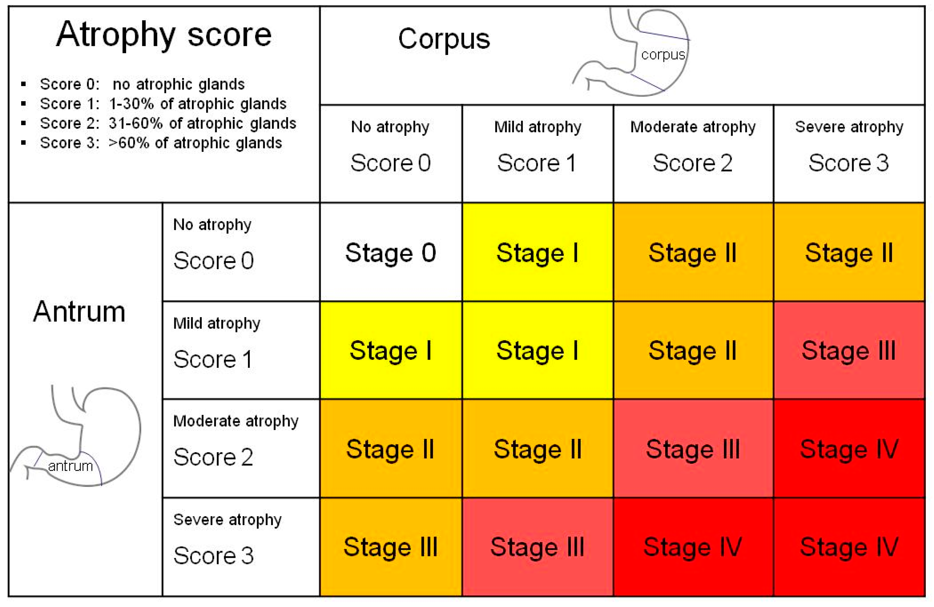
- Rugge, M.; Correa, P.; di Mario, F.; El-Omar, E.; Fiocca, R.; Geboes, K.; Genta, R.M.; Graham, D.Y.; Hattori, T.; Malfertheiner, P.; et al. OLGA staging for gastritis: A tutorial. Dig. Liver. Dis. 2008, 40, 650–658. [Google Scholar] [CrossRef] [PubMed]
- El Zimaity, H.M.; Ota, H.; Graham, D.Y.; Akamatsu, T.; Katsuyama, T. Patterns of gastric atrophy in intestinal type gastric carcinoma. Cancer 2002, 94, 1428–1436. [Google Scholar] [CrossRef] [PubMed]
- Epplein, M.; Xiang, Y.B.; Cai, Q.; Peek, R.M., Jr.; Lin, H.; Correa, P.; Gao, J.; Wu, J.; Michel, A.; Pawlita, M.; et al. Circulating cytokines and gastric cancer risk. Cancer Causes Control. 2013, 24, 2245–2250. [Google Scholar] [CrossRef] [PubMed]
- Lida, T.; Iwahashi, M.; Katsuda, M.; Nakamori, M.; Nakamura, M.; Naka, T.; Ojima, T.; Ueda, K.; Hayata, K.; Nakamura, Y. Tumor-infiltrating CD4+ Th 17 cells produce IL-17 in tumor microenvironment and promote tumor progression in human gastric cancer. Oncol. Rep. 2011, 25, 1271–1277. [Google Scholar]
- Meng, X.Y.; Zhou, C.H.; Ma, J.; Jiang, C.; Ji, P. Expression of interleukin-17 and its clinical significance in gastric cancer patients. Med. Oncol. 2012, 29, 3024–3028. [Google Scholar] [CrossRef] [PubMed]
- Dai, Z.M.; Zhang, T.S.; Lin, S.; Zhang, W.G.; Liu, D.; Cao, X.M.; Li, H.B.; Wang, M.; Liu, X.H.; Liu, K.; et al. Role of IL-17A rs2275913 and IL-17F rs763780 polymorphisms in risk of cancer development: An update meta-analysis. Sci. Rep. 2016, 6, 20439. [Google Scholar] [CrossRef] [PubMed]
- Muruyama, T.; Kono, K.; Mizukami, Y.; Kawaguchi, Y.; Mimura, K.; Watanabe, M.; Izawa, S.; Fujii, H. Distribution of Th17 cells and FoxP3(+) regulatory T cells in tumor-infiltrating lymphocytes, tumor-draining lymph nodes and peripheral blood lymphocytes in patients with gastric cancer. Cancer Sci. 2010, 101, 1947–1954. [Google Scholar] [CrossRef] [PubMed]
- Kuai, W.X.; Wang, Q.; Yang, X.Z.; Zhao, Y.; Yu, R.; Tang, X.J. Interleukin-8 associates with adhesion, migration, invasion and chemosensitivity of human gastric cancer cells. World J. Gastroenterol. 2012, 18, 979–985. [Google Scholar] [CrossRef] [PubMed]
- Bockerstett, K.A.; DiPaolo, R.J. Regulation of gastric carcinogenesis by inflammatory cytokines. Cell Mol. Gastroenterol. Hepatol. 2017, 4, 47–53. [Google Scholar] [CrossRef] [PubMed]
- Huang, F.Y.; Chan, A.O.; Rashid, A.; Wong, D.K.; Seto, W.K.; Cho, C.H.; Lai, C.L.; Yuen, M.F. Interleukin 1β increases the risk of gastric cancer through induction of aberrant DNA methylation in a mouse model. Oncol. Lett. 2016, 11, 2919–2924. [Google Scholar] [CrossRef] [PubMed] [Green Version]
- Al-Sammak, F.; Kalinski, T.; Winert, S.; Link, A.; Wex, T.; Malfertheiner, P. Gastric epithelial expression of IL-12 cytokine family in Helicobacter pylori infection in human: Is it head or tail of the coin? PLoS ONE 2013, 8, e75192. [Google Scholar] [CrossRef] [PubMed]
- Tsai, C.Y.; Wang, C.S.; Tsai, M.M.; Chi, H.C.; Cheng, W.L.; Tseng, Y.H.; Chen, C.Y.; Lin, C.D.; Wu, J.I.; Wang, L.H.; et al. Interleukin-32 increase human gastric cancer cell invasion associated with tumor progression and metastasis. Clin. Cancer Res. 2014, 20, 2276–2288. [Google Scholar] [CrossRef] [PubMed]
- Buzzelli, J.N.; Chalinor, H.V.; Pavlic, D.I.; Sutton, P.; Menheniott, T.R.; Giraud, A.S.; Judd, L.M. IL33 is a stomach alarmin that initiates a skewed Th2 response to injury and infection. Cell. Mol. Gastroenterol. Hepatol. 2015, 1, 203–221.e3. [Google Scholar] [CrossRef] [PubMed]
- Lahner, E.; Esposito, G.; Galli, G.; Annibale, B. Atrophic gastritis and pre-malignant gastric lesions. Transl. Gastrointest. Cancer 2015, 4, 272–281. [Google Scholar]
- Schneller, J.; Gupta, R.; Mustafa, J.; Villanueva, R.; Straus, E.W.; Raffaniello, R.D. Helicobacter pylori infection is associated with a high incidence of intestinal metaplasia in the gastric mucosa of patients at inner-city hospitals in New York. Dig. Dis. Sci. 2006, 51, 1801–1809. [Google Scholar] [CrossRef] [PubMed]
- Wong, B.C.; Lam, S.K.; Wong, W.M.; Chen, J.S.; Zheng, T.T.; Feng, R.E.; Lai, K.C.; Cheng, W.H.; Yuen, S.T.; Leung, S.Y.; et al. Helicobacter pylori eradication to prevent gastric cancer in a high-risk region of China: A randomized controlled trial. JAMA 2004, 291, 187–194. [Google Scholar] [CrossRef] [PubMed]
- Yamaoka, Y. Mechanisms of disease: Helicobacter pylori virulence factors. Nat. Rev. Gastroenterol. Hepatol. 2010, 7, 629–641. [Google Scholar] [CrossRef] [PubMed]
- Yong, X.; Tang, B.; Li, B.-S.; Xie, R.; Hu, C.J.; Luo, G.; Qin, Y.; Dong, H.; Yang, S.M. Helicobacter pylori virulence factor CagA promotes tumorigenesis of gastric cancer via multiple signaling pathways. Cell Commun. Signal. 2015, 13, 1–13. [Google Scholar] [CrossRef] [PubMed]
- Van Doorn, L.J.; Figueiredo, C.; Sanna, R.; Plaisier, A.; Schneeberger, P.; de Boer, W.; Quint, W. Clinical relevance of the cagA, vacA, and iceA status of Helicobacter pylori. Gastroenterology 1998, 115, 58–66. [Google Scholar] [CrossRef]
- Correa, P.; Piazuelo, M.B. The gastric precancerous cascade. J. Dig. Dis. 2012, 13, 2–9. [Google Scholar] [CrossRef] [PubMed]
- Elsborg, L.; Mosbech, J. Pernicious anaemia as a risk factor in gastric cancer. Acta Med. Scand. 1979, 206, 315–318. [Google Scholar] [CrossRef] [PubMed]
- Nikou, G.C.; Angelopoulos, T.P. Current concepts on gastric carcinoid tumors. Gastroenterol. Res. Pract. 2012, 2012, 287825. [Google Scholar] [CrossRef] [PubMed]
- Vanoli, A.; La Rosa, S.; Luinetti, O.; Klersy, C.; Manca, R.; Alvisi, C.; Rossi, S.; Trespi, E.; Zangrandi, A.; Sessa, F.; et al. Histologic changes in type A chronic atrophic gastritis indicating increased risk of neuroendocrine tumor development: The predictive role of dysplastic and severely hyperplastic enterochromaffin-like cell lesions. Hum. Pathol. 2013, 44, 1827–1837. [Google Scholar] [CrossRef] [PubMed]
- Zhou, K.; Ho, W. Gastric carcinoids: Classification and Diagnosis. In Management of Pancreatic Neuroendocrine Tumors; Pisegna, R.J., Ed.; Springer: New York, NY, USA, 2015; pp. 83–93. [Google Scholar]
- Burkitt, M.D.; Pritchard, D.M. Review article: Pathogenesis and management of gastric carcinoid tumours. Aliment Pharmacol. Ther. 2006, 24, 1305–1320. [Google Scholar] [CrossRef] [PubMed]
- Minalyan, A.; Benhammou, N.J.; Artashesyan, A.; Lewis, S.M.; Pisegna, J.R. Autoimmune atrophic gastritis: Current perspectives. Clin. Exp. Gastroenterol. 2017, 1, 19–27. [Google Scholar] [CrossRef] [PubMed]
- Visvader, J.E. Cells of origin in cancer. Nature 2011, 469, 314–322. [Google Scholar] [CrossRef] [PubMed]
- Shibata, W.; Sue, S.; Tsumura, S.; Ishii, Y.; Sato, T.; Kameta, E.; Sugimori, M.; Yamada, H.; Kaneko, H.; Sasaki, T.; et al. Helicobacter-induced gastric inflammation alters the properties of gastric tissue stem/progenitor cells. BMC Gastroenterol. 2017, 17, 145. [Google Scholar] [CrossRef] [PubMed]
- Tan, E.M. Autoantibodies as reporters identifying aberrant cellular mechanisms in tumorigenesis. J. Clin. Investig. 2001, 108, 1411–1415. [Google Scholar] [CrossRef] [PubMed]
- Werner, S.; Chen, H.; Tao, S.; Brenner, H. Systematic review: Serum autoantibodies in the early detection of gastric cancer. Int. J. Cancer 2015, 136, 2243–2252. [Google Scholar] [CrossRef] [PubMed]
- Macdonald, I.K.; Parsy-Kowalska, C.B.; Chapman, C.J. Autoantibodies: Opportunities for early cancer detection. Trends Cancer 2017, 3, 198–213. [Google Scholar] [CrossRef] [PubMed]
- Liu, W.; Peng, B.; Lu, Y.; Xu, W.; Qian, W.; Zhang, J.Y. Autoantibodies to tumor-associated antigens as biomarkers in cancer immunodiagnosis. Autoimmun. Rev. 2011, 10, 331–335. [Google Scholar] [CrossRef] [PubMed]
- Zaenker, P.; Ziman, M.R. Serologic autoantibodies as diagnostic cancer biomarkers—A review. Cancer Epidemiol. Biomarkers Prev. 2013, 22, 2161–2181. [Google Scholar] [CrossRef] [PubMed]
- Flammann, H.T.; Kuhn, HM. P53 autoantibodies and cancer: Specificity, diagnosis and monitoring. In Cancer and Autoimmunity; Shoenfeld, Y., Gershwin, M.E., Eds.; Elsevier Science: Amsterdam, The Netherlands, 2000; pp. 181–188. [Google Scholar]
- Saif, M.W.; Zalonis, A.; Syrigos, K. The clinical significance of autoantibodies in gastrointestinal malignancies: An overview. Expert Opin. Biol. Ther. 2007, 7, 493–507. [Google Scholar] [CrossRef] [PubMed]
- Crawford, L.V.; Pim, D.C.; Bulbrook, R.D. Detection of antibodies against the cellular protein p53 in sera from patients with breast cancer. Int. J. Cancer 1982, 30, 403–408. [Google Scholar] [CrossRef] [PubMed]
- Wurl, P.; Weigmann, F.; Meye, A.; Fittkau, M.; Rose, U.; Berger, D.; Rath, F.W.; Dralle, H.; Taubert, H. Detection of p53 autoantibodies in sera of gastric cancer patients and their prognostic relevance. Scand. J. Gastroenterol. 1997, 32, 1147–1151. [Google Scholar] [CrossRef] [PubMed]
- Soussi, T. p53 Antibodies in the sera of patients with various types of cancer: A review. Cancer Res. 2000, 60, 1777–1788. [Google Scholar] [PubMed]
- Shimada, H.; Ochiai, T.; Nomura, F. Japan p53 Antibody Research Group. Titration of serum p53 antibodies in 1085 patients with various types of malignant tumors: A multiinstitutional analysis by the Japan p53 Antibody Research Group. Cancer 2003, 97, 682–689. [Google Scholar] [CrossRef] [PubMed]
- Shiota, G.; Ishida, M.; Noguchi, N.; Takano, Y.; Oyama, K.; Okubo, M.; Katayama, S.; Harada, K.; Hori, K.; Ashida, K.; et al. Clinical significance of serum P53 antibody in patients with gastric cancer. Res. Commun. Mol. Pathol. Pharmacol. 1998, 99, 41–51. [Google Scholar] [PubMed]
- Maehara, Y.; Kakeji, Y.; Watanabe, A.; Baba, H.; Kusumoto, H.; Kohnoe, S.; Sugimachi, K. Clinical implications of serum anti-p53 antibodies for patients with gastric carcinoma. Cancer 1999, 85, 302–308. [Google Scholar] [CrossRef]
- Ura, Y.; Ochi, Y.; Hamazu, M.; Ishida, M.; Nakajima, K.; Watanabe, T. Studies on circulating antibody against carcinoembryonic antigen (CEA) and CEA-like antigen in cancer patients. Cancer Lett. 1985, 25, 283–295. [Google Scholar] [CrossRef]
- Albanopoulos, K.; Armakolas, A.; Konstadoulakis, M.M.; Leandros, E.; Tsiompanou, E.; Katsaragakis, S.; Alexiou, D.; Androulakis, G. Prognostic significance of circulating antibodies against carcinoembryonic antigen (anti-CEA) in patients with colon cancer. Am. J. Gastroenterol. 2000, 95, 1056–1061. [Google Scholar] [CrossRef] [PubMed]
- Nakamura, H.; Hinoda, Y.; Nakagawa, N.; Makiguchi, Y.; Itoh, F.; Endo, T.; Imai, K. Detection of circulating anti-MUC1 mucin core protein antibodies in patients with colorectal cancer. J. Gastroenterol. 1998, 33, 354–361. [Google Scholar] [CrossRef] [PubMed]
- Yagihashi, A.; Asanuma, K.; Nakamura, M.; Araya, J.; Mano, Y.; Torigoe, T.; Kobayashi, D.; Watanabe, N. Detection of anti-survivin antibody in gastrointestinal cancer patients. Clin. Chem. 2001, 47, 1729–1731. [Google Scholar] [PubMed]
- Cho-Chung, Y.S. Autoantibody biomarkers in the detection of cancer. Biochim. Biophys. Acta. 2006, 1762, 587–591. [Google Scholar] [CrossRef] [PubMed]
- Qiu, L.L.; Hua, P.Y.; Ye, L.L.; Wang, Y.C.; Qiu, T.; Bao, H.Z.; Wang, L. The detection of serum anti-p53 antibodies from patients with gastric carcinoma in China. Cancer Detect Prev. 2007, 31, 45–49. [Google Scholar] [CrossRef] [PubMed]
- Shimizu, K.; Ueda, Y.; Yamagishi, H. Titration of serum p53 antibodies in patients with gastric cancer: A single-institute study of 40 patients. Gastric Cancer 2005, 8, 214–219. [Google Scholar] [CrossRef] [PubMed]
- Shimada, H.; Noie, T.; Ohashi, M.; Oba, K.; Takahashi, Y. Clinical significance of serum tumor markers for gastric cancer: A systematic review of literature by the Task Force of the Japanese Gastric Cancer Association. Gastric Cancer 2014, 17, 26–33. [Google Scholar] [CrossRef] [PubMed]
- Di Mario, F.; Cavallaro, L.G. Non-invasive tests in gastric diseases. Dig. Liver Dis. 2008, 40, 523–530. [Google Scholar] [CrossRef] [PubMed]
- Majeed, W.; Iftikhar, A.; Khaliq, T.; Aslam, B.; Muzaffar, H.; Atta, K.; Mahmood, A.; Waris, S. Gastric carcinoma: Recent trends in diagnostic biomarkers and molecular targeted therapies. Asian Pac. J. Cancer Prev. 2016, 17, 3053–3060. [Google Scholar] [PubMed]
- Zayakin, P.; Ancāns, G.; Siliņa, K.; Meistere, I.; Kalniņa, Z.; Andrejeva, D.; Endzeliņš, E.; Ivanova, L.; Pismennaja, A.; Ruskule, A.; et al. Tumor-associated autoantibody signature for the early detection of gastric cancer. Int. J. Cancer 2013, 132, 137–147. [Google Scholar] [CrossRef] [PubMed]
- Werner, S.; Chen, H.; Butt, J.; Michel, A.; Knebel, P.; Holleczek, B.; Zörnig, I.; Eichmüller, S.B.; Jäger, D.; Pawlita, M.; et al. Evaluation of the diagnostic value of 64 simultaneously measured autoantibodies for early detection of gastric cancer. Sci. Rep. 2016, 6, 25467. [Google Scholar] [CrossRef] [PubMed]
- Zhou, S.L.; Ku, J.W.; Fan, Z.M.; Yue, W.B.; Du, F.; Zhou, Y.F. Detection of autoantibodies to a panel of tumor-associated antigens for the diagnosis values of gastric cardia adenocarcinoma. Dis. Esophagus 2015, 28, 371–379. [Google Scholar] [CrossRef] [PubMed]
- Wang, P.; Song, C.; Xie, W.; Ye, H.; Wang, K.; Dai, L.; Zhang, Y.; Zhang, J. Evaluation of diagnostic value in using a panel of multiple tumor-associated antigens for immunodiagnosis of cancer. J. Immunol. Res. 2014, 2014, 512540. [Google Scholar] [CrossRef] [PubMed]


| Clinical Presentation | No symptoms or dyspepsia | |
| Anemia (iron deficiency, vitamin B12 deficiency) | ||
| Coexisting autoimmune diseases: | Autoimmune thyroid diseases (Hashimoto and Graves) | |
| Type 1 diabetes | ||
| Addison disease | ||
| Polyglandular autoimmune syndromes type III | ||
| Serology | Gastrin 17 | >10 pmol/L |
| Pepsinogen I | <30 μg/L | |
| Pepsinogen II | normal (3–15 μg/L) | |
| Parietal cell autoantibodies | pos 90–95% | |
| Intrinsic factor autoantibodies | pos 30–50% | |
| Pathology | Corpus/fundus restricted gastritis | |
| Neoplastic Risk | Gastric carcinoid: increased according to gastric (oxyntic) atrophy score to the corpus and fundus of the stomach | |
| Gastric adenocarcinoma: increased according to pangastric atrophy score | ||
© 2018 by the authors. Licensee MDPI, Basel, Switzerland. This article is an open access article distributed under the terms and conditions of the Creative Commons Attribution (CC BY) license (http://creativecommons.org/licenses/by/4.0/).
Share and Cite
Bizzaro, N.; Antico, A.; Villalta, D. Autoimmunity and Gastric Cancer. Int. J. Mol. Sci. 2018, 19, 377. https://doi.org/10.3390/ijms19020377
Bizzaro N, Antico A, Villalta D. Autoimmunity and Gastric Cancer. International Journal of Molecular Sciences. 2018; 19(2):377. https://doi.org/10.3390/ijms19020377
Chicago/Turabian StyleBizzaro, Nicola, Antonio Antico, and Danilo Villalta. 2018. "Autoimmunity and Gastric Cancer" International Journal of Molecular Sciences 19, no. 2: 377. https://doi.org/10.3390/ijms19020377