Article

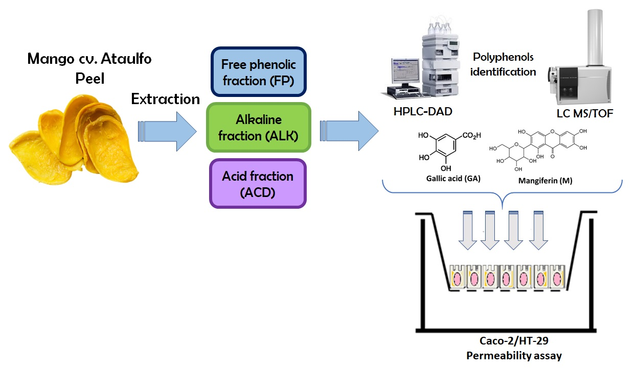
Intestinal Permeability and Cellular Antioxidant Activity of Phenolic Compounds from Mango (Mangifera indica cv. Ataulfo) Peels

by
Ramón Pacheco-Ordaz
1,
Marilena Antunes-Ricardo
2,
Janet A. Gutiérrez-Uribe
2,3,* and
Gustavo A. González-Aguilar
1,*
1
Centro de Investigación en Alimentación y Desarrollo, A.C., Carretera a la Victoria Km 0.6, La Victoria, Hermosillo 83000, Sonora, Mexico
2
Tecnologico de Monterrey, Centro de Biotecnologia-FEMSA., Av. Eugenio Garza Sada 2501 Sur, Monterrey C.P. 64849, Nuevo León, Mexico
3
Tecnologico de Monterrey, Department of Bioengineering and Science, Campus Puebla, Av. Atlixcáyotl 2301, Puebla C.P. 72453, Puebla, Mexico
*
Authors to whom correspondence should be addressed.
Int. J. Mol. Sci. 2018, 19(2), 514; https://doi.org/10.3390/ijms19020514
Submission received: 28 November 2017 / Revised: 8 January 2018 / Accepted: 22 January 2018 / Published: 8 February 2018
(This article belongs to the Special Issue Bioactive Phenolics and Polyphenols 2018)

Abstract

:
Mango (Mangifera indica cv. Ataulfo) peel contains bound phenolics that may be released by alkaline or acid hydrolysis and may be converted into less complex molecules. Free phenolics from mango cv. Ataulfo peel were obtained using a methanolic extraction, and their cellular antioxidant activity (CAA) and permeability were compared to those obtained for bound phenolics released by alkaline or acid hydrolysis. Gallic acid was found as a simple phenolic acid after alkaline hydrolysis along with mangiferin isomers and quercetin as aglycone and glycosides. Only gallic acid, ethyl gallate, mangiferin, and quercetin were identified in the acid fraction. The acid and alkaline fractions showed the highest CAA (60.5% and 51.5%) when tested at 125 µg/mL. The value of the apparent permeability coefficient (Papp) across the Caco-2/HT-29 monolayer of gallic acid from the alkaline fraction was higher (2.61 × 10⁻⁶ cm/s) than in the other fractions and similar to that obtained when tested pure (2.48 × 10⁻⁶ cm/s). In conclusion, mango peels contain bound phenolic compounds that, after their release, have permeability similar to pure compounds and exert an important CAA. This finding can be applied in the development of nutraceuticals using this important by-product from the mango processing industry.

Graphical Abstract

1. Introduction

The consumption of tropical fruits has increased in the last decade due to the concern that consumers have to live a healthier life style [1,2]. Mango (Mangifera indica L.) is one of the most consumed fruits worldwide for its sensory properties and for being considered a good source of bioactive compounds such as ascorbic acid, carotenoids, tocopherols, and phenolic compounds (PC). PC are involved in the aroma and flavor of plant foods, and are responsible for plant defense against infections [3]. In addition, these compounds are associated with different health benefits such as antioxidant, anti-inflammatory, anti-microbial, and anti-proliferative effects [4,5].
Mango cv. Ataulfo is a widely consumed Mexican variety, and has the highest PC content and antioxidant capacity compared with other varieties [6]. However, only the pulp of the fruit is consumed, and it is consumed either fresh or processed into purees, juices, jams, or canned slices. Mango processing generates a large amount of by-products such as peels and kernels, which account for 35–65% of the total fruit [7]. However, the peels and kernels are discarded into the environment as waste, making them a source of pollution.
Lately, the use of food by-products as a source of phytochemicals has increased. Quercetin, kaempferol, and mangiferin from the extracts of the leaves and bark extracts are extensively used as dietary supplements due to their health benefits. Unlike pulp, which contains mainly small molecules like phenolic acids [8], more complex molecules, such as quercetin, catechin, mangiferin, gallotannins, gallic acid, and other glycosylated compounds have been identified in mango peel, which indicates that it could be a source of functional ingredients [9,10]. Several authors have studied the potential benefits of mango waste.
In nature, most PCs exist linked to other complex structures such as proteins and fiber. Therefore, pre-treatments are necessary before conventional extraction to obtain the complete phenolic profile of the plant. Generally, alkali treatment can break the ester bonds that linked the PC to the cell wall components and release the majority of bound phenolics. In contrast, acid hydrolysis breaks glycoside bounds and therefore releases the aglycones [11].
The beneficial effects of the PC depend on their absorption throughout the gastrointestinal tract [12,13]. Several in vivo and in vitro studies describe the absorption of PC. Among them, the human colon adenocarcinoma (Caco-2) monolayer model has been widely used in recent years for the screening of the permeability of different drugs [14].
The major challenge in the development of nutraceuticals is their bioavailability, since, in nature, these compounds interact within the food matrix in their conjugated forms [15]. In addition, the plant matrix or the other components have been reported to alter the pharmacokinetics of PC and therefore their bioactivity as antioxidant agents. In this study, the PCs’ profile and the biological activities of three (03) different fractions of mango peel extract were analyzed. The aim of this study was to evaluate the intestinal permeability of free and bound PCs present in mango cv. Ataulfo fractions after an alkaline and acid hydrolysis using a Caco-2 monolayer model, as well as, to evaluate the contribution of these free and bound PCs in the cellular antioxidant activity of each fraction in order to determine the potential of this by-product as a source of nutraceuticals.

2. Results

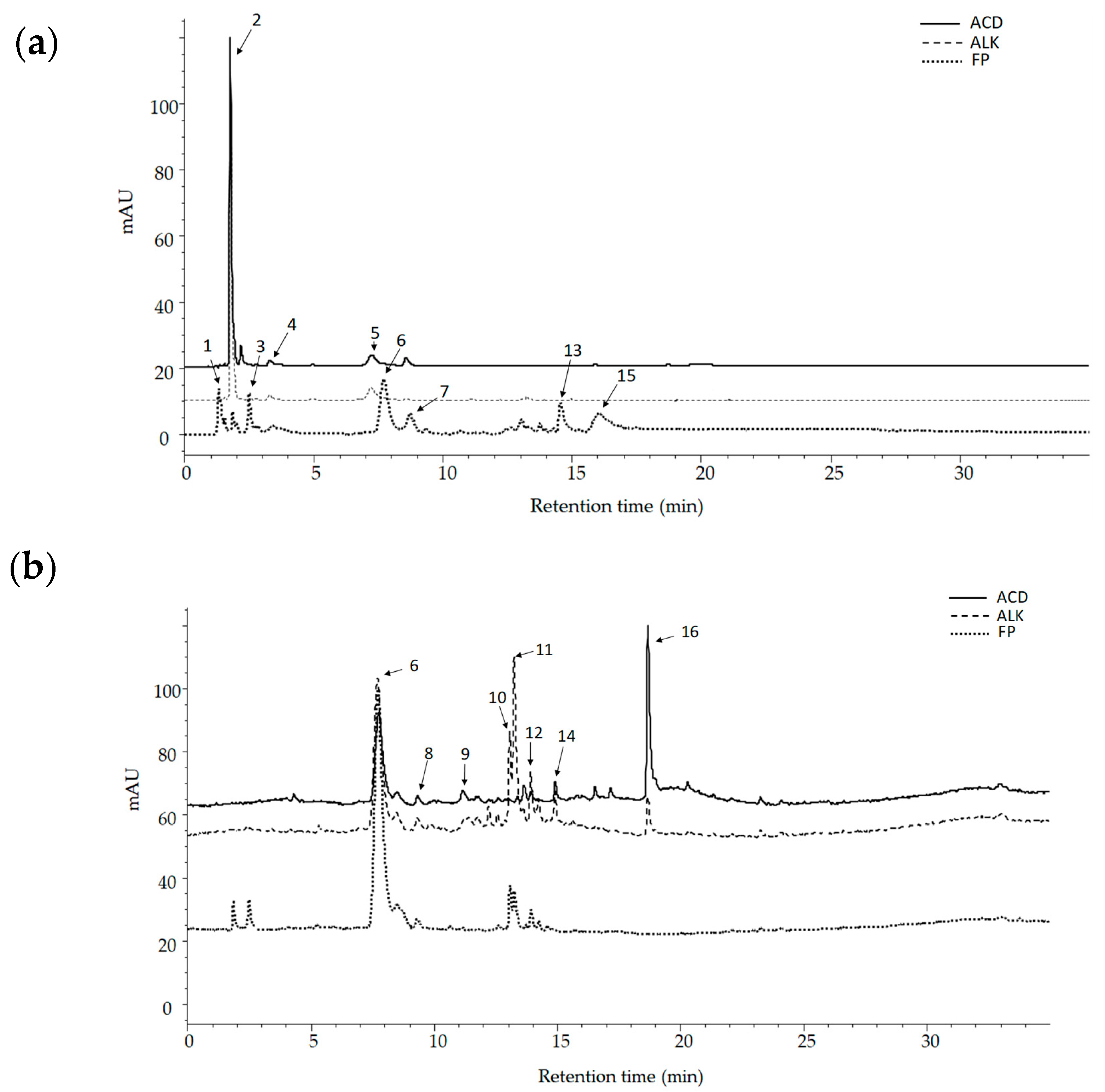
2.1. Identification and Quantification of PCs in Mango cv. Ataulfo Peel Extracts

Of the PC found in the alkaline (ALK) and acid (ACD) extracts, gallic acid (peak 2) was the major component (Figure 1a). Mangiferin (peak 6) was among the most abundant PC in the free phenolics extract (FP), and a significant increase in quercetin (peak 16) was observed in the extract following acid hydrolysis (ACD) (Figure 1b). Additionally, other compounds were identified in the different extracts based on their UV maxima and accurate mass (Table 1). In FP, compounds with complex structure such as gallotannins, xanthones, glycosylated flavonoids, and other phenolic acid derivatives were identified based on preliminary reports [16,17]. The gallotannins were identified as galloyl glycoside (peak 1) with an [M–H] ion at m/z 331, hyemaloside A (peak 7) with an [M–H] ion at m/z of 728, methyl digallate ester (peak 13) with an [M–H] ion at m/z of 335, and hexagalloyl glucoside (peak 15) with a [M–H] ion at m/z of 1091. The quercetin hexoside I and II (peak 10 and peak 12) [M–H] ion at m/z 463 were found in the FP extract.
Instead of the free gallic acid (peak 2) found in ALK fraction, three gallic acid derivatives were identified in FP: galloyl quinic acid (peak 3), digallic acid (peak 5), and ethyl gallate (peak 8) with a [M–H] ion at m/z of 343, 321, and 197, respectively. Three mangiferin isomers appeared at 365 nm (peaks 6, 11, and 14) with a [M–H] ion at m/z of 421 each one.
In addition to the gallic acid release, other phenolic acids, such as caffeoyl hydroxycitric acid (peak 4) and coumaric acid (peak 9), were extracted after alkaline hydrolysis. The alglycone of quercetin (peak 16) was also identified in this fraction and showed a [M–H] ion at m/z of 301. In contrast, acid hydrolysis released fewer compounds; only gallic acid, ethyl gallate, mangiferin, and quercetin were identified in ACD fraction.

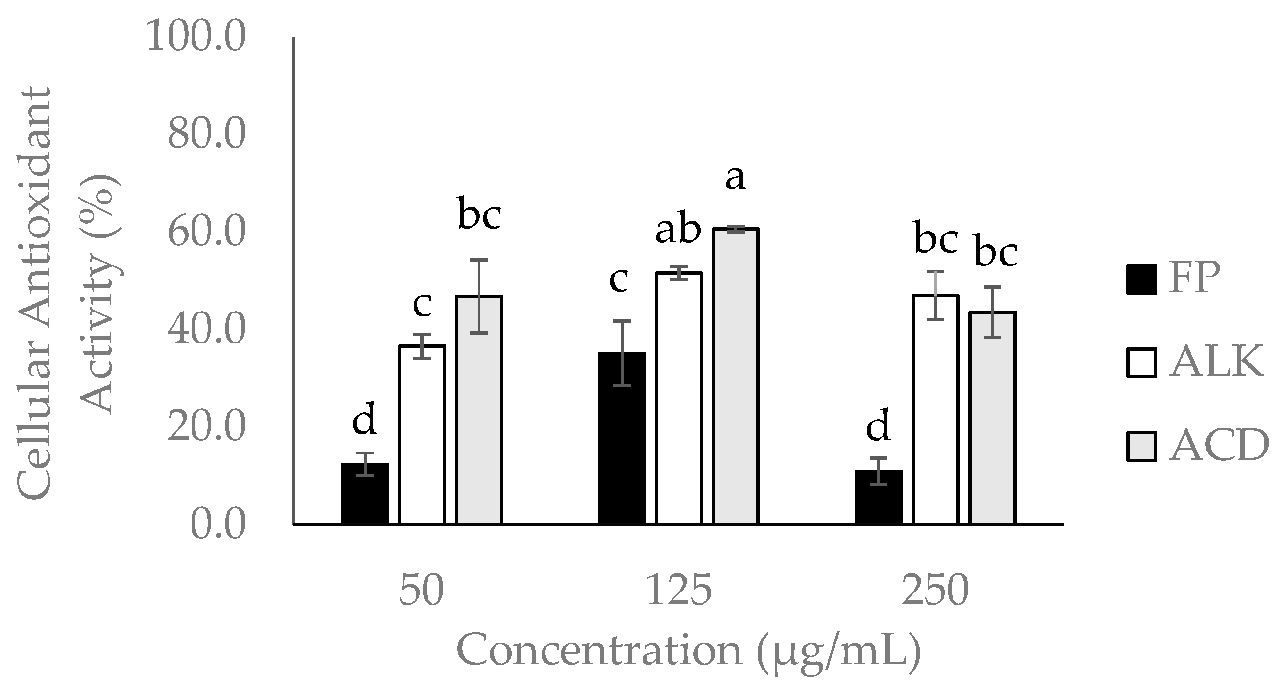
2.2. Cytotoxicity and Cellular Antioxidant Activity (CAA) of Mango cv. Ataulfo Peel Extracts

Among the three tested extracts (FP, ALK, and ACD), FP was the most cytotoxic followed by ALK (Table 2). Caco-2 and HT-29 cells in combination were more sensitive to the three mango peel extracts.
The ACD and ALK fractions showed the highest antioxidant potential (60.57 ± 0.55% and 51.56 ± 1.39%) at 125 µg/mL. Regarding to the FP fraction, it showed a value of IC50 about 2.0-fold lower than the maximum concentration assayed for CAA (250 µg/mL), and therefore Caco-2 cells viability was strongly affected, and in consequence it showed lower antioxidant activity (10.90%) than the obtained when tested at 125 µg/mL (35.17%) (Figure 2).

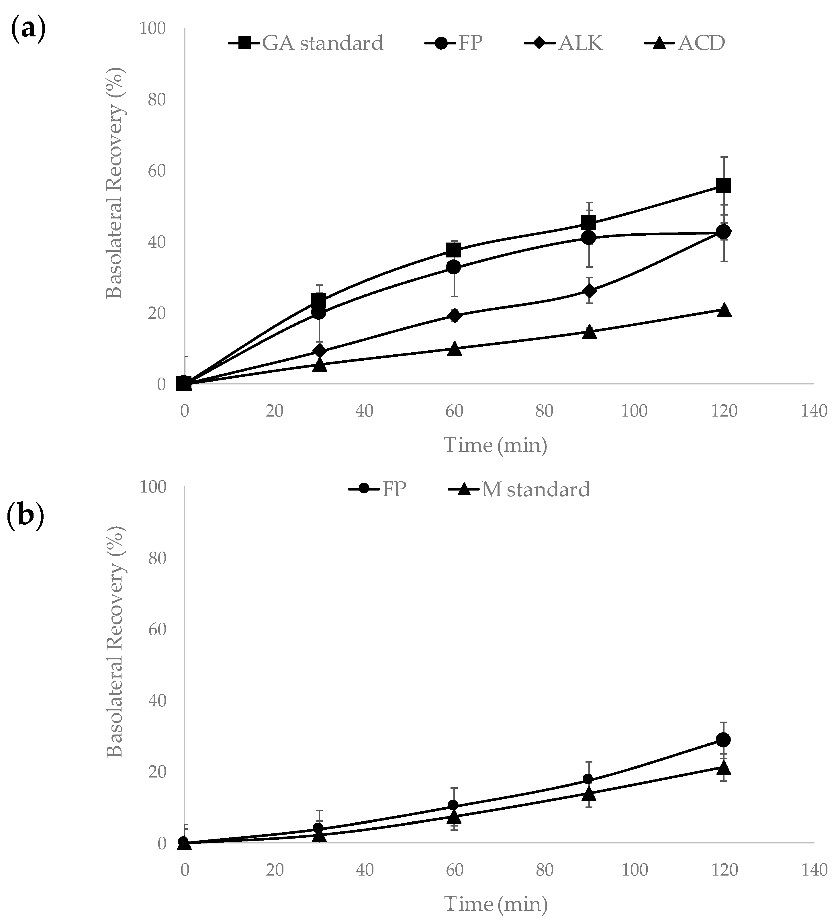
2.3. Intestinal Permeability Experiment

The basolateral side of the intestinal permeability experiment represents the basal surface of the membrane that mediates the transport of nutrients from cell to surrounding fluids that lead to the circulatory system. Gallic acid and mangiferin were detected in this side of the Caco-2/HT-29 (75:25%) monolayer after 30 min of incubation with the extract (Figure 3a,b). Basolateral recovery of gallic acid ranged between (20.8 ± 0.63%)–(55.6 ± 8.14%) for all samples (Figure 3a). In contrast, mangiferin was only detected in the FP fraction, with a recovery of 28.84 ± 0.28% (Figure 3b). The FP and ALK fractions presented similar recovery values for gallic acid 42.39 ± 1.76% and 43.02 ± 2.38%, respectively. The gallic acid standard showed the highest recovery percentage (Figure 3a). The mass balance obtained for gallic acid in ALK, ACD fractions, with gallic acid standard, was between 94–98%, and in the case of FP it was of 55 ± 2.5%. On the other hand, mangiferin standard exhibited an average mass balance of 88%.
The values of the apparent permeability coefficient (Papp) of gallic acid in the FP, ALK, and ACD using the Caco-2/HT-29 co-culture were 1.97, 2.61, and 1.16 × 10⁻⁶ cm/s, respectively (Figure 4a). The gallic acid standard showed a higher (Papp of 2.48 × 10⁻⁶ cm/s) value compared with the FP and ACD but was similar to the obtained for gallic acid found in ALK mango peel extract. On the other hand, the Papp value for the mangiferin standard was 1.49 × 10⁻⁶ cm/s, whereas the value of the FP fraction was 2.47 × 10⁻⁶ cm/s (Figure 4b).

3. Discussion

3.1. Identification of PCs of Mango cv. Ataulfo Peel Extract

Since phytochemicals are mainly found bound to proteins, fiber, or other complex structures in nature; alkaline and acid hydrolysis were carried out to fully characterize the mango cv. Ataulfo peel extract. The most abundant family of compounds identified in mango extract fractions were gallotaninns, mangiferin isomers, and flavonoids. Gallic acid derivatives such as galloyl glucose, methyl digallate ester, and methyl gallate were also detected. These results agree with those reported by López-Cobo et al. [10], which indicated that gallotanins and other gallic acid conjugates were found in high concentrations in the peels of mango of the Keitt, Osteen, and Sensation cultivars. Similarly, Dorta et al. [20] demonstrated the presence of gallotannins, ethyl gallate, methyl gallate, gallic acid, galloyl glucose, and theogallin, as well as the xanthone mangiferin, in mango peel. Additionally, López-Cobo et al. [10] identified quercetin glycosides such as isoquercitrin, quercetin-3-O-galactoside, and quercetin pentoside, which is consistent with the results of this study.
Gómez-Caravaca et al. [18] analyzed four parts of mango Keitt, and the peel of the tissue with the highest content of PC. The majority of the compounds identified in the peel were phenolic acids such as gallic, syringic, protocatechuic, and ferulic acid ellagic acid, as well as ellagic acid derivatives. However, in this study, only gallic and caffeic acids conjugates were found. Differences between the profiles of PC among mango varieties and cultivars depend on multiple factors including the type of soil, weather, temperature, hydric stress, and post-harvest damage, all of which affect the biosynthetic pathway of PC in plants [6,15].

3.2. Cytotoxicity of Mango Peel Extract

The FP fraction was the most cytotoxic to Caco-2, HT-29 cell lines, and the mixture of the two. This may be explained by the abundance of gallotanins in this fraction [23]. Urueña et al. [25] demonstrated that a gallotannin-rich fraction obtained from Caesalpinia spinosa reduced the proliferation of breast cancer through the activation of apoptosis pathway, resulting in the activation of caspases. Likewise, gallotanins may regulate the production of reactive oxygen species (ROS) by altering the redox balance in the cell, which activates the intrinsic mitochondrial apoptosis pathway [26]. Mangiferin can induced apoptosis by activating caspase-8, caspase-9, and caspase-3 and pro-apoptotic protein Bid. Also, reductions in the activation of nuclear factor-kappaB (NF-κB) can caused a decrease in matrix metalloproteinase-7 and -9, and inhibit the β-catenin pathway [27]. Additionally, the presence of some flavonoids and mangiferin could be causing the synergistic antiproliferative effects observed in this sample [28].

3.3. Cellular Antioxidant Activity (CAA)

The ACD fraction was the more effective in the reduction of intracellular reactive oxygen species (ROS) production, which was monitored based on the Dichloro-dihydro-fluorescein diacetate (DFFH-DA) fluorescence assay. The major compounds detected in ACD were quercetin and gallic acid, and both compounds have demonstrated a high antioxidant activity. Differences in CAA between FP and ACD can be explained by the abundance of glycosylated PC in the FP fraction. In previous studies, quercetin showed the highest antioxidant activity in comparison with its glycosylated form due to the availability of hydroxyl groups to participate in antioxidant reactions [24,25,26]. On the other hand, an hormesis effect was observed when the FP and ACD fraction were tested at the concentration of 250 µg/mL. Hormesis is defined as a biphasic dose-dependent response in which a compound in low doses results in an antioxidant effect, while it became pro-oxidant at high concentration levels [28]. PC can exert a pro-oxidant effect by formatting labile aroxyl radical and reacting with oxygen producing superoxide anion (O2) [29]. This effect has been observed in quercetin and in others PCs [30,31,32].
Abbasi et al. [33] assessed the cellular antioxidant activity of the mango pulp and peel from nine cultivars in the liver hepatocellular cell line (HepG2). Mango peel extracts showed the highest antioxidant activity (2986.5 ± 380 µmol QE/100 g FW.) The cultivar Xiao Tainang showed the highest CAA with an IC50 of 130 µg/mL, whereas in our study, ACD fraction of mango cv. Ataulfo peel at the concentration of 125 µg/mL showed 60% of CAA. PCs can exert their antioxidant activity in two ways; they can act at the cell membrane and break peroxyl radical chain reactions at the cell surface, or they can enter the cell and react intracellularly with ROS. Polar compounds can interact with the membrane surfaces by hydrogen bonding and protecting cell membranes from external and internal oxidative stress [34]. On the other hand, hydrophobic compounds can be embedded more easily in the membrane and influence the fluidity and disrupt oxidative chain reactions. The difference in CAA observed between FP, ALK, and ACD is related with the permeability of the phenolic profile of each fraction. This could be an explanation of why the ACD and ALK fractions in general showed more antioxidant potential than the FP fraction.

3.4. Instetinal Permeability Assay

Caco-2/HT-29 monolayer has been used to evaluate the permeability of new drugs and phytochemicals due to its similarity to the human intestinal epithelium, and the combination of the two cell lines decreased the expression of P-gp transporters in Caco-2 allowing the permeability of xenobiotics. The apparent permeability (Papp) values of tannin corilagin and gallic acid were higher when tested pure than in an extract, because PC can act antagonistically and obstruct the absorption of other compounds [35,36,37]. On the contrary, in our study ALK exhibited similar Papp to the gallic acid standard. This could be due to the fact that other specific compounds in the extract can improve the absorption of gallic acid as reported by Xie et al. [38].
In the present study, free gallic acid was not detected in the FP but it was found in the basolateral side 30 min into the monolayer permeability assay. An explanation could be that the FP is rich in gallotannins and these compounds could enter into the cell by the action of the specific transporters and become degraded intracellularly to gallic [39,40]. It has already been reported that gallotannins such as penta-galloylglucose can be degraded to tri- and tetra-galloylglucose by esterases from Caco-2 cells while they are being transported across cell monolayer [41].

4. Materials and Methods

4.1. Material and Methods

Mango cv. Ataulfo fruits (commercial ripeness stage) that were free from external defects were purchased from a local market (Hermosillo, Sonora, México) and transported to the laboratory. Mangos were washed with tap water, sanitized, and peeled. Fruit peels was freeze-dried (LABCONCO, Kansas City, MO, USA), subsequently ground, and stored at −35 °C until analysis.

4.2. Chemicals and Reagents

Gallic acid and mangiferin standards, 2,2′-azobis-(2-methylpropionamidine) dihydrochloride (AAPH), Lucifer yellow (LY), 2′,7′-dichlorofluorescin diacetate (DCFH-DA), water, formic acid, and acetonitrile HPLC grade were obtained from Sigma-Aldrich, Inc. (St. Louis, MO, USA). Dubelcco’s Modified Eagle Medium (DMEM-F12) and McCoy’s medium were obtained from Thermo Fisher Scientific (Waltham, MA, USA). Fetal bovine serum (FBS), Hank’s Balanced Salt Solution (HBSS), Phosphate Saline Buffer pH 7.4 (PBS), Trypsin-EDTA 0.25%, penicillin (10,000 Unit/mL), and streptomycin (10,000 μg/mL) were acquired from GIBCO (Grand Island, NY, USA). CellTiter 96® AQueous One Solution Cell Proliferation Assay was obtained from Promega Corporation (Madison, WI, USA).

4.3. Phenolics Extraction Procedure

The extraction of phenolic compounds was carried out following the method described by Mattila and Kumpulainen [42], with modifications (Figure 5). Dried mango cv. Ataulfo peel (500 mg) was weighted and homogenized with 7 mL of a mixture of ethanol and 10% acetic acid using a magnetic stirrer. Afterwards, peel sample was ultrasonicated for 30 min (Bransonic Ultrasonic Co., Danbury, CT, USA) and centrifuged at 10,000 rpm for 10 min, then the supernatant was recovered. This supernatant was considered the free phenolic rich-fraction (FP) and 1 mL was taken directly for HPLC analysis.
A mixture of 12 mL distilled water and 5 mL of 10 M NaOH were added to the remaining mango peel (pellet). The sample was immediately flushed with nitrogen, then sealed and stirred at 100 rpm and room temperature for 16 h. The solution was then adjusted to pH 2 with 12 N HCl and extracted three times with 15 mL of a mixture of diethyl ether and ethyl acetate (1:1; v/v). The supernatant of each extraction was recovered, pooled, and evaporated to dryness with a rotary evaporator (R-3000 Buchi, Flawil, Switzerland) and finally resuspended in 1.5 mL of ethanol. This sample was defined as alkaline fraction (ALK) and aliquot was taken for the HPLC analysis. The residual sample was subsequently hydrolyzed in acid by adding 2.5 mL of 10 M HCl and incubating the mixture in a water bath (Thermo Fisher Scientific, Waltham, MA, USA) at 85 °C for 30 min. The sample was left to cool down at room temperature and the pH was adjusted to 2. Same extraction described above with diethyl ether and ethyl acetate was used for the hydrolyzed product. This sample identified as acid fraction (ACD), and an aliquot was taken for the HPLC analysis.

4.4. High Performance Liquid Chromatography (HPLC) Analysis

PC of mango cv. Ataulfo peel were identified by HPLC coupled to DAD-UV detector (1200 Series, Agilent Technologies, Santa Clara, CA, USA) using the method reported by Acosta-Estrada [43] with some modifications. Separation were carried out using a Zorbax SB-Aq, 3.0 mm × 100 mm (3.5 μm) reverse phase column at a temperature of 25 °C with a flow rate 0.6 mL/min. The mobile phase used was (A) water pH 2 acidified with formic acid and (B) acetonitrile 100%. The gradient started with 5% of B and increased to 30% within the first 15 min, changed to 60% at 20 min, then to 80% 5 min after, and at 30 min B increased to 100%. Chromatograms were obtained at 280 nm and 365 nm after the injection of 5 μL of sample and integrated by HP-Agilent Software (Chemstation for LC Copyright Agilent Technologies, Santa Clara, CA, USA).
The identification of the PC was performed on a liquid chromatography coupled with time-of-flight mass spectrometry (LC/MS-TOF) (Agilent Technologies, Santa Clara, CA, USA) and equipped with a quaternary pump system with a vacuum degasser, a thermostated column compartment with an electrospray ionization source (ESI). The same conditions describe above were used. Mass spectra were acquired in negative mode over a range from 150 to 1500 m/z.

4.5. Cell Culture

Human colorectal adenocarcinoma cells (Caco-2 and HT-29) were obtained from American Type Culture Collection (ATCC) (Manassas, VA). Caco-2 cells were grown in DMEM-F12 supplemented with 5% fetal bovine serum, whereas HT-29 cells were grown in McCoy’s medium supplemented with 10% fetal bovine serum. Cells were incubated at 37 °C in a humidified atmosphere containing 5% CO2.

4.6. Cytotoxicity Assay

The effect of mango cv. Ataulfo peel free phenolics extract (FP) and the corresponding alkaline and acid hydrolysates (ALK and ACD) on cell viability was measured with CellTiter 96 Aqueous One Solution Cell Proliferation Assay (Promega, Madison, WI). Caco-2 and HT-29 were seeded individually and combined (Caco-2/HT-29; 75:25%) in a 96-well plate in a solution of 100 µL at 5 × 10⁵ cells/mL. After 24 h, the extracts were added at final concentrations ranging between 50 and 500 µg/mL. After 48 h of incubation, 20 µL of CellTiter 96 was added to each well so the cell viability could be determined by measuring the absorbance at 490 nm in a microplate reader (Synergy HT, Bio-Tek, Winooski, VT, USA). The IC50 values (concentration to inhibit by 50%) for each sample were determined.

4.7. Cellular Antioxidant Activity (CAA) Assay

The cellular antioxidant activity (CAA) assay was performed as reported for López-Barrios et al. [44]. A day before the experiment, Caco-2 cells were cultured in a black-walled, clear-bottom 96-well microplate (Costar, Corning Inc., Corning, NY, USA) at a density of 5 × 10⁵/mL. After 24 h, medium was removed and cells were washed with 100 µL of phosphate buffered saline (PBS). Afterwards, cells were treated with 100 µL of the FP, ALK, and ACD fractions of mango peel extract at different concentrations (50, 125, and 250 µg/mL) containing DCFH-DA (60 μM), and the cells were incubated at 37 °C for 20 min. Following incubation, the treatment solutions were removed and the cells were washed twice with PBS. Finally, 100 µL of 500 μM AAPH solution was added to each well, except for blank and negative control wells. The microplate was placed in the microplate reader (Synergy HT, Bio-Tek, Winooski, VT, USA). Fluorescence emitted at 538 nm with excitation at 485 nm was measured every 2 min for 90 min at 37 °C. The CAA values of mango peel extract at each concentration were calculated using the following Equation (1).
CAA   unit = 1 ( S A / C A )
where S A is the integrated area under the sample fluorescence versus time curve and C A is the integrated area from the control curve.

4.8. Caco-2/HT-29 Monolayer Permeability Assay

A Caco-2 and HT-29 co-culture (ratio 75:25%, respectively) was seeded at a density of 1 × 10⁵ cells/insert in trans-well inserts (polycarbonate membrane, 12 mm i.d., 1.12 cm2 growth area, 0.4 µm pore size (Corning, NY, USA)) and placed in 6 well plates with 1.5 mL of medium at the apical side (AP) and 2.5 mL at the basolateral side (BL). Cells were allowed to grow and differentiate for 21 days to form a monolayer, and the culture medium was replaced three times per week. The permeability experiment was carried out according to Antunes-Ricardo et al. [45]. The medium was removed and cell monolayers were washed with Hank’s balanced salt solution (HBSS). FP, ALK, and ACD, as well as gallic acid and mangiferin standards, were inoculated in the apical side of the differentiated Caco-2/H-T29 monolayers. The experiment was carried out for 120 min, and the samples were withdrawn from apical side and along with the basolateral samples taken at 30, 60, 90, and 120 min they were analyzed by HPLC-DAD-UV.
The apical side was filled with 0.4 mL of LY solution (100 µM) and the basolateral side was filled with HBSS. After 2 h, 100 µL of basolateral and apical media were transferred to a 96-well plate and the fluorescence of each sample was measured at 530 nm (emission) and 485 nm (excitation) using a microplate reader. Cell monolayers’ integrity was monitored by lucifer yellow (LY) permeation [46] using the apparent permeability coefficient (Papp) value, which is calculated with the following Equation (2):
Papp = (dQ/dt) × (V/A*C0)
where dQ/dt is the change in drug concentration in the receiver solution (µM/s), V represents the volume of the solution in the receiving compartment (mL), A denotes the membrane surface area (cm2), and C0 is the initial concentration in the donor compartment (µM). Data from membranes displaying an LY apparent permeability coefficient (Papp) >1 × 10⁻⁶ cm/s were excluded. The LY concentration was calculated on the basis of a standard curve in the concentration range of 1.56 to 100 µM.

5. Statistical Analysis

All experiments were performed at least three times, and results are expressed as the mean ± standard deviation. Statistical analyses were performed using the statistical software JMP 13.0 (SAS Institute Inc., Cary, NC, USA). Data were analyzed by ANOVA followed by Tukey’s HSD test with a significance level of p < 0.05.

6. Conclusions

Mangiferin, gallic acid, and quercetin were the main compounds identified in mango peel extracts. Gallic acid released after alkaline hydrolysis had similar permeability to that obtained with a pure standard. The ALK fraction showed better permeability properties than the other fraction and a greater antioxidant potential, due to its high content of PCs. The dose tested and the structure of the PC play an important role in their permeability and therefore in their antioxidant activity. The use of three different extracts allowed us to obtain a better characterization of PC profile and compare their biological activity. Although the extraction after alkaline hydrolysis will need to be evaluate to determine if its suitable for industrial set up, due the large amount of solvents and recovery yield, this finding provides novel and valuable information that can be applied in the development of nutraceuticals from mango peels. Based on our results, compounds such as tannins and glycosides were not absorbed, which indicate that they can reach the colon and possibly modify the bacteria populations present in this part of the gastro-intestinal tract. In this sense, further studies are needed in order to assess the prebiotic potential of mango cv. Ataulfo peel.

Acknowledgments

Authors acknowledge the financial support from the Consejo Nacional de Ciencia y Tecnología (CONACYT) through the Research Project number 563: “Un enfoque multidisciplinario de la farmacocinética de polifenoles de mango Ataulfo: Interacciones moleculares, estudios preclínicos y clínicos” and the Tecnologico de Monterrey (NutriOmics Research Group).

Author Contributions

Ramón Pacheco-Ordaz as the principal author was involved in the design and execution of all experiments, as well as the data analysis and the manuscript writing. Marilena Antunes-Ricardo participated in the phytochemical identification and in the execution of the in vitro experiments, the data analysis, and the writing of the manuscript. Janet A. Gutiérrez-Uribe and Gustavo A. González-Aguilar Gustavo, as corresponding authors, contributed to the design of the study, the data analysis, and the edition of the manuscript.

Conflicts of Interest

The authors declare no conflict of interest.

Abbreviations

CAACellular antioxidant activity
PCPhenolic compounds
FPFree phenolic fraction
ALKAlkaline fraction
ACDAcid fraction
GAGallic acid
MMangiferin
ROSReactive oxygen species
DFFH-DADichloro-dihydro-fluorescein diacetate
AAPH2,2′-azobis-(2-methylpropionamidine) dihydrochloride
PappApparent permeability coefficient
PappAP-BLApparent permeability coefficients from apical to basolateral direction

References

  1. Do Socorro, M.R.; Alves, R.E.; de Brito, E.S.; Perez-Jimenez, J.; Saura-Calixto, F.; Mancini-Filho, J. Bioactive compounds and antioxidant capacities of 18 non-traditional tropical fruits from Brazil. Food Chem. 2010, 121, 996–1002. [Google Scholar] [CrossRef]
  2. Da Silva, L.M.R.; de Figueiredo, E.A.T.; Ricardo, N.M.P.S.; Vieira, I.G.P.; de Figuereido, R.W.; Brasil, I.M.; Gomes, C.L. Quantification of bioactive compounds in pulps and by-products of tropical fruits from Brazil. Food Chem. 2014, 143, 398–404. [Google Scholar] [CrossRef] [PubMed]
  3. Bravo, L. Polyphenols: Chemistry, dietary sources, metabolism, and nutritional significance. Nutr. Res. 1998, 56, 317–333. [Google Scholar] [CrossRef]
  4. De Pascual-Teresa, S.; Moreno, D.A.; García-Viguera, C. Flavanols and anthocyanins in cardiovascular health: A review of current evidence. Int. J. Mol. Sci. 2010, 11, 1679–1703. [Google Scholar] [CrossRef] [PubMed] [Green Version]
  5. Quideau, S.; Deffieux, D.; Douat-Casassus, C.; Pouységu, L. Plant polyphenols: Chemical properties, biological activities, and synthesis. Angew. Chem. Int. Ed. 2011, 50, 586–621. [Google Scholar] [CrossRef] [PubMed]
  6. Manthey, J.A.; Perkins-Veazie, P. Influences of harvest date and location on the levels of β-carotene, ascorbic acid, total phenols, the in vitro antioxidant capacity, and phenolic profiles of five commercial varieties of mango (Mangifera indica L.). J. Agric. Food Chem. 2009, 57, 10825–10830. [Google Scholar] [CrossRef] [PubMed]
  7. Ayala-Zavala, J.F.; Vega-Vega, V.; Rosas-Dominguez, C.; Palafox-Carlos, H.; Villa-Rodriguez, J.A.; Siddiqui, W.; Dávila-Aviña, J.E.; González-Aguilar, G.A. Agro-industrial potential of exotic fruit byproducts as a source of food additives. Food Res. Int. 2011, 44, 1866–1874. [Google Scholar] [CrossRef]
  8. Palafox-Carlos, H.; Yahia, E.M.; González-Aguilar, G.A. Identification and quantification of major phenolic compounds from mango (Manifera indica, cv. Ataulfo) fruit by HPLC-DAD-MS/MS-ESI and their individual contribution to the antioxidant activity during ripening. Food Chem. 2012, 135, 105–111. [Google Scholar] [CrossRef]
  9. Jahurul, M.H.A.; Zaidul, I.S.M.; Ghafoor, K.; Al-Juhaimi, F.; Nyam, K.; Norulandi, N.A.N.; Sahena, F.; Omar, A.K.M. Mango (Mangifera indica L.) by-products and their valuable components: A review. Food Chem. 2015, 183, 173–180. [Google Scholar] [CrossRef] [PubMed]
  10. López-Cobo, A.; Verado, V.; Diaz-de-Cerio, E.; Segura-Carretero, A.; Fernandez-Gutiérrez, A.; Gomez-Caravaca, A.M. Use of HPLC-and GC-QTOF to determine hydrophilic and lipophilic phenols in mango fruit (Mangifera indica L.) and its by-products. Food Res. Int. 2017, 100, 423–434. [Google Scholar] [CrossRef] [PubMed]
  11. Blancas-Benitez, F.J.; Mercado-Mercado, G.; Quirós-Sauceda, A.E.; Montalvo-González, E.; González-Aguilar, G.A.; Sáyago-Ayerdi, S.G. Bioaccessibility of polyphenols associated with dietary fiber and in vitro kinetics release of polyphenols in Mexico ‘Ataulfo’ mango (Mangifera indica L.) by-products. Food Funct. 2015, 6, 859–868. [Google Scholar] [CrossRef] [PubMed]
  12. Quirós-Sauceda, A.E.; Chen, C.Y.O.; Blumberg, J.B.; Astiazaran-Garcia, H.; Wall-Medrano, A.; González-Aguilar, G.A. Processing ‘Ataulfo’ Mango into juice preserves the bioavailability and antioxidant capacity of its phenolic compounds. Nutrients 2017, 9, 1082. [Google Scholar] [CrossRef] [PubMed]
  13. Acosta-Estrada, B.A.; Gutiérrez-Uribe, J.A.; Serna-Saldívar, S.O. Bound phenolics in foods, a review. Food Chem. 2014, 152, 46–55. [Google Scholar] [CrossRef] [PubMed]
  14. Artursson, P.; Palm, K.; Luthman, K. Caco-2 monolayers in experimental and theoretical predictions of drug transport. Adv. Drug Deliv. Rev. 2012, 64, 280–289. [Google Scholar] [CrossRef]
  15. Manach, C.; Scalbert, A.; Morand, C.; Rémésey, C.; Jiménez, L. Polyphenols: Food sources and bioavailability. Am. J. Clin. Nutr. 2004, 79, 727–747. [Google Scholar] [CrossRef] [PubMed]
  16. Ramirez, J.E.; Zambrano, R.; Sepúlveda, B.; Simigiortis, M.J. Antioxidant properties and hyphenated HPLC-PDA-MS profiling of chilean Pica mango fruits (Mangifera indica L. cv. piqueño). Molecules 2013, 19, 438–458. [Google Scholar] [CrossRef] [PubMed]
  17. Geerkens, C.H.; Schweigger, R.M.; Steingass, H.; Boguhn, J.; Rodehutscord, M.; Carle, R. Influence of apple and citrus pectins, processed mango peels, a phenolic mango peel extract, and gallic acid as potential feed supplements on in vitro total gas production and rumen methanogenesis. J. Agric. Food Chem. 2013, 61, 5727–5737. [Google Scholar] [CrossRef] [PubMed]
  18. Gómez-Caravaca, A.M.; Lopez-Cobo, A.; Verardo, V.; Segura-Carretero, A.; Fernández-Gutiérrez, A. HPLC-DAD-q-TOF-MS as a powerful platform for the determination of phenolic and other polar compounds in the edible part of mango and its by-products (peel, seed, and seed husk). Electrophoresis 2016, 37, 1072–1084. [Google Scholar] [CrossRef] [PubMed]
  19. Kapepula, P.M.; Kabamba Ngombe, N.; Tshisekedi Tshibangu, P.; Tsumbu, C.; Franck, T.; Mouithys-Mickalad, A.; Mumba, D.; Tshala-Katumbay, D.; Serteyn, D.; Tits, M.; et al. Comparison of metabolic profiles and bioactivities of the leaves of three edible Congolese Hibiscus species. Nat. Prod. Res. 2017, 31, 2885–2892. [Google Scholar] [CrossRef] [PubMed]
  20. Dorta, E.; González, M.; Lobo, G.; Sánchez-Moreno, C.; de Ancos, B. Screening of phenolic compounds in by-product extracts from mangoes (Mangifera indica L.) by HPLC-ESI-QTOF-MS and multivariate analysis for use as a food ingredient. Food Res. Int. 2014, 57, 51–60. [Google Scholar] [CrossRef]
  21. Schieber, A.; Ullrich, W.; Carle, R. Characterization of polyphenols in mango puree concentrate by HPLC with diode array and mass spectrometric detection. Innov. Food Sci. Emerg. Technol. 2000, 1, 161–166. [Google Scholar] [CrossRef]
  22. Heinrich, M.; Lorenz, P.; Daniels, R.; Stintzing, F.C.; Kammerer, D.R. Lipid and phenolic constituents from seeds of Hypericum perforatum L. and Hypericum tetrapterum Fr. and their antioxidant activity. Chem. Biodivers. 2017, 14, e1700100. [Google Scholar] [CrossRef] [PubMed]
  23. Sáyago-Ayerdi, S.G.; Moreno-Hernández, C.L.; Montalvo-González, E.; García-Magaña, M.L.; Mata-Montes de Oca, M.; Torres, J.L.; Perez-Jiménez, J. Mexican ‘Ataulfo’mango (Mangifera indica L.) as a source of hydrolyzable tannins. Analysis by MALDI-TOF/TOF MS. Food Res. Int. 2013, 51, 188–194. [Google Scholar] [CrossRef]
  24. Ajila, C.M.; Rao, U.P. Mango peel dietary fibre: Composition and associated bound phenolics. J. Funct. Foods 2013, 5, 444–450. [Google Scholar] [CrossRef]
  25. Ureña, C.; Mancipe, J.; Hernandez, J.; Castañeda, D.; Pombo, L.; Gomez, A.; Asea, A.; Florentino, S. Gallotannin-rich Caesalpinia spinosa fraction decrease the primary tumor and factors associated with poor prognosis in a murine breast cancer model. BMC Complement. Altern. Med. 2013, 13, 74. [Google Scholar] [CrossRef] [PubMed]
  26. Banerjee, N.; Kim, H.; Krenek, K.; Talcott, S.T.; Mertens-Talcott, S.U. Mango polyphenolics suppressed tumor growth in breast cancer xenografts in mice: Role of the PI3K/AKT pathway and associated microRNAs. Nutr Res. 2015, 35, 744–751. [Google Scholar] [CrossRef] [PubMed]
  27. Rajendran, P.; Rengarajan, T.; Nandakumar, N.; Divya, H.; Nishigaki, I. Mangiferin in cancer chemoprevention and treatment: Pharmacokinetics and molecular targets. J. Recept. Signal Trasduct. Res. 2015, 35, 76–84. [Google Scholar] [CrossRef] [PubMed]
  28. Sak, K. Cytotoxicity of dietary flavonoids on different human cancer types. Pharmacogn. Rev. 2014, 8, 122–146. [Google Scholar] [CrossRef] [PubMed]
  29. Vargas, A.J.; Burd, R. Hormesis and synergy: Pathways and mechanisms of quercetin in cancer prevention and management. Nutr. Rev. 2010, 68, 418–428. [Google Scholar] [CrossRef] [PubMed]
  30. Hodnick, W.F.; Milosavljevic, E.B.; Nelson, J.H.; Pardini, R.S. Relationships between redox potentials, inhibition of mitochondrial respiration, and production of oxygen radicals by flavonoids. Biochem. Pharmacol. 1988, 37, 2607–2611. [Google Scholar] [CrossRef]
  31. León-González, A.J.; Auger, C.; Schini-Kerth, V.B. Pro-oxidant activity of polyphenols and its implication on cancer chemoprevention and chemotherapy. Biochem. Pharmacol. 2015, 98, 371–380. [Google Scholar] [CrossRef] [PubMed]
  32. Fernández-Ponce, M.T.; López-Biedma, A.; Sánchez-Quesada, C.; Casas, L.; Mantell, C.; Gaforio, J.J.; de la Ossa, E.M. Selective antitumoural action of pressurized mango leaf extracts against minimally and highly invasive breast cancer. Food Funct. 2017, 8, 3610–3620. [Google Scholar] [CrossRef] [PubMed]
  33. Abbasi, A.M.; Liu, F.; Guo, X.; Fu, X.; Li, T.; Liu, R.H. Phytochemical composition, cellular antioxidant capacity and antiproliferative activity in mango (Mangifera indica L.) pulp and peel. Int. J. Food Sci. 2017, 52, 817–826. [Google Scholar] [CrossRef]
  34. Su, D.; Ti, H.; Zhang, R.; Zhang, M.; Wei, Z.; Deng, Y.; Guo, J. Structural elucidation and cellular antioxidant activity evaluation of major antioxidant phenolics in lychee pulp. Food Chem. 2014, 158, 385–391. [Google Scholar] [CrossRef] [PubMed]
  35. Wolfe, K.L.; Liu, R.H. Structure-activity relationships of flavonoids in the cellular antioxidant activity assay. J. Agric. Food Chem. 2008, 56, 8404–8411. [Google Scholar] [CrossRef] [PubMed]
  36. Tammela, P.; Laitinen, L.; Galkin, A.; Wennberg, T.; Heczko, R.; Vuorela, H.; Slotte, J.P.; Vuorela, P. Permeability characteristics and membrane affinity of flavonoids and alkyl gallates in Caco-2 cells and in phospholipid vesicles. Arch. Biochem. Biophys. 2004, 425, 193–199. [Google Scholar] [CrossRef] [PubMed]
  37. Mao, X.; Wu, L.F.; Zhao, H.J.; Liang, W.Y.; Chen, W.J.; Han, S.X.; Qi, Q.; Cui, Y.P.; Li, S.; Yang, G.H.; et al. Transport of corilagin, gallic acid, and ellagic acid from Fructus phyllanthi tannin fraction in Caco-2 cell monolayers. Evid. Based Complement. Altern. Med. 2016, 2016, 2088–2098. [Google Scholar] [CrossRef] [PubMed]
  38. Xie, Y.; Luo, H.; Hong, C.; Ma, P.; Li, G.; Zhang, T.; Wu, T.; Ji, G. Phytic acid enhances the oral absorption of isorhamnetin, quercetin, and kaempferol in total flavones of Hippophae rhamnoides L. Fitoterapia 2014, 93, 216–225. [Google Scholar] [CrossRef] [PubMed]
  39. Farrell, T.; Poquet, L.; Dionisi, F.; Barron, D.; Williamson, G. Characterization of hydroxycinnamic acid glucuronide and sulfate conjugates by HPLC–DAD–MS: Enhancing chromatographic quantification and application in Caco-2 cell metabolism. J. Pharm. Biomed. Anal. 2011, 55, 1245–1254. [Google Scholar] [CrossRef] [PubMed]
  40. Kosińska-Cagnazzo, A.; Diering, S.; Prim, D.; Andlauer, W. Identification of bioaccessible and uptaken phenolic compounds from strawberry fruits in in vitro digestion/Caco-2 absorption model. Food Chem. 2015, 170, 288–294. [Google Scholar] [CrossRef] [PubMed]
  41. Kiss, A.K.; Piwowarski, J.P. Ellagitannins, gallotannins and their metabolites-the contribution to the anti-inflammatory effect of food products and medicinal plants. Curr. Med. Chem. 2016, 23, 1–22. [Google Scholar] [CrossRef]
  42. Mattila, P.; Kumpulainen, J. Determination of free and total phenolic acids in plant-derived foods by HPLC with diode-array detection. J. Agric. Food Chem. 2002, 50, 3660–3667. [Google Scholar] [CrossRef] [PubMed]
  43. Acosta-Estrada, B.A.; Serna-Saldívar, S.O.; Gutiérrez-Uribe, J.A. Chemopreventive effects of feruloyl putrescines from wastewater (Nejayote) of lime-cooked white maize (Zea mays). J. Cereal Sci. 2015, 64, 23–28. [Google Scholar] [CrossRef]
  44. López-Barrios, L.; Antunes-Ricardo, M.; Gutiérrez-Uribe, J.A. Changes in antioxidant and antiinflammatory activity of black bean (Phaseolus vulgaris L.) protein isolates due to germination and enzymatic digestion. Food Chem. 2016, 203, 417–424. [Google Scholar] [CrossRef] [PubMed]
  45. Antunes-Ricardo, M.; Rodríguez-Rodríquez, C.; Gutiérrez-Uribe, J.A.; Cepeda-Cañedo, E.; Serna-Saldívar, S.O. Bioaccessibility, intestinal permeability and plasma stability of isorhamnetin glycosides from Opuntia ficus-indica (L.). Int. J. Mol. Sci. 2017, 18, 1816. [Google Scholar] [CrossRef] [PubMed]
  46. Konishi, Y.; Kobayashi, S.; Shimizu, M. Transepithelial transport of p-coumaric acid and gallic acid in Caco-2 cell monolayers. Biosci. Biotechnol. Biochem. 2003, 67, 2317–2324. [Google Scholar] [CrossRef] [PubMed]
Figure 1. Base peak HPLC chromatograms at (a) 280 nm and (b) 365 nm with the identification of phenolic compounds detected in free phenolic (FP), alkaline (ALK), and acid (ACD) fractions obtained from mango cv. Ataulfo peel.
Ijms 19 00514 g001
Figure 2. Cellular antioxidant activity (CAA) of the free phenolic (FP), alkaline (ALK), and acid (ACD) fractions obtained from mango cv. Ataulfo peel. Caco-2 cells were treated with 50, 125, and 250 µg/mL of each fraction for 20 min. Value are presented as mean ± standard deviation; a, b, c, d different letters above the bars indicate significant differences in the PC content between mango peel fractions (p < 0.05).
Ijms 19 00514 g002
Figure 3. Basolateral recovery (%) of phenolic compounds from free phenolic (FP), alkaline (ALK) and acid (ACD) fractions obtained from mango cv. Ataulfo peel, gallic acid (GA), and mangiferin (M) standards, after permeability experiments in a Caco-2/HT-29 cell monolayer. (a) Basolateral recovery of gallic acid and (b) basolateral recovery of mangiferin.
Ijms 19 00514 g003
Figure 4. Apparent permeability (Papp) of (a) gallic acid (GA) and (b) mangiferin (M) present in free phenolic (FP), alkaline (ALK), and acid (ACD) fractions obtained from mango cv. Ataulfo peel, and their respective standards from apical to basolateral compartment (AP-BL).
Ijms 19 00514 g004
Figure 5. Schematic description of the extraction procedure to obtain free phenolic (FP), alkaline (ALK), and acid (ACD) fractions from mango cv. Ataulfo peel.
Ijms 19 00514 g005
Table 1. Phenolic compounds found in free phenolics (FP), alkaline (ALK), and acid (ACD) hydrolysis extracts from mango Mangifera indica cv. Ataulfo peel.
Peak NumberUV MaxAccurate Massm/z (M–H)Molecular FormulaTentative IdentificationConcentration (µg/mg)Reference
FPALKACD
1272332.07331C13H16O10Galloyl glycoside a0.81NQNQ[16]
2270170.02169C7H6O5Gallic acid aNQ271.46184.56[18]
3282344.07343C14H16O10Galloyl quinic acid a3.24NQNQ[19]
4280370.05369C15H14O11Caffeoyl hydroxycitric acid aNQ10.716.82[19]
5275322.03321C14H10O9Digallic acid aNQ40.2923.22[18]
6238, 256, 318, 365422.08421C19H18O11Mangiferin b36.02NQ7.11[18]
7280728.12727C33H28O19Hyemaloside A a3.19NQNQ[17]
8271198.05197C9H10O5Ethyl gallate aNQ1.6910.07[20]
9227, 309164.05163C9H8O3Coumaric acid aNQ2.231.39[21]
10257, 352464.09463C21H20O12Quercetin hexoside I c0.540.56NQ[10]
11241, 259, 317, 367422.08421C19H18O11Mangiferin isomer I a2.335.38NQ[22]
12256, 351464.09463C21H20O12Quercetin hexoside II c0.230.37NQ[16]
13280336.05335C15H12O9Methyl digallate ester a4.22NQNQ[16]
14236, 261, 318, 369422.08421C19H18O11Mangiferin isomer II aNQ0.710.07[22]
152791092.131091C48H36O30Hexagalloyl glucose a7.28NQNQ[23]
16254, 370302.04301C15H10O7Quercetin c NQ0.271.44[24]
Sum57.86333.67234.68
NQ: The compound was detected under the limits of quantification; a Quantified as µg of gallic acid equivalents; b Quantified as µg of mangiferin equivalents; c Quantified as µg of quercetin equivalents.
Table 2. Antiproliferative activity of Mango cv. Ataulfo peel extracts fractions, against Caco-2, HT-29 cell lines, and in combination 75:25%. Values were expressed as the half maximal inhibitory concentration (IC50) in µg/mL.
Mango ExtractCell Line
Caco-2 HT-29 Caco-2/HT-29 (75:25)
Free Phenolic135.03±23.39Cb190.50±11.45Ca135.84±13.34Bb
Alkaline H246.46±4.83Ba227.86±3.32Bb157.38±6.64Bc
Acid H.327.91±12.89Aa261.05±5.03Ab235.29±3.84Ac
A, B, C Capital letters indicate difference between the extracts. a, b, c Small letters indicate differences between cell lines (p < 0.05).

