The Antioxidant Content and Protective Effect of Argan Oil and Syzygium aromaticum Essential Oil in Hydrogen Peroxide-Induced Biochemical and Histological Changes

Abstract
:1. Introduction
2. Results
2.1. Chemical Composition of Syzygium aromaticum Essential Oil and Argan Oil
2.2. Antioxidant Content and Activity of Syzygium aromaticum Essential Oil and Argan Oil
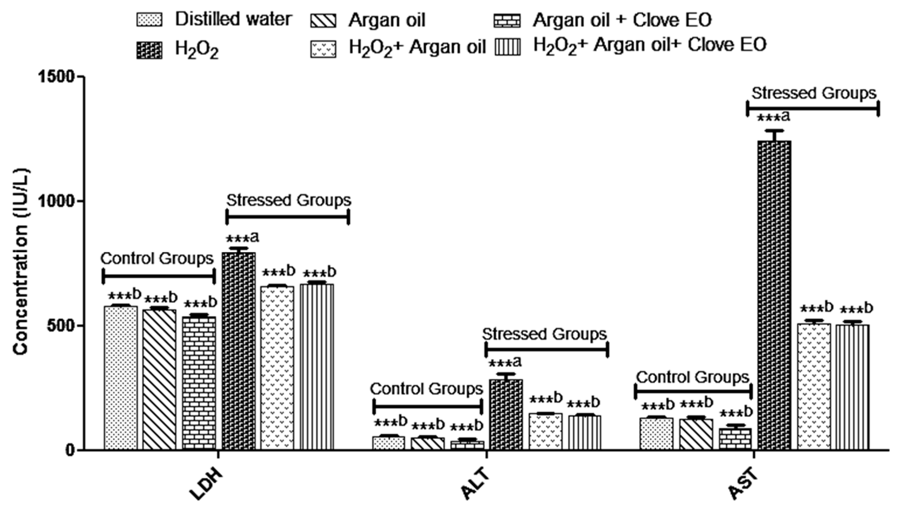
2.3. Effect of the Interventions on Enzymatic Markers
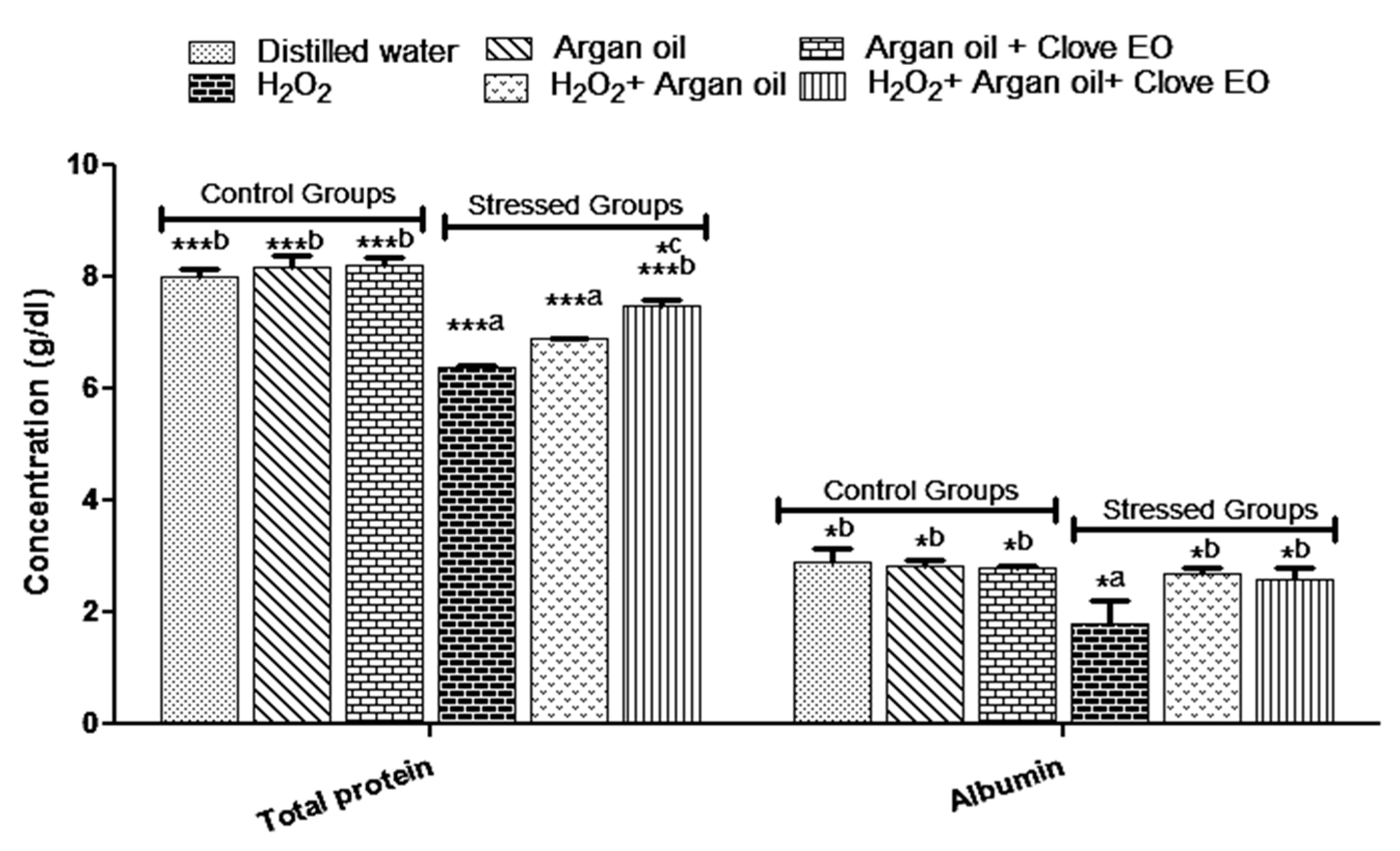
2.4. Effect of the Interventions on Total Protein and Albumin Levels
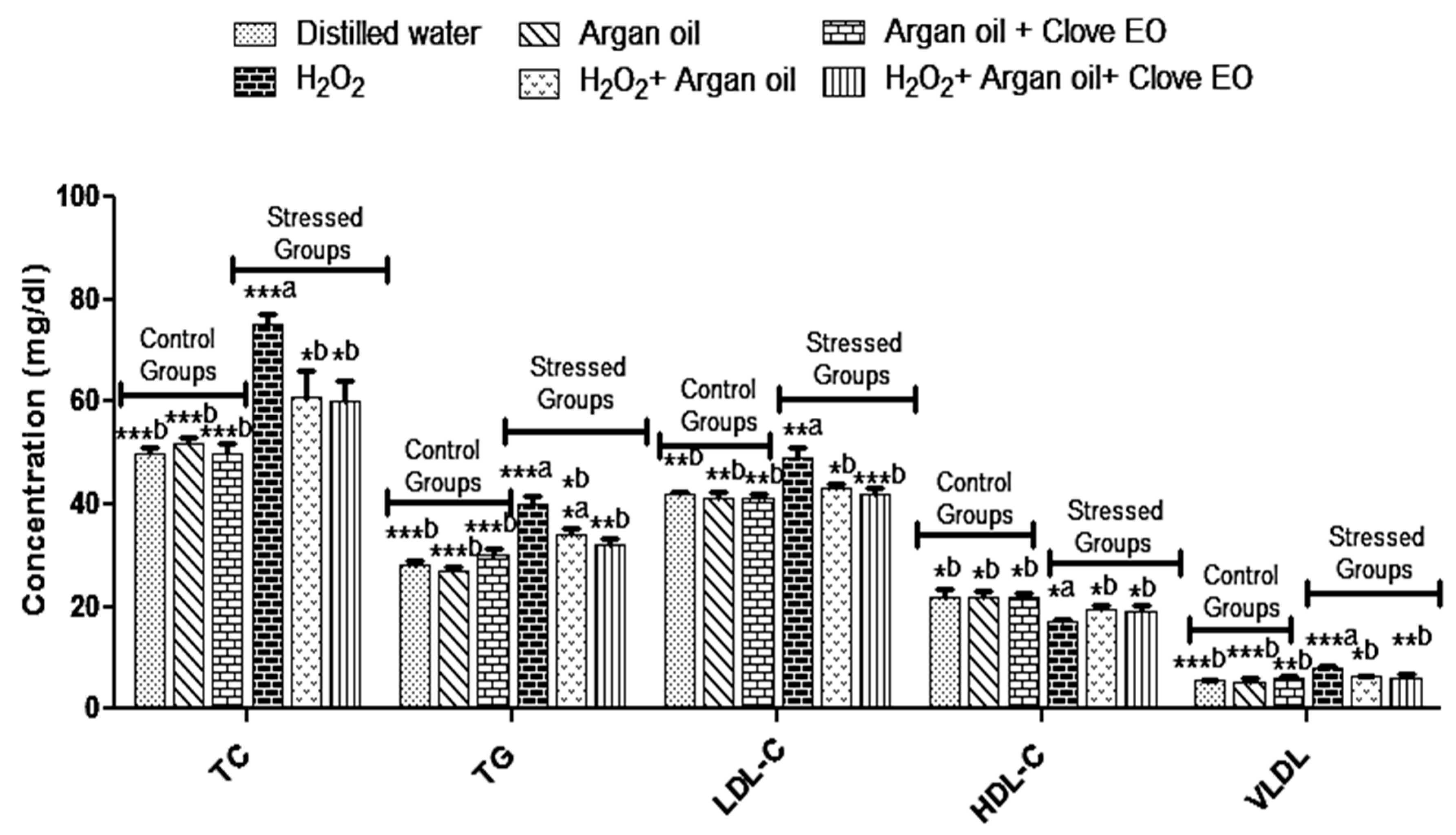
2.5. Effect of the Interventions on TC, TG, LDL-C, HDL-C and VLDL Levels
2.6. Effect of the Interventions on Serum Electrolytes
2.7. Effect of the Interventions on Blood Urea and Creatinine Levels
2.8. Effect of the Interventions on Organs Weights
2.9. Effects of the Interventions on Histopathological Changes
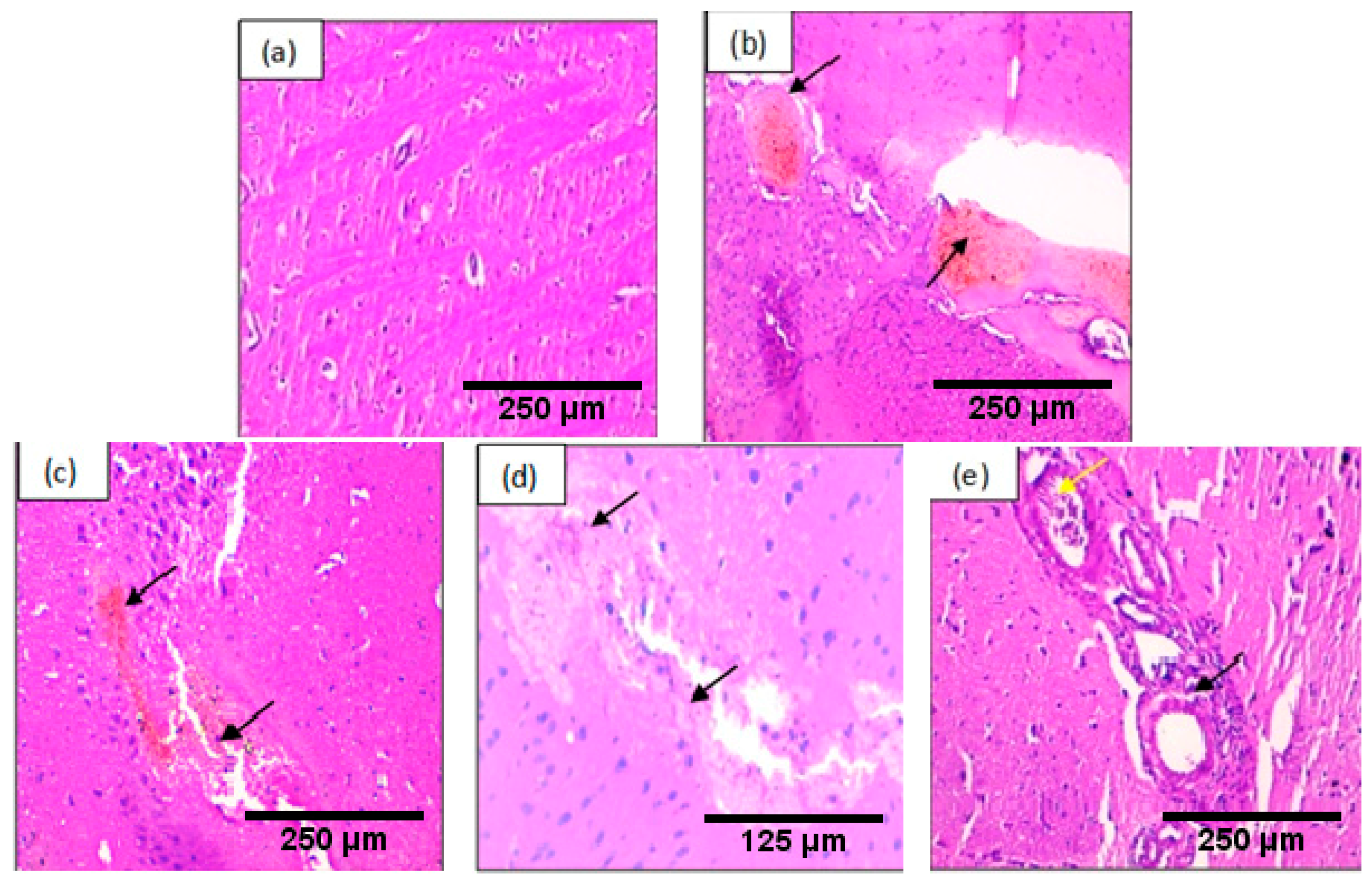
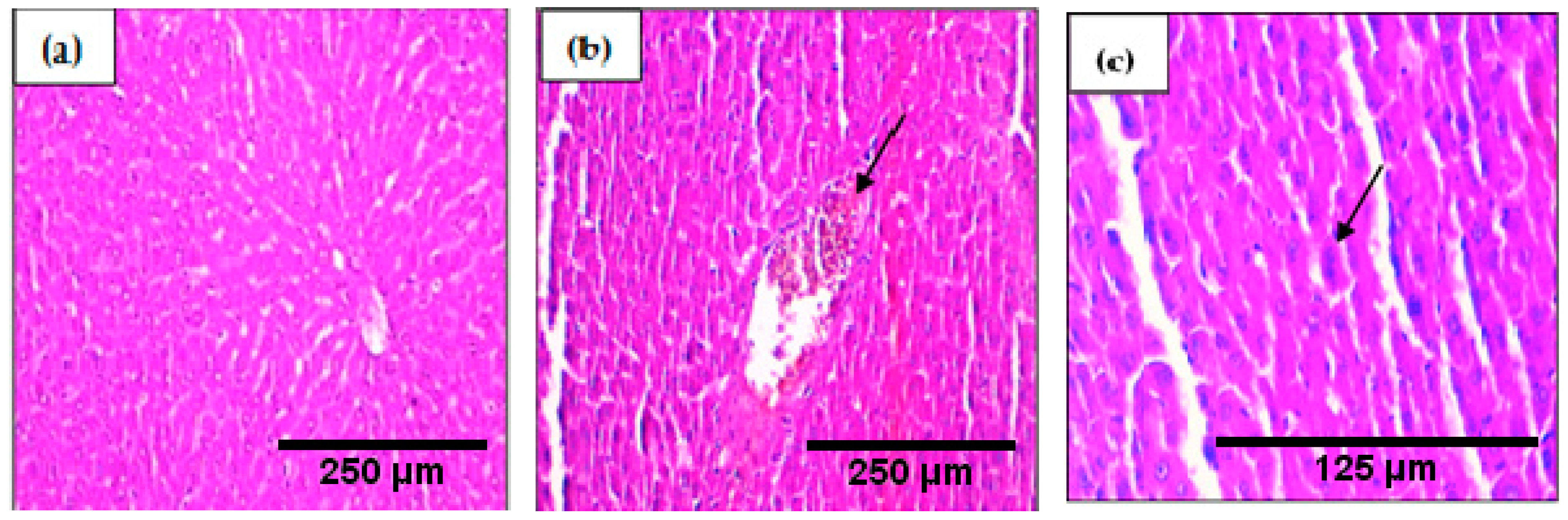
2.9.1. Brain
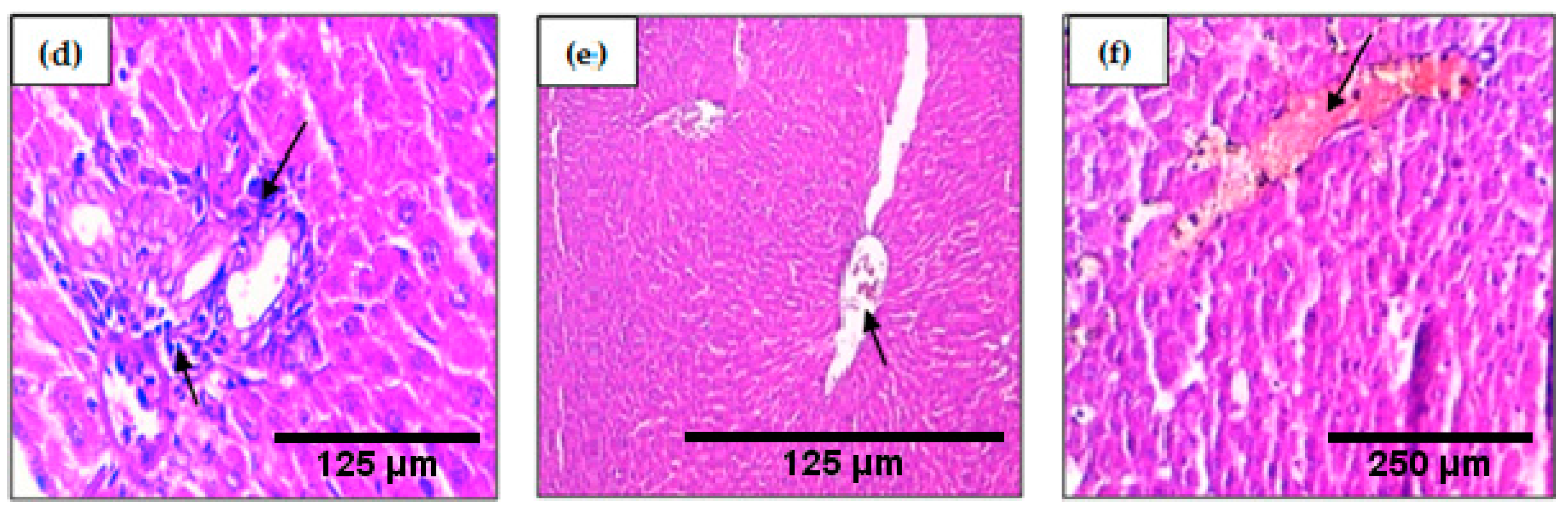
2.9.2. Liver
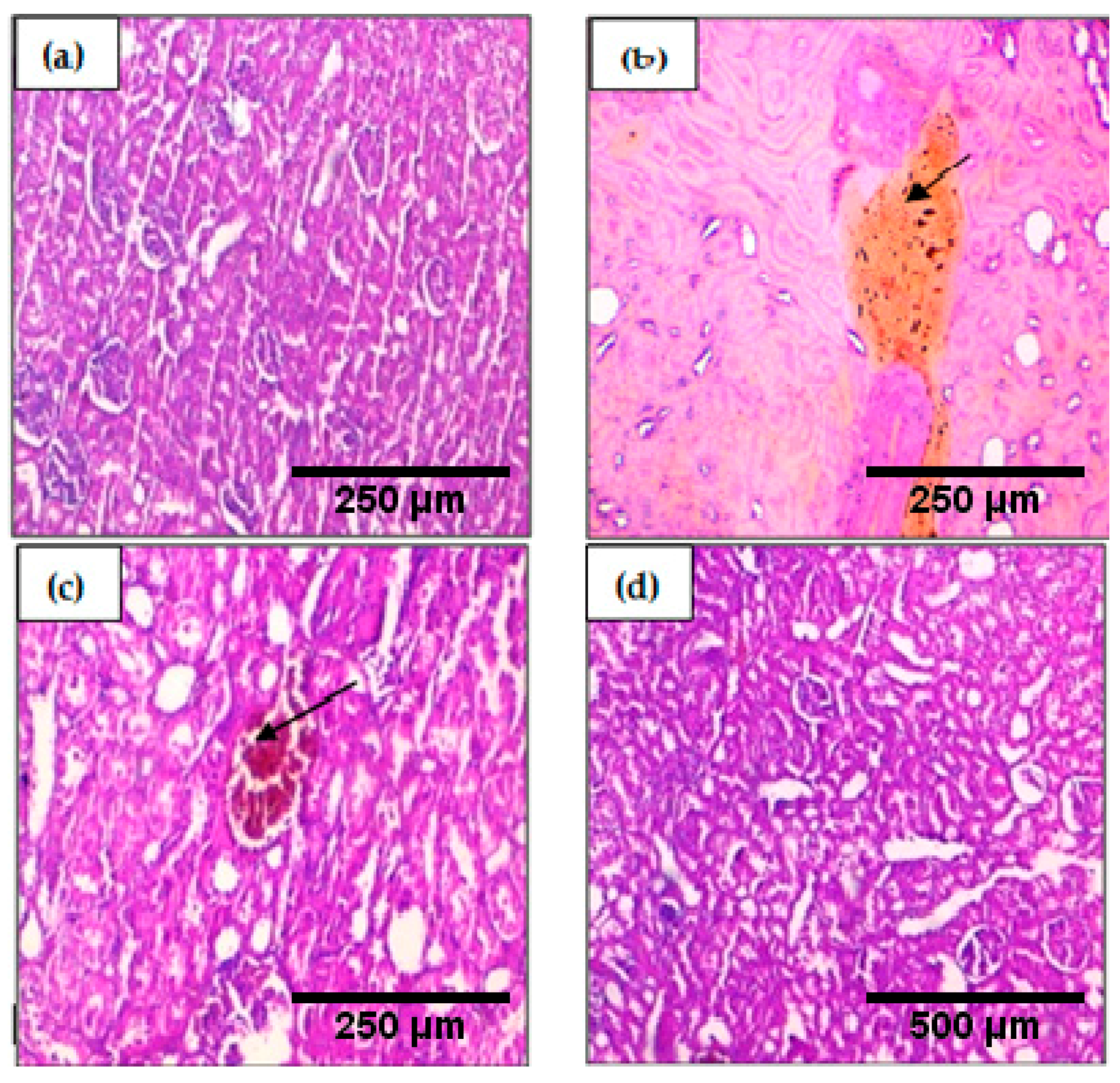
2.9.3. Kidney
3. Discussion
4. Materials and Methods
4.1. Argan Oil
4.2. Essential Oil Extraction
4.3. Characterization andChemical Composition of Syzygium aromaticum Essential Oil
4.3.1. Gas Chromatography Analysis
4.3.2. Gas Chromatography–Mass Spectrometry (GC–MS)
4.4. In Vitro Antioxidant Activities of Argan Oil and Syzygium aromaticum Essential Oil
4.4.1. Total Phenolic Content
4.4.2. Flavones and Flavonols
4.4.3. Total Flavonoids Content
4.4.4. Total Antioxidant Capacity (TAC)
4.5. Experimental Animals
4.6. Experimental Design
4.7. Blood Analysis
4.8. Histopathological Study
4.9. Statistical Analysis
Acknowledgments
Author Contributions
Conflicts of Interest
Abbreviations
| ROS | Reactive oxygen species |
| RNS | reactive nitrogen species |
| GC/MS | Gas chromatography–mass spectrometry |
| DPPH | 1,1-diphenyl-2-picrylhydrazyl |
| TAC | Total antioxidant capacity |
| LDH | Lactate dehydrogenase |
| ALT | Alanine aminotransferase |
| AST | Aspartate aminotransferase |
| TC | Total cholesterol |
| TG | Triglycerides |
| LDL-C | Low Density Lipoprotein |
| HDL-C | High Density Lipoprotein-cholesterol |
| VLDL | Very Low Density Lipoprotein |
| BHT | Butylated hydroxytoluene |
| H & E | hematoxylin and eosin |
References
- Korovila, I.; Hugo, M.; Castro, J.P.; Weber, D.; Hohn, A.; Grune, T.; Jung, T. Proteostasis, oxidative stress and aging. Redox Biol. 2017, 13, 550–567. [Google Scholar] [CrossRef] [PubMed]
- Torres-Cuevas, I.; Parra-Llorca, A.; Sánchez-Illana, A.; Nuñez-Ramiro, A.; Kuligowski, J.; Cháfer-Pericás, C.; Cernada, M.; Escobar, J.; Vento, M. Oxygen and oxidative stress in the perinatal period. Redox Biol. 2017, 12, 674–681. [Google Scholar] [CrossRef] [PubMed]
- Kleniewska, P.; Pawliczak, R. The participation of oxidative stress in the pathogenesis of bronchial asthma. Biomed. Pharmacother. 2017, 94, 100–108. [Google Scholar] [CrossRef] [PubMed]
- Scicchitano, B.M.; Pelosi, L.; Sica, G.; Musarò, A. The physiopathologic role of oxidative stress in skeletal muscle. Mech. Ageing Dev. 2017. [Google Scholar] [CrossRef] [PubMed]
- Kelly, F.J.; Fussell, J.C. Role of oxidative stress in cardiovascular disease outcomes following exposure to ambient air pollution. Free Radic. Biol. Med. 2017, 110, 345–367. [Google Scholar] [CrossRef] [PubMed]
- Andersen, J.K. Oxidative stress in neurodegeneration: Cause or consequence? Nat. Rev. Neurosci. 2004, 10, S18–S25. [Google Scholar] [CrossRef] [PubMed]
- Cheignon, C.; Tomas, M.; Bonnefont-Rousselot, D.; Faller, P.; Hureau, C.; Collin, F. Oxidative stress and the amyloid beta peptide in Alzheimer’s Disease. Redox Biol. 2017. [Google Scholar] [CrossRef] [PubMed]
- Crotty, G.F.; Ascherio, A.; Schwarzschild, M.A. Targeting urate to reduce oxidative stress in Parkinson disease. Exp. Neurol. 2017. [Google Scholar] [CrossRef] [PubMed]
- Andrisic, L.; Dudzik, D.; Barbas, C.; Milkovic, L.; Grune, T.; Zarkovic, N. Short overview on metabolomics approach to study pathophysiology of oxidative stress in cancer. Redox Biol. 2018, 14, 47–58. [Google Scholar] [CrossRef] [PubMed]
- Saed, G.M.; Diamond, M.P.; Fletcher, N.M. Updates of the role of oxidative stress in the pathogenesis of ovarian cancer. Gynecol. Oncol. 2017, 145, 595–602. [Google Scholar] [CrossRef] [PubMed]
- Mani, R.S.; Amin, M.A.; Li, X.; Kalyana-Sundaram, S.; Veeneman, B.A.; Wang, L.; Ghosh, A.; Aslam, A.; Ramanand, S.G.; Rabquer, B.J.; et al. Inflammation-Induced Oxidative Stress Mediates Gene Fusion Formation in Prostate Cancer. Cell Rep. 2016, 17, 2620–2631. [Google Scholar] [CrossRef] [PubMed]
- Pisoschi, A.M.; Pop, A. The role of antioxidants in the chemistry of oxidative stress: A review. Eur. J. Med. Chem. 2015, 97, 55–74. [Google Scholar] [CrossRef] [PubMed]
- El Abbassi, A.; Khalid, N.; Zbakh, H.; Ahmad, A. Physicochemical Characteristics, Nutritional Properties, and Health Benefits of Argan Oil: A Review. Crit. Rev. Food Sci. Nutr. 2014, 54, 1401–1414. [Google Scholar] [CrossRef] [PubMed]
- Charrouf, Z.; Guillaume, D. Argan oil: Occurrence, composition and impact on human health. Eur. J. Lipid Sci. Technol. 2008, 110, 632–636. [Google Scholar] [CrossRef]
- Charrouf, Z.; Guillaume, D. Phenols and polyphenols from Argania spinosa. Am. J. Food Technol. 2007, 2, 679–683. [Google Scholar]
- Bennani, H.; Drissi, A.; Giton, F.; Kheuang, L.; Fiet, J.; Adlouni, A. Antiproliferative effect of polyphenols and sterols of virgin argan oil on human prostate cancer cell lines. Cancer Detect. Prev. 2007, 31, 64–69. [Google Scholar] [CrossRef] [PubMed]
- El Midaoui, A.; Haddad, Y.; Couture, R. Beneficial effects of argan oil on blood pressure, insulin resistance, and oxidative stress in rat. Nutrition 2016, 32, 1132–1137. [Google Scholar] [CrossRef] [PubMed]
- Khallouki, F.; Younos, C.; Soulimani, R.; Oster, T.; Charrouf, Z.; Spiegelhalder, B.; Bartsch, H.; Owen, R.W. Consumption of argan oil (Morocco) with its unique profile of fatty acids, tocopherols, squalene, sterols and phenolic compounds should confer valuable cancer chemopreventive effects. Eur. J. Cancer Prev. 2003, 12, 67–75. [Google Scholar] [CrossRef] [PubMed]
- Berrougui, H.; Ettaib, A.; Gonzalez, M.D.H.; Sotomayor, M.A.; de Bennani-Kabchi, N.; Hmamouchi, M. Hypolipidemic and hypocholesterolemic effect of argan oil (Arganiaspinosa L.) in Merionesshawi rats. J. Ethnopharmacol. 2003, 89, 15–18. [Google Scholar] [CrossRef]
- Monfalouti, H.E.; Guillaume, D.; Denhez, C.; Charrouf, Z. Therapeutic potential of argan oil: A review: Therapeutic potential of argan oil. J. Pharm. Pharmacol. 2010, 62, 1669–1675. [Google Scholar] [CrossRef] [PubMed]
- Hafsé, M.; Benbrahim, K.F.; Saidi, A.; Farah, A. Volatile Components and Antibacterial Profile of Essential Oils Extracted from Leaves and Twigs of Pistacialentiscus L. Br. Microbiol. Res. J. 2013, 3, 602–611. [Google Scholar]
- Andrade, M.; das Graças Cardoso, M.; de Andrade, J.; Silva, L.; Teixeira, M.; ValérioResende, J.; da Silva Figueiredo, A.; Barroso, J. Chemical Composition and Antioxidant Activity of Essential Oils from CinnamodendrondinisiiSchwacke and SiparunaguianensisAublet. Antioxidants 2013, 2, 384–397. [Google Scholar] [CrossRef] [PubMed]
- Liu, Y.; Zhang, Y.; Lin, K.; Zhang, D.; Tian, M.; Guo, H.; Wang, Y.; Li, Y.; Shan, Z. Protective effect of piperine on electrophysiology abnormalities of left atrial myocytes induced by hydrogen peroxide in rabbits. Life Sci. 2014, 94, 99–105. [Google Scholar] [CrossRef] [PubMed]
- Kumar, S.; Srivastava, N.; Gomes, J. The effect of lovastatin on oxidative stress and antioxidant enzymes in hydrogen peroxide intoxicated rat. Food Chem. Toxicol. 2011, 49, 898–902. [Google Scholar] [CrossRef] [PubMed]
- Na, J.-Y.; Song, K.; Kim, S.; Kwon, J. Rutin protects rat articular chondrocytes against oxidative stress induced by hydrogen peroxide through SIRT1 activation. Biochem. Biophys. Res. Commun. 2016, 473, 1301–1308. [Google Scholar] [CrossRef] [PubMed]
- Sies, H. Hydrogen peroxide as a central redox signaling molecule in physiological oxidative stress: Oxidative eustress. Redox Biol. 2017, 11, 613–619. [Google Scholar] [CrossRef] [PubMed]
- Tudor, E. Temperature dependence of the retention index for perfumery compounds on a SE-30 glass capillary column I. Linear equations. J. Chromatogr. A 1997, 779, 287–297. [Google Scholar] [CrossRef]
- Hoskovec, M.; Grygarová, D.; Cvačka, J.; Streinz, L.; Zima, J.; Verevkin, S.P.; Koutek, B. Determining the vapour pressures of plant volatiles from gas chromatographic retention data. J. Chromatogr. A 2005, 1083, 161–172. [Google Scholar] [CrossRef] [PubMed]
- Adams, R.P. Identification of Essential Oil Components by Gas Chromatography/Mass Spectrometry; Allured Publishing Corporation: Carol Stream, IL, USA, 1995. [Google Scholar]
- Högnadóttir, Á.; Rouseff, R.L. Identification of aroma active compounds in orange essence oil using gas chromatography-olfactometry and gas chromatography-mass spectrometry. J. Chromatogr. A 2003, 998, 201–211. [Google Scholar] [CrossRef]
- De Oliveira, M.S.; da Costa, W.A.; Pereira, D.S.; Botelho, J.R.S.; de Alencar Menezes, T.O.; de Aguiar Andrade, E.H.; da Silva, S.H.M.; da Silva Sousa Filho, A.P.; de Carvalho, R.N. Chemical composition and phytotoxic activity of clove (Syzygium aromaticum) essential oil obtained with supercritical CO2. J. Supercrit. Fluids 2016, 118, 185–193. [Google Scholar] [CrossRef]
- Fayemiwo, K.A.; Adeleke, M.A.; Okoro, O.P.; Awojide, S.H.; Awoniyi, I.O. Larvicidal efficacies and chemical composition of essential oils of Pinussylvestris and Syzygium aromaticum against mosquitoes. Asian Pac. J. Trop. Biomed. 2014, 4, 30–34. [Google Scholar] [CrossRef]
- Nam, H.; Kim, M.-M. Eugenol with antioxidant activity inhibits MMP-9 related to metastasis in human fibrosarcoma cells. Food Chem. Toxicol. 2013, 55, 106–112. [Google Scholar] [CrossRef] [PubMed]
- Gülçin, İ.; Elmastaş, M.; Aboul-Enein, H.Y. Antioxidant activity of clove oil—A powerful antioxidant source. Arab. J. Chem. 2012, 5, 489–499. [Google Scholar] [CrossRef]
- Abozid, M.M.; El-Sayed, S.M. Antioxidant and Protective Effect of Clove Extracts and Clove Essential Oil on Hydrogen Peroxide Treated Rats. Int. J. ChemTech Res. 2013, 5, 1477–1485. [Google Scholar]
- Ganie, S.A.; Haq, E.; Hamid, A.; Masood, A.; Zargar, M.A. Long dose exposure of hydrogen peroxide (H2O2) in albino rats and effect of Podophyllumhexandrum on oxidative stress. Eur. Rev. Med. Pharmacol. Sci. 2011, 15, 906–915. [Google Scholar] [PubMed]
- Bakour, M.; Al-Waili, N.S.; El Menyiy, N.; Imtara, H.; Figuira, A.C.; Al-Waili, T.; Lyoussi, B. Antioxidant activity and protective effect of bee bread (honey and pollen) in aluminum-induced anemia, elevation of inflammatory makers and hepato-renal toxicity. J. Food Sci. Technol. 2017. [Google Scholar] [CrossRef] [PubMed]
- Jagadeesan, G.; Bharathi, E. In vivo restoration of hepatic and nephro protective potential of hesperidin and ellagic acid against mercuric chloride intoxicated rats. Biomed. Aging Pathol. 2014, 4, 219–222. [Google Scholar] [CrossRef]
- Akirov, A.; Masri-Iraqi, H.; Atamna, A.; Shimon, I. Low Albumin Levels Are Associated with Mortality Risk in Hospitalized Patients. Am. J. Med. 2017. [Google Scholar] [CrossRef] [PubMed]
- Samarghandian, S.; Samini, F.; Azimi-Nezhad, M.; Farkhondeh, T. Anti-oxidative effects of safranal on immobilization-induced oxidative damage in rat brain. Neurosci. Lett. 2017, 659, 26–32. [Google Scholar] [CrossRef] [PubMed]
- Hussein, S.; El-Saba, A.-A.; Galal, M.K. Biochemical and histological studies on adverse effects of mobile phone radiation on rat’s brain. J. Chem. Neuroanat. 2016, 78, 10–19. [Google Scholar] [CrossRef] [PubMed]
- Shukri, R.; Mohamed, S.; Mustapha, N.M. Cloves protect the heart, liver and lens of diabetic rats. Food Chem. 2010, 122, 1116–1121. [Google Scholar] [CrossRef]
- Drissi, A. Evidence of hypolipemiant and antioxidant properties of argan oil derived from the argan tree (Arganiaspinosa). Clin. Nutr. 2004, 23, 1159–1166. [Google Scholar] [CrossRef] [PubMed]
- Pirisi, F.M.; Cabras, P.; Cao, C.F.; Migliorini, M.; Muggelli, M. Phenolic Compounds in Virgin Olive Oil. 2. Reappraisal of the Extraction, HPLC Separation, and Quantification Procedures. J. Agric. Food Chem. 2000, 48, 1191–1196. [Google Scholar] [CrossRef] [PubMed]
- Adams, R.P. Identification of essential oil components by gas chromatography/mass spectrometry. J. Am. Soc. Mass Spectrom. 1997, 6, 671–672. [Google Scholar]
- Goodarzi, S.; Hadjiakhoondi, A.; Yassa, N.; Khanavi, M.; Tofighi, Z. Essential oils chemical composition, antioxidant activities and total phenols of Astrodaucuspersicus. Iran. J. Basic Med. Sci. 2016, 19, 159. [Google Scholar] [PubMed]
- Paradiso, V.M.; Clemente, A.; Summo, C.; Pasqualone, A.; Caponio, F. Towards green analysis of virgin olive oil phenolic compounds: Extraction by a natural deep eutectic solvent and direct spectrophotometric detection. Food Chem. 2016, 212, 43–47. [Google Scholar] [CrossRef] [PubMed]
- Boulanouar, B.; Abdelaziz, G.; Aazza, S.; Gago, C.; Miguel, M.G. Antioxidant activities of eight Algerian plant extracts and two essential oils. Ind. Crops Prod. 2013, 46, 85–96. [Google Scholar] [CrossRef]
- Park, Y.-S.; Jung, S.-T.; Kang, S.-G.; Heo, B.G.; Arancibia-Avila, P.; Toledo, F.; Drzewiecki, J.; Namiesnik, J.; Gorinstein, S. Antioxidants and proteins in ethylene-treated kiwifruits. Food Chem. 2008, 107, 640–648. [Google Scholar] [CrossRef]
- Amzad Hossain, M.; Shah, M.D. A study on the total phenols content and antioxidant activity of essential oil and different solvent extracts of endemic plant Merremiaborneensis. Arab. J. Chem. 2015, 8, 66–71. [Google Scholar] [CrossRef]
- El-Hadarm, A.; Hassanien, M. Hepatoprotective effect of cold-pressed Syzygium aromaticum oil against carbon tetrachloride (CCl4)-induced hepatotoxicity in rats. J. Pharm. Biol. 2016, 54, 1364–1372. [Google Scholar] [CrossRef] [PubMed]
- Rim, R.; Rhazali, L.; Harmouch, A.; Lotfi, H.; Benazzouz, B.; El Hessni, A.; Ouichou, A.; Akhouayri, O.; Mesfioui, A. Does argan oil supplementation affect metabolic parameters and behavior in Wistar rats? Food Nutr. Sci. 2015, 6, 816. [Google Scholar]


| Compounds | Kovats Index | Area (%) | Chemical Formula | Kovats Index (Literature) |
|---|---|---|---|---|
| Eugenol | 1353.00 | 87.03 | C10H12O2 | 1327.70 [27] |
| β-Caryophyllene | 1428.00 | 0.69 | C15H24 | 1433.90 [28] |
| Eugenyl acetate | 1538.00 | 11.25 | C12H14O3 | 1524.00 [29] |
| Caryophyllene oxide | 1689.00 | <0.10 | C15H24O | 1606.00 [30] |
| Sample | Phenolics 1 | Flavones and Flavonols 2 | Flavonoids 2 | TAC 3 |
|---|---|---|---|---|
| Argan oil (mg Eq/100 g) | 41.28 ± 0.40 * | 1.80 ± 0.07 * | 8.31 ± 1.06 * | 90.90 ± 4.53 * |
| Syzygium aromaticum essential oil (mg Eq/100 g) | 165.52 ± 9.71 | 29.60 ± 1.02 | 44.08 ± 5.34 | 3235.50 ± 237.40 |
| Minerals | Distilled Water | Argan Oil | Argan Oil + Clove Essential Oil | H2O2 | H2O2 + Argan Oil | H2O2 + Argan Oil + Clove Essential Oil |
|---|---|---|---|---|---|---|
| Sodium (mmol/L) | 140 ± 30 | 139 ± 2.1 | 138 ± 2 | 143 ± 2 | 142 ± 1.5 | 145 ± 2.1 |
| Potassium (mmol/L) | 6 ± 1 | 5.8 ± 0.8 | 5.6 ± 1.4 | 6.3 ± 1.2 | 5.6 ± 1.7 | 6 ± 0.9 |
| Chloride (mmol/L) | 103 ± 1.2 | 105 ± 3 | 100 ± 4 | 106 ± 2.5 | 102 ± 2.3 | 102 ± 2 |
| Renal Markers | Distilled Water | Argan Oil | Argan Oil + Clove Essential | H2O2 | H2O2 + Argan Oil | H2O2 + Argan Oil + Clove Essential Oil |
|---|---|---|---|---|---|---|
| Creatinine (mg/dL) | 0.7 ± 0.05 | 0.6 ± 0.03 | 0.7 ± 0.03 | 0.75 ± 0.2 | 0.6 ± 0.04 | 0.6 ± 0.08 |
| Urea (mg/dL) | 24 ± 1 **,b | 22 ± 0.5 ***,b | 23 ± 1.5 ***,b | 30 ± 0.2 **,a | 22 ± 1.5 ***,b | 21 ± 0.9 ***,b |
| Parameters | Distilled Water | Argan Oil | Argan Oil + Clove EO | H2O2 | H2O2 + Argan Oil | H2O2 + ArganOil + Clove Essential Oil |
|---|---|---|---|---|---|---|
| Body weight (g) | 190 ± 10 | 198 ± 3 | 192.5 ± 3.53 | 181 ± 2 | 198.5 ± 1.5 | 203 ± 5 |
| Brain weight (g) | 1.87 ± 0.1 ***,b | 1.92 ± 0.12 ***,b | 1.8 ± 0.15 ***,b | 1.29 ± 0.11 ***,a | 1.62 ± 0.04 ***,a, ***,b | 1.71 ± 0.09 ***,a, ***,b, *,c |
| Liver weight (g) | 6.45 ± 0.04 ***,b | 6.4 ± 0.1 ***,b | 6.32 ± 0.1 ***,b | 9.18 ± 0.11 ***,a | 6.78 ± 0.05 **,a, ***,b | 6.65 ± 0.06 *,a, ***,b |
| Kidney weight (g) | 0.75 ± 0.04 ***,b | 0.76 ± 0.02 ***,b | 0.735 ± 0.01 ***,b | 1.195 ± 0.01 ***,a | 0.975 ± 0.02 ***,a | 0.8 ± 0.02 ***,b, **,c |
| Brain relative weight (g/100 g BW) | 0.984 ± 0.05 ***,b | 0.969 ± 0.06 ***,b | 0.935 ± 0.077 ***,b | 0.71 ± 0.06 ***,a | 0.816 ± 0.02 ***,a, ***,b | 0.842 ± 0.04 ***,a, ***,b, *,c |
| Liver relative weight (g/100 g BW) | 3.394 ± 0.02 ***,b | 3.383 ± 0.06 ***,b | 3.283 ± 0.05 ***,b | 5.07 ± 0.06 ***,a | 3.41 ± 0.025 **,a, ***,b | 3.27 ± 0.029 *,a, ***,b |
| Kidney relative weight (g/100 g BW) | 0.394 ± 0.02 ***,b | 0.383 ± 0.01 ***,b | 0.381 ± 0.05 ***,b | 0.660 ± 0.05 ***,b | 0.491 ± 0.01 ***,a | 0.394±0.009 ***,b, **,c |
© 2018 by the authors. Licensee MDPI, Basel, Switzerland. This article is an open access article distributed under the terms and conditions of the Creative Commons Attribution (CC BY) license (http://creativecommons.org/licenses/by/4.0/).
Share and Cite
BAKOUR, M.; SOULO, N.; HAMMAS, N.; FATEMI, H.E.; ABOULGHAZI, A.; TAROQ, A.; ABDELLAOUI, A.; AL-WAILI, N.; LYOUSSI, B. The Antioxidant Content and Protective Effect of Argan Oil and Syzygium aromaticum Essential Oil in Hydrogen Peroxide-Induced Biochemical and Histological Changes. Int. J. Mol. Sci. 2018, 19, 610. https://doi.org/10.3390/ijms19020610
BAKOUR M, SOULO N, HAMMAS N, FATEMI HE, ABOULGHAZI A, TAROQ A, ABDELLAOUI A, AL-WAILI N, LYOUSSI B. The Antioxidant Content and Protective Effect of Argan Oil and Syzygium aromaticum Essential Oil in Hydrogen Peroxide-Induced Biochemical and Histological Changes. International Journal of Molecular Sciences. 2018; 19(2):610. https://doi.org/10.3390/ijms19020610
Chicago/Turabian StyleBAKOUR, Meryem, Najoua SOULO, Nawal HAMMAS, Hinde EL FATEMI, Abderrazak ABOULGHAZI, Amal TAROQ, Abdelfattah ABDELLAOUI, Noori AL-WAILI, and Badiaa LYOUSSI. 2018. "The Antioxidant Content and Protective Effect of Argan Oil and Syzygium aromaticum Essential Oil in Hydrogen Peroxide-Induced Biochemical and Histological Changes" International Journal of Molecular Sciences 19, no. 2: 610. https://doi.org/10.3390/ijms19020610